Chemical characterization, antioxidant and
antimicrobial activity of propolis obtained from
Melipona quadrifasciata quadrifasciata
and
Tetragonisca angustula
stingless bees
A.R. Torres
1,2, L.P. Sandjo
3, M.T. Friedemann
2, M.M. Tomazzoli
4, M. Maraschin
4, C.F. Mello
1and
A.R.S. Santos
1,21Programa de Pós-graduac¸ão em Farmacologia, Centro de Ciências da Saúde, Universidade Federal de Santa Maria, Santa Maria, RS, Brasil 2Laboratorio de Neurobiologia da Dor e In
flamac¸ão, Departamento de Ciências Fisiológicas, Centro de Ciências Biológicas, Universidade Federal de Santa Catarina, Florianópolis, SC, Brasil 3Departamento de Ciências Farmacêuticas, Centro de Ciências da Saúde, Universidade Federal de Santa Catarina, Florianópolis, SC, Brasil 4Laboratório de Morfogênese e Bioquímica Vegetal, Centro de Ciências Agrárias, Universidade Federal de Santa Catarina, Florianópolis, SC, Brasil
Abstract
In this study, we investigated the chemical composition, and antioxidant and antibacterial properties of ethanolic extracts of propolis (EEP) fromMelipona quadrifasciata quadrifasciataandTetragonisca angustula. Chemical composition of EEP was determined by colorimetry and chromatographic (HPLC-DAD and UPLC-Q/TOF-MS/MS) analysis. Antimicrobial activity of EEP was evaluated against gram-positive (S. aureus, methicillin-resistantS. aureus,E. faecalis) and gram-negative (E. coliand
K. pneumoniae) bacteria by the minimal inhibitory concentration (MIC) test using the microdilution method. Furthermore, the growth curve and integrity of cell membrane ofS. aureusandE. coliwere investigated using standard microbiological methods. HPLC-DAD analysis showed that the EEP ofM. quadrifasciata quadrifasciatahas a more complex chemical composition than the EEP ofT. angustula. Moreover, UPLC-MS analyses ofM. quadrifasciata quadrifascitaindicated flavonoids and terpenes as major constituents. The bactericidal activity of both EEPs was higher against gram-positive bacteria than for gram-negative bacteria. The EEP fromM. quadrifasciata quadrifasciatapresented MIC values lower than the EEP fromT. angustulafor all tested bacteria. The EEP fromM. quadrifasciata quadrifasciatacaused lysis of the bacterial wall and release of intracellular components from bothE. coliandS. aureus. Ourfindings indicate that the chemical composition of propolis from stingless bees is complex and depends on the species. The extract fromM. quadrifasciata quadrifascitawas more effective against gram-positive than gram-negative strains, especially againstS. aureusand methicillin-resistantS. aureuscompared toT. angustula
extract, by a mechanism that involves disturbance of the bacterial cell membrane integrity.
Key words: Stingless bees; Propolis; Antimicrobial activity;S. aureus;M. quadrifasciata quadrifasciata;T. angustula
Introduction
Propolis is a complex mixture of pollen and resinous and balsamic substances collected by bees from buds,
flowers, and plant exudates, and bee salivary secretions (1). Since propolis is a bee product of plant origin, its chemical composition and biological activity depends on the specificity of the localflora, season of harvest, and bee species (2–4).
Different biological and therapeutic properties have been reported for propolis, including antioxidant (3,5), anti-inflammatory (5,6), immunomodulatory (7,8), antitumoral (8,9),
and antimicrobial activities (2,7,10,11) among others. It has been shown that propolis has bactericidal and bacterio-static activity against various gram-positive bacteria, such asS. aureus,S. mutansandB. subtilis, and gram-negative bacteria, includingE. coli,K. pneumoniaeandP. aeruginosa (6,7,9,10). Moreover, a synergistic inhibitory effect of prop-olis and antibiotics on the growth ofS. aureus has been reported (11). Such an antimicrobial activity of propolis is particularly relevant if one considers the increasing emergence
Correspondence: A.R.S. Santos:<adair.santos@ufsc.br>
of antibiotic-resistant microorganisms in hospitals and in the community (11). This situation is aggravated by the inadequate use and prescription of antibiotics and the scarcity of new drugs (12).
Most of the studies in the literature have investigated the antimicrobial activity of the propolis produced byApis mellifera. However, little is known about the biological effects of the propolis produced by other bees, such as the Meliponines. Melipona quadrifasciata quadrifasciata Lepeletier and Tetragonisca angustula Letreille stingless bees belong to the Meliponini tribe, and are two among more than 200 species of Brazilian native stingless bees (13). Native from tropical and subtropical regions,M. quadrifasciata quadrifasciata and Tetragonisca angustula are locally known as Mandac¸aia and Jataí, respectively. Interestingly, the propolis fromM. quadrifasciata quadrifasciatais known as geopropolis because it presents soil traces in its com-position (14). Due to the unique behavioral and morpho-logical characteristics of these bees, one might reasonably hypothesize that the propolis produced by them has distinct composition and biological activity. Thus, the aim of this work was to characterize the chemical composition of the ethanolic crude extract of propolis (EEP) produced by M. quadrifasciata quadrifasciataand T. angustulaand investigate its potential antioxidant and antibacterial activity against gram-negative and gram-positive bacteria, includ-ing methicillin-resistantS. aureus.
Material and Methods
Chemicals and reagents
DPPH (2,2-diphenyl-1-picrylhydrazyl), resazurin, Folin-Ciocalteu phenol reagent (2N), gallic acid monohydrate (C7H6O5.H2O), quercetin, aluminum chloride (AlCl3) and sodium carbonate (Na2CO3) were purchased from Sigma (USA). Ethanol and methanol were obtained from Merck (Brazil). Acetonitrile was from Tedia (Brazil). The culture medium Brain Heart Infusion was obtained from Himedia (India). The bacteria strains were obtained from Laborclin (Brazil) and Microbiology Laboratory of the Federal Uni-versity of Santa Maria (Brazil). All other chemicals were of analytical grade and purity. Aqueous solutions were prepared in ultrapure water produced by a Milli-Q system (18.2 MO, Millipore, France).
Propolis samples and ethanolic extract preparation The samples were collected in September of 2014 in Rio das Antas, Brazil. Five samples of the propolis from M. quadrifasciata quadrifasciata and three samples from T. angustula were obtained from the inner parts of the beehives. The ethanolic extract was prepared as reported by Park et al. (15). Two grams of the powder was mixed with 25 mL of 80% ethanol in a sealed container protected from light (to avoid loss of volatile and photosensitive com-pounds), under agitation in a water bath at 70°C for 30 min. After extraction, the mixture wasfiltered (grade 1 Whatman)
to obtain the EEP at concentration of 80 mg/mL (propolis: ethanol 80%, w/v).
Total polyphenol andflavonoid contents
The total polyphenol content of EEP was determined using the Folin-Ciocalteu colorimetric method described by Frozza et al. (16). Briefly, 100 mL of the hydroalcoholic extract (1 mg/mL) was mixed with 500mL of Folin-Ciocalteu and after 5 min in dark, 400mL sodium carbonate (7.5%) was added. After incubation in the dark at room tempera-ture for 30 min, the absorbance of the reaction mixtempera-ture was measured at 765 nm in a spectrophotometer (model FlexStation, Molecular Devices, USA). Gallic acid standard solutions (0.25–4.0 mL/mL) were used for the calibration
curve. The average of three readings was used to deter-mine the total polyphenol content, reported as mg of gallic acid equivalents per g of propolis (GAEs).
The total flavonoid content in EEP was determined by the method described by Campos et al. (9). For this, 0.5 mL of EEP (100mg/mL) was mixed with 4.5 mL of 2% aluminum chloride hexahydrate in methanol. After 30-min incubation at room temperature in the dark, the absorbance was read at 415 nm using a plate spectrophotometer (FlexStation, Molecular Device). Quercetin (0.4–11mg/mL)
was used as standard. Triplicates were used to determine the flavonoid content, reported as mg of QE per g of propolis.
High performance liquid chromatography (HPLC-DAD) analysis
Briefly, 10-mL samples of EEP were injected in the liquid chromatographer (Thermo Scientific Dionex UltiMate 3000, USA), equipped with a C18 reverse phase column (BioBasic-18, 150 mm 4.6 mm Ø, 5mm) thermostatized at 40°C and diode array detector. Elution occurred with a
flow rate of 0.8 mL/min using a linear gradient of a formic acid aqueous solution 0.5% (v/v) (solvent A) and methanol (solvent B) as follows: (0–10 min) 15% B, (10–55 min)
gradual increase to 70% B and (55–60 min) gradual
reduction to 15% B. The identification of the phenolic com-pounds was carried out by comparing the retention time of the samples with pinocembrin, quercetin,r-coumaric acid, chrysin, gallic acid, and artepillin C standards.
Ultra performance liquid chromatography (UPLC) analysis/ESI-QTOF-mass spectrometry (MS)
99.9/0.1 (v/v)] and B (acetonitrile). The elution was made as follows; 0.0–4.9 min 50% of A; 5–9 min 40% of A;
9.1–12 min 10% of A; 12.1–14.9 min 5% of A; 15–20 min
95% of A. The injection volume was 2mL.
Mass data were recorded on a Xevo G2-S QTof (Waters) equipped with an electrospray ionization source operating in positive (ESI+) and negative (ESI–) ion modes using
the following instrument settings: nebulizer gas: nitro-gen; cone gasflow 10 L/h; desolvation gasflow 900 L/h; sampling cone 40 V; source offset 80 V; collision gas: argon; Lockspray reference sample: Leucine enkephalin. Lock masses are m/z 556.2771 (ESI+) and m/z 554.2615 (ESI–).
LC infusion (ESI+). The desolvation and source temperatures were set at 300 and 90°C, respectively. The capillary voltage was set to 3 kV. Data were collected between 100 and 1200 Da, with a scan time of 1.0 sec over an analysis time of 20 min. The LC-MS/MS analyses were performed with a collision energy of 25 eV.
LC infusion (ESI–). The desolvation and source
temperatures were set at 300 and 90°C, respectively. The capillary voltage was set to 2.5 kV. Data were col-lected between 100 and 1200 Da, with a scan time of 1.0 sec over an analysis time of 20 min. The LC-MS/MS analyses were performed with a collision energy of 25 eV. Data was processed with the MassLynx V4.1 software (Waters).
Antioxidant activity
The DPPH free radical scavenging activity was meas-ured according to Campos et al. (9), with minor modifi -cations. Briefly, 150mL of various concentrations of EEP were mixed with 150mL of DPPH stock solution [80mmol/L in ethanol at 80% (v/v)]. The mixture was incubated at room temperature in the dark for 20 min and absorbance was measured at 517 nm in a plate spectrophotometer (FlexStation, Molecular Devices). Extract concentrations were plotted against respective inhibition of DPPH reduc-tion and IC50was estimated by nonlinear regression using data from three independent experiments carried out in triplicate.
Determination of minimal inhibitory concentration (MIC) The MIC of EEP against S. aureus (ATCC 25923), methicillin-resistant S. aureus (MRSA, clinic isolate), E. faecalis (ATCC 29212), E. coli (ATCC 25922), and K. pneumoniae(ATCC 23883) was determined by the broth microdilution method, which was performed according to the Clinical and Laboratory Standards Institute - CLSI M.07-A.9 (17), with minor modifications. The bacterial strains were inoculated in Brain Heart Infusion (BHI) broth with different concentrations of EEP (16–0.25 mg/mL) in
96-well microplates and incubated at 37°C for 24 h. The bacterial inoculum density was adjusted to 108 CFU/mL according to the 0.5 MacFarland scale and diluted to obtain a final concentration of 5105 CFU/mL. After 24 h of
incubation, 30 mL of resazurin at 0.01% (w/v) was added and after 30 min the samples were visually inspected (18). The color change from purple to pink was recorded as positive bacterial growth. The inoculated medium was used as positive control (growth control), culture medium was used as negative control (sterility control), and a diluent control was made in each experiment. The MIC was considered as the lowest concentration of EEP that inhibited growth. Five independent experiments were performed for each bacterial strain.
Growth curve
The growth curve assay was used to investigate the bactericidal effects of EEP (0, 0.5, 1, or 2 MIC) over time (0, 2, 4, 6, 8, 12, and 24 h intervals). For this, 100mL of EEP and 100 mL of bacterial inoculum, both previously diluted in BHI broth. The inoculum was diluted to obtain a
final concentration of 5105CFU/mL. After each incuba-tion, 10mL resazurin (0.01%) was added to the withdrawn sample and the mixture was incubated at room tempera-ture in the dark for 5 min. The mixtempera-ture was then centrifuged at 10,000gfor 10 min at room temperature and the absorb-ance of the supernatant was measured at 550 nm.
Integrity of cell membrane
The bacterial cell membrane integrity was assessed by measuring the release of cell constituents into super-natant according to Diao et al. (19), with minor modifi -cations. Bacterial cultures (100 mL) were incubated overnight at 37°C and centrifuged at 3500gfor 15 min at room temperature, washed three times and resuspended in 0.1 M phosphate buffer solution (PBS, pH 7.4). The cell suspension absorbance was adjusted to 0.5 at 620 nm with PBS. Two hundred microliters of 0.1 M PBS (negative control, 0 MIC) or EEP (1 MIC) were added to 1.8 mL of bacterial suspension. The suspensions were incubated at 37°C for 4 h, with periodic agitation. Samples were then centrifuged at 11,000gfor 5 min at 4°C and 200mL of the supernatant was removed to assess the released content (largely nucleic acids) by measuring absorbance at 260 nm (SpectraMax, Molecular Devices). Absorbance values were corrected using adequate control blanks contain-ing EEP and PBS (pH 7.4).
Statistical analysis
Data were analyzed by thet-test or one-way ANOVA followed by Bonferroni’s test depending on the number of groups. IC50was determined by nonlinear regression. All analyses were performed using GraphPad Prism version 6.07 for Windows, GraphPad Software, USA.
Results
Total polyphenol andflavonoid content
quadrifasciata and T. angustula, respectively. The fl avo-noid content was 0.14±0.03 mg QE/g of propolis for M. quadrifasciata quadrifasciataand 0.15±0.02 mg QE/g of propolis for T. angustula. Only the polyphenol content was significantly different between the EEPs of the two bee species (Po0.001).
HPLC-DAD analysis
HPLC chromatograms are presented in Figure 1A and 1B. The analysis of the propolis fromM. quadrifas-ciata quadrifasquadrifas-ciatarevealed the presence of gallic acid, vanillin, r-coumaric acid, and quercetin (retention times: 2.68, 7.67, 12.73, and 24.45 min, respectively). The analysis of the propolis fromT. angustularevealed the presence of gallic acid (retention time: 2.68 min).
UPLC analysis/ESI-QTOF-MS
UPLC-ESI-QTOF-MS/MS techniques showed a good separation profile for the EEP from M. quadrifasciata quadrifasciata. The analysis in positive and negative ioni-zation modes revealed the presence of 26 diterpenes skeletons as major components, of which 17 were char-acterized (Table 1). The identification was supported by data found in the literature, based on which some of these propolis constituents were found to be sesquiterpene metab-olites, triterpenes, stilbenes, and polyphenols.
the neutral species H2and the OH radical while the other was found after considering the decarboxylation (loss of CO2), the elimination of CH4 and C2H4 resulting in the opening of the left ring of the decalin portion.
The structure of hinokiol, 3b-hydroxytotarol, or totara-8,11,13-triene-7a,13-diol (21,22) was proposed for the major compound detected in the positive mode at 6.95 min (m/z 303.2305: [C20H30O2+H]+). In fact, all three
compounds could successively loose two molecules of H2O to generate m/z 285.2242 [M+H-H2O]+and m/z 267.2139 [M+H-2H2O]+, respectively. Their phenol could also iso-merize to a ketone and a ring constriction could occur by elimination of carbon monoxide to give m/z 255.2142 [M+H-H2O-CO]+. Furthermore, an isopropylene moiety could also be eliminated from the precursor m/z 255.2142 yielding m/z 215.1833 [M+H-H2O-CO-C3H6]+.
Table 1.Identification of compounds in ethanolic crude extract from the propolis ofM. quadrifasciata quadrifasciataby UPLC-MS/MS and ESI-QTOF/MS analysis, in negative and positive mode.
tR(min) Mol. weight (m/z)
Calc. mass (m/z)
Elem. comp.
Fragments (m/z) Proposed structure Ref.
ESI- ([M-H]-)
4.71 291.1586 291.1596 C17H24O4 273.1490, 245.1571, 229.1601, 213.1283
pinusenocarp 35
5.34 335.2220 335.2222 C20H32O4 317.2103, 299.1998 junicedric acid or salvicin 36 5.48 331.1913 331.1909 C20H28O4 313.1810, 269.1823, 255.1380,
227.1429
inumakiol D 37
6.95 319.2274 319.2273 C20H32O3 300.2069, 231.1707 isocupressic acid 20
7.36 317.2103 317.2117 C20H30O3 299.1998, 271.1856, 221.1538 agathalic acid 20 7.50 317.2103 317.2117 C20H30O3 299.1998, 287.1987, 273.2234,
271.2092, 257.1837, 255.2166, 253.2015
15-oxolabda-8(17), 13Z-diene-19-oic acid or
(15-oxolabda-(17),13E-dien-19-oic acid) or agathalic acid
20, 36
9.45 347.2197 347.2222 C21H32O4 – 15-agathic acid methyl ester 20
11.28 301.2157 301.2171 C20H30O2 – trans-communic acid or
pimaric acid
36
ESI+([M+H]+)
6.95 303.2305 303.2324 C20H30O2 285.2144, 267.2139, 257.2272, 215.1833, 201.1677
hinokiol, or 3b-hydroxytotarol or totara-8,11,13-triene-7a,13-diol
21, 22
7.36 301.2183 301.2168 C20H28O2 283.2144, 255.2140, 199.1512, 185.1365, 173.1339
angustanoic acid A 38
7.50 301.2183 301.2168 C20H28O2 283.2144, 255.2140, 199.1512, 185.1365, 173.1339
Related to angustanoic acid A –
7.91 315.1970 315.1960 C20H26O3 271.2100, 227.1460, 213.1305, 199.1137, 187.1145, 175.1145,
171.0843, 149.0993
artepillin C methyl ether –
8.09 327.1584 327.1596 C20H22O4 –
(E)-4-(3-methyl-2-buten-1-yl)-3,30,5-trihydroxy-40 -methoxystilbene or (E)-2-(3-methyl-2-buten-1-yl)-30,40, 5-trihydroxy-3-methoxystilbene
39
8.35 327.1584 327.1596 C20H22O4 – related to
(E)-4-(3-methyl-2-buten-1-yl)-3,30 ,5-trihydroxy-40-methoxystilbene
39
8.53 287.2378 287.2375 C20H30O – trans-totarol or trans-communal 36
11.83 303.2305 303.2324 C20H30O2 257.2305, 255.2142, 201.1677, 187.1508, 173.1366, 149.1343,
135.1183, 123.1199
related totrans-communic acid or pimaric acid
36
13.96 441.3735 441.3733 C30H48O2 –
24(E)-3b-hydroxycycloart-24-ene-26-al
Antioxidant activity
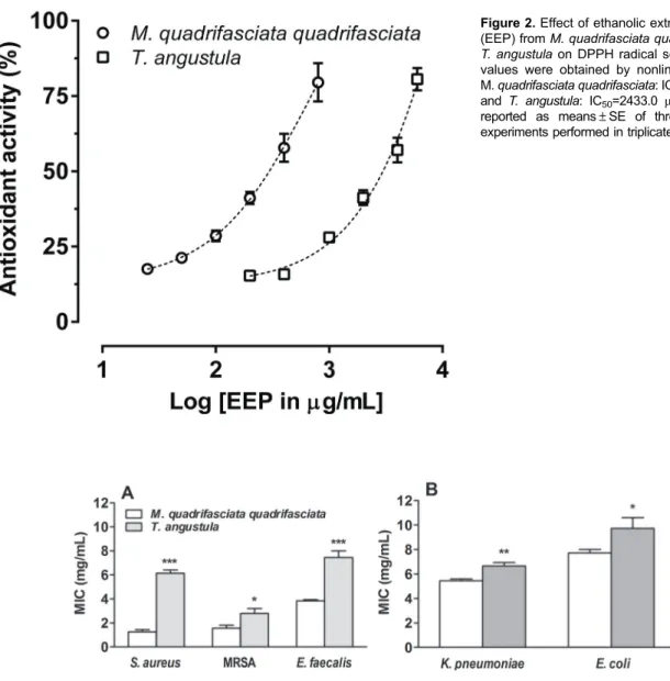
The results reported in Figure 2 show that both EEPs had dose-dependent antioxidant activity. Moreover, the EEP from M. quadrifasciata quadrifasciata [IC50= 241.8 (203.1 to 287.7)mg/mL] was ten-fold more potent than the EEP fromT. angustula[IC50= 2433.0 (2086.0 to 2838.0)mg/mL)].
Determination of MIC
The MIC values of the extracts for gram-positive and gram-negative bacteria are shown in Figure 3A and 3B. Gram-positive bacteria (E. faecalis,S. aureusand MRSA) were more sensitive than gram-negative bacteria (E. coli
and K. pneumoniae) to both EEPs. In addition, the EEP from M. quadrifasciata quadrifasciata was more potent and efficacious than the EEP ofT. angustulashowing the lowest MIC values for all tested bacteria.
Growth curve
Considering the promising results in the MIC assay, we decided to investigate the effect of the EEP from M. quadrifasciata quadrifasciataon the growth ofS. aureus (ATCC 25923) andE. coli(ATCC 25922) over time. Figure 4 shows that the inhibitory effect of M. quadrifasciata quadrifasciata (1 MIC) EEP on S. aureus growth was Figure 2.Effect of ethanolic extracts of propolis (EEP) fromM. quadrifasciata quadrifasciataand T. angustulaon DPPH radical scavenging. IC50 values were obtained by nonlinear regression; M.quadrifasciata quadrifasciata: IC50=241.8mg/mL and T. angustula: IC50=2433.0 mg/mL. Data are reported as means±SE of three independent experiments performed in triplicate.
Figure 3.Susceptibility of bacterial strains to ethanolic extracts of propolis in minimal inhibitory concentration (MIC).A, Gram-positive bacteria:S. aureus(ATCC 25923), methicillin-resistantS. aureus(MRSA, clinical isolate), andE. faecalis(ATCC 29212);B, Gram-negative bacteria:K. pneumoniae(ATCC 23883) andE. coli(ATCC 25922). Data are reported as means±SE of 3–5 independent
time-dependent and occurred in about 6 h. On the other hand, the inhibitory effect of the extract on the growth of E. colitook 12 h to occur.
Integrity of cell membrane
The results presented in Table 2 show that the EEP from M. quadrifasciata quadrifasciata (1 MIC) increased 6.6- and 5.6-fold the leakage of cell constituents ofS. aureus and E. coli, respectively, suggesting that it causes an irreversible damage of the bacterial cell membrane, leading to cell death.
Discussion
The current study revealed that EEPs fromM. quadri-fasciata quadriquadri-fasciataandT. angustulahad antimicrobial activity against gram-positive and gram-negative bacteria and antioxidant activity. The chemical analysis of the EEPs revealed the presence of terpenoids, flavonoids, and polyphenols, which were more abundant in the EEP fromM. quadrifasciata quadrifasciata(Figure 1).
The more prominent effect of EEPs against gram-positive than against gram-negative bacteria, as assessed
by the MIC assay, agrees with previous studies that have shown that propolis from stingless bees (7,9,23) and fromApis mellifera(6) has antimicrobial activity, particu-larly against gram-positive bacteria. In this regard, the currently reported activity against MRSA is particularly interesting due to the present scenario of recrudescence of resistant S. aureusstrains (12). The MICs estimated in the current study for gram-positive bacteria are similar to the estimated MICs of EEPs from other stingless bees, around 2–3 mg/mL for S. aureus (9,23),
includ-ing MRSA. Previous studies that have used the same experimental protocol of MIC determination used in our study could not determine a MIC for EEP against gram-negative bacteria (9). However, we found MIC values for EEP against gram-negative bacteria between 5 and 7 mg/mL in our samples, also indicating some activity of EEP fromM. quadrifasciata quadrifasciataandT. angustula against gram-negative bacteria. Considering the esti-mated MICs in our assays, the EEP from M. quad-rifasciata quadquad-rifasciatawas more potent than the EEP from T. angustula as an antimicrobial agent. Although the EEPs showed important antimicrobial activity for all tested strains, MICs values obtained (2 to 7 mg/mL) can Figure 4.Effect of ethanolic extracts of propolis fromM. quadrifasciata quadrifasciataon growth curve assay. Absorbance readings for assays withS. aureus(A) andE. coli(B). Data are reported as means±SE of three independent experiments performed in triplicate. *Po0.05 compared to respective control (t-test).
Table 2. Effect of ethanolic extracts of propolis (EEP) from M. quadrifasciata quadrifasciataon cell constituents release ofS. aureusandE. coliafter 4 h.
Bacterial Strains Cell Constituents Release (OD260 nm)
Control EEP (1 x MIC)
Relative Release (EEP/Control)
S. aureus 0.041±0.004 0.260±0.033* 6.61±1.211 E. coli 0.149±0.017 0.811±0.054** 5.65±0.585
be considered high, conferring a reasonable antimicrobial activity.
Due to its better antimicrobial activity, the EEP from M. quadrifasciata quadrifasciatawas chosen for additional tests: growth curve, release of cell constituents, and mass spectrometry experiments. To analyze the effect of the EEP fromM. quadrifasciata quadrifasciataagainstS. aureus andE. coliover time, a growth curve assay was performed in the absence or presence of the EEP (1 MIC).S. aureus was more susceptible to EEP thanE. colialso in this assay. Accordingly, while a significant growth reduction was found at 6 hours forS. aureus, 12 hours were necessary to show a significant growth reduction forE. coli, compared to their respective controls (0 MIC).
Although some authors attribute the bacteriostatic and bactericidal activity of propolis to the inhibition of protein synthesis and prevention of cell division (24), its nature and complexity complicate the identification of a mechan-ism of action. In this study, we performed a cell constituent release assay to investigate a possible mechanism of action for EEP, i.e. disruption of the cell membrane, which would cause the release of large molecules to the medium. The assay revealed a significant release of intracellular constituents of S. aureus and E. coli to the incubation medium in the presence of the EEP fromM. quadrifasciata quadrifasciata(Table 2), supporting that it causes cell lysis. Aiming to further elucidate the composition of EEP from M. quadrifasciata quadrifasciata, an UPLC coupled with mass spectrometry assay was carried out. The assay showed 26 diterpene skeletons as major components and, based on the literature, it was possible to suggest 17 structures. Among these, the following compounds are particularly relevant: one of elemental composition C20H30O2, which may be a hinokiol or totarol deriva-tive, isocupressic acid, and artepillin C methyl ester. The presence of totarol and possibly a derivative is consistent with our antibacterial findings. Totarol is a highly hydro-phobic diterpenoid with a high phospholipid/water partition coefficient, capable of interfering with the structural integ-rity of the membrane of bacteria and causing cell lysis (25). In addition, it decreases the expression of penicillin bind-ing protein 2a, a protein involved in penicillin resistance of MRSA (26). Recent evidence supports that totarol inhibits hemolytic proteins and enterotoxins secreted byS. aureus (27) and has potential application in clinical therapy and food decay prevention. In line with this view, hinokiol, also an identified component of EEP from M. quadrifasciata quadrifasciata, has been described as having antimicro-bial, antitumoral, antioxidant and anti-inflammatory activity (28,29). Therefore, hinokiol may also be involved in the antimicrobial action of EEP fromM. quadrifasciata quadri-fasciata. In addition, isocupressic acid, also a component of propolis, has antimicrobial activity (30) and may play a role in the antibiotic effect of EEP fromM. quadrifasciata quadrifasciata. The UPLC-MS also revealed the pres-ence of artepillin C in the EEP from M. quadrifasciata
quadrifasciata. Artepillin C has been pointed out as the possible active component responsible for the antimicro-bial and antioxidant activity of green propolis (31), similarly to totarol, interacting with cell membrane and creating point defects in its structure (32). Therefore, one might consider that artepillin C is involved in the current antimicrobial effect of EEP fromM. quadrifasciata quadrifasciata.
It is well known that propolis from different bee species contain significant amount of antioxidants (5). Therefore, we decided to comparatively assess the antioxidant acti-vity and total content of phenols andflavonoids in the EEPs from M. quadrifasciata quadrifasciata and T. angustula. The EEP fromM. quadrifasciata quadrifasciatapresented higher antioxidant activity than the EEP formT. angustula in the DPPH assay (IC50=241.8 and 2433.0mg/mL, respec-tively). Interestingly, Bonamigo et al. (33) also demon-strated that ethanol extracts of propolis obtained from the stingless beesM. quadrifasciata anthidioideshad a higher antioxidant capacity in the DPPH (IC50=60.9mg/mL) and ABTS (IC50=13.4 mg/mL) assay compared to Scapto-trigona depilis. Considering the antioxidant profile of the propolis extract obtained from the M. quadrifasciata anthidioides and M. quadrifasciata quadrifasciata in the DPPH test, we can observe that the M. quadrifasciata anthidioides was about 3.9-fold more potent than the M. quadrifasciata quadrifasciata. Based on the above results, we can also suggest that the antioxidant activity present in propolis seemed to depend on the genus and species of bees, considering that the potency and efficacy of the propolis obtained from the bees belonging to the Melipona genus (M. quadrifasciata anthidioidesandM. quadrifasciata quadrifasciata) were higher than Tetragonisca (T. angustula) and Scaptotrigona (S. depilis), respectively.
The differences in the chemical composition of prop-olis extracts in the same region may be related to species of bees and the preference for a particular plant species to elaborate the propolis (2,33). Moreover, the genetic vari-ability of bee species influences the chemical composition of propolis, resulting in different biological activities (2). Accordingly, the EEP from M. quadrifasciata quadrifas-ciata presented a higher concentration of total phenols andflavonoids, reinforcing the direct correlation between phenol concentration and antioxidant activity established in the literature (34).
Acknowledgments
We thank Luis Celso Stafaniak for providing the propolis samples and his dedication in divulgating the importance of preserving the various species of stingless
bees, and Dr. Rosmari Horner for providing the clinical isolate strain of MRSA. This research was supported by Conselho Nacional de Desenvolvimento Científico e Tecnológico (CNPq)/Brazil. ARSS, MM, and CFM are recipients of CNPq productivity fellowships.
References
1. Brasil, Ministério da Agricultura. Instruc¸ão Normativa n°3– ANEXO VI–Regulamento técnico para afixac¸ão de
identi-dade e qualiidenti-dade de própolis.Diário Oficial da República Federativa do Brasil. Brasilia, Jan 19, 2001. http://extranet. agricultura.gov.br/sislegis-consulta/consultarLegislacao.do? operacao=visualizar&id=1798.
2. Bankova V, Popova M, Trusheva B. Propolis volatile compounds: chemical diversity and biological activity: a review.Chem Cent J2014; 8: 28, doi: 10.1186/1752-153X-8-28.
3. Miguel MG, Nunes S, Dandlen SA, Cavaco AM, Antunes MD. Phenols and antioxidant activity of hydro-alcoholic extracts of propolis from Algarve, South of Portugal.Food Chem Toxicol2010; 48: 3418–3423, doi: 10.1016/j.fct.2010. 09.014.
4. Tomazzoli MM, Neto RDP, Moresco R, Westphal L, Zeggio ARS, Specht L, et al. Discrimination of Brazilian propolis according to the seasoning using chemometrics and machine learning based on UV-Vis scanning data.J Integr Bioinform 2015; 12: 279–291, doi: 10.1515/jib-2015-279.
5. Franchin M, da Cunha MG, Denny C, Napimoga MH, Cunha TM, Koo H, et al. Geopropolis from Melipona scutellaris decreases the mechanical inflammatory hypernociception by inhibiting the production of IL-1beta and TNF-alpha.J Ethno-pharmacol 2012; 143: 709–715, doi: 10.1016/j.jep.2012.
07.040.
6. Silva JC, Rodrigues S, Feas X, Estevinho LM. Antimicrobial activity, phenolic profile and role in the inflammation of propolis. Food Chem Toxicol 2012; 50: 1790–1795, doi:
10.1016/j.fct.2012.02.097.
7. Liberio SA, Pereira AL, Dutra RP, Reis AS, Araujo MJ, Mattar NS, et al. Antimicrobial activity against oral patho-gens and immunomodulatory effects and toxicity of geopro-polis produced by the stingless bee Melipona fasciculata Smith. BMC Complement Altern Med2011; 11: 108, doi: 10.1186/1472-6882-11-108.
8. Sforcin JM. Propolis and the immune system: a review. J Ethnopharmacol2007; 113: 1–14, doi: 10.1016/j.jep.2007. 05.012.
9. Campos JF, dos Santos UP, Macorini LF, de Melo AM, Balestieri JB, Paredes-Gamero EJ, et al. Antimicrobial, antioxidant and cytotoxic activities of propolis from Melipona orbignyi (Hymenoptera, Apidae).Food Chem Toxicol2014; 65: 374–380, doi: 10.1016/j.fct.2014.01.008.
10. Choudhari MK, Punekar SA, Ranade RV, Paknikar KM. Antimicrobial activity of stingless bee (Trigona sp.) propolis used in the folk medicine of Western Maharashtra, India. J Ethnopharmacol2012; 141: 363–367, doi: 10.1016/j.jep.
2012.02.047.
11. Wojtyczka RD, Dziedzic A, Idzik D, Kepa M, Kubina R, Kabala-Dzik A, et al. Susceptibility of Staphylococcus aureus clinical isolates to propolis extract alone or in combination
with antimmicrobial drugs.Molecules2013; 18: 9623–9640, doi: 10.3390/molecules18089623.
12. Smith R, Coast J. The true cost of antimicrobial resistance. BMJ2013; 46: 1493, doi: 10.1136/bmj.f1493.
13. Velikova M, Bankova V, Marcucci MC, Tsvetkova I, Kujumgiev A. Chemical composition and biological activity of propolis from Brazilian meliponinae.Z Naturforsch C2000; 55: 785–789.
14. Nogueira-Neto P.Vida e criac¸ão de abelhas indı´genas sem ferrão. 1stedn. São Paulo, Editora Nogueirapis, 1997. 15. Park YK, Ikegaki M, Abreu JAS, Alcici NMF. Estudo da
preparac¸ão dos extratos de própolis e suas aplicac¸ões. Food Sci Technol1998; 18: 313–318, doi: 10.1590/S0101-20611998000300011.
16. Frozza CO, Garcia CS, Gambato G, de Souza MD, Salvador M, Moura S, et al. Chemical characterization, antioxidant and cytotoxic activities of Brazilian red propolis.Food Chem Toxicol2013; 52: 137–142, doi: 10.1016/j.fct.2012.11.013. 17. CLSI. Methods for dilution antimicrobial susceptibility tests
for bacteria that grow aerobically; Approved Standard – Ninth Edition.CLSI document M07-A9. WaynePA: Clinical and Laboratory Standards Institute; 2012.
18. Bueno-Silva B, Alencar SM, Koo H, Ikegaki M, Silva GV, Napimoga MH, et al. Anti-inflammatory and antimicrobial evaluation of neovestitol and vestitol isolated from Brazilian red propolis. J Agric Food Chem2013; 61: 4546–4550,
doi: 10.1021/jf305468f.
19. Diao WR, Hu QP, Zhang H, Xu JG. Chemical composition, antibacterial activity and mechanism of action of essential oil from seeds of fennel (Foeniculum vulgare Mill.).Food Cont. 2014; 35: 109–116, doi: 10.1016/j.foodcont.2013.06.056.
20. Banskota AH, Tezuka Y, Prasain JK, Matsushige K, Saiki I, Kadota S. Chemical constituents of Brazilian propolis and their cytotoxic activities.J Nat Prod1998; 61: 896–900, doi: 10.1021/np980028c.
21. Enzell CR, Wahlberg I. Mass spectrometric studies of diter-penes. 7. Aromatic diterditer-penes.Acta Chem Scand1970; 24: 2498–2510, doi: 10.3891/acta.chem.scand.24-2498. 22. Wang YZ, Tang CP, Ke CQ, Weiss HC, Gesing ER, Ye Y.
Diterpenoids from the pericarp of Platycladus orientalis. Phytochemistry2008; 69: 518–526, doi: 10.1016/j.phytochem.
2007.07.023.
23. Miorin PL, Levy Junior NC, Custodio AR, Bretz WA, Marcucci MC. Antibacterial activity of honey and propolis from Apis mellifera and Tetragonisca angustula against Staphylococcus aureus. J Appl Microbiol 2003; 95: 913–
920, doi: 10.1046/j.1365-2672.2003.02050.x.
25. Micol V, Mateo CR, Shapiro S, Aranda FJ, Villalain J. Effects of (+)-totarol, a diterpenoid antibacterial agent, on phos-pholipid model membranes. Biochim Biophys Acta 2001; 511: 281–290, doi: 10.1016/S0005-2736(01)00284-X.
26. Nicolson K, Evans G, O’Toole PW. Potentiation of methicillin activity against methicillin-resistant Staphylococcus aureus by diterpenes.FEMS Microbiol Lett 1999; 179: 233–239, doi: 10.1111/j.1574-6968.1999.tb08733.x.
27. Shi C, Zhao X, Li W, Meng R, Liu Z, Liu M, et al. Inhibitory effect of totarol on exotoxin proteins hemolysin and entero-toxins secreted by Staphylococcus aureus.World J Microbiol Biotechnol2015; 31: 1565–1573, doi:
10.1007/s11274-015-1905-3.
28. Starks CM, Norman VL, Willians RB, Goering MG, Rice SM, O’Neil-Johson M., et al. Antibacterial activity of Taxodium ascendens diterpenes against methicillin-resistant Staphy-lococcus aureus.Nat Prod Commun2014; 9: 1129-1130. 29. Wang SY, Wu JH, Shyur LF, Kuo YH, Chang ST. Antioxidant
Activity of Abietane-Type Diterpenes from Heartwood of Taiwania cryptomerioides Hayata.Holzforschung2002; 56: 487–492, doi: 10.1515/HF.2002.075.
30. Bankova V, Marcucci MC, Simova S, Nikolova N, Kujumgiev A, Popov S. Antibacterial diterpenic acids from Brazilian propolis.Z Naturforsch C1996; 51: 277–280.
31. Veiga RS, Mendonc¸a S, Mendes PB, Paulino N, Mimica NJ, Neto AA, et al. Artepillin C and phenolic compounds respon-sible for antimicrobial and antioxidant acticity of green propolis and Baccharis dracunculifolia DC.J Appl Microbiol 2017; 122: 911–920, doi: 10.1111/jam.13400.
32. Pazin WM, Olivier DD, Vilanova N, Ramos AP, Voets IK, Soares AE, et al. Interaction of Artepillin C with model
membranes.Eur Biophys J2016; 46: 383–393, doi: 10.1007/
s00249-016-1183-5.
33. Bonamigo T, Campos JQ, Alfredo TM, Balestieri JBP, Cardoso CAL, Paredes-Gamero EJ, et al. Antioxidant, cytotoxic, and toxic activities of propolis from two native bess in Brazil: Scaptotrigona depilis and Melipona quad-rifasciataanthiodioides.Oxid Med Cell Longev2017; 2017: 1038153, doi: 10.1155/2017/1038153.
34. Duthie GG, Gardner PT, Kyle JA. Plant polyphenols: are they the new magic bullet?Proc Nutr Soc2003; 62: 599–603, doi: 10.1079/PNS2003275.
35. Yang X, Zhang YC, Zhang H, Dong AJ, Zhao HT, et al. Diterpenoid acids fromPinus koraiensis.Chem Nat Compd 2010; 46: 227–229, doi: 10.1007/s10600-010-9575-8.
36. Popova MP, Chimou IB, Marekov IN, Bankova VS. Terpenes with antimicrobial activity fromCretan propolis. Phytochem-istry2009; 70: 1262–1271, doi: 10.1016/j.phytochem.2009.
07.025.
37. Zhang L-C, Wu X-De, He J, Li Y, Zhang R-P, Zhao Q-S. Three new abietane diterpenoids fromPodocarpus fleury. Phytochem Lett 2013; 6: 364–367, doi: 10.1016/j.phytol.
2013.04.003.
38. Sy LK, Brown GD. Abietane diterpenes from Illicium angustisepalum.J Nat Prod1998; 61: 907–912.
39. Duke CC, Tran VH, Duke RK. Prenylated hydroxystilbenes. Patent No. WO2012/149608A1. The University of Sidney; 2012.