Artigo
*e-mail: aflach@gmail.com
CHEMICAL COMPOSITIONS AND ANTIOXIDANT AND ANTIMICROBIAL ACTIVITIES OF PROPOLIS PRODUCED BY Frieseomelitta longipes AND Apis mellifera BEES
Edineide Cristina A. de Souzaa, Etyene Janyne G. da Silvaa, Hayron Kalil C. Cordeirob, Nauara M. Lage Filhob, Felipe
M. A. da Silvac, Diany Lucy S. dos Reisd, Carla Portod, Eduardo J. Pilaud, Luiz Antonio M. A. da Costaa, Afonso D. L. de
Souzac, Cristiano Menezese and Adriana Flacha,*¤
aDepartamento de Química, Universidade Federal de Roraima, 69310-000, Boa Vista – RR, Brasil bUniversidade Federal Rural da Amazônia, 66075-110 Belém – PA, Brasil
cDepartamento de Química, Universidade Federal da Amazônia, 69077-000, Manaus – AM, Brasil dDepartamento de Química, Universidade Estadual de Maringá, 87020-900, Maringá – PR, Brasil eEmbrapa Amazônia Oriental, 66095-903 Belém – PA, Brasil
Recebido em 24/08/2017, aceito em 15/01/2018, web 28/02/2018
In this study, we investigated the chemical compositions and antioxidant and antimicrobial activities of propolis produced by the stingless bee Frieseomelitta longipes and the honeybee Apis mellifera collected from colonies in North Brazil. In terms of volatile composition, both mono- and sesquiterpenes were detected in the propolis of F. longipes while only sesquiterpenes were detected in that of A. mellifera. Out of 50 volatiles identified in all samples, 26 were found exclusively in F. longipes propolis and 8 were found exclusively in A. mellifera propolis. The chemical profiles of the propolis extracts were determined by atmospheric pressure chemical ionization-mass spectrometry and liquid chromatography-electrospray ionization-tandem mass spectrometry allowed to identify several prenylated benzophenones. A. mellifera extracts exhibited major antioxidant activity as assessed by the 2,2-diphenyl-1-picrylhydrazyl method and all extracts exhibited antioxidant activity as assessed by the β-carotene/linoleic acid method. The ethanolic extracts of the propolis showed promisor activity against all tested microorganisms.
Keywords: volatiles; phenolics; terpenes; stingless bee; honeybee.
INTRODUCTION
Since ancient times, propolis has been used as an alternative medicine owing to its biological action. Besides its antimicrobial activity, it has several therapeutic properties, such as antitumor, antioxidative, and anti-inflammatory activities.1 Propolis has become
popular as an alternative medicine and is a constituent of several cosmetics and health products. Many studies have been conducted to elucidate the chemical compositions and biological activities of propolis from different regions of the world.2,3
Propolis is a resinous material found in colonies of many eusocial bee species. It is derived from plant resins collected by foragers and used to close gaps in hives, trapping the bodies of small invaders. It works as a physical barrier against natural enemies and also as a biochemical weapon against pathogenic or opportunistic microbes. Recent findings have also revealed its important role in colony immunity.4
The propolis used worldwide is harvested from the same bee species, Apis mellifera, also known as honeybees. Although from the same bee, the product is not standard because its composition can differ depending on the plants of the region where, and the season when, it is produced. In Brazil, for example, there are two well-known types of propolis: green propolis, produced in Southeast and Central Brazil from Baccharis dracunculifolia, a plant of the Asteraceae family, and red propolis, produced in the littoral regions of Northeast Brazil from Dalbergia ecastaphyllum, a plant of the Leguminosae family.2,5,6
The chemical composition of propolis can also differ depending on the bee species. Several other eusocial bees can be managed in hives, such as those of the Meliponini tribe (also known as stingless bees), which produce pure propolis with great potential for commercial use.7
Stingless bees occur in all tropical and subtropical areas of the globe and
consist of more than 500 different species. Unlike honeybees, stingless bees mix propolis with wax to produce a building material, known as cerumen, which is used to construct all the structures in their nests, such as brood combs, entrances, and food pots. Recent studies have found that propolis from stingless bees has interesting characteristics and sometimes stronger biological activities than propolis from honeybees.8-11 However, its commercial use remains limited because
little is known about its composition or whether its properties make propolis production from these species economically viable.
Propolis contains volatile and fixed compounds. The volatile fraction corresponds to what are also called essential oils and contains mono- and sesquiterpenes, as well as alkanes and various aromatic compounds.9,12,13 The fixed compounds in propolis comprise
triterpenoids, flavonoids, lignans, phenolic esters of caffeic and coumaric acids, diterpenes, and prenylated benzophenones, among others.2,3
In the present study, we aimed to investigate whether the propolis produced by different bee species in the same area and at the same time are different.
We hypothesized that each bee species would use different resin sources and have specific processing methods, therefore producing unique propolis. To test this hypothesis, we compared the chemical compositions of propolis produced by two bee species, Apis mellifera (Apidae: Apini) and Frieseomelitta longipes (Hymenoptera: Apidae: Meliponini), located in the same area of the Amazon region of Brazil over the same time period.
MATERIALS AND METHODS Propolis collection
Embrapa Eastern Amazon, in the city of Belém, Brazil. Three colonies of F. longipes and one colony of A. mellifera were used. The colonies of the stingless bees and A. mellifera were separated by 50 meters. The surrounding area is predominantly composed of native secondary forest of the Amazonian ecosystem, but there are also urban areas and experimental Embrapa fields close to the apiary. To stimulate the production of propolis, two 2-cm-wide louvers were made in the side of the hives between the cover and the colony nest and sealed with adhesive transparent tape. Propolis was collected 15 days after the introduction of the louvers. Three samples of propolis collected from F. longipes colonies (FL-1, FL-2, and FL-3) and one collected from the A. mellifera colony (AM) were sent to the Laboratory of Biotechnology and Fine Chemistry at the Federal University of Roraima for analysis.
Volatiles extraction
The propolis samples were chilled and powdered using a pestle and mortar. Portions of 15 to 20 g were subjected to hydrodistillation for 3 h in a modified Clevenger apparatus and the essential oil collected was dried over anhydrous sodium sulfate. The yield from the extraction was determined by the proportional relationship between the weight of the propolis samples and the weight of the obtained oil.
Volatile compound analysis by gas chromatography-mass spectrometry (GC-MS)
A Shimadzu gas chromatograph (model GC-2010) coupled to a mass spectrometer from the same manufacturer (model QP2010 Plus) was used for the analysis of volatile compounds. Separation was performed using a fused silica capillary column (RTX-5MS, 30 m × 0.25 mm × 0.25 µm). A 1 µL aliquot of a 15 mg mL-1 ethyl
acetate solution of the oil was injected into the chromatograph. The injector temperature was 220 °C, the interface temperature was 280 °C, and the column temperature was programmed to increase from 60 °C at 3 °C min-1 to 280 °C. Helium was used as the carrier gas
at a constant flow rate of 1.02 mL min-1. Mass spectra were acquired
in the m/z range 40–600 using electron ionization with an ionization power of 70 eV and the ion source at 260 °C.
Volatile constituent identification
The compositions of the essential oils were determined by comparing the values of their retention indexes with those obtained for a homologous series of n-alkanes (C7-C30) under the same conditions,
according to the method of Van den Dool and Kratz.14 Later, the
experimental mass spectra were verified by comparison with those in the Wiley 8 and FFNSC 1.2 digital libraries and with data from existing literature.15
Extract preparation
Powdered propolis samples (4.10–34.29 g) were subjected to extraction at 25ºC three times using 250 mL absolute ethanol (99.5%) over a period of 24 h. The extracts were combined, concentrated on a rotary evaporator, and stored in a desiccator at reduced pressure.
Chemical ionization at atmospheric pressure analysis
The mass spectra were acquired using an ion-trap type spectrometer (Thermo-LCQ fleet) equipped with an atmospheric pressure chemical ionization (APCI) source programmed to operate in positive and negative acquisition modes. The information was
recorded using acquisition continuum mode using LCQ Fleet Tune. The monitored track was m/z 100–1000. Stock solutions were diluted at 20 ppm in HPLC-grade methanol and applied by direct insertion from a 5-µL loop using a pump syringe as a transport vehicle. The operational conditions were: APCI(-): discharge voltage, 3 kV; vaporizer temp, 300 °C; IRS gas, 30 arbs; auxiliary gas, 10 arbs; sweep gas, 0 arb; capillary temp, 200 °C; capillary voltage, -40 V; tube lens, -115 V; syringe pump, 30 µL min-1.
Liquid chromatography-electrospray ionization tandem mass spectrometry (LC-ESI-MS/MS) analysis
Propolis extracts were reconstituted in 500 µL of methanol, and 50 µL of this extract was diluted in 450 µL of the same solvent and analyzed using by Ultra High Performance Liquid Chromatography (Shimadzu, Nexera X2) coupled to a quadrupole time-of-flight mass spectrometer (Bruker, Impact II UHR-QTOF) controlled by the Otof Control and Hystar software packages (Bruker Daltonics) and equipped with an electrospray source operating in negative ionization mode. The parameters used for ESI(-) mode were: capillary voltage, 3.0 KV; nebulizer gas pressure, 4.0 Bar; dry heater temperature 220 °C. Full-scan MS spectra (m/z 100–1300) were acquired, and the ions of interest were selected by auto MS/MS scan fragmentation. Chromatographic separation was performed with a gradient mixture of solvents A (H2O/0.1 % formic acid, v/v) and B (methanol) using a
C18 column (Waters Acquity UPLC® CSH™ 1.7 µm, 2.1 × 100 mm).
The column was maintained at 40 °C, the flow rate was 0.2 mL min-1,
and the sample injection volume was 2 µL. The separations ran for 20 min using the program: 0-1 min: 40% B, 1–7 min: 90% B, 7-13 min: 97% B, followed by an isocratic elution in the period from 13 to 16 min at 97% B, and finally 16-18 min 40% B , keeping 40% until 20 min to re-equilibrate the column prior to another run. Sodium formiate solution was injected as calibrant at the start of each chromatographic run.
2,2-Diphenyl-1-picryl-hydrazyl (DPPH) radical-scavenging activity analysis
The analyses were performed according to the methodology described by Mensor et al.16 with some modifications. For the
extracts of F. longipes propolis, a 1 mg mL-1 sample was used, and for A. mellifera propolis, a 0.1 mg mL-1 sample was used. Different
sized aliquots of these solutions were withdrawn (for F. longipes, 20, 30, 40, 50, 60, and 70 µL, and for A. mellifera, 25, 50, 75, 100, 125, and 150 µL), followed by addition of 1.5 mL DPPH (1 mmol L-1).
A control solution consisting of 1.5 mL of a solution of DPPH and methanol was also prepared. The blank was formed by the same aliquots of samples and methanol. The determinations were performed using a UV-Vis spectrophotometer (Shimadzu UV-mini model 1240). The percentage uptake of the DPPH radicals was used to calculate percentage antioxidant activity (AA%). IC50 values were determined
using linear regression analysis.
Antioxidant activity analysis using the β-carotene/linoleic acid system
For the oxidation of samples, we used the methodology described by Emmos et al.,17 whereby 50 µL of a β-carotene solution
(1 mg mL-1), 40 µL of linoleic acid, and 265 µL of Tween 80 were
mixed and solubilized in 1 mL of chloroform. The chloroform was removed with nitrogen (N2) before the residue was then dissolved
the mixture was a water bath at 40 °C. Oxidation was monitored in a spectrophotometer at 470 nm. A β-carotene/linoleic acid emulsion without antioxidant was used for control samples, and a 1 mg mL-1
solution of 2,6-tert-butyl-1-hydroxy-toluene (BHT) was used as a positive control. The antioxidant activity (AA%) was expressed as the percentage of inhibition compared with that of the control after 120 min.
Antimicrobial activity
The gram-positive bacteria Bacillus cereus INCQS 00003 (ATCC 11778) and Staphylococcus aureus INCQS 0057 (ATCC 43300), the gram-negative bacteria Pseudomonas aeruginosa INCQS 00025 (ATCC 15442) and Escherichia coli INCQS 00051 (ATCC 13863), and the yeasts Candida albicans (ATCC 90028) and Candida tropicalis (ATCC 28707) were utilized for antimicrobial tests. The strains were supplied by the Coleção de Microrganismos de Referência em Vigilância Sanitária (CMRVS, FIOCRUZ-INCQS, Rio de Janeiro, RJ, Brazil). The bacteria were cultivated in brain-heart infusion medium (BHI) at 36 ± 1 °C for 24 h and the yeasts were cultivated in Sabouraud agar at 36 ± 1 °C for 36 h.
The cultures were adjusted to 0.5 McFarland (1x10⁸) standard
turbidity scale equivalents (105 CFU mL-1), then a dilution (1:10) was
performed to achieve a dilution of 107 CFU mL-1. After the suspension
was inoculated into the broth, the final bacterial concentration of the test was 105 CFU mL-1, as recommended by CLSI (2012). The
minimum inhibitory concentration values (MICs) of the ethanolic extracts were determined with 96-well plates as per a method established by the Clinical and Laboratory Standards Institute (CLSI).18 The negative control was 80% ethanol and the positive
controls were ampicillin and fluconazole. The extract concentrations tested were 1000, 500, 250, 125, 62.5, 31.3, 15.6, and 7.8 µg mL-1
and 2,3,5-triphenyl tetrazolium chloride (TCC) was used to assess the viability of the microorganisms.
RESULTS AND DISCUSSION Volatiles
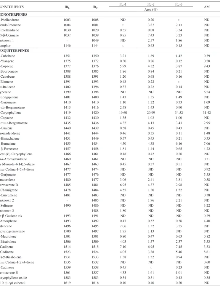
The relative abundances of essential oils in the F. longipes propolis varied between the colonies sampled. FL-1 had an essential oil percentage of 0.07%, while FL-2 and FL-3 had much higher values of 0.47 and 0.78%, respectively. In contrast, A. mellifera contains only 0.12% essential oils.
The essential oil constituents of propolis from F. longipes and A. mellifera are listed in Table 1. In the propolis of F. longipes, around 40 compounds were identified as mono- and sesquiterpenes. There is much less variety in the compounds found for in A. mellifera propolis, which comprises exclusively sesquiterpenes.
Six monoterpenes were identified in F. longipes. Sample FL-1 presented lower concentration of monoterpenes (1.40%) compared to those of FL-2 and FL-3 (14.58 and 10.74%, respectively). The most abundant monoterpene was (Z)-ocimene (7.43%, in FL-2).
Among the sesquiterpenes, β-caryophyllene presented the highest percentages in all the processed samples (19.6% in FL-1, 20.99% in FL-2, 34.52% in FL-3 and 31.42% in AM). The sesquiterpenes that presented contents of > 5% in A. mellifera were α-copaene (9.43%), cyperene (6.24%), α-humulene (7.06%), γ-gurjunene (5.55%), β-bisabolene (5.53%), and δ-cadinene (8.61%). Of these, cyperene and γ-gurjunene were not detected in F. longipes propolis. F. longipes propolis presented significant percentages of α-copaene (3.87-5.99%), α-humulene (4.38-6.16%), germacrene D (2.98-6.95%), δ-cadinene
(6.95-7.45%), and γ-cadinene (3.38–5.46%). Of these, germacrene D was not detected in A. mellifera propolis.
Patricio et al. identified components in the tibia of three Frieseomelitta species collected in different places.19 When
comparing the data obtained in their study to that obtained in the current study for the propolis of F. longipes, it is confirmed that no component is present in all the samples analyzed for this genus of bees, demonstrating that there is no uniformity in their compositions. α-phellandrene in F. longipes, F. silvestrii, and F. s. languida, and α-cubebene and α-copaene in F. longipes, F. silvestrii, and F. varia were detected. Comparing F. longipes and F. silvestrii, eight common components were identified: α−phellandrene, α-cubebene, α-copaene, β-caryophyllene, γ-muurolene, α-muurolene, γ-cadinene, and δ-cadinene. When F. longipes wascompared with F. s. languida, only one component was identified in both. In a comparison between F. longipes and F. varia, six common components were identified: α-cubebene, γ-yalangene, α-copaene, α-gurjunene, E- β-farnesene, and germacrene D.
There are far fewer studies on the volatile compounds from stingless bees compared to those on the volatile compounds from honey bees. Studies performed with the propolis of A. mellifera and Melipona beecheii obtained in Yucatan, Mexico and extracted with a Likens-Nickerson micro-apparatus identified similar compositions with some qualitative differences. Some compounds were only present in the propolis of A. mellifera and others in that of M. beecheii. The authors concluded that the behavior of these two bee species must be different.9
Leonhardt et al. showed that the resin from certain tree species did not attract bees, although these species produced large amounts of resin and were often located close to trees from which bees collected resin.20,21 Leonhardt et al showed that Borneo stingless bees use
olfactory cues to find trees from which to collect resins. Specifically, they use mono- and sesquiterpenes to localize and recognize the source of resins.22 In the current study, different components were
found between the propolis of F. longipes and A. mellifera, even though they are exposed to the same flora in the same period. Thus, they must have different preferences regarding the collection of resins.
Chemical profiles of the extracts by APCI-MS
Figure 1 shows profiles of the propolis extracts obtained by direct injection of samples from honeybees and stingless bees in negative ion mode.
The chemical profile of propolis from F. longipes showed greater variety of compounds compared to that of A. mellifera. However there were common ions in all samples, as is the case for m/z 109, 533, 601, and 669. FL-1 and FL-2 exhibit very similar profiles, although FL-3 presents the same ions. Ishida et al.,23 in studies
with propolis samples from A. mellifera of Amazonia, identified the polyprenylated benzophenones 7-epi-nemorosone (m/z 501) and xanthochymol (m/z 601) as the main constituents. These compounds have already been isolated from species of the family Clusiaceae.24, 25 The ion at m/z 669 has been previously identified in
Table 1. Volatile constituents of F. longipes and A. mellifera propolis
CONSTITUENTS IRL IRC
FL-1 FL-2 FL-3
AM Area (%)
MONOTERPENES
α-Phellandrene 1003 1008 ND 0.20 t ND
Pseudolimonene 1004 1001 t 3.87 2.13 ND
β-Phellandrene 1030 1020 0.55 0.08 3.34 ND
(Z)-β-Ocimene 1037 1039 0.85 7.43 3.23 ND
Unknown 1 - 1049 ND 2.57 1.86 ND
Camphor 1146 1144 t 0.43 0.15 ND
SESQUITERPENES
α-Cubebene 1351 1350 3.21 1.89 1.42 0.39
α-Ylangene 1375 1372 0.30 0.26 0.12 0.28
α-Copaene 1377 1376 5.99 4.32 3.87 9.43
β-Bourbonene 1388 1385 1.86 0.84 0.21 ND
β-Cubebene 1388 1391 1.20 0.68 0.16 ND
β-Elemene 1391 1393 0.48 0.22 t ND
Iso-Italicene 1402 1396 0.37 0.22 0.14 ND
Cyperene 1399 1398 ND ND ND 6.24
β-Longipinene 1401 1400 1.43 1.55 1.49 ND
α-Gurjunene 1410 1410 1.10 1.22 0.33 1.09
α-cis-Bergamotene 1413 1416 2.58 1.43 0.90 ND
β-Caryophyllene 1419 1420 19.60 20.99 34.52 31.42
β-Copaene 1432 1430 1.35 1.02 1.00 ND
β-trans-Bergamotene 1435 1436 4.32 4.13 3.43 2.95
α-Guaiene 1440 1439 0.58 0.45 0.43 ND
Aromadendrene 1441 1444 0.46 0.35 0.11 1.48
α-Himachalene 1451 1449 0.57 0.45 0.21 0.35
α-Humulene 1455 1454 4.50 4.38 6.16 7.06
E-β-Farnesene 1457 1458 1.81 1.44 0.65 4.22
9-epi-(E)-Caryophyllene 1466 1461 0.48 0.42 0.26 ND
allo-Aromadendrene 1460 1461 ND ND ND 0.51
cis Muurola-4(14),5-diene 1467 1463 0.45 0.37 0.51 ND
trans Cadina-1(6),4-diene 1477 1474 ND ND ND 0.92
γ−Gurjunene 1477 1476 ND ND ND 5.55
γ-muurolene 1480 1477 3.06 2.41 3.81 0.58
Germacrene D 1485 1481 6.95 4.37 2.98 ND
β-Chamigrene 1478 1484 4.55 1.30 1.52 ND
α-Curcumene 1481 1483 ND ND ND 0.38
Unknown 2 - 1485 ND 1.96 2.21 ND
β-Selinene 1490 1486 ND ND ND 3.22
Unknown 3 - 1488 1.80 ND ND ND
cis β-Guaiene cis 1493 1491 ND ND ND 0.29
γ-Amorphene 1493 1492 0.47 0.52 0.36 4.40
Valencene 1496 1495 2.06 1.52 3.25 ND
Bicyclogermacrene 1500 1497 1.75 1.13 ND ND
α-Muurolene 1501 1501 0.80 0.47 0.63 0.82
β-Bisabolene 1506 1509 4.03 1.57 2.37 5.53
γ-Cadinene 1514 1515 7.10 6.95 7.45 3.33
δ-Cadinene 1523 1524 4.05 3.38 5.46 8.61
(E)-γ-Bisabolene 1531 1533 1.38 1.52 0.94 ND
trans Cadina-1(2),4-diene 1535 1532 ND ND ND 0.60
α-Cadinene 1539 1538 0.45 t 0.23 ND
Germacrene B 1561 1557 4.33 1.61 1.01 ND
Caryophyllene oxide 1583 1583 0.54 0.51 0.43 0.35
1,10-di-epi-cubenol 1619 1616 0.40 0.40 0.20 ND
LC-ESI-MS/MS analysis
The LC-ESI-MS/MS analyses were performed in a negative ionization mode to obtain more information about the chemical compositions of the propolis displayed on APCI-MS fingerprint. Ions
with m/z 451, 501, 533, 601, and 669 were fragmented, and their fragmentation profiles were compared with data described in the literature.23-26 This evidence suggests the presence of benzophenones
derivatives in all propolis samples (Table 2).
In the sample FL-1, a peak with retention time (RT) 11.65 min
Figure 1. APCI-MS fingerprints of F. longipes and A. mellifera propolis extracts in negative ion mode
Table 2. Compounds detected in the propolis samples using LC-ESI-MS/MS in negative ion mode.
Peak Time (min)Retention Sample Putative Identification Molecular Weight Precursor Ion (m/z) Errorn (ppm) Fragment Ions
1 11.65 FL-1,FL-2, FL3, AM 7- epi-Nemorosone C33H42O4 501.3002 0.59
432.2296 [M-H-C5H9]-, 417.2064 [M-H-C5H9-CH3]-,
363.1586 [M-H-2(C5H9)]-, 327.1952 [M-H- C7H5O-C5H9]-, 309.1128 [M-H-C7H5O-C5H9-H2O]
-2 14.74 FL-1,FL-2, FL3, AM Xanthochymol C38H50O6 601.3520 0.49
465.3363 [M-H-C7O3H4]-, 449.1949 [M-H-C10H16O]-, 177.1040 [M-H-C10H16OC15H12O5]-,
109.0288 [C6O2H5]
-3 16.57 FL-1,FL-2, FL3, AM Guttiferone C or D C43H58O6 669.4147 0.29
533.3986 [M-H-2(C5H8)]- , 177.0185 [M-H-C10H16O-C15H12O5]-,
109.0295 [C6H5O]
-4 10.61 FL-1, FL-2, FL-3 Gambogenone C27H3206 451.2099 4.87
315.1945 [M-H-C7O3H4]-, 109.0279 [C6O2H2]
-5 11.37 FL-1, FL-2, FL-3, AM Aristophenone A C33H42O6 533.2898 3.37 464.2198 [M-H-C5H9]
-6 12.95 FL-1,FL-2, FL3, AM Polyprenylated benzophenone derivative C33H42O4 501.3003 *
363.1597 [M-H-2(C5H9)]-, 309.1127 [M-H-C7H5O-C5H9-H2O]
-7 13,16 FL-1,FL-2, FL3, AM
(1R,5R,7R)-3-Benzoyl-7-[(2E)- 3,7-dimethyl-2,6-octadien-1- yl]-4-hydroxy-8,8-dimethyl-5-(3-methyl-2-buten-1-yl)
bicyclo[3.3.1]non-3-ene-2,9-dione
C33H42O4 501.2998 0.59
433,2365 [M-H-C5H9] -336,1356 [M-H-C10H17-C2H5] -309.1126 [M-H-C7H5O-C5H9-H2O]
showed a deprotonated molecular ion at m/z 501.3002 [M-H]-, and a fragments at m/z 432.2296, m/z 417.2064, and m/z 363.1586 (Table 2), which is consistent with the presence of 7-epi-Nemorosone considering a 0.59 ppm mass error.23 This ion was common to all
samples from F. longipes and A. mellifera and was detected at similar retention times. In the samples FL-1, FL-2, FL-3, and AM, another benzophenones were detected by ESI-MS/MS. The signal at m/z 601.3520 (C38O6H49) corresponds to a deprotonated molecular
ion [M-H]-, with a similar retention time (14.74 min) and the same
fragmentation pattern in all four samples, producing daughter ions at m/z 465.3363, m/z 449.1949, m/z 177.1040, and m/z 109.0288, which is consistent with the xanthochymol molecule considering a mass error of 0.49 ppm (Table 2).23 The molecular ion [M-H]- at m/z
669.4147 (16.57 min) was tentatively identified as guttiferone C or D derivatives,26 corresponding to the mass of the xanthochymol plus
an isopentenyl unit (C5H8, 68 Da) showing the same fragmentation
pattern, within a 0.29 ppm mass error (Table 2).
Other probable benzophenone was detected in FL-1, FL-2 and FL-3 samples (RT 10.61 min) consistent with gambogenone, with deprotonated molecular ion at m/z 451.2093 [M-H]- (C27H32O6),
producing daughter ions at m/z 315.1945 and m/z 109.0279, with mass error of 4.87 ppm.23 The peak at retention time 11.37 min present in all samples was tentatively characterized as aristophenone A, which produced a deprotonated ion m/z 533.2898 [M-H]- (C33O6H41) and a fragment at m/z 464.2198 (loss of the prenyl
group), with mass error of 3.37 ppm.23 In addition, other probable polyprenylated benzophenones were are detected by LC-ESI-MS/ MS. These compounds showed a deprotonated molecular ion [M-H]- at m/z 501 (C33H42O4), and in all cases, the fragmentation
pattern showed ions at m/z 309, probably indicating the presence of a prenyl portion. The ion at m/z 501.3003 with RT 12.95 min (Table 2) yielded fragments at m/z 363.1587 and 309.1127. The compound at m/z 501.2996 with RT 13.16 min produced fragments at m/z 433,2365 [M-H-C5H9]-, 336,1356 [M-H-C10H17-C2H5]- e
309.1126 [M-H-C7H5O-C5H9-H2O]- (Table 2) was tentatively
characterized as (1R,5R,7R)-3-benzoyl-7-[(2E)-3,7-dimethyl-2,6-octadien-1-yl]-4-hydroxy-8,8-dimethyl-5-(3-methyl-2-buten-1-yl) bicyclo[3.3.1]non-3-ene-2,9-dione. The fragmentation pattern of both compound showed the loss of C5H9 group, indicating the presence
of a prenyl moiety. Polyprenylated benzophenones predominated in these samples making up a chemical profile very few reported for Brazilian propolis. The chromatograms of LC-ESI-MS/MS analysis and the mass spectra of the detected benzophenones are shown in Supplementary material (Figures 1S–8S).
Determination of the antioxidant activity of the extracts The antioxidant activities of the propolis extracts were evaluated by different methods. The results of the analyses are listed in Table 3. In the free radical DPPH sequestration method, the extract of
the propolis from A. mellifera presented high antiradical action, with values above those for the propolis of F. longipes. Using the β-carotene/linoleic acid oxidation method, both the propolis of A. mellifera and F. longipes exhibited strong protection capacity of a lipid substrate, presenting comparable percentages to that of the reference standard, BHT. Mendonça et al.26 in a study on red propolis of A. mellifera determined the IC50 using the radical DPPH
sequestration method for several extracts among which the ethanolic extract presented a value of 8.01 µg mL-1, similar to those found for
F. longipes. Campos et al.28 evaluated the biological activities of Melipona orbignyi propolis and obtained an IC50 of 40.0 µg mL-1, a
much greater value than those for the samples from F. longipes and A. mellifera. Propolis has been reported to be a source of compounds with phenolic functionalities. Consequently, its antioxidant activities have been intensely investigated because phenolic substances are recognized as potent antioxidants. The antioxidant activity of phenolic compounds is related to their structural arrangements, which promote the inactivation of free radicals through the donation of hydrogen atoms or in the complexation of metals, which occurs due to the presence of hydroxyl groups, conjugated double bonds, and carbonyl groups.29, 30
The results obtained in this study can be attributed to the presence of prenylated benzophenones, and the antioxidant activities of 7-epi-nemorosone, xanthochymol, gambogenone and aristophenone A identified in this study have already been determined.31,32 Trusheva et
al.32 have isolated several compounds with high antioxidant activities, among them prenylated benzophenones, from red propolis. The data obtained in the present study show the potential antioxidant properties of propolis by two separate methods used to measure antioxidant activity.
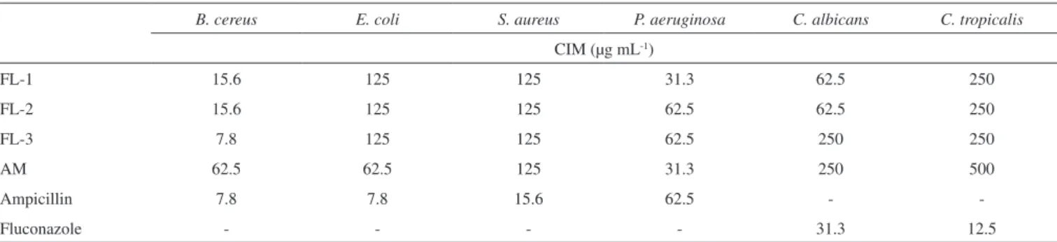
Antimicrobial activities of the extracts
Table 4 shows the results regarding the antimicrobial activities of ethanolic propolis extracts. All extracts are active against all microorganisms used.
There is currently no antimicrobial activity data for F. longipes propolis in the literature. The sample FL-3 exhibited activity against B. cereus (ATCC 11778) that is comparable to that of
Table 3. Antioxidant activities of F. longipes and A. mellifera propolis extracts (mean ± SD; n = 3).
Propolis DPPH IC50 (µg mL-1) β-carotene/linoleic acid (%)
FL-1 8.47±0.00040 75.6±0.7
FL-2 8.64±0.00001 75.1±1.1
FL-3 8.81±0.00002 73.1±1.1
AM 3.74±0.00020 75.5±1.1
BHT 6.00±0.00020 86.4±0.7
Table 4. Antimicrobial activities of F. longipes and A. mellifera propolis extracts.
B. cereus E. coli S. aureus P. aeruginosa C. albicans C. tropicalis
CIM (µg mL-1)
FL-1 15.6 125 125 31.3 62.5 250
FL-2 15.6 125 125 62.5 62.5 250
FL-3 7.8 125 125 62.5 250 250
AM 62.5 62.5 125 31.3 250 500
Ampicillin 7.8 7.8 15.6 62.5 -
ampicillin. A. mellifera (AM) showed the highest activity against E. coli (ATCC 13863) of all the samples. S. aureus (ATCC 43300) is resistant to methicillin and oxacillin but was inhibited by all the extracts tested. All extracts exhibited strong antimicrobial activity for all microorganisms and new studies must be carried out to determine active compounds.33
CONCLUSIONS
The study revealed that the volatile compositions of propolis were very different between the two different bee species examined, as F. longipes propolis contained both mono- and sesquiterpenes, whereas the propolis of A. mellifera contained only sesquiterpenes. The chemical profiles of the extracts showed a greater diversity of ionizable compounds in the samples of the stingless bee, and although the colonies sampled were found in the same environment, the individual samples had several constituents that were unique to a particular sample. The antioxidant and antimicrobial activities of the extracts were significant, and could be better explained upon the determination of the biological activities of isolated constituent compounds in future studies.
ACKNOWLEDGEMENT
The authors are grateful for the financial support provided by the Conselho Nacional de Desenvolvimento Científico e Tecnológico, Brazil (Proc. 472917/2011-0) and to the Coordenação de Aperfeiçoamento de Pessoal de Nível Superior for granting a scholarship (Edineide Cristina Alexandre de Souza) the and finantial suport (CAPES - PRÓ-AMAZÔNIA – Auxílio 3260/2013).
SUPPLEMENTARY MATERIAL
Figures 1S to 8S are available for free download at http:// quimicanova. sbq.org.br in pdf format.
REFERENCES
1. Viuda-Martos, M.; Ruiz-Navajas, Y.; Fernández-López, J; Pérez-Álvarez, J. A.; J Food Sci. 2008, 73, R117.
2. Miguel, M. G.; Antunes, M. D.; J Pharm Bioallied Sci. 2011, 3, 479. 3. Huang, S.; Zhang, C.; Wang, K.; Li, G.Q.; Hu, F. L.; Molecules. 2014,
19, 19610.
4. Simone-Finstrom, M.; Spivak, M.; Apidologie 2010, 41, 295.
5. Salatino, A.; Teixeira, E. W.; Negri, G.; Message, D.; Evid Based Complement Alternat Med. 2005, 2, 33.
6. Silva, B. B.; Rosalen, P. L.; Cury, J. A.; Ikegaki, M.; Souza, V. C.; Esteves, A.; Alencar S.M.; Evid Based Complement Alternat Med. 2008, 5, 313.
7. Cortopassi-Laurino, M.; Imperatriz-Fonseca, V. L.; Roubik, D. W.; Dollin, A.; Heard, T.; Aguilar, I.; Venturieri, G. C.; Eardley, C.; Nogueira-Neto, P.; Apidologie 2006, 37, 275.
8. Fernandes Jr., A.; Leomil, L.; Fernandes, A. A. H.; Sforcin, J. M.; J. of Venomous Anim. Toxins 2001, 7, 173.
9. Pino, J. A.; Marbot, R.; Delgado, A.; Zumárraga, C.; Sauri, E.; J. Essent. Oil Res. 2006, 18, 53.
10. Farnesi, A. P.; Aquino-Ferreira, R.; De Jong, D.; Bastos, J. K.; Soares, A. E.; Genet Mol Res. 2009, 8, 635.
11. Liberio, A. S.; Pereira, A. L. A.; Dutra, R. P.; Reis, A. S.; Araújo, M. J. A. M.; Mattar, N. S.; Silva, L. A.; Ribeiro, M. N. S.; Nascimento, F. R. F.; Guerra, R. N. M.; Monteiro-Neto, V.; BMC Complementary Altern. Med. 2011, 11, 108.
12. Bankova, V. S.; Christov, R.; Popov, S.; Marcucci, M. C.; Tsvetkova, I.; Kujumgiev, A.; Fitoterapia 1999, 70, 190.
13. Bankova, V.; Popova, M.; Trusheva, B.; Chem. Cent. J. 2014, 8, 28. 14. Van Den Dool, H.; Kratz, P. D. A.; J. Chromatogr. 1963, 2, 463. 15. Adams, R.P.; Identification of Essential Oil Components by Gas
chromatography/quadrupole Mass Spectroscopy. 4rd ed., Publisher: Allured Publishing Corporation, 2007, pp.15-804.
16. Mensor, L. L.; Menezes, F. S.; Leitão, G. G.; Reis, A. S.; Santos, T. C.; Coube, C. S.; Leitão, S. G.; Phytother. Res. 2001, 15, 127.
17. Emmons, C. L.; Peterson, D. M.; Paul, G. L.; J. Agric. Food Chem.
1999, 47, 4894.
18. Clinical and Laboratory Standards Institute. Methods for Dilution Antimicrobial Susceptibility Tests for Bacteria That Grow Aerobically; Approved Standard—Ninth Edition. CLSI document M07-A9. Wayne, PA: CLSI; 2012.
19. Patricio, E. F. L. R. A.; Cruz-López, L.; Maile, R.; Morgan, E. D.; Apidologie 2003, 34, 359
20. Leonhardt, S. D.; Bluthgen, N.; Biotropica 2009, 41, 730.
21. Leonhardt, S. D.; Schmitt, T.; Bluthgen, N.; PLoS One 2011; 6, 23445. 22. Leonhardt, S. D.; Zeilhofer, S.; Bluthgen, N.; Schmitt, T.; Chem. Senses.
2010, 35, 603.
23. Ishida, V. F. C.; Negri, G.; Salatino, A.; Bandeira, M. F. C. L.; Food Chem. 2011, 12, 966.
24. Chattopadhyay, S. K.; Kumar, S.; J Chromatogr. B. 2006, 844, 67. 25. Kumar, S.; Chattopadhyay, S. K.; Biomed. Chromatogr. 2007, 21, 139. 26. Mendonça, I. C. G.; Porto, I. C. C. M.; Nascimento, T. G.; Souza, N. S.;
Oliveira, J. M. S.; Arruda, R. E. S.; Mousinho, K. C.; dos Santos, A. F.: Basílio-Júnior, I. D.; Parolia, A.; Barreto F. S.; BMC Complement Altern. Med. 2015, 15, 357.
27. Tomás-Barberán, F. A.; Garcia-Viguera, C.; Vit-Olivier, P.; Ferreres, F.; Tomas-Lorente, F.; Phytochemistry 1993, 34, 191.
28. Campos, J.F.; Santos, U.P.; Rocha, P.S.; Damião, M.J.; Balestieri, J.B.P.; Cardoso, C.A.L.; Paredes-Gamero, E. J.; Estevinho, L. M.; Souza, K. de Picolo; dos Santos, E. L.; Evid Based Complement Alternat Med. 2015; Article ID 296186, 1.
29. Cook, N.; Samman, S. J. Nutr. Biochem. 1996, 7, 66.
30. Silva, J. F. M.; Souza, M. C.; Matta, S. R.; Andrade, M. R.; Vidal, F. V. N.; Food Chem. 2006, 99, 431.
31. Piccinelli, A. L.; Cuesta-Rubio, O.; Chica, M. B.; Mahmood, N.; Pagano, B.; Pavone, M.; Barone, V.; Rastrelli, L.; Tetrahedron 2005, 61, 8206.
32. Baggett, S.; Protiva, P.; Mazzola, E. P.; Yang, H.; Ressler, E. T.; Basile, M. J.; Weinstein, B.; Kennelly, E. J.; J. Nat. Prod. 2005,68, 354. 33. Duarte, M. C. T; Leme, E. E.; Delarmelina, C.; Soares, A. A.; Figueira,
G. M.; Sartoratto, A.; J. Ethnopharmal. 2007, 111, 197.