Multiple genes contribute to anhydrobiosis (tolerance to extreme desiccation)
in the nematode
Panagrolaimus superbus
Cláudia Carolina Silva Evangelista
1,2, Giovanna Vieira Guidelli
1, Gustavo Borges
1, Thais Fenz Araujo
1,
Tiago Alves Jorge de Souza
1,2, Ubiraci Pereira da Costa Neves
3, Alan Tunnacliffe
4and Tiago Campos
Pereira
1,21
Departamento de Biologia, Faculdade de Filosofia, Ciências e Letras de Ribeirão Preto, Universidade de
São Paulo (USP), Ribeirão Preto, SP, Brazil.
2Programa de Pós-Graduação em Genética, Faculdade de Medicina de Ribeirão Preto, Universidade de São
Paulo (USP), Ribeirão Preto, SP, Brazil.
3
Departamento de Física, Faculdade de Filosofia, Ciências e Letras de Ribeirão Preto, Universidade de São
Paulo (USP), Ribeirão Preto, SP, Brazil.
4Deptartment of Chemical Engineering and Biotechnology, University of Cambridge, Cambridge, UK.
Abstract
The molecular basis of anhydrobiosis, the state of suspended animation entered by some species during extreme desiccation, is still poorly understood despite a number of transcriptome and proteome studies. We therefore con-ducted functional screening by RNA interference (RNAi) for genes involved in anhydrobiosis in the holo-anhydrobiotic nematode Panagrolaimus superbus. A new method of survival analysis, based on staining, and proof-of-principle RNAi experiments confirmed a role for genes involved in oxidative stress tolerance, while a novel medium-scale RNAi workflow identified a further 40 anhydrobiosis-associated genes, including several involved in proteostasis, DNA repair and signal transduction pathways. This suggests that multiple genes contribute to anhydrobiosis inP. superbus.
Keywords: peroxiredoxin, kinase, RNAi, proteostasis.
Received: February 07, 2017; Accepted: July 20, 2017.
Introduction
A few species of bacteria, yeasts, plants and small in-vertebrates are capable of surviving extreme desiccation through a unique and outstanding strategy: anhydrobiosis. When facing severe drought, these species begin to dehy-drate and, instead of dying, they accumulate intrinsically disordered proteins (such as LEA and TDPs; Boothbyet al., 2017) and non-reducing disaccharides (such as treha-lose and sucrose), which promote the vitrification of the in-ternal cellular environment. This process results in a bioglass – an amorphous organic scaffold that completely arrests metabolism and preserves internal contents (Crowe et al., 1998). It is also proposed that such disaccharides act as `water-replacement molecules’, directly interacting with proteins and membranes and helping to maintain their na-tive structures (Sakuraiet al., 2008). However, it is possible
that other factors also contribute to the protection of the or-ganism. In this “dry state” (anhydrobiosis itself), the organ-ism is tolerant to several other physical stresses such as extremes of temperature and pressure, ultraviolet light and radiation (Tunnacliffe and Lapinski, 2003; Watanabeet al., 2006a,b).
In rotifers, anhydrobiosis arrests the “biological clock”, meaning that they do not age when in suspended an-imation. Therefore, the average lifespan is unaltered by desiccation, regardless of the time spent in the dry state (Ricci et al., 1987). However, once a dried rotifer is rehydrated, the same animal needs an interval of at least 24 h before being subjected to desiccation again to be able to survive (Schramm and Becker, 1987). Notably, variations in the life histories of different anhydrobiotic species may occur (Ricci and Caprioli, 1998).
Anhydrobiotic organisms are exposed to extreme wa-ter stress, which causes delewa-terious effects in the cell, in-cluding oxidative damage (Leprinceet al., 1994; Françaet al., 2007). Oxidative stress refers to a biological condition in which there is an imbalance in the concentrations of oxi-dant species and antioxioxi-dants (Sies, 2000). Organisms have
Send correspondence to Tiago Campos Pereira, Departmento de Biologia, Faculdade de Filosofia, Ciências e Letras de Ribeirão Preto, University of São Paulo (USP), Avenida Bandeirantes 3900, 14040-901 Ribeirão Preto, SP, Brazil. E-mail: tiagocampospereira@ffclrp.usp.br.
developed adaptive mechanisms for cellular detoxification, including systems that repair or prevent damage caused by oxidants (Michielset al., 1994). Among such defense sys-tems in the anhydrobiotic nematodePanagrolaimus super-busare: protein DJ-1 (Culletonet al., 2015), glutathione peroxidases (GP114) and peroxiredoxin (PER).
Although anhydrobiosis in animals was first de-scribed more than three centuries ago (Van Leeuwenhoek, 1702), its molecular basis is poorly understood. Transcriptome and proteome analyses in tardigrades (Milnesium tardigradum, Richtersius coronifer and Hypsibius dujardini), bdelloid rotifers (Adineta ricciae), nematodes (Aphelenchus avenae, Ditylenchus africanus, Plectus murrayi and Panagrolaimus superbus), insects (Polypedilum vanderplanki), algae and plants (Pyropia orbicularis, Myrothamnus flabellifolia and Boea hygrometrica) identified several genes and proteins that were up- or down-regulated by water loss (Adhikariet al., 2009; Haegemanet al., 2009; Maliet al., 2010; Schokraie et al., 2010, 2012; Boschettiet al., 2011; Tysonet al., 2012; Yamaguchiet al., 2012, Wang et al., 2014; López-Cris-toffaniniet al., 2015; Ma et al., 2015; Zhu et al., 2015; Ryabovaet al., 2017). Such studies are, by their nature, cor-relative, and do not provide evidence for a functional role of the genes or proteins concerned. In the model nematode, Caenorhabditis elegans, whose dauer larvae alone are des-iccation tolerant, a large number of mutants are available that can be used to study anhydrobiosis in this species (Erkutet al., 2011, 2013). However, in other nematodes, the lack of such mutants has prompted researchers to use RNA interference (RNAi) techniques instead (Reardonet al., 2010), an approach which is also possible inC. elegans (Galet al., 2004; Erkutet al., 2013).
Identification of anhydrobiosis-related genes is a cen-tral requirement in the development of anhydrobiotic engi-neering, which aims to confer desiccation tolerance on dehydration-sensitive biological samples (cells, tissues, or-gans) (Chenet al., 2009; García De Castroet al., 2000; Liet al., 2012). Successful anhydrobiotic engineering would have multiple applications in agriculture (e.g., by rendering plants tolerant to drought) and medicine (e.g., preservation in the dry state of organs for transplant).
In this study we examined anhydrobiosis inP. super-bus, a free-living nematode nearly 1 mm long that feeds on bacteria and was first described by Fuchs (1930). Members of the genusPanagrolaimusinhabit diverse niches, from the Antarctic, volcanic islands, temperate and semi-arid soils to terrestrial mosses (Shannonet al., 2005; McGillet al., 2015).P. superbusandC. elegansbelong to the same order (Rhabditida) and are anatomically similar. However, the former is dioecious while the latter is typically her-maphroditic and has a faster populational growth rate. We have focused onP. superbus, rather than C. elegans, be-cause the former nematode is: (i) holo-anhydrobiotic (Jöns-son, 2005) i.e., able to enter anhydrobiosis at any life stage,
(ii) robustly desiccation tolerant (Shannonet al., 2005) and (iii) does not demand extra/special laboratory procedures to obtain specific larval stages.
We first developed a new method for rapid and accu-rate assessment of survival to desiccation that could be used in a scalable screening procedure. To test this method and also to gain further information on the functional roles of glutathione peroxidase (a protein previously shown to be involved in anhydrobiosis; Reardonet al., 2010) and pero-xiredoxin (a biochemically related protein), we performed RNA interference by feeding on populations ofP. super-bus. We then developed a new medium-scale RNAi screen-ing protocol to screen a panel of 97 target genes previously shown to be regulated during extreme desiccation in other anhydrobiotic species and found that knockdown of 40 of these genes adversely affects desiccation tolerance inP. superbus.
Materials and Methods
Nematode maintenance
Panagrolaimus superbus(strain DF5050) used in this study was first isolated from Surtsey Island (Iceland) by Björn Sohlenius (Sohlenius, 1988; Shannonet al., 2005) and was maintained in incubators at 21 °C, in the dark, on NGM (Nematode Growth Medium) agar plates and fed with a layer ofEscherichia coli(strain OP50).
Evaluation of staining for the determination of survival percentages
P. superbusworms were collected from maintenance plates (NGM agar covered with a layer of OP50) by rinsing with 5 mL of M9 buffer, transferred to 50 mL test tubes and left for 10 min to precipitate. Then, the supernatant was dis-carded and worms were transferred to new 50 mL tubes containing 10 mL of M9 buffer. This washing procedure was done three times to reduce the amount of OP50 bacte-ria. Worms were then separated into two 0.2 mL vials. One sample was heated at 70 °C for 10 min in a thermocycler with subsequent decanting for 10 min. Heating at this tem-perature is lethal to worms, providing a positive control for staining. The second sample was kept at rest for 20 min at room temperature.
For each treatment, three plates were produced: i) a sample of live worms, ii) a sample of dead worms, and iii) a mixture of live and dead worms. These plates were analyzed via light microscopy and images were captured using the program ScopePhoto to verify whether stained worms (bluish by trypan blue, pink by erythrosin B) were active, thus revealing any false positives. Similarly, a total and permanent absence of movements in unstained (live worms), representing false negatives, was also considered. The whole procedure was also performed forC. elegans worms. Staining for only one hour was also tested. Three biological replicates were performed (N > 100 worms per group, per replicate).
RNAi by feeding - PER and GP114
Partial cDNAs corresponding to a glutathione pero-xidase (designated GP114, GenBank Accession Number GR881191) and peroxiredoxin (designated PER, GenBank Accession Number GR881190) in the L4440 vector (ampi-cillin resistance) were propagated inE. coliHT115 (Rear-don et al., 2010). These feeding strains, designated dsGP114 and dsPER, were grown in 50 mL tubes of liquid LB ampicillin (50mg/mL) under agitation (210 rpm, 37 °C) overnight. Subsequently, tubes were centrifuged for 10 min at 3,500 xg. Pellets were resuspended with 600mL of liquid LB ampicillin and inverted on petri dishes containing NGM agar and IPTG (1 mM). Plates were left for two days at room temperature to induce double-stranded RNA (dsRNA) expression by the bacteria.
Worms were then collected from OP50 and washed with M9, as previously described. Subsequently, a small amount of worms was transferred to either plates contain-ing bacteria expresscontain-ing dsRNA against GP114 or PER. They were left for nearly 15 days to ensure silencing of the entire population. HT115 bacteria containing a GFP gene cloned in the L4440 vector (L4440::GFP, referred to as “GFP”) was used as a negative control. Populations fed with L4440::GP114 bacteria are referred to as “dsGP114”, “GP114 knockdown” or “GP114-silenced”; similarly for PER, mutatis mutandis. The terms “GP114” and “PER” were used to refer to the corresponding genes/cDNAs. Three biological replicates were performed (N = 200 worms for each treatment, for each replicate).
Desiccation challenge
Worms were submitted to desiccation challenge ac-cording to Shannonet al.(2005). Briefly, silenced worms were immobilized on 0.45 mm Supor filter membranes (Sigma Aldrich) by vacuum filtration with a Sartorius fun-nel, placed in 1.5 mL test tubes and then subjected to the following conditions: 98% relative humidity (RH) for 24 h over a saturated solution of copper sulphate (unless other-wise stated); 10% RH for 24 h over dry silica gel and pre-hydration in 100% RH for 24 h in distilled water va-pour. Rehydration was achieved by adding 1.5 mL of M9
buffer to the samples. Survival percentage was measured by staining with erythrosin B. Three biological replicates were performed (N > 100 worms per group, per replicate).
Assessing the roles of PER and GP114 as antioxidants
PER- and GP114-silenced worms (in M9 buffer) were subjected to oxidative stress by adding hydrogen per-oxide (H2O2, Synth) to the following final concentrations: 0
mM (zero), 1mM, 10mM, 100mM, 1 mM, 10 mM, 20 mM and 40 mM. The final volume in all tubes was 100 mL. These values were selected according to previous studies onC. elegans(Larsen, 1993).
Samples were then homogenized by mild agitation and incubated at 20 °C for 24 h. After this period, the supernatant was removed and 1 mL of erythrosin B (0.4% w/v) was added and left for four hours. Worms were then washed three times with M9 buffer and survival percent-ages were determined (number of unstained worms/total number of worms). Three biological replicates were per-formed (N = 200 per group, per replicate).
Screening for anhydrobiosis-related genes inP. superbus
Selection of targets
A total of 97 potential targets were considered for the screening experiments. The first group comprised 33 kinase-related cDNAs, obtained from a mixed population of P. superbus and cloned in the pDNR-Lib vector (Clontech), kindly provided by Dr. Trevor Tyson (Van Andel Institute, USA). They correspond to all genes whose “target codes” end with a “K”, in Table 1. These targets were selected because signaling processes are likely to be very important for entry into anhydrobiosis. The second group (all other genes in Table 1) comprised 64 genes shown to be up-regulated during anhydrobiosis in other an-imal species (Adhikariet al., 2009; Haegemanet al., 2009; Maliet al., 2010; Schokraieet al., 2010, 2012; Cornetteet al., 2010; Boschettiet al., 2012; Tysonet al., 2012; Yama-guchiet al., 2012). These targets were selected by consider-ing the followconsider-ing aspects: (i) they should be induced in at least one species during anhydrobiosis and (ii) there should be homolog(s) within theP. superbusEST library (Tysonet al., 2012).
Production of long double-stranded RNAs (dsRNAs)
ex-tension step at 72 °C for 10 min. The resulting amplicons were precipitated with isopropanol, resuspended with ultra-pure water and submitted toin vitrotranscription (Yuet al., 2002) using TranscriptAid T7 High yield transcription kit; ThermoScientific, followed by DNase I treatment, accord-ing to the manufacturer’s instructions. dsRNAs were di-luted to 1mg/mL with ultrapure water and Tris HCl (pH 6.8) was added to a final concentration of 5 mM.
dsRNAs were also generated for 14 other genes from P. superbus(Table S1, target codes 1-4, 6-11, 15-17 and 21). Initially, their corresponding cDNAs were obtained by RT-PCR (using the same conditions described in section 2.6.5) and cloned into vector pCR2.1 TOPO (Invitrogen). These cloned sequences were then used as templates for a second round of PCR (performed as for pDNR-Lib, de-scribed above), but now using T7-gene specific primers (sequences on Table S1; targets 1 to 21). The resulting amplicons could be readily used forin vitrotranscription (as previously described), yielding dsRNAs.
Design of dicer substrates
Dicer substrates were designed for 50 targets (genes whose target codes start with “si”, on Table S2), using the freeware Strand Analysis (Pereira et al., 2007) and ex-tended three nucleotides at each end. These molecules are 27 RNA duplexes, with two nucleotide 3’ overhangs and phosphate groups at the 5’ ends. As a negative control, we designed a dicer substrate against GFP (accession number X83960). Dicer substrates were purchased from Sigma-Aldrich; their sequences are listed in Table S2.
RNA interference by soaking with siRNAs/dsRNAs
RNAi was triggered by soaking 200 - 600 worms (per biological replicate per target) for 24 h, in the dark, with long dsRNAs at a final concentration of 0.8mg/mL (soaking volume: 35mL) or dicer substrates (siRNAs) at final con-centration of 1mM (soaking volume: 100mL) and kept in the dark for 24 h without agitation at 21 °C. Three biologi-cal replicates were performed.
Confirmation of gene silencing by semi-quantitative RT-PCR
We selected a few representative targets to perform semi-quantitative RT-PCR to evaluate gene silencing. We also assessed a representative gene shown not to be in-volved in anhydrobiosis (si86 – whose silencing did not lead to decrease in survival) to show that a lack of decrease in survival after desiccation is not due to ineffective gene silencing.
Initially, total RNA from worms was extracted using the TRIzol reagent (Invitrogen) according to the manufac-turer’s guidelines. RNA samples were quantified by spectrophotometry and subsequently diluted in ultra pure water (RNase free) to yield a final concentration of 1
mg/mL. All RNA samples were pre-treated with DNase I
(Fermentas) following a modified version of the manufac-turer’s protocol: one unit of enzyme (1 h at 37 °C), followed by addition of another unit of enzyme (1 h at 37 °C). Re-verse transcription reactions (RT) were performed using ImProm-IITMkit (Promega) and random primers (500 ng) in a final volume of 20mL, according to the manufacturer. PCR was then performed using the GoTaqRDNA Polymer-ase kit (Promega) according to the manufacturer’s instruc-tions. PCR was performed using 2 mL of RT and 25 picomoles of each gene-specific primer (forward or re-verse) orb-actin (separate tubes) in a final volume of 50
mL.
All PCR reactions were performed under the follow-ing conditions: 94 °C for 5 min, followed by 33 cycles of 94 °C for 30 s, 57 °C for 30 s, 72 °C for 1 min, and a final ex-tension at 72 °C for 10 min. Amplifications curves were an-alyzed to guarantee that a plateau was not reached under these conditions for the tested genes. PCR products were resolved in 1% agarose gel stained with Sybr Safe (Invi-trogen). Band densitometry was done using IMAGE J soft-ware. Data normalization was done by dividing the value obtained for the silenced gene by the value found forb -actin for the corresponding sample. For each target, semi-quantitative RT-PCR was performed in technical tripli-cates, each one consisting of a pool of nearly 600 worms.
Nucleotide sequences of each primer are listed in Ta-ble S1. PCR conditions were the same for all genes ana-lyzed, except for target si86: 23 cycles.
Lethality assay
In order to determine whether the knockdown alone causes a decrease in viability, worms were soaked for 24 h with RNA duplexes for all 97 targets (or fed with dsPER/dsGP114) and survival percentages were deter-mined by staining. This procedure aims to guarantee that any decrease in survival percentage (compared to control group) is due to the disruption of the process of anhydrobiosis rather than an unrelated lethality. Three bio-logical replicates were performed (N = 200 per group, per replicate).
Statistical analyses
All experiments were performed in biological tripli-cates (or quadruplitripli-cates) and data are presented as mean values and standard deviations. Statistical analyses were performed using Student’s t-test, z-test or one-way ANOVA (with Tukey’s or Dunn’s post-hoc tests) or Mann-Whitney Rank Sum test with SigmaStat software. Statistical differences were considered significant whenp£
Results
Evaluation of staining for the determination of survival percentages
Both tested compounds (erythrosin B and trypan blue) are commonly used for staining cells, and were suc-cessfully used to indicate viability in whole worms (Figure S1 A-F). Heat-killed worms were strongly and completely stained, while live animals remained unstained even after four hours of soaking in dye solution. In a very few cases, faint local staining was observed in live nematodes, proba-bly indicating local tissue damage.
After desiccation challenge, all developmental stages (eggs, larvae and adult worms) were stained when dead. No false negatives were observed. However, staining patterns varied: in many cases we observed intense whole-body staining, but in some cases partial staining (of the anterior, middle or posterior body regions) were seen in moving worms (false positives). Light staining occurred in some larvae and adults, without movement, which we considered dead. Staining for four hours or one hour was equally effec-tive. It is possible that desiccation might make membranes leaky without killing worms (generating the few observed false positives). However, we believe that the observed “whole-body staining” reflects a degree of membrane dam-age that is incompatible with life, and thus, these worms can be considered dead. Additionally, such categorisation was applied to all groups (experimental and control), allow-ing an unbiased analysis.
Involvement of a peroxiredoxin gene inP. superbus
anhydrobiosis
Previously, a glutathione peroxidase gene (GP114) was shown by RNAi to be involved inP. superbus anhy-drobiosis (Reardonet al., 2010). To test whether the stain-ing method could demonstrate the effect of gene silencstain-ing on survival after desiccation, we first fed nematodes bacte-ria expressing GP114 dsRNA. In addition, we also used RNAi to examine the role of anotherP. superbus antioxi-dant gene, encoding a peroxiredoxin (PER), in anhydro-biosis.
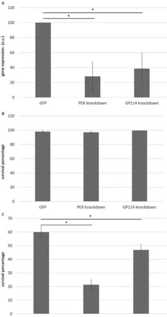
Gene silencing was confirmed by semi-quantitative RT-PCR, revealing an average reduction in mRNA levels of 71% for PER and 61% for GP114 transcripts, compared to a control group (Figure 1A). Prior to desiccation, gene si-lencing had no effect on nematode viability (Figure 1B), demonstrating that PER and GP114 are not essential genes under the tested conditions. After desiccation, all groups displayed increased mortality (Figure 1C), but PER and GP114 groups were significantly more sensitive than the GFP control. In particular, PER knockdown resulted in a 66% decrease in viability compared to the control. As well as confirming a role for glutathione peroxidase, these data also show for the first time the participation of a peroxire-doxin (PER) in nematode anhydrobiosis. An EST-based
study onP. superbusidentified two clusters for peroxire-doxin genes and three for glutathione peroxidase (Tysonet al., 2012). Therefore, it is possible that the lower survival percentage observed when silencing peroxiredoxin, pared to glutathione peroxidase, is due to the lower com-pensation capacity within the first protein family. These
Figure 1- Involvement of peroxiredoxin and glutathione peroxidase in anhydrobiosis inP. superbus. A) Molecular analysis by semi-quantitative RT-PCR revealed an average reduction of 71% of PER RNA transcripts and 61% of GP114 in worms subjected to RNAi by feeding compared to the control group corresponding to worms fed with bacteria expressing dsRNA against GFP gene, which is not associated with anhydrobiosis (*p
findings are consistent with oxidative stress being a signifi-cant component of the various stress vectors experienced by desiccating nematodes, as indicated for other organisms (Haegemanet al., 2009; Cornetteet al., 2010). This experi-ment also validates the staining method for the assessexperi-ment of survival of desiccation.
PER and GP114 act as antioxidants
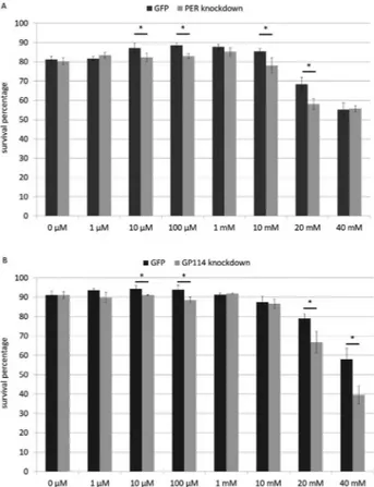
Knockdown of PER and GP114 had little or no effect on the morphology, development, fertility and behaviour of P. superbus(data not shown), as anticipated. However, we would expect gene silencing to compromise the ability of nematodes to combat oxidative stress, and we therefore tested this by exposing control and experimental groups to increasing concentrations of hydrogen peroxide for 24 h.
Slightly fluctuating responses (around the value ob-served for 0mM) are noted for both treatments (PER and GP114) up to 10 mM and statistically significant differ-ences (between control and respective experimental group) may be observed from 10mM. Curiously, although a de-crease in survival can be observed at 10 and 20 mM for PER, it is not seen at 40 mM, possibly due to compensation by other members of this gene family at higher concentra-tions (Figure 2A). On the other hand, GP114 also shows a
decrease between experimental group and respective con-trol at 20 mM, which is still present at 40 mM (Figure 2B), suggesting that a similar compensatory mechanism is not present, or is less effective. Taken together, these findings suggest that both PER and GP114 are involved in control-ling oxidative stress, a situation known to occur during de-hydration.
Evaluation of soaking as a means of triggering RNAi inP. superbus
We decided to determine whether immersing P. superbusin solutions containing long (>100 bp) and short (27 bp) RNA duplexes was also effective in promoting knockdown. We tested a few representative targets to show that, as judged by semi-quantitative RT-PCR, successful gene silencing was achieved by soaking with 27 bp RNA duplexes (known as dicer substrates) at 1mM for 24 h (Fig-ure 3 and Fig(Fig-ure S2). Soaking in solutions of RNA du-plexes resulted in gene silencing in a dose-dependent manner (up to 1mM), although nonspecific effects began to emerge at high concentrations (10mM, data not shown). Successful RNAi using dicer substrates was confirmed by its effect on target mRNA levels and by phenotypical anal-yses (knockdown ofifb-1andactingenes, data not shown).
Functional identification of anhydrobiosis-related
genes inP. superbus
We combined the use of the staining method for as-sessment of nematode viability with the soaking method for induction of RNAi in a medium-scale screening experi-ment to identifyP. superbusgenes associated with anhy-drobiosis. We selected a panel of 97 genes, from which 40 genes (Figure 4 and Table 1) showed reduced survival (20% on average) after knockdown and desiccation chal-lenge. This level of decrease has been previously observed
Figure 2- PER and GP114 enzymes combat oxidative stress. Statistically significant decreases in survival percentages were observed in both treat-ments (glutathione peroxidase- and peroxiredoxin-silenced worms) ex-posed to hydrogen peroxide. (* p<0.05; t-test between control group (GFP) and corresponding experimental group).
Figure 4- Identification of anhydrobiosis-related genes inP. superbus. All the 40 genes here indicated lead to statistically significant reductions >10% in survival percentage (z-test, p<0.05 compared to control group) after knockdown and desiccation.
Table 1- Functional identification of anhydrobiosis-related genes via RNAi. All the 40 genes whose knockdown lead to statistically significant decreases in survival percentage³10% compared to control group (i.e., worms soaked with RNA duplexes against GFP, normalized as 100% survival) are listed here in gray. Nine genes (marked with asterisks) presented statistically significant decreases lower than 10% (One-Way ANOVA). The remaining 48 tar-gets that did not lead to statistically significant reductions in the initial screenings and are listed in white.
Target Code Target identity
1K putative serine threonine-protein kinase (7e-25)
2K Cyclic AMP-dependent protein kinase
3K Casein kinase II regulatory subunit
4K Protein kinase
5K casein kinase I isoform gamma-1 (9e-19)
6K C2 domain containing protein (4e-62); CBR-FER-1 pro-tein (2e-46); myoferlin (8e-20)
7K TKL/LISK/TESK protein kinase (2e-64)
8K diacylglycerol kinase (5e-83)
9K PREDICTED: serine/threonine-protein kinase Nek6-like (1e-62)
10K CK1/WORM6 protein kinase (2e-141)
11K SH3-domain kinase binding protein
12K Serine/threonine-protein kinase
13K testis-specific serine threonine-protein kinase 2 (5e-58)
15K malonyl-acyl carrier protein (2e-22). ADP-specific phosphofructokinase/glucokinase conserved region fam-ily protein (5e-22)
16K serine threonine protein kinase-related domain containing protein (4e-49)
17K putative tyrosine-protein kinase kin-31 (6e-83); SH2 mo-tif and tyrosine protein kinase and protein of unknown function DUF595 domain containing protein (5e-81)
18K gastrulation defective protein 1 (9e-54); protein kinase domain containing protein (6e-50)
19K adenylate kinase 1 (6e-28)
20K serine threonine-protein kinase pelle (1e-58); CBR-PIK-1 protein (1e-57)
21K Er (fms/fps related) protein kinase
22K Cyclin-dependent kinases regulatory subunit
23K Guanylate kinase family protein
24K protein MAK-1, isoform c (7e-11)
Target Code Target identity
26K Protein kinase domain containing protein
27K CAMK/CAMKL/MELK protein kinase (2e-27)
28K serine threonine-protein kinase akt-1 (3e-90)
33K putative tyrosine-protein kinase kin-31 (1e-12)
34K protein kinase domain-containing protein (7e-78); casein kinase I isoform gamma-1 (3e-76)
36K TK/FER protein kinase (5e-61)
37K tyrosine-protein kinase fer (1e-47)
38K CK1/TTBKL protein kinase (7e-48)
39K serine threonine protein kinase-related domain containing protein (6e-53)
40K PLK/PLK1 protein kinase (5e-51)
si23 Pinin/SDK/memA/ protein conserved region containing protein
si24* glutamate dehydrogenase (1e-06)
si25 cathepsin L-like cysteine proteinase (4e-150)
si26 A - heat shock protein 70
si27/28 B/C - Heat shock 70 kDa protein
si29 Ras-related protein Rab-1A
si30 Ras-related protein Rab-11B
si31 cuticle collagen protein LON-3 (3e-27)
si32 CBR-RPS-0 protein (40S ribosomal protein AS) (9e-29)
si33 Immunodominant antigen Ov33-3 / Pepsin inhibitor Dit33
si34 Ubiquitin-conjugating enzyme H1
si35* histone H2B 2 (7e-53)
si36 cytochrome P450 like_TBP (3e-29)
si37 CRE-RPL-9 protein
si38 ribosomal protein L44 (4e-30)
si39 A - euk. Transl. Elong. factor 1A
in other nematodes (Reardon et al., 2010; Erkut et al., 2013) and revealed to be consistent with involvement in anhydrobiosis.
Approximately half of the positive targets (24 genes) were related to cell signalling (kinase domain-containing proteins). The remaining sixteen genes encode several clas-ses of proteins, including proteaclas-ses, ribosomal proteins, structural proteins, aquaporins, DNA repair enzymes and molecular chaperones.
Discussion
P. superbusis a well-studied anhydrobiotic nematode that is amenable to RNAi (Shannonet al., 2008) and there-fore a gene silencing approach can be taken to test the in-volvement of candidate genes in anhydrobiosis. However, to achieve this on anything more than a small scale requires some improvement to screening procedures, particularly in assessing survival of desiccation. Prior to the work in this report, desiccation tolerance was determined by observa-tion under the microscope of “movement” as a survival
cri-terion. However, this has its drawbacks, since absence of mobility is not necessarily evidence of death, and therefore the surviving fraction may be underestimated; it is also time- and labor-intensive. We describe here an alternative staining method, which is simple, fast, cheap and provides unequivocal survival data; it is particularly suited to me-dium- or large-scale screening experiments. Of the two stains tested, erythrosin B may be slightly preferable, as it has been used as a food colorant for many years and there-fore carries fewer safety concerns.
The developed approach could be used to deepen our understanding on the functional roles of twoP. superbus genes, involved in oxidative stress, in the process of desic-cation tolerance. GP114 has previously been validated as an anhydrobiosis-related gene using RNAi (Reardonet al., 2010); we confirmed this here and also showed the involve-ment of PER, demonstrating a role for peroxiredoxins in anhydrobiosis for the first time. As their name suggests, peroxiredoxins reduce hydrogen peroxide levels and thus help to limit oxidative damage caused by water loss, includ-Target Code includ-Target identity
si41 Elongation factor 1 beta
si42 DNA repair protein RAD51 homolog 1 (4e-110)
si43 Pv-hsp60
si44 Pv-p23
si45 putative heat shock protein 90 (2e-143)
si46 60S ribosomal protein L4 (1e-147)
si47 40S ribosomal protein S8 (2e-71)
si48 60S ribosomal protein L7a (3e-120)
si76 oxidoreductase, aldo/keto reductase family protein (8e-77)
si77 zinc finger domain containing protein (8e-39) (AN1-like Zinc finger, 7e-37)
si78 channel protein, MIP family (3e-74); aquaporin (3e-69)
si79 autophagy-related protein 2-like protein A (1e-14)
si80 peptidyl-prolylcis-transisomerase domain containing protein (3e-12); cyclophilin-type peptidyl-prolylcis-trans isomerase-15 (9e-09)
si81* chaperonin Cpn60 TCP-1 domain containing protein (3e-63)
si82 Derlin-2
si83 DJ-1
si84 Ezrin Radixin Moesin family member (erm-1)
si85 HSP70 cochaperone BAG1
si86 LC3, GABARAP and GATE-16 family member (lgg-1)
si87* ATP-dependent protease La (1e-75); lon protease homolog, mitochondrial precursor (7e-74)
si88* isocitrate dehydrogenase, NADP-dependent (7e-102)
si89* prefoldin subunit 2, PFD-2 (3e-10)
si90 Probable E3 ubiquitin-protein ligase
Target Code Target identity
si91 Proteasome subunit alpha type 4
si92* CRE-PBS-1 protein (5e-52); proteasome domain contain-ing protein (2e-50)
si93 Protein disulfide isomerase
si94 RIC1 Putative stress responsive protein
si95 Small heat shock proteinalpha crystallin family
si96 tetratricopeptide TPR-1 domain containing protein (3e-43); hsp70-interacting protein, putative (1e-23)
si97 THaumatiN family member
si98 Ubiquitin conjugating enzyme (E2) family member (ubc-3)
si99* ubiquitin (2e-112)
si100* ubiquitin-activating enzyme E1 (4e-68)
1 Novel protein (PREDICTED: 1 2-dihydroxy-3-keto-5-methylthiopentene dioxygenase-like)
2 (Lamin Receptor / ribosomal Protein AS)
3 Large subunit ribosomal protein 23
4 Proteasome 26S subunit subunit 4 ATPase
6 Sterol carrier protein
7 Aspartyl protease protein 6
8 Thymidylate synthase
9 ATP synthase subunit family member
10 ADP/ATP translocase
11 Bi-functional glyoxylate cycle protein
15 40S ribosomal protein S12
16 Proteosome subunit alpha
17 Glutathione s-transferase
21 Heat shock protein
ing lipid peroxidation, protein oxidation and DNA muta-tions, which may otherwise compromise cell function, eventually culminating in cell death (Hansenet al., 2006). The decrease in viability (~60%, compared to control group) after desiccation of PER-silenced nematodes re-veals that peroxiredoxin activity is important for successful anhydrobiosis inP. superbus. However, the complexity of this phenomenon means that many other processes must be involved, and many studies have highlighted the impor-tance of non-reducing disaccharide accumulation, LEA proteins, heat shock proteins and other molecular adapta-tions (see Erkutet al., 2013 for a systems approach in C. elegansand Burnell and Tunnacliffe (2011) for an earlier review of nematode anhydrobiosis).
Once we had validated the staining protocol by show-ing the involvement of GP114 and PER in P. superbus anhydrobiosis, our next step was to develop a practical ap-proach to screen a larger set of genes. Therefore, we de-cided to evaluate the efficiency of RNAi by soaking in this species, since it is faster than feeding methods (24 h, in-stead of several days on feeding plates), uses less space (0.2 - 1.5 mL tubes, instead of 60 - 90 mm plates), demands fewer consumables and does not require cloning cDNAs in special feeding vectors. Original studies in C. elegans showed that SID-1, a transmembrane protein expressed in the pharynx, is responsible for the uptake of long dsRNA (Feinberg and Hunter, 2003) and short siRNAs at high con-centrations (Issaet al., 2005; Shihet al., 2009). The obser-vation of nonspecific effects at extremely high concentra-tions of dicer substrates (10mM) is probably due to: (i) interference in endogenous pathways, such as microRNA biosynthesis (Grimmet al., 2006), (ii) off-targeting,i.e., si-lencing other genes, or/and (iii) general interference in the transcriptome (Jacksonet al., 2003).
We then initiated a medium-scale screening experi-ment inP. superbuswith 97 genes implicated in anhydro-biosis using RNAi by soaking and observed a decrease in survival for nearly half of them. Many of these gene se-quences, together with their possible roles in anhydro-biosis, were first discussed by Tysonet al. (2012) who identified them inP. superbusafter generating an EST li-brary. Since this library was constructed using mixed popu-lations of worms (no specific developmental stages) under normal humidity conditions, this study was not able to de-termine whether the genes identified were in fact involved in anhydrobiosis. Here we will discuss some of the genes which were not explicitly mentioned in previous work (Tysonet al., 2012) and which our RNAi experiments sug-gest to have a functional role inP. superbusanhydrobiosis.
The most abundant EST found inP. superbus(Tyson et al., 2012) encodes sxp/Ral-2 protein, a small (16–21 kDa) basic protein with a common domain of unknown function, which is highly expressed in parasitic nematodes and is secreted onto the surface of the worm cuticle. Several cuticle proteins are differentially expressed during
desicca-tion in diverse species (Adhikariet al., 2009; Cornette et al., 2010), which are possibly involved in modification of cuticle permeability and which, along with aquaporins, sur-face lipids (Whartonet al., 2008) and behavioral responses (worm coiling/clumping), might promote controlled water loss during dehydration. We were able to validate an ana-tomically related polypeptide: the `cuticle collagen protein LON-3’ (target code “si31”), a polypeptide involved inC. elegansbody shape and probably targeted by TGF-beta sig-naling (Suzuki et al., 2002). Notably, the surface of C. elegansdisplays indentations in its circumference spaced about one micrometer apart, defining rings called annuli. It is suggested that annuli may function as pleats, allowing the cuticle to fold on the inner radius of a bend and extend over the outer radius (Riddleet al., 1997). It was recently shown thatC. elegans lon-2mutants present with wider annuli and a decrease in furrow depth (Essmannet al., 2017). There-fore, morphological changes promoted bylon-3(which is closely related to lon-2) possibly involve alterations in annuli also, thereby altering the total body surface and con-trolling water loss.
Schokraie et al. (2010) found three different cathepsins (K, Z and L1) during a proteomic study in the tardigradeM. tardigradum. One of them, cathepsin-L-like cysteine proteinase (target code “si25”), which is a ubiqui-tous protease in eukaryotes, is associated with desiccation tolerance in P. superbus. The parasitic nematode Parelaphostrongylus tenuisexpresses a cathepsin B cys-teine protease homolog which is abundant in larval stages. Although it is less abundant in larval stage L3, it is still pre-dominant during this developmental phase. Curiously, L3 is the phase when the parasite leaves the intermediate host (snail) and it was observed that, to some extent, L3 is desic-cation tolerant, allowing persistence in the environment (Duffy et al., 2006). Although most cathepsins degrade autophagosomal content, cathepsin L also degrades lyso-somal membrane components (Kaminskyy and Zhivo-tovsky, 2012). Therefore, this enzyme could be involved in a general turnover of damaged proteins after desiccation. Several other proteases have been implicated in desiccation tolerance in diverse species, including (i) serine endopepti-dases and aminopeptiendopepti-dases in the resurrection plant Ramonda serbica(Kidricet al., 2014), (ii) ATP-dependent ClpXP protease in the bacteriumStaphylococcus aureus (Chaibenjawong and Foster, 2011), and (iii) carboxy-termi-nal protease (CtpA) inRhizobium leguminosarum(Gilbert et al., 2007), indicating an important role in proteome turn-over mediated by proteinases.
proteome, by phosphorylating their substrates within a very short period, allowing rapid entry in the dry state. Other studies have also demonstrated a high number of such pro-teins involved in dehydration/drought/desiccation toler-ance in different plant species, including 229 kinases in chrysanthemum plants (Xuet al., 2013) and over 460 kin-ases in the resurrection plantMyrothamnus flabellifoliathe (Maet al., 2015). Still within this context, adenylate kinase (target code “19K”), an enzyme involved in the biosyn-thesis of ATP, was demonstrated to be up-regulated in a drought-tolerant genotype of tomato (Gonget al., 2010). In P. superbus, accumulation of ATP during dehydration is probably a key aspect of the whole process, providing a readily available source of energy during rehydration, when cells need energy to resume activities, but are not fully capable of generating it.
An important aspect of our approach relies on the fact that since our panel of 97 targets were genes differentially expressed during desiccation in different anhydrobiotic species (the nematode Plectus murrayi, the dipteran Polypedilum vanderplankiand two tardigradesHypsibius dujardiniandMilnesium tardigradum), one might expect that most (if not all) of them would be shown to be anhy-drobiosis-related inP. superbus. However, our study con-firmed the association of only 40 of them. This result may reflect that different anhydrobiotic species adopt different biochemical strategies and/or molecular programs to pro-mote desiccation tolerance. For example, although many anhydrobiotic animals accumulate trehalose during desic-cation, bdelloid rotifers do not (Lapinski and Tunnacliffe, 2003), while plants often accumulate sucrose (Zhanget al., 2016). Moreover,P. superbusis a fast strategist (i.e., it is able to enter anhydrobiosis in the absence of precondition-ing), while other species demand a slow desiccation proto-col (Shannonet al., 2005). Thus, we cannot unequivocally rule out the involvement of the other 57 assessed targets in anhydrobiosis (Table 1, in white) since their roles may be minor (secondary) within anhydrobiosis, or be compen-sated by other genes, demanding other genetic analyses (e.g., CRISPR-mediated single- or multiple-knockouts, which is not established for this species) to determine it.
The set of genes identified here, along with other sim-ilar functional studies, might be used for the development of anhydrobiotic engineering (García De Castro et al., 2000), a research field which aims to render cells and whole organisms tolerant to dehydration. Several anhydrobiosis-based approaches have recently been developed in order to preserve biological samples at room temperature for longer periods, including the stabilization of RNA molecules (Hernandezet al., 2009) and poxviral/adenoviral vaccines without refrigeration (Alcocket al., 2010). Further chal-lenges encompass the development of transgenic plants tol-erant to extreme drought, a recurrent and increasing challenge in agriculture and food production. This may be even more relevant considering the expected population
growth over the next decades and the predictions of a rise in global temperature. Heterologous expression of just one desiccation-related protein is sufficient to promote a signif-icant increase in drought tolerance (Wanget al., 2017). For example, Liu et al. (2009) produced transgenic tobacco plants expressing LEA proteins derived from the resurrec-tion plantBoea hygrometrica.Transgenic plants express-ing BhLEA1 (Boea hygrometricaLEA1) or BhLEA2 were submitted to water stress and, compared to a control group, displayed (i) higher water content, (ii) higher activities of photosystem II, superoxide dismutase and peroxidase, (iii) lower membrane permeability and (iv) stabilization of sev-eral proteins including ribulose-bisphosphate carboxylase (large subunit).
On the other hand, medicine might also benefit from discoveries on the molecular basis of anhydrobiosis. This might be achieved via strategies based on the concept of ‘DNA vaccines’, which promote the transient expression of heterologous proteins within the human body. This ap-proach allows, for example, the expression of virus-derived proteins in the body, which, in turn, trigger the immune sys-tem to produce corresponding antibodies and promote pro-tection (Dowdet al., 2016). Such an approach might be used to express anhydrobiosis-related genes for novel pur-poses. For example, Hashimotoet al.(2016) reported sev-eral unique genes in the anhydrobiotic tardigrate Ramazzottius varieornatus. One of these was shown to sup-press X-ray-induced DNA damage by nearly 40% and to improve radiotolerance when expressed in human cell cul-tures. Therefore, heterologous expression of the “DNA re-pair protein RAD51 homolog 1” (target si42, identified in our present study) via strategies based on the concept of `DNA vaccines’ might eventually be useful for people sub-mitted to radiotherapy during treatment against cancer. More elaborate and complex strategies might allow, in the future, the expression of several anhydrobiosis-related pro-teins and the preservation in the dry state and at room tem-perature of human organs for transplant until a compatible patient is found.
Finally, the current work, along with the impending publication of the P. superbus genome by other groups, helps to establish P. superbus as a nematode model for anhydrobiosis. Comparative studies betweenP. superbus andC. elegans, which have a fundamentally different ap-proach to desiccation tolerance, may shed light on the evo-lution of desiccation tolerance in these proximal species.
Conclusions
Acknowledgments
This work was supported by Fundação de Amparo a Pesquisa do Estado de São Paulo (FAPESP, process 2008/54236-7) and Conselho Nacional de Desenvolvimen-to Científico e Tecnológico (CNPq, process 472427/2008-3). CCSE and TAJS were recipients of student fellowships from CAPES (Coordenação de Aperfeiçoamento de Pes-soal de Nível Superior); TCP, GVG, TFA and GB were re-cipients of fellowships from FAPESP (Processes 2009/01520-2; 2010/14905-7; 2010/11756-0; 2012/06441-6). AT received support from the European Research Council in the form of an Advanced Investigator Award (AdG 233232). We also thank Trevor Tyson for providingP. superbuskinase cDNAs.
References
Adhikari BN, Wall DH and Adams BJ (2009) Desiccation sur-vival in an Antarctic nematode: Molecular analysis using expressed sequenced tags. BMC Genomics 10:69.
Alcock R, Cottingham MG, Rollier CS, Furze J, De Costa SD, Hanlon M, Spencer AJ, Honeycutt JD, Wyllie DH, Gilbert SC, et al. (2010) Long-term thermostabilization of live poxviral and adenoviral vaccine vectors at supraphysiological temperatures in carbohydrate glass. Sci Transl Med 2:19ra12.
Boschetti C, Carr A, Crisp A, Eyres I, Wang-Koh Y, Lubzens E, Barraclough TG, Micklem G and Tunnacliffe A (2012) Bio-chemical diversification through foreign gene expression in bdelloid rotifers. PLoS Genet 8:e1003035.
Boschetti C, Pouchkina-Stantcheva N, Hoffmann P and Tunna-cliffe A (2011) Foreign genes and novel hydrophilic protein genes participate in the desiccation response of the bdelloid rotiferAdineta ricciae. J Exp Biol 214:59-68.
Boothby TC, Tapia H, Brozena AH, Piszkiewicz S, Smith AE, Giovannini I, Rebecchi L, Pielak GJ, Koshland D and Goldstein B (2017) Tardigrades use intrinsically disordered proteins to survive desiccation. Mol Cell 65:975-984. Burnell AM and Tunnacliffe A (2011) Gene induction and
desic-cation stress in nematodes. In: Perry RN and Wharton (eds) Molecular and Physiological Basis of Nematode Survival. CABI Press, Wallingford, pp 126-156.
Chaibenjawong P and Foster SJ (2011) Desiccation tolerance in Staphylococcus aureus. Arch Microbiol 193:125-135. Chen W, Zhang X, Liu M, Zhang J, Ye Y, Lin Y, Luyckx J and Qu
J (2009) Trehalose protects against ocular surface disorders in experimental murine dry eye through suppression of apoptosis. Exp Eye Res 89:311-318.
Cornette R, Kanamori Y, Watanabe M, Nakahara Y, Gusev O, Mitsumasu K, Kadono-Okuda K, Shimomura M, Mita K, Kikawada T, et al. (2010) Identification of anhydrobio-sis-related genes from an expressed sequence tag database in the cryptobiotic midgePolypedilum vanderplanki(Diptera; Chironomidae). J Biol Chem 285:35889-35999.
Crowe JH, Carpenter JF and Crowe LM (1998) The role of vitrifi-cation in anhydrobiosis. Annu Rev Physiol 60:73-103. Culleton BA, Lall P, Kinsella GK, Doyle S, McCaffrey J,
Fitzpatrick DA and Burnell AM (2015) A role for the Par-kinson’s disease protein DJ-1 as a chaperone and
antioxi-dant in the anhydrobiotic nematode Panagrolaimus superbus. Cell Stress Chaperones 20:121-137.
Garcia de Castro A, Lapinski J and Tunnacliffe A (2000) Anhy-drobiotic engineering. Nat Biotechnol 18:473.
Dowd KA, Ko SY, Morabito KM, Yang ES, Pelc RS, DeMaso CR, Castilho LR, Abbink P, Boyd M and Nityanandam R,et al.(2016) Rapid development of a DNA vaccine for Zika vi-rus. Science 354:237-240.
Duffy MS, Cevasco DK, Zarlenga DS, Sukhumavasi W and Appleton JA (2006) Cathepsin B homologue at the interface between a parasitic nematode and its intermediate host. In-fect Immun 74:1297-1304.
Erkut C, Penkov S, Khesbak H, Vorkel D, Verbavatz JM, Fahmy K and Kurzchalia TV (2011) Trehalose renders the dauer larva ofCaenorhabditis elegansresistant to extreme desic-cation. Curr Biol 21:1331-1336.
Erkut C, Vasilj A, Boland S, Habermann B, Shevchenko A and Kurzchalia TV (2013) Molecular strategies of the Caenorhabditis elegansdauer larva to survive extreme des-iccation. PLoS One 8:e82473.
Essmann CL, Elmi M, Shaw M, Anand GM, Pawar VM and Srinivasan MA (2017)In-vivohigh resolution AFM topo-graphic imaging ofCaenorhabditis elegansreveals previ-ously unreported surface structures of cuticle mutants. Nanomedicine 13:183-189.
Feinberg EH and Hunter CP (2003) Transport of dsRNA into cells by the transmembrane protein SID-1. Science 301:1545-1547.
França MB, Panek AD and Eleutherio EC (2007) Oxidative stress and its effects during dehydration. Comp Biochem Physiol A Mol Integr Physiol 146:621-631.
Fuchs G (1930) Neue an Borkenkäfer und Russelkäfer gebundene Nematoden, halbparasitische und Wohnungseinmieter. Zool Jb 59:586-608.
Gal TZ, Glazer I and Koltai H (2004) An LEA group 3 family member is involved in survival ofC. elegansduring expo-sure to stress. FEBS Lett 577:21-26.
García De Castro A, Bredholt H, Strøm AR and Tunnacliffe A (2000) Anhydrobiotic engineering of gram-negative bacte-ria. Appl Environ Microbiol 66:4142-4124.
Gilbert KB, Vanderlinde EM and Yost CK (2007) Mutagenesis of the carboxy terminal protease CtpA decreases desiccation tolerance in Rhizobium leguminosarum. FEMS Microbiol Lett 272:65-74.
Gong P, Zhang J, Li H, Yang C, Zhang C, Zhang X, Khurram Z, Zhang Y, Wang T, Fei Z,et al.(2010) Transcriptional pro-files of drought-responsive genes in modulating transcrip-tion signal transductranscrip-tion, and biochemical pathways in to-mato. J Exp Bot 61:3563-3575.
Grimm D, Streetz KL, Jopling CL, Storm TA, Pandey K, Davis CR, Marion P, Salazar F and Kay MA (2006) Fatality in mice due to oversaturation of cellular microRNA/short hair-pin RNA pathways. Nature 441:537-541.
Haegeman A, Jacob J, Vanholme B, Kyndt T, Mitreva M and Gheysen G (2009) Expressed sequence tags of the peanut pod nematode Ditylenchus africanus: The first transcrip-tome analysis of an Anguinid nematode. Mol Biochem Parasitol 167:32-40.
Hashimoto T, Horikawa DD, Saito Y, Kuwahara H, Kozuka-Hata H, Shin-I T, Minakuchi Y, Ohishi K, Motoyama A, Aizu T, et al. (2016) Extremotolerant tardigrade genome and im-proved radiotolerance of human cultured cells by tardi-grade-unique protein. Nat Commun 7:12808.
Hernandez GE, Mondala TS and Head SR (2009) Assessing a novel room-temperature RNA storage medium for compati-bility in microarray gene expression analysis. Biotechniques 47:667-670.
Issa Z, Grant WN, Stasiuk S and Shoemaker CB (2005) Develop-ment of methods for RNA interference in the sheep gastroin-testinal parasite, Trichostrongylus colubriformis. Int J Parasitol 35:935-940.
Jackson AL, Bartz SR, Schelter J, Kobayashi SV, Burchard J, Mao M, Li B, Cavet G and Linsley PS (2003) Expression profiling reveals off-target gene regulation by RNAi. Nat Biotechnol 21:635-637.
Jönsson KI (2005) The evolution of life histories in holo-anhy-drobiotic animals: A first approach. Integr Comp Biol 45:764-770.
Kaminskyy V and Zhivotovsky B (2012) Proteases in autophagy. Biochim Biophys Acta 1824:44-50.
Kidric M, Sabotic J and Stevanovic B (2014) Desiccation toler-ance of the resurrection plantRamonda serbicais associated with dehydration-dependent changes in levels of proteolytic activities. Plant Physiol 171:998-1002.
Lapinski J and Tunnacliffe A (2003) Anhydrobiosis without trehalose in bdelloid rotifers. FEBS Lett 553:387-390. Larsen PL (1993) Aging and resistance to oxidative damage in
Caenorhabditis elegans. Proc Natl Acad Sci U S A
90:8905-8909.
Leprince O, Atherton NM, Deltour R and Hendry GAF (1994) The involvement of respiration in free radical processes dur-ing loss of desiccation tolerance in germinatdur-ingZea maysL. (an electron paramagnetic resonance study). Plant Physiol 104:1333-1339.
Li S, Chakraborty N, Borcar A, Menze MA, Toner M and Hand SC (2012) Late embryogenesis abundant proteins protect human hepatoma cells during acute desiccation. Proc Natl Acad Sci U S A 109:20859-20864.
Liu X, Wang Z, Wang LL, Wu RH, Phillips J and Deng X (2009) LEA 4 group genes from the resurrection plant Boea hygrometricaconfer dehydration tolerance in transgenic to-bacco. Plant Sci 176:90-98.
López-Cristoffanini C, Zapata J, Gaillard F, Potin P, Correa JA and Contreras-Porcia L (2015) Identification of proteins in-volved in desiccation tolerance in the red seaweedPyropia orbicularis(Rhodophyta, Bangiales). Proteomics 15:3954-3968.
Ma C, Wang H, Macnish AJ, Estrada-Melo AC, Lin J, Chang Y, Reid MS and Jiang CZ (2015) Transcriptomic analysis re-veals numerous diverse protein kinases and transcription factors involved in desiccation tolerance in the resurrection plantMyrothamnus flabellifolia. Hortic Res 22:15034. Mali B, Grohme MA, Förster F, Dandekar T, Schnölzer M, Reuter
D, Welnicz W, Schill RO and Frohme M (2010) Trans-criptome survey of the anhydrobiotic tardigradeMilnesium tardigradum in comparison withHypsibius dujardiniand Richtersius coronifer. BMC Genomics 11:168.
McGill LM, Shannon AJ, Pisani D, Félix MA, Ramløv H, Dix I, Wharton DA and Burnell AM (2015) Anhydrobiosis and
freezing-tolerance: Adaptations that facilitate the establish-ment ofPanagrolaimusnematodes in polar habitats. PLoS One 10:e0116084.
Michiels C, Raes M, Toussaint O and Remacle J (1994) Impor-tance of Se-glutathione peroxidase, catalase, and Cu/Zn-SOD for cell survival against oxidative stress. Free Radic Biol Med 17:235-248.
Pereira TC, Pascoal VDB, Secolin R, Rocha CS, Maia IG and Lopes-Cendes I (2007) Strand Analysis, a free online pro-gram for the computational identification of the best RNA interference (RNAi) targets based on Gibbs free energy. Genet Mol Biol 30:1206-1208.
Reardon W, Chakrabortee S, Pereira TC, Tyson T, Banton MC, Dolan KM, Culleton BA, Wise MJ, Burnell AM and Tunna-cliffe A (2010) Expression profiling and cross-species RNA interference (RNAi) of desiccation-induced transcripts in the anhydrobiotic nematode Aphelenchus avenae. BMC Mol Biol 11:6.
Ricci C and Caprioli, M (1998) Stress during dormancy: Effect on recovery rates and life-history traits of anhydrobiotic ani-mals. Aquat Ecol 32:353-359.
Ricci C, Vaghi L and Manzini ML (1987) Desiccation of rotifers (Macrotrachela quadricornifera) - Survival and reproduc-tion. Ecology 68:1488-1494.
Riddle DL, Blumenthal T, Meyer BJ and Priess JR (1997).C. elegans II. 2nd edition. Cold Spring Harbor Laboratory Press, New York, 1222 p.
Ryabova A, Mukae K, Cherkasov A, Cornette R, Shagimar-danova E, Sakashita T, Okuda T, Kikawada T and Gusev O (2017) Genetic background of enhanced radioresistance in an anhydrobiotic insect: Transcriptional response to ioniz-ing radiations and desiccation. Extremophiles 21:109-120. Sakurai M, Furuki T, Akao K, Tanaka D, Nakahara Y, Kikawada
T, Watanabe M and Okuda T (2008) Vitrification is essential for anhydrobiosis in an African chironomid,Polypedilum vanderplanki. Proc Natl Acad Sci U S A 105:5093-5098. Schramm U and Becker W (1987) Anhydrobiosis of the bdelloid
rotiferHabrotrocha rosa(Aschelminthes). Z Mikrosk Anat Forsch 101:1-17.
Schokraie E, Hotz-Wagenblatt A, Warnken U, Mali B, Frohme M, Förster F, Dandekar T, Hengherr S, Schill RO and Schnölzer M (2010) Proteomic analysis of tardigrades: To-wards a better understanding of molecular mechanisms by anhydrobiotic organisms. PLoS One 5:e9502.
Schokraie E, Warnken U, Hotz-Wagenblatt A, Grohme MA, Hengherr S, Förster F, Schill RO, Frohme M, Dandekar T and Schnölzer M (2012) Comparative proteome analysis of Milnesium tardigradum in early embryonic state versus adults in active and anhydrobiotic state. PLoS One 7:e45682.
Shannon AJ, Browne JA, Boyd J, Fitzpatrick DA and Burnell AM (2005) The anhydrobiotic potential and molecular phylo-genetics of species and strains of Panagrolaimus (Nematoda, Panagrolaimidae). J Exp Biol 208:2433-2445. Shannon AJ, Tyson T, Dix I, Boyd J and Burnell AM (2008)
Sys-temic RNAi mediated gene silencing in the anhydrobiotic nematodePanagrolaimus superbus. BMC Mol Biol 9:58. Shih JD, Fitzgerald MC, Sutherlin M and Hunter CP (2009) The
Sies H (2000) What is oxidative stress? In: Keaney JF (ed) Oxida-tive Stress and Vascular Disease. Kluwer Academic Pub-lishers, Dordrecht, pp 1-8.
Sohlenius B (1988) Interactions between two species of Panagrolaimusin agar cultures. Nematologica 34:208-217.
Suzuki Y, Morris GA, Han M and Wood WB (2002) A cuticle col-lagen encoded by the lon-3 gene may be a target of TGF-beta signaling in determining Caenorhabditis elegans body shape. Genetics 162:1631-1639.
Tunnacliffe A and Lapinski J (2003) Resurrecting Van Leeu-wenhoek’s rotifers: A reappraisal of the role of disaccha-rides in anhydrobiosis. Philos Trans R Soc Lond B Biol Sci 358:1755-1771.
Tyson T, O’Mahony Zamora G, Wong S, Skelton M, Daly B, Jones JT, Mulvihill ED, Elsworth B, Phillips M, Blaxter M, et al.(2012) A molecular analysis of desiccation tolerance mechanisms in the anhydrobiotic nematodePanagrolaimus superbususing expressed sequenced tags. BMC Res Notes 5:68.
Van Leeuwenhoek A (1702) Letter to Hendrik van Bleyswijk, dated 9 February 1702.
Wang C, Grohme MA, Mali B, Schill RO and Frohme M (2014) Towards decrypting cryptobiosis - analyzing anhydrobiosis in the tardigradeMilnesium tardigradumusing transcripto-me sequencing. PLoS One 9:e92663.
Wang B, Du H, Zhang Z, Xu W and Deng X (2017) BhbZIP60 from Resurrection PlantBoea hygrometricais an mRNA splicing-activated endoplasmic reticulum stress regulator involved in drought tolerance. Front Plant Sci 8:245.
Watanabe M, Sakashita T, Fujita A, Kikawada T, Nakahara Y, Hamada N, Horikawa DD, Wada S, Funayama T, Kobayashi Y,et al.(2006a) Estimation of radiation toler-ance to high LET heavy ions in an anhydrobiotic insect, Polypedilum vanderplanki. Int J Radiat Biol 82:835-842.
Watanabe M, Sakashita T, Fujita A, Kikawada T, Horikawa DD, Nakahara Y, Wada S, Funayama T, Hamada N, Kobayashi Y,et al.(2006b) Biological effects of anhydrobiosis in an
African chironomid,Polypedilum vanderplankion radiation tolerance. Int J Radiat Biol 82:587-592.
Wharton DA, Petrone L, Duncan A and McQuillan AJ (2008) A surface lipid may control the permeability slump associated with entry into anhydrobiosis in the plant parasitic nematode Ditylenchus dipsaci. J Exp Biol 211:2901-2908.
Xu Y, Gao S, Yang Y, Huang M, Cheng L, Wei Q, Fei Z, Gao J and Hong B (2013) Transcriptome sequencing and whole genome expression profiling of chrysanthemum under dehy-dration stress. BMC Genomics 14:662.
Yamaguchi A, Tanaka S, Yamaguchi S, Kuwahara H, Takamura C, Imajoh-Ohmi S, Horikawa DD, Toyoda A, Katayama T, Arakawa K,et al.(2012) Two novel heat-soluble protein families abundantly expressed in an anhydrobiotic tardi-grade. PLoS One 7:e44209.
Yu JY, DeRuiter SL and Turner DL (2002) RNA interference by expression of short-interfering RNAs and hairpin RNAs in mammalian cells. Proc Natl Acad Sci U S A 99:6047-6052. Zhang Q, Song X and Bartels D (2016) Enzymes and metabolites
in carbohydrate metabolism of desiccation tolerant plants. Proteomes 4:40.
Zhu Y, Wang B, Phillips J, Zhang ZN, Du H, Xu T, Huang LC, Zhang XF, Xu GH, Li WL, et al. (2015) Global trans-criptome analysis reveals acclimation-primed processes in-volved in the acquisition of desiccation tolerance inBoea hygrometrica. Plant Cell Physiol 56:1429-1441.
Supplementary material
The following online material is available for this article:
Table S1:Nucleotide sequences of primers.
Table S2: Nucleotide sequences of all dicer substrates.
Figure S1: Staining P. superbus with erythrosin B and trypan blue.
Figure S2:Gel densitometry.
Associate Editor: Igor Schneider