Anais da Academia Brasileira de Ciências (2011) 83(1): 329-354 (Annals of the Brazilian Academy of Sciences)
Printed version ISSN 0001-3765 / Online version ISSN 1678-2690 www.scielo.br/aabc
New information on
Riograndia guaibensis
Bonaparte, Ferigolo & Ribeiro, 2001
(Eucynodontia, Tritheledontidae) from the Late Triassic of southern Brazil:
anatomical and biostratigraphic implications
MARINA B. SOARES, CESAR L. SCHULTZ and BRUNO L.D. HORN
Departamento de Paleontologia e Estratigrafia, Instituto de Geociências, Universidade Federal do Rio Grande do Sul Av. Bento Gonçalves, 9500, Caixa Postal 15.001, 91501-970 Porto Alegre, RS, Brasil
Manuscript received on December 20, 2010; accepted for publication on January 24, 2011
ABSTRACT
The tritheledontidRiograndia guaibensiswas the first cynodont described for the “Caturrita Formation” fauna from the Late Triassic of southern Brazil (Santa Maria 2 Sequence). The type materials did not preserve anatomical in-formation regarding braincase, occiput, basicranium, zygomatic arch, postdentary bones and craniomandibular joint. Here new materials are described and supply the missing information. Riograndiashows a suite of important anatomi-cal features quite derived among the non-mammaliaform eucynodonts, such as the partial closure of the medial orbital wall and braincase, extensive secondary osseous palate, wide primary palate, basicranium with jugular foramen sepa-rated from the periphery of fenestra rotunda, narrow zygomatic arch and much reduced postdentary bones. Many of these features constitute synapomorphies shared only with the other members of mammaliamorpha. Thus, the almost complete cranial, mandibular and dental information from the new fossils ofRiograndiacan bring a significant improve in the understanding of the anatomy and phylogenetic relationships of the tritheledontids and help to elucidate the transformational steps involved in the cynodont-mammal transition. Additionally,Riograndiais a key taxon in refining the “Caturrita Formation” biostratigraphy, enabling the connection of several fossiliferous outcrops that have a rich tetrapod fauna that can be correlated with other Triassic faunas from Gondwana and Laurasia.
Key words:Caturrita Formation, Cynodontia, Late Triassic,Riograndia, Tritheledontidae.
INTRODUCTION
The upper Triassic beds of southern Brazil assigned as “Caturrita Formation” (e.g. Andreis et al. 1980) or the “upper portion of the Santa Maria 2 Sequence” (Zer-fass et al. 2003) became renowed for their primitive di-nosaur containning, represented by the prosauropod Un-aysaurus tolentinoiLeal, Azevedo, Kellner & Da Rosa,
2003, and the theropod (sensuLanger et al. 2009) Guai-basaurus candelariensisBonaparte, Ferigolo & Ribeiro,
1999. Other than these dinosaurs, these beds also pro-duce the dinosauriformSacisaurus agudoensisFerigolo
& Langer, 2006, the dicynodontJachaleria
candelar-Proceedings of the Third Gondwanan Dinosaur Symposium Correspondence to: Marina Bento Soares
E-mail: marina.soares@ufrgs.br
iensisAraújo & Gonzaga, 1980, an indeterminate
phy-tosaur (Lucas and Kischlat 2003), isolated teeth of ar-chosaurs (Dornelles 1990) and a stereospondyl amphib-ian (Dias-da-Silva et al. 2009). However, the most sur-prising findings from these layers are related with a new and extremely rich association of small tetrapods, which are less than 15 cm length. This fauna has yielded the procolophonid Soturnia caliodon Cisneros & Schultz,
2003, the sphenodontidClevosaurus brasiliensis
Bona-parte & Sues, 2006, the lepidosaurCargninia enigmat-icaBonaparte, Schultz, Soares & Martinelli, 2010, the
pterosaur Faxinalipterus minima Bonaparte, Schultz,
Among these, Riograndia guaibensis Bonaparte,
Feri-golo & Ribeiro, 2001 was the first one reported, fol-lowed by Brasilodon quadrangularis, Brasilitherium riograndensis Bonaparte, Martinelli, Schultz &
Ru-bert, 2003, Irajatherium hernandezi Martinelli,
Bona-parte, Schultz & Rubert, 2005, andMinicynodon maieri
Bonaparte, Schultz, Soares & Martinelli, 2010. We judge important to emphasize here that, unlike Liu and Olsen (2010), we follow the view of Bonaparte et al. (2003, 2005, 2010) who considerBrasilodonand Brasi-litherium different from each other and, therefore, as
valid taxa. This whole fauna indicates a Norian age for the layers where it occurs (see Geological Setting and Biostratigraphy section).
Previous knowledge about Riograndia guaibensis
is based on the description of the holotype MCN-PV-2264, represented by an incomplete skull with denti-tion, and its related materials, which consist of a left mandible with dentition, but lacking post dentary bones (MCN-PV2265), and a fragmented right mandible with three postcanines teeth (MCN-PV2271) (Bonaparte et al. 2001) (Fig. 1). Bonaparte et al. (2001) recognized several synapomorphies shared by Riograndiaand the “ictidosaurs” (or Tritheledontidae sensu Hopson and Kitching 1972), but proposed the monotypic family Riograndidae to include the only species Riograndia guaibensis.
The type series of Riograndia (Bonaparte et al.
2001) did not provide anatomical information on the orbital wall, braincase, basicranium, primary palate, occiput, zygomatic arch, postdentary bones and cranio-mandibular joint. Fortunately, new and more complete materials of Riograndiawere collected, supplying the missing information concerning these anatomical re-gions. The new materials include four well-preserved skulls with teeth and several mandibular specimens with teeth, two of them with the postdentary bones preserved. It is noteworthy that although many postcranial ele-ments have been found in field works, none of them can be confidently assigned toRiograndia. These new
cra-nial, mandibular and dental materials show important de-rived features, many of them suggesting a closer affin-ity among Riograndia,Pachygenelus(and other
trithe-ledontids) and Mammaliaformes (sensu Rowe 1988,
Kielan-Jaworowska et al. 2004, Luo 2007).
Fig. 1 –Riograndia guaibensisMCN-PV2264, holotype. (a) dorso-lateral view; (b) ventral view; (c) MNCP-PV2265, medial view of the left mandible with dentition. Scale bar: 10 mm. anp, angular process; ap, articular process; cp, coronoid process; d, dentary; f, frontal; gld, groove for dental lamina; if, infraorbital foramen; i1, lower incisor 1; j, jugal; l, lacrimal; lf, lacrimal foramina; m, maxilla; mc, Meckel canal; n, nasal; p, parietal; pa, palatine; pm, premaxilla; sm, septomaxilla; sy, symphysis. Modified from Bonaparte et al. (2001).
NEW INFORMATION ONRiograndia guaibensis FROM THE LATE TRIASSIC OF SOUTHERN BRAZIL 331
and incomplete specimens. Thus, the cranial, mandibu-lar and dental information from the new specimens of
Riograndiashould greatly improve our understanding
of the anatomy of the Tritheledontidae, should help to elucidate the intermediate transformational steps in the cynodont-mammal transition, and provides new data to unravel the interrelationships within Cynodontia.
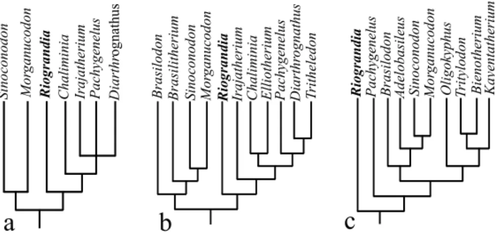
Martinelli et al. (2005) showed Riograndiato be
the most basal taxon within Tritheledontidae (Fig. 2a), while Martinelli and Rougier (2007) placedRiograndia
as the most basal taxon of the monophyletic Ictidosau-ria Clade, and as sister taxon of the most inclusive Tri-theledontidae, composed by Irajatherium,Chaliminia, Elliotherium,Pachygenelus,Diarthrognathus, and Tri-theledon(Fig. 2b). Recently, Liu and Olsen (2010) used, among the tritheledontids, onlyRiograndiaand Pachy-genelusin their analysis and did not recognize Trithe-ledonta as a monophyletic group (Fig. 2c). Neverthe-less, the authors support the close relationship between
RiograndiaandPachygenelus. They emphasize that the
possibility of a monophyletic Ictidosauria (sensu
Mar-tinelli and Rougier 2007) group cannot be excluded and needs additional tests. In any case,Riograndiais closely
related to other tritheledontids (or tritheledontians) than other non-mammaliaform cynodonts; the new materials presented here reinforce this phylogenetic hypothesis. In this paper we adopted the view of Martinelli et al. (2005), who placeRiograndiaas the most basal
Trithe-ledontidae.
Riograndiaalso plays an important role in the
en-lightenment of the biostratigraphic context of the Up-per Triassic sequence from southern Brazil, because this non-mammaliaform cynodont is commonly repre-sented in the faunal association of the “Caturrita Forma-tion”, both in number of individuals and in wide geo-graphic distribution.
INSTITUTIONALABBREVIATIONS
MCN– Museu de Ciências Naturais, Fundação
Zoobo-tânica do Rio Grande do Sul (FZBRS), Rio Grande do Sul, Brazil.
UFRGS-PV-T– Universidade Federal do Rio Grande do
Sul, Rio Grande do Sul, Brazil.
UNISINOS– Universidade do Vale do Rio dos Sinos, Rio
Grande do Sul, Brazil.
SYSTEMATIC PALAEONTOLOGY
THERAPSIDABroom, 1905 CYNODONTIAOwen, 1861
EUCYNODONTIAKemp, 1982 PROBAINOGNATHIAHopson, 1990
TRITHELEDONTIDAEBroom, 1912
RiograndiaBonaparte, Ferigolo & Ribeiro, 2001
Riograndia guaibensis
Bonaparte, Ferigolo & Ribeiro, 2001
Holotype: MCN-PV2264, anterior portion of the skull,
from the tip of snout to the fronto-parietal contact, with complete dentition.
Hipodigm: MCN-PV2265, an almost complete lower
jaw with complete dentition lacking the postdentary bones and MCN-PV2271, fragment of the middle por-tion of right lower jaw, with three postcanines.
Referred specimens:UFRGS-PV-0569-T, almost
com-plete skull lacking zygomatic arches occluding with the lower jaws and teeth; UFRGS-PV-0601-T, almost com-plete skull lacking zygomatich arches, with incomcom-plete dentition; UFRGS-PV-0833-T, skull in palatal view with teeth and a left mandibular ramus with postdentary bones; UFRGS-PV-0622-T, left dentary with teeth in lin-gual view; UFRGS-PV-0623-T, right mandibular ramus with teeth, preserved in buccal view; UFRGS-PV-0624-T, right mandibular ramus with part of the postdentary bones and teeth; UNISINOS-4881, anterior portion of skull with teeth; UFRGS-PV-0788-T, fragment of a left maxilla with the canine and seven postcanines; T, isolated canine tooth; UFRGS-PV-01062-T, isolated incisor tooth; UFRGS-PV-0842-T and MCN-PV10204, isolated postcanine teeth.
Locality and horizon: The specimens
MCN-PV22-64, MCN-PV2265, MCN-PV2271, UFRGS-PV-0596-T and UFRGS-PV-0601-T are from Sesmaria do Pinhal 1 outcrop (Candelária municipality); UFRGS-PV-0624-T, UFRGS-PV-0833-UFRGS-PV-0624-T, UFRGS-PV-0842-T and UNI-SINOS-4881 are from Linha São Luiz outcrop (Faxi-nal do Soturno municipality). The specimens UFRGS-PV-01062-T and MCN-PV10204 are from Botucaraí outcrop (Candelária municipality) andSacisaurusSite
Fig. 2 – Partial cladograms for Eucynodontia showing the relationships betweenRiograndiaand the other Mammaliamorpha (sensuKielan-Jaworowska et al. 2004, Luo 2007). (a) adapted from Martinelli et al. (2005); (b) adapted from Martinelli and Rougier (2007); (c) adapted from Liu and Olsen (2010).
capital of Rio Grande do Sul State, Brazil. “Caturrita Formation” (Santa Maria 2 Sequence), Norian age (see Geological Setting and Biostratigraphy).
Emended diagnosis: small tritheledontid (sensu
Mar-tinelli et al. 2005) with the combination of the follow-ing derived characters: three upper and three lower in-cisors; reduced upper incisor 1 and hypertrophied lower incisor 1; upper canines larger than the lower ones; post-canines blade-like with 5-9 small, sharp and subequal cuspules evenly distributed on the almost semicircular border of the crown in the upper postcanine crown and in the posterodorsal border of the lower ones; postcanine teeth forming an angle with the long axis of the palate; elongated septomaxilla bordering the posterior margin of the external nares; frontal with an anterolateral pro-jection contacting the nasal medially; absence of pre-frontal; postorbital bar absent; dorsoventrally deep lac-rimal; contact of the ventral process of frontal with the ascending process of palatine; sphenopalatine foramen bordered by the ascending process of palatine; ossified orbitosphenoid; weak anterior portion of the zygomatic arch; dorsoventrally narrow zygomatic arch; wider am-plitude of the zygomatic arch at the half of the arch; quadrate suspended by the squamosal; premaxilla bor-dering the posterior margin of the incisive foramen; max-illa participating of the anterior margin of the subtem-poral fossae in palatal view; maxilla excluded from the ventral margin of the subtemporal fossa by the jugal in lateral view; secondary osseous palate extending poste-riorly to the last postcanine; presence of the lesser
pala-tine foramina in the secondary osseous palate; wide pri-mary palate; intermediate pterygopalatine ridges reach-ing the basisphenoid; broad interpterygoid vacuities; ba-sisphenoid wing excluded from the margin of the fen-estra ovalis; jugular foramen separated from the fenes-tra rotunda; two hypoglossal foramina outside the mar-gin of the jugular foramen; prootic and opisthotic un-fused; ascending process of the alisphenoid moderately expanded; open pterygoparoccipital foramen; postden-tary bones reduced in a rod-like bar; denpostden-tary symphysis unfused; the articular process of the dentary in contact with the squamosal.
GEOLOGICAL SETTING AND BIOSTRATIGRAPHY
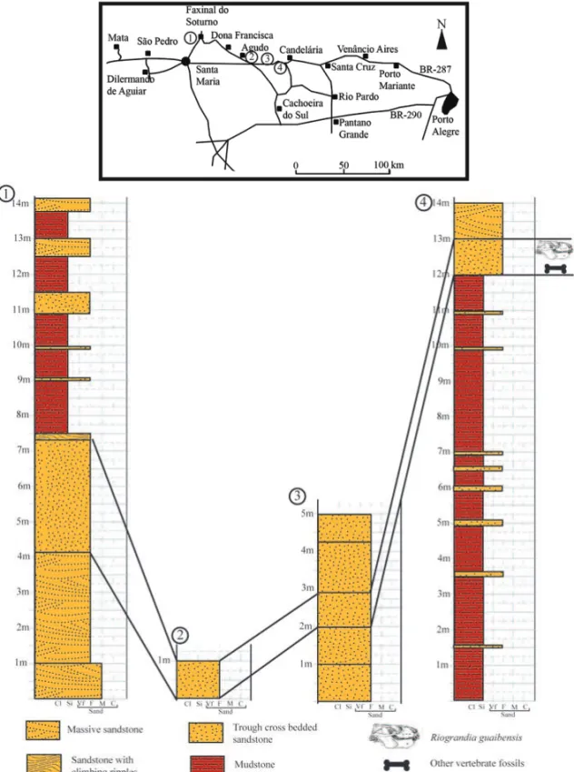
Several specimens ofRiograndia were collected from
four outcrops (Fig. 3) of Rio Grande do Sul State, south-ern Brazil, making possible to attempt the temporal cor-relation among them.
This taxon occurs predominantly in the Linha São Luiz outcrop (29◦33′29′′S; 53◦26′55′′W), located near
Faxinal do Soturno city and in Sesmaria do Pinhal 1 outcrop (29◦41′08
.5′′S; 52◦50′45′′W), near Candelária
city. An isolated first lower incisor (UFRGS-PV-01062-T), diagnostic ofRiograndia, was found in the Botucaraí
outcrop (29◦40′53′′S; 52◦50′28′′W), also located near
Candelária city, and another postcanine tooth (MCN-PV10204) was collected in theSacisaurussite, in Agudo city (19◦43′12′′S; 47◦45′04′′W).
“Catur-NEW INFORMATION ONRiograndia guaibensis FROM THE LATE TRIASSIC OF SOUTHERN BRAZIL 333
Fig. 3 – Location map and stratigraphic correlation of the four fossiliferous outcrops whereRiograndia guaibensisoccurs.
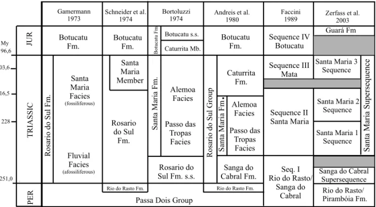
rita Formation” (sensuAndreis et al. 1980) (Fig. 4). In 1989, U.F. Faccini (unpublished data) firstly studied the Triassic sedimentary sequence of southern Brazil by us-ing the sequence stratigraphy and demonstrated that the “Caturrita Formation” (sensu Andreis et al. 1980) had
two distinct depositional sequences (both Triassic) that he called Sequence II and Sequence III (Faccini et al. 1995). The fossils here discussed came from the upper layers of the Sequence II.
Zerfass et al. (2003) refin this allostratigraphic framework, establishing two second-order superse-quences for the south Brazilian Triassic (Fig. 4). The upper one, named Santa Maria Supersequence (Middle-Late Triassic) was divided into three third-order se-quences, numbered from 1 to 3, which correspond to the Sequence II of U.F. Faccini (unpublished data). The Santa Maria 2 Sequence (sensu Zerfass et al. 2003)
with red mudstones interbedded with small-scale trough cross-bedded sandstone lenses. Rhytmites and sigmoidal massive to climbing cross-laminated sandstone bodies are also present. This facies association is interpreted as a lacustrine-deltaic depositional system. This basal portion of the Santa Maria 2 Sequence encompasses theHyperodapedonAssemblage Zone (sensuAbdala et al. 2001), in which the most abundant components are the rhynchosaurHyperodapedonand the traversodontid
cynodont Exaeretodon. They permit to correlate these
levels with those of the Ischigualasto Formation from Argentina, whose basal layer was dated as 230.3-231.4 ±0.3My (Rogers et al. 1993, Furin et al. 2006, Martinez et al. 2011).
Upwards, the sandstone content of the Santa Maria 2 Sequence increases. The arenous layers occur as nar-row, massive or stratified (horizontal and trough cross bedding) lenses interpreted as amalgamated sandstone bodies related to high width/depth ratio channels. This succession is interpreted as the progressive replacement of a lacustrine basin by a fluvial system.
H. Zerfass (personal communication) does not ex-clude the hypothesis that the upper layers of the Santa Maria 2 Sequence could in fact constitute another depo-sitional sequence, but more detailed stratigraphic work in the area is needed to test this hypothesis, once the cur-rently available evidences do not suggest any significant depositional gap inside this package. Nowadays, seven fossiliferous outcrops are assigned to the upper portion of the Santa Maria 2 Sequence in the Rio Grande do Sul State, as shown in the Table I.
The individualization of a biostratigraphic unit for the upper portion of the Santa Maria 2 Sequence was first proposed by C.M.S. Scherer (unpublished data) who have recognized that the Botucaraí outcrop, from which were recovered the dicynodont Jachaleria can-delariensisand isolated archosaur’s teeth, had distinct
sedimentary faces and represented a younger horizon than the Rhynchosauria Cenozone (= Hyperodapedon
AZ) (Schultz et al. 2000). In such a stratigraphic frame-work, the Botucaraí outcrop should be correlated with the basal layers of the Los Colorados Formation of Ar-gentina, whereJachaleria colorataBonaparte, 1970 was recorded and that overlaps the Ischigualasto Formation (marked by the presence of hyperodapedontidae
rhyn-chosaurs). This correlation indicated (at that time) a No-rian age for the Botucaraí fauna and allowed the recog-nition of an informal biostratigraphic unit to the Brazil-ian beds containing that dicynodont genus, named “ Ja-chaleria Interval” (C.M.S. Scherer unpublished data).
Some years later, other outcrops were found near the Botucaraí Outcrop, and in Agudo and Faxinal do So-turno cities (ca. 100 km west from the Botucaraí region). These outcrops correspond to the upper levels of the Santa Maria 2 Sequence (sensuZerfass et al. 2003), and
have produced the dinosaurGuaibasaurus candelarien-sis(Bonaparte et al. 1999, 2006) and small vertebrates.
Based on these new discoveries, especially on the abun-dant materials of the “ictidosaur”Riograndia
(Bonapar-te et al. 2001), Rubert and Schultz (2004) proposed a formal biostratigraphic name – Ictidosauria Assemblage Zone – to replace the “Jachaleria Interval”. Further, this denomination was changed to Mammaliamorpha Assemblage Zone by Schultz and Soares (2006) based on the argument that this last name better reflects the phylogenetic status of the small non-mammaliaform cynodonts (e.g.Riograndia, Irajatherium, Brasilodon, Brasilitherium) of the upper levels of the Santa Maria 2
Sequence.
Langer et al. (2007) discussed the possible cor-relations of this Assemblage Zone, claiming that “the whole fauna might be intermediate between those sampled at localities of La Chilca and La Esquina, both from Los Colorados Formation, Argentina (Bonaparte 1982, Abdala et al. 2001), including the earliest records of certain clevosaurid, tritheledontian and leptopleurine clades. Alternatively, it might congregate temporally separate assemblages. Given their separated occurrences and based on their phylogenetic affinities, Jachaleria candelariensis and Sacisaurus agudoensis would be assigned to an older fauna, whileIrajatherium hernan-deziandClevosaurus brasiliensiswould characterize a
younger one. In this case, forms that occur together with most of these taxa, such asRiograndiaand
brasilo-dontid cynodonts would have longer temporal ranges, occurring along that entire time interval”. Moreover, the presence of the prosauropodUnaysaurusdoes not allow
NEW INFORMATION ONRiograndia guaibensis FROM THE LATE TRIASSIC OF SOUTHERN BRAZIL 335
Fig. 4 – Comparative chart showing six stratigraphic proposes for the Brazilian Triassic.Riograndia guaibensisoccurs in the lower portion of the Caturrita Formation (sensuAndreis et al. 1980) or in the upper portion of the Santa Maria 2 Sequence (sensuZerfass et al. 2003). Fm., Formation; JUR, Jurassic; Mb, member; My, millions of years; PER, Permian;s.s., sensu stricto. Modified from Scherer et al. (2000). Geological Time Scale based on Gradstein and Ogg (2004).
TABLE I
Fossil record from the Santa Maria 2 Sequence, Upper Triassic of Rio Grande do Sul, Brazil.
Outcrop Localization Fossiliferous content
Botucaraí Candelária
Jachaleria, indeterminate phytosaur, stereospondyl amphibian, archosaur teeth,
Guaibasaurus(?),Riograndia Sesmaria
do Pinhal 1 Candelária
Clevosaurus, Brasilodon, Brasilitherium,
Irajatherium,Riograndia Sesmaria
do Pinhal 2 Candelária Guaibasaurus Sesmaria
do Pinhal 3 Candelária indeterminate archosaur,Brasilitherium
Linha São Luiz Faxinal do Soturno
Clevosaurus, Soturnia, Cargninia, Faxinalipterus,
Guaibasaurus, Brasilodon, Brasilitherium, Minicynodon, Irajatherium,Riograndia
SacisaurusSite Agudo Sacisaurus, Brasilitherium,Riograndia
UnaysaurusSite São Martinho
da Serra Unaysaurus
suggests a Norian age (Langer et al. 2007).
Following this point of view, Abdala and Ribeiro (2010) used the denominationRiograndiaAssemblage Zone (AZ) to include the faunas of the outcrops Ses-maria do Pinhal 1, SesSes-maria do Pinhal 2 and Linha São Luiz, not including the faunas of the other outcrops above listed (Table I). According to these authors, the
RiograndiaAZ could be correlated with the lower fauna
of the Los Colorados Formation (e.g. Jachaleria colo-rata), thus been interpreted as Norian, Late Norian or Rhaetic in age.
is no strong stratigraphic evidence for a significant dis-cordance between this sequence and the underlying se-quences that include the HyperodapedonAZ. Besides,
if a stratigraphic gap inside the Santa Maria 2 Sequence occurs, it is likely to be positioned belowJachaleriaand Sacisaurus, but not between these layers and the Rio-grandiaAZ (sensuAbdala and Ribeiro, 2010). More-over, if the levels containingJachaleria(as well as the
phytosaur and the stereospondyl amphibian) and Saci-saurus, whose outcrops revealed also teeth of Riogran-diaandBrasilitherium(Ferigolo and Langer 2006),
rep-resent a fauna with a distinct age from that of the Rio-grandiaAZ, the choice of this taxon as a guide to name
this biozone is not so suitable.
In this paper we preferred to assume that the whole fauna of the upper layers of the Santa Maria 2 Sequence constitutes a different faunal association from that oc-curring in the lower part of the sequence ( Hyperoda-pedonAZ) and that of the most representative taxon of
this fauna, which is Riograndia guaibensis. A
synthe-sis of our biostratigraphic opinion regarding the bio-stratigraphy of the South Brazilian Triassic is presented in Figure 5.
DESCRIPTION AND COMPARATIVE ANATOMY
For purposes of description, only the anatomical regions that were not mentioned by Bonaparte et al. (2001) will be presented. Also, the reinterpretation of some ele-ments not so clear in the holotype was possible thanks to the analysis of the new materials.
MEDIALORBITALWALL
The medial orbital wall ofRiograndiawas described by Bonaparte et al. (2001). However, some new informa-tion on the ascending process of palatine and orbitosphe-noid was provided by the specimens UFRGS-PV-0596-T and UFRGS-PV-0601-UFRGS-PV-0596-T.
Palatine(Fig. 7a, b, c, d). In the medial orbital wall, the
ascending process of the palatine contacts the lacrimal anteriorly and the frontal dorsally. This is very similar to that ofDiarthrognathus(Crompton 1958), Elliothe-rium(Sidor and Hancox 2006)Therioherpeton, Prozos-trodon (Bonaparte and Barberena 2001), Brasilodon
(Bonaparte et al. 2003), Sinoconodon (Crompton and
Luo 1993) and Morganucodon(Kermack et al. 1981).
Although this pattern is a derived condition among non-mammaliaform cynodonts, the palatine of Riograndia
does not reach the development degree of tritylodon-tids, in which this bone is wider and contributes for the total closure of the medial orbital wall. The sphenopala-tine foramen (not reported by Bonaparte et al. 2001) is enclosed by the ascending process of palatine, at the level of sixth postcanine (UFRGS-PV-0596-T). This foramen is for the passage of the major palatine nerves and vessels to the palate and the caudal nasal nerves and vessels to the nasal cavity (Wible et al. 2004). Among the non-mammaliaform cynodonts, mentions about the sphenopalatine foramen are scarce. Bonaparte and Bar-berena (2001) described that this opening is completely enclosed by the ascending process of palatine in the me-dial orbital wall ofProzostrodon, and the same pattern is observed in the tritylodontids (e.g. Bienotheroides, Sun 1984;Tritylodon, Rowe 1988;Kayentatherium, Luo
1994) andSinoconodon(Wible 1991, Crompton and Luo
1993). In another way, Bonaparte et al. (2003) described the sphenopalatine foramen in Brasilodon, which was
placed between the ascending process of palatine and orbitosphenoid as inMorganucodon.
Orbitosphenoid. No reference on the orbitosphenoid
was made by Bonaparte et al. (2001) because this struc-ture was not preserved in the holotype. None of the new materials shows evidence of an ossified orbitosphe-noid, but Rodrigues et al. (2006) have confirmed the presence of this element in two specimens of Riogran-dia(UFRGS-PV-0596T and UFRGS-0601-T) through a
comparative investigation using C.T. Scanning method. According to Rodrigues et al. (2006), the orbitosphenoid ofRiograndiais less developed than that of Prozostro-don(UFRGS-PV-0248-T) andBrasilitherium (UFRGS-PV-0760-T), but none of these taxa reaches the ossifica-tion degree of the tritylodontids and mammaliaforms, in which this element forms the floor of the anterior por-tion of the braincase (Kielan-Jaworowska et al. 2004).
LATERALWALL OFBRAINCASE
NEW INFORMATION ONRiograndia guaibensis FROM THE LATE TRIASSIC OF SOUTHERN BRAZIL 337
Fig. 5 – Biostratigraphic chart of terrestrial Triassic faunas from Gondwana showing theRiograndiaAZ. Dinodon-tosaurusAZ (sensuBarberena et al. 1985),SantacruzodonAZ (sensuAbdala and Ribeiro 2010);HyperodapedonAZ (sensuAbdala et al. 2001). Ans, Anisian; AZ, Assemblage Zone; Crn, Carnian; Ind, Induan; Lad, Ladinian; Mad, Madagascar; Nor, Norian; Ol, Olenekian; Rht, Rhaetic; S. Africa, South Africa. Modified from Abdala and Ribeiro (2010).Geological Time Scale based on Gradstein and Ogg (2004).
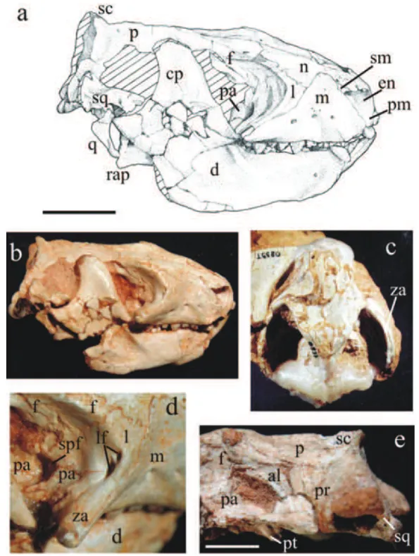
Parietal(Fig. 6a, b, e). The sutural relationships of the
ventral border of the parietal with the elements of the braincase can be better observed in the specimen UFR-GS-PV-0601-T. In lateral view, posterior to the contact with the frontal, the parietal contacts the alisphenoid and the prootic ventrally. Despite the sutures are not clear, it can be inferred that, from the frontal-parietal contact, the parietal delineates a descending bend that covers the dorsal margin of the alisphenoid and the prootic until it reaches the squamosal. Along the ventral margin of the parietal, in contact with the prootic and the alisphe-noid, a narrow and shallow open groove is placed. The groove is not well defined in its origin, but its poste-rior end appears to be near the position of the ptery-goparoccipital foramen (in the prootic). The position of this groove suggests that it corresponds to the sinus canal or orbitotemporal canal, which passes the ramus
supraor-bitalis of the stapedial artery. Riograndiashares the
ple-siomorphic condition, an opened orbitotemporal canal, with other non-mammaliaform cynodonts (Wible and Hopson 1993) excepting tritylodontids (Hopson 1994).
Squamosal(Fig. 6a, b, e). The cranial area comprised
by the squamosal is very restricted. In the posterior-most portion of the temporal region, the squamosal con-tacts the anterior lamina of the prootic anteriorly and the parietal dorsally. Its anterodorsal limit is situated at the level of the anterior opening of the posttempo-ral canal, which is placed in the prootic-squamosal su-ture. Posteriorly, the squamosal borders dorsally on the obliquely descending parietal. There is no evidence of a place to the articulation with the quadrate-jugal in the squamosal ofRiograndia. The remains from the glenoid
a concave area, more anteriorly rather than ventrally directed, resembling that of Pachygenelus (Luo and
Crompton 1994), Sinoconodon (Crompton and Luo
1993) andHadrocodium(Luo et al. 2001).
Quadrate (Fig. 6a, b). In the holotype ofRiograndia
(Bonaparte et al. 2001) the quadrate is not preserved. The information of this element comes from a frag-mented right quadrate, which is displaced from its nat-ural position, of UFRGS-PV-0596-T. When observed in anterior or posterior view, the quadrate shows a triangu-lar shape, resembling that ofProbainognathus(Romer
1970), but being slender. In Riograndia, the trochlear
area is less expanded than that of Probainognathus, a fact that gives an elongated aspect for the quadrate that is unusual among non-mammaliaform cynodonts. The trochlea presents a cylindrical shape and its convex ar-ticular facet contacts the concave area of the arar-ticular bone. In its ventral surface, the trochlea bears a pair of elongated condyles, the medial trochlear condyle, and the lateral one, which is wider than the former as in Pro-cynosuchus(Kemp 1979),Thrinaxodon (Fourie 1974), Probainognathus(Romer 1970), among others. A lat-eral notch is situated above the latlat-eral trochlear condyle, which separates the trochlea of the lateral margin of the articular facet of the dorsal plate without forming a neck, as in ThrinaxodonandProbainognathus. The
dorsal plate of Riograndia’s quadrate shows a sharper
dorsal angle than in Pachygenelusand Morganucodon
(whose quadrate exhibits a rounded dorsal margin), re-sembling more the pattern ofProbainognathus. The
ar-ticular facet of the dorsal plate is concave as in Probain-ognathus,Massetognathus, tritylodontids,Pachygenelus
andMorganucodon(Luo and Crompton 1994). Differ-ing fromProbainognathus, in which the quadrate
articu-lates with the alisphenoid, the quadrate ofRiograndiais
exclusively suspended by the squamosal as in the trithele-dontids DiarthrognathusandPachygenelus (Crompton
1958, Crompton and Hylander 1986, Allin and Hop-son 1992, Luo 1994, Luo and Crompton 1994). More-over, the quadrate of Riograndia was not covered by
the squamosal in lateral view, as in Diarthrognathus,
Chiniquodonand traversodontids; this bone is exposed as in Probainognathus, which would provide a better
mobility (Romer 1970). There is no stapedial process in the quadrate ofRiograndia. Regarding this character
it is similar to other tritheledontids (Luo and Crompton 1994), but different from Brasilodon and Brasilithe-rium(Bonaparte et al. 2005, Luo 2007).
Alisphenoid (Fig. 6e). The alisphenoid is identified
in two specimens (0596-T and UFRGS-PV-0601-T). In lateral view, the ascending process of the alisphenoid is a spatulated plate. In its dorsal border, the ascending process shows an anterior expansion that meets the ventroposterior margin of the frontal. It seems that the alisphenoid-frontal contact inRiograndia occu-pies a wider area than that ofPachygenelus(Wible and
Hopson 1993), Brasilodon and Brasilitherium
(Bona-parte et al. 2003, 2005), similarly to the patterns ob-served inDiartrognathus (Crompton 1958), Sinocono-don(Crompton and Luo 1993) andMorganucodon
(Ker-mack et al. 1981). The major part of the anterior mar-gin of the alisphenoid delimitates the posterior border of the sphenorbital fissure, as in most non-mammaliaform cynodonts, excepting in tritylodontids (Luo 1994). Be-low the contact with the frontal, the ascending process of alisphenoid becomes narrower near its base as in
Morganucodon(Kermack et al. 1981). Throughout its
anterior margin, a well-defined thin vertical ridge is de-limited, resembling that ofDiarthrognathus(Crompton
1958). The suture between the alisphenoid and the parietal has been already mentioned. The specimen UFRGS-PV-0601-T shows thatRiograndiaexhibits the
general feature of non-mammaliaform cynodonts that is the ascending process of the alisphenoid being antero-posteriorly broad and widely in contact with the anterior lamina of the prootic. The alisphenoid quadrate ramus is preserved in the specimens UFRGS-PV0601-T and UFRGS-PV-0833-T, and will be described together with other basicranial elements.
Prootic(Fig. 6e). The prootic of Riograndiais
later-ally projected, forming a lateral flange that is supported by the quadrate ramus of the alisphenoid. This lateral flange is well developed as inPachygenelus(Wible and
indi-NEW INFORMATION ONRiograndia guaibensis FROM THE LATE TRIASSIC OF SOUTHERN BRAZIL 339
Fig. 6 –Riograndia guaibensis. UFRGS-PV-0596-T in lateral view: (a) drawing and (b) photo; (c) UFRGS-PV-0833-T in dorsal view; (d) UFRGS-PV-0596-T, detail of the medial orbital wall; (e) UFRGS-PV-0601-T in lateral view. Scale bar = 10 mm. al, alisphenoid; cp, coronoid process; d, dentary; en, external naris; f, frontal; l, lacrimal; lc, lambdoidal crest; lf, lacrimal foramen; m, maxilla; n, nasal; p, parietal; pa, palatine; pm, premaxilla; pr, prootic; pt, pterygoid; q, quadrate; rap, retroarticular process; sc, sagittal crest; sm, septomaxilla; so, supraoccipital; spf, sphenopalatine foramen; sq, squamosal; za, zygomatic arch.
cation of the location for the exit for the maxillar (V2) and mandibular (V3) branches of the trigeminal nerve. In lateral view, the ventrally opened pterygoparoccipi-tal foramen is reached by the alisphenoid quadrate ra-mus in its anteroventral margin, but in most of its part this opening is enclosed by the anterior lamina of the
an-terior opening of the posttemporal canal communicated through this groove, which corresponds to the open chan-nel for the passage of the superior ramus of the stape-dial artery and other vessels that exit through the ptery-goparoccipital foramen (Rougier et al. 1992). Thus, in
Riograndiait seems that these blood vessels should run in an open channel in the lateral flange of the prootic until they join the sinus canal system (orbitotemporal canal) and posttemporal veins (Wible and Hopson 1993). This is the same configuration as inPachygenelus(Wible
and Hopson 1993). However, the open ascending chan-nel for the superior ramus of the stapedial artery is dif-ferent and more primitive than the enclosed channel of this vessel in mammaliaforms, such as Morganucodon
(Wible and Hopson 1993) and Hadrocodium (Luo et al. 2001).
ZYGOMATICARCH(FIG. 6C,D)
At least in one of the new specimens, UFRGS-PV-0833-T, the zygomatic arches are preserved in all of their length. Some information, related to the root of the zygomatic arch, was also provided by the specimen UFRGS-PV-0596-T. The specimen UFRGS-PV-0833-T confirms the suggestion of Bonaparte et al. (2001) that the zygomatic arch ofRiograndiais slender. In a
gen-eral way, the zygomatich arch resembles that of Pachy-genelus, whose arch delineates, in lateral view, a straight
line. The prominent zygomatic process of the maxilla, at the expense of the anterior process of the jugal, con-stitutes an important element of the suborbital region of the skull of Riograndia. This is a condition also
pre-sented by other tritheledontids (Hopson and Barghusen 1986), Brasilodon (Bonaparte et al. 2003) and mam-mals (Luo 1994). In the root of the zygomatic arch, the maxilla contacts posteriorly the jugal and medially the lacrimal. The lacrimal of Riograndiaoccupies a large
area in the root of the zygomatic arch as inChaliminia
(Bonaparte 1980, Martinelli and Rougier 2007), Diar-thrognathus(Crompton 1958) andPachygenelus
(Bona-parte et al. 2003). The lacrimal-jugal contact in the root of the zygomatic arch is much reduced in lateral view due to the presence of the zygomatic process of maxilla that covers the jugal laterally. The lacrimal of Riogran-dia seems to contribute in the root of the zygomatic
arch in the same way as Brasilodon (Bonaparte et al.
2003),Morganucodon(Kermack et al. 1981), Sinocon-odon(Crompton and Luo 1993) and the docodont Hal-danodon(Lillegraven and Krusat 1991), in which this
bone and the maxilla rather than the jugal are the dom-inant elements. The weak representation of the jugal in the root of the zygomatic arch showed by Riograndia
contrasts with the general non-mammaliaform cynodont pattern whose jugal is the dominant element. Regard-ing the dorsal extension of the zygomatic arch, Riogran-diapresents the same condition of most
non-mammalia-form cynodonts (excepting traversodontids and tritylo-dontids) and mammaliaforms, in which the maximal dorsal height of the arch is situated in a plane below half of the orbit (Hopson and Kitching 2001). The squamosal zygomatic portion is positioned in the mid-dle of the the arch, overlying the jugal, and configures a dorso-ventrally compressed bar exhibiting the same depth of the jugal. Due to the lateral curvature of the squamosal and the jugal, the zygomatic arch of Rio-grandiashows, in dorsal view, a round shape, with its
maximal lateral projection placed in the middle of the arch, as inThrinaxodon(Fourie 1974),Probainognathus
(Romer 1970),Pachygenelus(Allin and Hopson 1992),
andBrasilodon(Bonaparte et al. 2003), among others.
PALATE
The secondary osseous palate was described by Bona-parte et al. (2001); the specimens UNISINOS-4881 and UFRGS-PV-0596-T bring additional information. The primary palate has received little description by previous authors and is well represented in UFRGS-PV-0596-T, UFRGS-PV-0833-T and UNISINOS-4881.
Palatine(Fig. 7a, b). In its posteriormost portion, the
palatal plate becomes broader, following the general trend of the whole skull. The contribution, in extension, of the palatine in the secondary palate is greater than that of the maxilla. The former makes up to 50% of the length of the postcanine row, the same derived con-dition of other tritheledontids, tritylodontids (Kielan-Jaworowska et al. 2004), Brasilodon (Bonaparte et al.
NEW INFORMATION ONRiograndia guaibensis FROM THE LATE TRIASSIC OF SOUTHERN BRAZIL 341
the palate, the greater palatine foramen is symmetri-cally positioned, very close to the suture between the maxilla and the palatine (but completely enclosed by the palatine). These structures, considered apomorphic for epicynodonts (Hopson and Barghusen 1986, Kielan-Jaworowska et al. 2004), served as a passage of the greater palatine branch of the sphenopalatine nerve (a branch of the maxillary ramus of the trigeminal nerve) and the greater palatine artery (Kermack et al. 1981, Rougier et al. 1992). In front of the anterior border of these foramina, there is a shallow groove that surpasses the anterior limit of the palatine and reaches the max-illa, where the nerve and the artery should run forward together. Posteriorly to the greater palatine foramina, in the same symmetric position, there are two smaller foramina, which should correspond to the lesser pala-tine foramina, only reported in tritylodontids among non-mammaliaform cynodonts (Kemp 1983, Kielan-Jaworowska et al. 2004). In this group, the foramina are placed in the same position of those fromSinoconodon
(Crompton and Luo 1993) andMorganucodon(Kermack et al. 1981), that is, near the posterolateral limit of the palatine, behind the last postcanine level. InRiograndia
this second pair of palatine foramina is placed more medially behind the great palatine foramina at the level of the last postcanine. Both pairs of foramina are clearly present in the secondary palate of the two specimens of
Riograndia(UFRGS-PV-0596-T and UNISINOS-4881)
and the holotype, which was confirmed in the re-examination done by the senior author. In this sense,
Riograndiashares with Tritylodontidae and
Mammali-aformes the derived condition of the presence of a lesser palatine foramen (Luo 1994, Kielan-Jaworowska et al. 2004). The posterior palatal plate of the palatine forms the floor and the lateral walls of the posterior portion of the nasopharyngeal cavity (Kermack et al. 1981, Hop-son and Barghusen 1986). This region is well preserved in the specimen UNISINOS-4881, but some informa-tion was also provided by UFRGS-PV-0596-T. In the medial region of the primary palate of the last speci-men, the palatine projects posteriorly, contacting medi-ally the vomer. The posteriormost portion of the pala-tine is enclosed medial and laterally by the pterygoids. At the limit between these two bones, in both sides of the primary palate, two lateral ridges are defined. These ridges converge medially until reaching the posterior margin of the palatine. The lateral pterygopalatine
ridges ofRiograndiaare parts of a system of ridges that
will be discussed in the description of the pterygoid.
Pterygoid (Figs. 6e; 7a, b). The pterygoids are
rela-tively well preserved in three specimens, UFRGS-PV-0596-T, UFRGS-PV-0601-T and UNISINOS-4881. No specimen shows the contact of pterygoid with the max-illa or the vomer. Laterally, in the anterior portion of the pterygoids, the pterygoid flanges are placed. The pterygoid flanges ofRiograndiaare smaller than those ofPachygenelus(Bonaparte et al. 2003) and have a
tri-angular shape, with the apex directed ventroposteriorly, which allows a lateral contact with the mandible. In the specimen UFRGS-PV-0596-T the pterygoid flanges are in close contact with the region of the supposed coro-noid bone of the lower jaw. In the medial region of its anterior portion the pterygoids are a little concave. The meeting of the two pterygoids in the sagittal plane de-fines a medial ridge. This ridge reaches anteriorly the internal nares area. At the level of the interpterygoid vacuity the medial ridge suffer an interruption, but it is reconstituted posteriorly until it reaches the basisphe-noid. Symmetrically placed in each side of the me-dial ridge, the pterygopalatine (or intermediate) ridges run parallel to it without keeping contact. These struc-tures correspond to the site of the attachment of the anterior pterygoid muscle, which promotes a stronger union between palate and neurocranium (Kemp 1982). The pterygopalatine ridges border laterally the inter-pterygoid vacuities and reach the anterior margin of the basisphenoid as inPachygenelusandMorganucodon
(Luo 1994). Kermack et al. (1981) affirm that the me-dial ridge of Morganucodon does not reach the
inter-nal nares region, but they suggest that this region would be divided by a cartilaginous septum. In Riograndia
there is no sign of a bone wall between the internal nares either. Thus, we can infer thatRiograndiashould also present a kind of connective tissue dividing the air pas-sages. Between the medial ridge and both pterygopala-tine ridges, two channels are formed. Another pair of channels is delimited in each side of the pterygopalatine ridges. Riograndiapresents a palatal system of ridges
and channels very similar to those ofPachygenelus(Luo 1994), tritylodontids (Young 1947), andMorganucodon
1986, Maier et al. 1996) infer that this system would be covered by soft tissue, extending the length of the secondary palate and dividing the nasopharyngeal cavity into two air passages: lateral and medial. The interptery-goid vacuities of Riograndia correspond to a
well-de-fined structure that was crossed by the cultriform pro-cess of the basisphenoid. Some very thin and small bone fragments dispersed in the interpterygoid vacuity area of UFRGS-PV-0596-T could indicate that the aperture was covered by an incipient sheet of bone. Among epi-cynodonts, the interpterygoid vacuities were observed in juveniles specimens of Thrinaxodon(Martinelli and
Rougier 2007), and among eucynodonts this feature is present in juveniles of Probelesodon andLumkuia, in adult specimens of tritheledontids, asDiarthrognathus
(Crompton 1958),Chaliminia(Bonaparte 1980,
Marti-nelli and Rougier 2007) andPachygenelus(Hopson and
Barghusen 1986), and inBrasilodonandBrasilitherium
(Bonaparte et al. 2005). The presence of the interpte-rygoid vacuities in epicynodonts has been interpreted as a retention of a primitive character or a reversion to a plesiomorphic therapsid condition, which was still conserved in the pre-epicynodontsDviniaand Procyno-suchus (Hopson and Barghusen 1986). According to
Kemp (1979), the vacuities are a remnant structure re-lated to some skull kinetism of basal therapsids still present in these Permian cynodonts. Martinelli and Rougier (2007) have considered that the re-acquisition of the interpterygoid vacuities in the derived non-mammaliaform cynodonts, which bear a high resem-blance with the pattern present in most basal forms (e.g.
Procynosuchus) and in immature individuals of more derived forms (e.g. Thrinaxodon, Lumkuia, Probele-sodon, Kayentatherium), could be the result of
hete-rochronic processes like paedomorphosis. We consider that, in the case ofRiograndiaand other tritheledontids,
it is plausible to accept that the presence of the interpte-rygoid vacuity is a consequence of the new remodeling of the primary palate, in connection with the increase of the nasopharyngeal system and brain, then to assume that the vacuity is related with skull kinetism as in prim-itive cynodonts. So, instead of being interpreted as a homoplastic feature shared with pre-epicynodonts, the interpterygoid vacuities of Riograndia and other tritheledontids should be interpreted as an evolutionary novelty. Despite the presence of the interpterygoid va-cuities, the primary palate of Riograndia, formed by
transversely enlarged pterygoids, is similar to that of other tritheledontids andMorganucodon. The
broaden-ing of the primary palate is related with an evolution-ary trend observed in the advanced non-mammaliaform eucynodonts, which includes an increase in the cerebral volume and an inflation of the nasopharyngeal system, promoting the posterior displacement of the choanas and the separation of the pterygopalatine ridges (Rowe 1993). Regarding the pterygoid quadrate ramus, it is possible to observe in the specimens UFRGS-PV-0601-T and UNISINOS-4881 that each pterygoid shows a lat-eral inflexion, becoming more convex posterioly. At this point, the pterygoids seem to be fused to the alisphe-noid and, from there, a posterolateral projection can be observed. As the suture between these two bones is in-distinguishable, it is probable that the mentioned projec-tion corresponds to the alisphenoid quadrate ramus. This condition is in agreement with the eucynodontia gen-eral pattern, which is the reduction or lack of the ptery-goid quadrate ramus (Hopson and Barghusen 1986). No sign of an ectopterygoid was observed in the specimens of Riograndia, as in other thitheledontids,
tritylodon-tids and basal mammaliaforms (e.g. Sinoconodon and Megazostrodon).
BASICRANIUM
Three studied skulls (0596-T, UFRGS-PV-0833T, UNISINOS-4881) reveal information on the ba-sicranium.
Basisphenoid-parasphenoid complex (Fig. 7a, b).
NEW INFORMATION ONRiograndia guaibensis FROM THE LATE TRIASSIC OF SOUTHERN BRAZIL 343
was not completely preserved in any of the specimens, we can infer that it was reduced because the area sit-uated in both sides of the basisphenoid is occupied by the prootic in the specimens UFRGS-PV-0596-T and UFRGS-PV-0833-T. Riograndia shares with Probain-ognathus (Romer 1970, Allin 1986), chiniquodontids
(Romer 1969, Teixeira 1982),Diarthrognathus
(Crom-pron 1958), Pachygenelus (Luo 1994) and
tritylodon-tids (Sues 1986) the derived condition that is a reduced basisphenoid wing excluded from the margin of the fenestra ovalis. The basisphenoid wing ofRiograndiais
more reduced than that ofDiarthrognathus(Crompton
1958), resembling more that of Pachygenelus
(Bona-parte et al. 2003). However, it is not as reduced as in mammaliaforms, such asAdelobasileus(Lucas and Luo 1993, Luo et al. 1995), and Hadrocodium (Luo et al.
2001). The cultriform process of the parasphenoid was not conserved in any of the specimens, although signs of ossification of the basisphenoid-parasphenoid com-plex, medial to the cavum epiptericum, reveal a short ridge. Probably the ossification that forms this ridge corresponds to the basisphenoid, which was originally fused to the parasphenoid and projects anteriorly through its cultriform process. Based on the medial pterygoid ridges, we can infer that the cultriform pro-cess ofRiograndiawould extend anteriorly between the pterygoids, lying over the referred ridge and crossing the interpterygoid vacuities until joining the vomer. In the area of origin of the medial pterygoid ridge of UFRGS-PV-0596-T it is possible to observe remnants of bone ad-hered in both sides, which may correspond to pterygoid fragments that were originally fused to the parasphenoid and basisphenoid. The fusion of these elements is char-acteristic of advanced non-mammaliaform eucynodonts andMorganucodon(Kermack et al. 1981). This feature
is also related to the cranial anatomical remodeling ob-served along the evolutionary history of the Cynodontia, in function of the increase of the cerebral and nasopha-ryngeal system volume as discussed in the case of the interpterygoid vacuities. The transversal enlargement of the pterygoids and its fusion to the cultriform process of the parasphenoid inRiograndiaillustrates this
evolution-ary trend, which is fully established inMorganucodon
(Kermack et al. 1981). About the position of the inter-nal carotid foramina, the specimen UFRGS-PV-0596-T shows clearly these small openings, placed symmetri-cally in the medial portion of the basisphenoid.
Alisphenoid(Fig. 7d). The anterior margin of the
ali-sphenoid cannot be observed because it is broken in the only specimen that bears this bone partially preserved (UFRGS-PV-0601-T). The quadrate ramus of the ali-sphenoid projects posteriorly and covers the pterygoid and the prootic laterally. In ventral view, the quadrate ramus of the alisphenoid participates on the formation of the lateral border of the cavum epiptericum and pro-jects posteriorly until the level of the anterior margin of the basioccipital, but without surpassing it. Riogran-dia shares this feature with other tritheledontids and
non-mammaliaform cynodonts, excepting tritylodon-tids (Rowe 1988), which present the plesiomorphic condition.
Basioccipital (Fig. 7e). About the basioccipital, no
previous information was provided by the holotype of
Riograndia(Bonaparte et al. 2001). The basioccipital, preserved in the specimens UFRGS-PV-0596-T and UFRGS-PV-0601-T, presents a typical hexagonal shape, but wider anteriorly. In its median portion there is a short prominent and thickened ridge that runs along the bone surface. In consequence, the basiocipital lateral margins are depressed. In the central portion of the bone there are two small nutritional foramina. Based on the anterior contact facet of the basioccipital, we can see that this bone meets the basisphenoid-parasphenoid complex through an anteriorly concave suture line. Lat-erally, the basioccipital meets the prootic through a di-agonal suture that converges anteriorly to the sagittal plane. The posteriormost sector of the basioccipital par-ticipates on the ventral border of the foramen magnum. The sutures between basioccipital and exoccipitals are wide and run anterioly diagonal to the sagittal plane.
Exoccipital (Fig. 7c, e). The exoccipitals of Riogran-diaare preserved in the specimens UFRGS-PV-0596-T,
non-mammalia-form cynodonts is the hypoglossal foramina in conflu-ence with the jugular foramen. The derived condition presented by Riograndiais also observed in Oligoky-phus (Kühne 1956), Pachygenelus (Bonaparte et al. 2003) andBrasilitherium(Bonaparte et al. 2005). How-ever, one can verify that the ossified area between the jugular foramen and the hypoglossal foramina (“jugu-lar process” of Kermack et al. 1981) in Pachygenelus, Brasilitheriumand mammaliaforms is wider than that in Riograndia(Lucas and Luo 1993, Luo et al. 2001).
Prootic and opisthotic(Fig. 7d, e). The prootic and the
opisthotic are partially represented in the basicranium of PV-0596-T, PV-0601-T and UFRGS-PV-0833-T.Riograndiapresents the primitive condition of unfused prootic and opisthotic, a feature evidenced by the specimen UFRGS-PV-0833-T, in which both ele-ments are preserved around the pterygoparoccipital men. Perpendicular to the posterior margin of this fora-men, we can observe a vertical straight line that should correspond to the suture between the prootic and the opisthotic. The supposed suture is situated in a simi-lar position to the suture between the prootic and the opisthotic of Probainognathus and Chiniquodon
(Ab-dala 1996). Regarding tritheledontids, Wible (1991) has made reference to a visible suture between these two bones in a juvenile specimen of Pachygenelus, but no
conclusion about the character state of adult individuals was possible to establish. InDiarthrognathus,
Cromp-ton (1958) affirms that there is no sign of suture dividing the prootic and the opisthotic. InBrasilodon(Bonaparte
et al. 2005), the prootic and the opisthotic are sepa-rated. The tritylodontidsOligokyphus(Crompton 1964)
and Kayentatherium (Sues 1986) show these two
ele-ments fused, as inBrasilitherium(Bonaparte et al. 2005)
and mammaliaforms (e.g. Morganucodon,Sinoconodon
andHadrocodium), whose fused prootic and opisthotic form the petrosal bone (Luo et al. 1995). The suture between the paroccipital process and the exoccipital is placed laterally to the occipital condyle (UFRGS-PV-0833T). The paroccipital process does not meet the quadrate because the latter bone is in contact exclusively with the squamosal. This is the same plesiomorphic state presented by Probainognathus, Massetognathus,
DiarthrognathusandPachygenelus(Luo and Crompton
1994), in which the paroccipital process is laterally cov-ered by the squamosal. The laterally opened
pterygo-paroccipital foramen ofRiograndia
(UFRGS-PV-0833-T) is anteriorly bordered by the prootic and posteriorly by the paroccipital process, as inPachygenelus (Wible and Hopson 1993). Running laterally to the prootic, the alisphenoid quadrate ramus posteriormost limit reaches the anterior margin of the pterygoparocipital foramen, but without covering it. Riograndiafollows the same
pattern ofPachygenelus(Wible and Hopson 1993),
trity-lodontds (Lucas and Luo 1993, Luo 1994), morganu-codontids andSinoconodon(Wible and Hopson 1993).
In Riograndia, the jugular foramen is separated
from the fenestra rotunda (or perilymphatic foramen) by a narrow bone bar. This separation is a derived condi-tion among non-mammaliaform cynodonts, being shared with tritylodontids (e.g.Tritylodon,Oligokyphus) (Rowe 1988), Pachygenelus (Wible 1991) and Brasilitherium
(Bonaparte et al. 2005). However, in the last two taxa, the distance between the jugular foramen and the fen-estra rotunda is wider than inRiograndia. The
poste-rior and lateral margins of the jugular foramen are dom-inated by the paroccipital process, while the anterior and medial ones are formed by the prootic. The fen-estra ovalis is a large opening (greater than the jugular foramen and the fenestra rotunda), totally enclosed by the prootic and positioned lateroanteriorly to the jugu-lar foramen and the fenestra rotunda (without contact-ing the basisphenoid wcontact-ing), a derived state shared with
Probainognathus(Luo 1994), chiniquodontids (Romer
1969),Diarthrognathus(Crompton 1958),Pachygenelus
(Allin 1986) and mammaliaforms (Luo 1994). Despite this derived condition, the fenestra ovalis ofRiograndia
still has an osseous ring, which is a primitive condition of non-mammaliaform cynodonts (Luo et al. 1995). This ring, which contacts the stapes, is a common feature among non-mammaliaform cynodonts (e.g. Probainog-nathus, Luangwa, Massetognathus, Kayentatherium, Diarthrognathus). The cavum epiptericum is postero-medially bordered by the prootic, laterally by the ali-sphenoid quadrate ramus and anteriorly by the basi-pterygoid process of the basisphenoid. Only a very nar-row ossified portion separates the anterior margin of the fenestra ovalis from the cavum epiptericum. Thus, as in most part of the non-mammaliaform cynodonts [ex-ceptingProbainognathus(Luo 1994),Exaeretodon
(Ab-dala et al. 2002) and tritylodontids (Hopson 1964, Kemp 1983)], the cavum epiptericum ofRiograndiadoes not
NEW INFORMATION ONRiograndia guaibensis FROM THE LATE TRIASSIC OF SOUTHERN BRAZIL 345
Squamosal. In UFRGS-PV-0596-T the squamosal
notch, to lodge the quadrate, is placed laterally to the contact between the squamosal and the paroccipital pro-cess, and medially to the glenoid cavity. The facet on the paroccipital process for the squamosal is parallel to the sagittal plane. Its cranial moiety does not extend an-teriorly to the level of the posterior margin of the ptery-goparoccipital foramen, which is not encircled by the squamosal.
OCCIPITALREGION
Comparisons with other tritheledontids are difficult be-cause the occipital region is poorly preserved in Diar-thrognathus (Crompton 1958), Chaliminia (Bonaparte
1980) and Elliotherium (Sidor and Hancox 2006); no published data about Pachygenelus is available. The specimens UFRGS-PV-0596-T and UFRGS-PV-0601-T have conserved the whole occipital region. In lateral view, the ventral margin of the occipital plate is more posteriorly projected than the dorsal one. In posterior view, the occipital plate shows a triangular shape, with the apex delimited by the union of the sagittal and lamb-doidal crests.
Interparietal. Due to the high level of fusion of
the occipital plate, the presence of an interparietal can-not be observed in UFRGS-PV-0596T, which has the best preserved skull roof and occipital region. Riogran-dia seems to follow the pattern showed by other
ad-vanced non-mammaliaform cynodonts as Diarthrogna-thus(Crompton 1958),Pachygenelus(Luo 1994), The-rioherpeton(Bonaparte and Barberena 1975, 2001),
tri-tylodontids (Rowe 1988) and mammaliaforms (Lucas and Luo 1993), which lack an independently ossified interparietal.
Supraoccipital(Fig. 7c). The supraoccipital is strongly
fused to the other occipital plate elements. The sagit-tal and lambdoidal crests project over the supraoccip-ital; throughout the medial portion of the bone a broad and short ridge is delineated, supporting the sagittal crest and promoting a greater robustness of this portion of the occipital plate. In each side of the medial ridge there are two shallow depressed areas that correspond to sites of muscular attachment. The supraoccipital seems to be restricted to the dorsal margin of the foramen magnum, whose lateral margins are delimitated by the exoccipi-tals. The suture between the supraoccipital and the
tabulars is not clear, but the former contacts laterally the latter.
Basioccipital (Fig. 7e). The posteriormost portion of
the basioccipital participates on the ventral border of the foramen magnum. In occipital view, the basioccipi-tal separates the two occipibasioccipi-tal condyles through a shal-low notch. This feature represents a derived condition shared with Chiniquodon (Abdala 1996), Lumkuia
(Hopson and Kitching 2001),Pachygenelus(Bonaparte et al. 2003),BrasilodonandBrasilitherium(Bonaparte
et al. 2003, 2005), among others. However, in Rio-grandiaand in these mentioned taxa, a well-developed
notch for the odontoid process of the axis, like that of
Adelobasileus (Lucas and Luo 1993), Sinoconodon
(Crompton and Luo 1993) and Morganucodon (Ker-mack et al. 1981), has not been formed yet.
Tabular(Fig. 7c). The tabular ofRiograndiaoccupies
the whole area lateral to the supraoccipital, being lim-ited dorsolaterally by the descending lambdoidal crests of parietals and laterally by the squamosal. Ventrolat-erally each tabular makes contact with the paroccipi-tal process, and ventromedially contacts the exoccipi-tals. Both posttemporal canals, through which the ar-teria and the vena diploetica magna pass (Wible and Hopson 1993), are dorsally encircled by the tabulars very close to the squamosal margin. The ventral mar-gin of each posttemporal canal touches the suture be-tween the tabular and the paroccipital process, being, thus, bordered by the exoccipitals. This condition, shared withProbainognathus(Romer 1970), Diarthrog-nathus(Crompton 1958),Pachygenelus(Lucas and Luo 1993),Brasilodon(Bonaparte et al. 2003), and Oligoky-phus(Crompton 1964), among others, is considered by
Luo (1994) as intermediate between the plesiomorphic condition, characterized by a posttemporal canal totally encircled by the tabular; and the derived condition ex-hibited by mammaliaforms, in which the posttemporal canal is dorsally bordered by the squamosal.
Exoccipital(Fig. 7c, e). The suture between the
NEW INFORMATION ONRiograndia guaibensis FROM THE LATE TRIASSIC OF SOUTHERN BRAZIL 347
the occipital condyles ofRiograndiais oval, similar to
the pattern observed inPachygenelus (Bonaparte et al.
2003). The condyles do not reach the top of the dorsal border of the foramen magnum, being restricted to its ventral one-third, which is a primitive condition (Rowe 1988). As mentioned above, the occipital condyles are well separated by the posterior margin of the basioccip-ital. Following the general pattern of the whole occipi-tal plate, we can observe that the ventral portion of the condyles is more posteriorly projected than the dorsal one. This condition is also observed in the occipital plate ofAdelobasileus(Lucas and Luo 1993). The
non-bifurcated paroccipital process occupies the base of the occipital plate, lateral to each occipital condyle. Dor-sally, the paroccipital process makes an irregular contact with the tabular and borders the ventral margin of the posttemporal canal, as mentioned before.
LOWERJAW
Bonaparte et al. (2001) have described the almost com-plete lower jaw of the specimen MCN-PV2265 in the medial view, but nothing was reported about the post-dentary bones ofRiograndia. All the new specimens
(0596-T, 0624T, UFRGS-PV-0833T, UFRGS-PV-0623T) confirm the characteristics observed in the type series (Bonaparte et al. 2001): a mandible with rather robust construction with a high and thick unfused symphysis, a high coronoid process and a distinct angular process. The postdentary bones com-plex ofRiograndiais described for the first time in this
contribution.
Postdentary bones (Fig. 8e). The information about
the postdentary bones of Riograndia comes from the
specimens UFRGS-PV-0596-T, UFRGS-PV-0624-T and UFRGS-PV-0833-T. The postdentary trough is dorsally limited by the medial ridge of the dentary that runs anteriorly until the level of pc6. Posteriorly, the medial ridge becomes wider and projects above the articular process, which represents the posteriormost edge of the dentary. However, the articular process of the dentary does not form a condyle as seen in mammaliaforms. The relative position of the articular process of the den-tary of Riograndia (Fig. 1c) is more dorsal than the dorsal margin of the postcanines row, a derived feature shared with traversodontids, tritylodontids (Bonaparte 1962, Crompton and Sun 1985), Pachygenelus (Allin
and Hopson 1992),Diarthrognathus(Crompton 1963), Chaliminia (Bonaparte 1980, Martinelli and Rougier
2007) andMorganucodon(Kermack et al. 1981). All the three specimens that have preserved the postdentary bones show some degree of fragmentation in these ele-ments, but in general we can observe that the surangu-lar, angusurangu-lar, articular and prearticular compose a frag-ile unity, lodge in the postdentary trough, as in other advanced non-mammaliaform eucynodonts and mam-maliaforms (Crompton and Luo 1993). The articular, prearticular and angular are fused, as in other trithele-dontids (Allin and Hopson 1992). Supposedly, the sur-angular runs parallel over the rod formed by the artic-ular, prearticular and angartic-ular, and finishes at the poste-riormost level of the retroarticular process. The artic-ular seems to form most part of the retroarticartic-ular pro-cess, which is concave and exhibits the ventral margin posteriorly projected. The reflected lamina of the an-gular was not preserved, and there is no evidence of a contact between the surangular and the squamosal in any of the analyzed specimens. Regardind the cranio-mandibular joint, it seems that Riograndia presents
the same condition of other tritheledontids (Luo and Crompton 1994) and Brasilitherium (Bonaparte et al.
2003), that is the articular process of the dentary in contact with the squamosal, instead of the surangular, as in most eucynodonts (Crompton 1958, 1972). Thus, the dentary-squamosal articulation (or contact) repre-sents a synapomorphy of Tritheledontidae, Brasilithe-riumand mammaliaforms (Luo 1994, Luo 2007). The
splenial, which should cover medially the Meckelian groove, was not preserved in any of the specimens. Only in the UFRGS-PV-0596-T was possible to distinguish a most medially pronunciated area in the medial face of the mandible that can suggest the presence of the coro-noid bone.
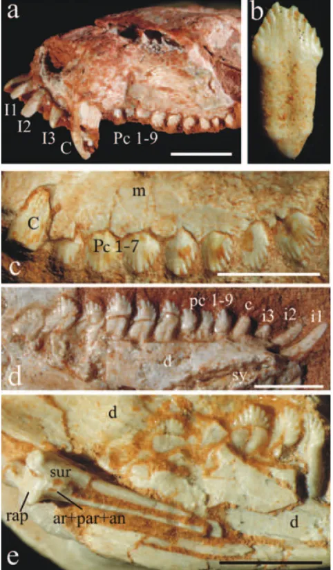
DENTITION(FIG. 8A,B,C,D,E)
The dental anatomy ofRiograndiahas received special
treatment by Bonaparte et al. (2001). The authors have considered thatRiograndiashows a plesiomorphic tooth
re-Fig. 8 – Riograndia guaibensis. (a) UNISINOS-4881 in lateral view; (b) UFRGS-PV-0842-T in lingual view; (c) UFRGS-PV-0788-T, right maxilla in lateral view with detail of the upper dentition; (d) UFRGS-PV- 0622-T, right mandible in lingual view with detail of the lower dentition; (e) UFRGS-PV-0833-T, right mandible in lingual view with detail of the postdentary bones. Scale Bar = 10 mm. ar, articular; an, angular; c, lower canine; C, upper canine; d, dentary; i, lower incisor; I, upper incisor; m, maxilla; par, prearticular; pc, lower postcanine; Pc, upper postcanine; rap, retroarticular process; sur, surangular.
duced in size. The postcanines are blade-like, with six to nine aligned cuspules, each of them bordered by a shallow groove. On the buccal side the central cuspules are usually the largest. The postcanine roots presented an incomplete subdivision, suggesting an incipient
bi-furcation. There is no cingulum in the postcanines. The new specimens ofRiograndiashow the same dental