Peripheral biomarkers of oxidative
stress in aging and Alzheimer’s disease
Tania Marcourakis
1, Rosana Camarini
2, Elisa Mitiko Kawamoto
2,
Leandro Rodrigues Scorsi
1, Cristoforo Scavone
2Abstract – Aging is associated with a greatly increased incidence of a number of neurodegenerative disorders, including Alzheimer’s disease (AD), Parkinson’s disease (PD) and amyotrophic lateral sclerosis (ALS). These con-ditions are associated with chronic inlammation, which generates oxygen reactive species, ultimately responsible for a process known as oxidative stress. It is well established that this process is the culprit of neurodegeneration, and there are also mounting evidences that it is not restricted to the central nervous system. Indeed, several studies, including some by our group, have demonstrated that increased peripheral oxidative stress markers are associated to aging and, more speciically, to AD. Therefore, it is very instigating to regard aging and AD as systemic conditions that might be determined by studying peripheral markers of oxidative stress.
Key words: Alzheimer’s disease, aging, oxidative stress, peripheral markers.
Biomarcadores periféricos do estresse oxidativo no envelhecimento e na doença de Alzheimer
Resumo – O envelhecimento está associado a uma alta incidência de doenças neurodegenerativas, incluindo doença de Alzheimer (DA), doença de Parkinson (DP) e esclerose lateral amiotróica (ELA). Estas condições estão relacionadas à inlamação crônica que gera espécies reativas de oxigênio, responsáveis por um processo denomi-nado de estresse oxidativo. Está bem estabelecido que este processo está envolvido na neurodegeneração, e existem várias evidências de que ele não é restrito ao sistema nervoso central. De fato, muitos estudos, inclusive alguns de nosso grupo, demonstraram que o aumento de marcadores do estresse oxidativo periférico está associado ao envelhecimento e, mais intensamente, à DA. Assim, é muito instigante pensar no envelhecimento e na DA como doenças sistêmicas que possam ser investigadas por meio de marcadores periféricos de estresse oxidativo.
Palavras-chave: doença de Alzheimer, envelhecimento, estresse oxidativo, marcadores periféricos.
1Department of Clinical and Toxicological Analysis, Faculty of Pharmaceutical Sciences and Neurology Investigation Center, School of Medicine (LIM-15). 2Department of Pharmacology, Biomedical Sciences Institute. University of São Paulo, São Paulo, Brazil.
Tania Marcourakis – Departamento de Análises Clínicas e Toxicológicas / Faculdade de Ciências Farmacêuticas da Universidade de São Paulo / Avenida Professor Lineu Prestes, 580 / Bloco 13B - 05508-900 São Paulo SP - Brazil. E-mail: [email protected]
Received 01/24/2008. Received in inal form 02/14/2008. Accepted 02/14/2008.
Several studies have shown that the aging process is a consequence of progressive accumulation of deleterious biochemical changes during life span, leading to an im-balance of body regulatory systems, including hormonal, immune and neuroendocrine mechanisms.1-4 It is believed that these changes can be more intense in neurodegenera-tive disorders.5-8
Aging is associated with a greatly increased incidence of a number of degenerative conditions, including Alzheimer’s disease (AD), Parkinson’s disease (PD), amyotrophic lateral sclerosis (ALS), atherosclerosis, and myocardial infarction. Currently, 26.6 million people worldwide have AD and this igure could rise to more than 100 million people by 2050.9
Most of those patients will have late-stage disease that re-quires a high level of care, increasing inancial and personal costs with a devastating effect on the world’s economies, health-care systems and families.
complex phenomenon, and other factors, such as genetic background, level of physical activity and nutrition might also be affecting aging, characterizing this as a complex multi-factorial process.12 Nevertheless, there is agreement that free radicals can damage proteins, lipids and DNA, leading to oxidative stress, lipid peroxidation and to the formation of DNA adducts.6,13
Free radicals which are derivatives of oxygen (O2), such as superoxide anion (O2•-) and hydroxyl radical (OH•), are called reactive oxygen species (ROS). However, ROS, such as H2O2 are not free radicals but are also harmful. ROS are normal byproducts of the mitochondrial electron trans- port chain produced during respiration of aerobic organ- isms. When ROS are overproduced and where this produc-tion overwhelms antioxidant defense systems, cells can be damaged and this process is called oxidative stress.14,15
The most abundant source of ROS in the central nervous system (CNS) is the respiratory burst system of activated microglia. When the system is activated, large quantities of O2•- are generated on the microglial external membrane, from which point they are released as a pur- poseful attack system. The amount of ROS produced dur-ing the process of oxidative phosphorylation represents 2% of the total oxygen consumed during respiration, but may vary depending on several parameters. The brain is partic-ularly sensitive to oxidative damage because: 1. it is rich in the more easily peroxidizable fatty acids; 2. it is responsible for about 20% of total oxygen consumption; 3. it has high levels of iron that predispose it to Fenton reaction with the formation of OH•; and 4. it has high neuronal calcium input and the presence of excitatory aminoacids, such as
glutamate. On the other hand, in comparison to other or-gans such as the liver and kidney, the brain has lower levels of the antioxidant enzymes superoxide dismutase (SOD), glutathione peroxidase (GPx) and catalase.14,16,17 A perfect balance among these three enzymes is necessary to main-tain cell survival. Moreover, neurons also contain low levels of glutathione (GSH), a major antioxidant responsible for the elimination of cytosolic peroxides.18
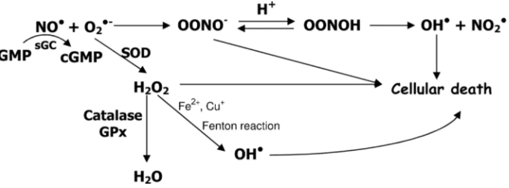
The rapid reaction of O2•- and nitric oxide (NO•) leads to the formation of peroxynitrite anion (ONOO-), and much attention has been given to this powerful oxidant agent, involved in oxidation and nitration of lipids, DNA strand leakage, nitration of proteins, including the forma- tion of 3-nitrotyrosine and disruption of structural pro-teins such as actin and neuroilament L. Figure 1 shows the route of production of ONOO- as well as its antioxidant mechanism.
Aging, neurodegeneration and oxidative stress
During the aging process, mitochondrial oxidative phosphorylation becomes less eficient, which probably contributes to an increase in the production of free radi-cals.22 The increase in O
2•- production would lead to an accumulation of intracellular calcium23 and activation of calcium dependent enzymes, such as nitric oxide synthase, which is responsible for the formation of NO. As a con-sequence of calcium accumulation, activation of protein kinase C may occur, one of the elements responsible for tau phosphorylation with the formation of the neuroibrillary tangles seen in AD24, or, in more extreme cases, triggers neuron apoptosis. Moreover, O2•- formation can increase brain H2O2 production, with microglia activation leading to the formation of more free radicals and the starting of
Figure 1. Nitric oxide (NO•) in the presence of superoxide anion (O
2•-) forms peroxynitrite anion (ONOO-), which is quite instable (T
1/2 less than 1 second) leading to the formation of hydroxyl radical
(OH•), one of the elements responsible for cellular death. NO• activates soluble guanylyl cyclase (sGC)
an inlammatory process.25,26 Most of the neurodegenera- tive conditions are associated with a chronic inlamma-tion. Although there is controversy whether inlammation is causative or a consequence of the disease process, it is now clear that it can greatly inluence its pathogenesis.16,27,28 Even though aging and neurodegeneration share the same basic mechanisms, it is dificult to establish the limits be-tween these two processes; there are mounting evidences that neurodegeneration might be an extension of the nor- mal aging process, which might in turn increase suscepti-bility to neurotoxic events.29,30
Each neurodegenerative process has its own neuro-pathological hallmarks but it has long been suspected that oxidative stress contributes to neuronal death in diseases such as AD, PD, and ALS.15,31-34 Moreover, it has been shown that oxidative stress is the earliest event in AD, occurring even before the development of the amyloid-β peptide (Aβ ) deposit in senile plaques and accumulation of ab-normal tau ilaments in neuroibrillary tangles.35-39
The neurotoxic nature of Aβ is not well understood, however, there is evidence of the involvement of OH• and O2•-.32,40-42 Aβ not only can increase the levels of free radi-cals, but can also lead to the depletion of antioxidant agents that ultimately will determine neuronal death.7,43 Although the deposit of insoluble Aβ is one of the hallmarks of AD pathology, the presence of higher levels of soluble oligo-mers of Aβ in the brains and cerebrospinal fluid of AD patients is receiving considerable attention. Moreover, it has been shown that these soluble oligomers, which have been considered neurotoxins involved in the early patho-genesis in AD, can stimulate excessive formation of ROS through the activation of N-methyl-D-Aspartate (NMDA) glutamate receptors.44-46
Evidence of free radical attack in AD cortex, PD sub-stantia nigra, and ALS spinal cord includes the presence of: 1. proteins that have been modiied by glycation; 2. the existence of low molecular weight compounds that have been oxidized and nitrated (such as 3-nitrotyrosine, 3-ni- tro-4-hydroxyphenylacetic acid, 5-nitrotocopherol, 4-hy-droxynonenal, and malondialdehyde); 3. the identiication of lipids that have been peroxidated and biomarkers of DNA oxidative damage (such as 8-hydroxy-deoxyguano-sine). Of those markers, 3-nitrotyrosine is quite stable and mostly derived from ONOO-, being a reliable indicator of its production.6,33
In order to investigate NO participation in the aging process, Özdemir et al.47 studied rats of different ages. They showed that brain thiobarbituric reactive substance (TBARS) production, a marker of lipid peroxidation, and nitrite levels were signiicantly increased with age. These increases were higher when L-NAME, a NOS inhibitor, was
administered to the rats and decreased when L-arginine, a NO precursor, was given. Their conclusion was that NO had a protector effect. Whether NO will have a protector or deleterious effect, is determined by its redox status. Under physiologic conditions, NO can be present as a nitrogen monoxide (NO•) and/or as nitrozonium cation (NO+). If the cell environment is conducive for NO• formation, a neurotoxic effect will be observed; however, if there is NO+ formation, NMDA receptor will be inhibited and a neuroprotective effect will result.48,49 NO behavior as a free radical or as an antioxidant agent will be dependent on O2•- levels. If the concentration of this anion is high, NO will lead to lipid peroxidation, otherwise NO will have an antioxidant behavior.50-53
Peripheral markers of oxidative stress in aging and AD
The study of peripheral biological samples in order to identify biomarkers either in aging or in neurodegenerative disorders seems an interesting approach and several groups have been working in this ield.54 Praticó55 pointed out the importance of studies on peripheral biological samples in living patients with a clinical diagnosis of AD, as it is dif-icult to address the question of whether oxidative stress is an early component in the pathogenesis of AD or a com-mon inal step of the neurodegenerative process based on post-mortem investigations. A follow-up of these patients in different stages of the disease could help to understand, for instance, the evolution of the antioxidant system. This kind of study received more attention in the light of the evidence that oxidative stress chronologically precedes Aβ deposit, and onset of AD symptoms. Moreover, as such
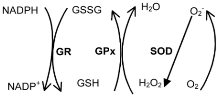
Figure 2. Superoxide anion (O2•
-) is formed in oxidative
changes can be detected peripherally, this points to a wide-spread disturbance.36,56
To better understand this issue, we shall summarize some studies addressing oxidative stress in aging and AD, performed on peripheral cells. As most of the studies ad- dressed the glutathione redox cycle, an important antioxi-dant system, Figure 2 can clarify the mechanisms involved.
Kasapoglu & Özben12 investigated 100 healthy subjects with ages ranging from 20 to 70 years and showed an age related increase in serum TBARS, a lipid peroxidation marker; however, of the erythrocytes antioxidant enzymes, only GPx had an age related reduced activity, since catalase and SOD activities were increased. Solichova et al.57 showed that nonagenarians have higher serum TBARS levels than subjects below 90 years’ old. Whether this oxidative state was due to a decline in antioxidant defenses or to an in-creased generation of ROS remains an open question.
One of the irst studies to address the variability of the antioxidant enzymes SOD, GPx, and catalase during life span was performed by Guemouri et al.,58 in a study in- volving 1836 healthy subjects from 4 to 97 years of age. Be-low the age of 65, enzyme activities remained unchanged. However, the group detected a decrease in the activity of three enzymes, both in plasma and erythrocytes, of subjects older than 65 years of age. A similar inding was described by King et al.,59 who found no change in erythrocytes an-tioxidant enzymes of healthy volunteers with ages ranging from 35 to 69 years. Junqueira et al.60 evaluated 503 healthy subjects aged from 20 to more than 70 years’ old. They showed an increase in plasma TBARS levels in individuals over 50 and of erythrocyte GPx activity in those over 40 years of age. The authors reported a strong positive correla-tion between age and these two measures, suggesting that erythrocyte GPx could be used as a marker of oxidative stress in aging.
In a bid to understand whether healthy centenarians have a peculiar proile of non-enzymatic and enzymatic antioxidants that could explain longevity, Mecocci et al.61 compared the results of centenarians to subjects aged 80-99, 60-79 and below 60 years. Non-centenarians had lower levels of plasmatic non-enzymatic antioxidants (vitamins A, C and E) and increased levels of antioxidants enzymes with age. Centenarians were characterized as having the highest vitamin A and E concentrations and lower SOD activity in plasma and erythrocytes. On the basis of these results, the authors suggested that the antioxidant activity of lipid vi-tamins, such as A and E, is of greater relevance to longevity. Andersen et al.62 described a similar result as centenarians had lower SOD and higher GR activities in erythrocytes compared to 60-79 year-old controls. The centenarians with better functional capacity were the ones with higher
GR activity. Paolisso et al.63 had the same objective and evaluated the plasma of 82 healthy subjects separated into three groups: <50 years’ old, 70 to 99 years’ old and cente-narians. Centenarians had lower TBARS compared to aged subjects and showed increased GSH/GSSG ratio, vitamin C and E compared to aged subjects, which pointed to re-duced oxidative stress in this group. These studies indicated that to reach extreme ages it is necessary to have a good antioxidant defense system and low oxidative stress insult.
Although not a consensus,64 the presence of increased oxidative stress at peripheral level in AD has been evidenced in some studies. Kawamoto et al.65 detected the involvement of peroxynitrite anion in platelets and erythrocyte with ag-ing and AD. Aging was associated to an increase in TBARS and NOS activity, a decrease in basal cyclic GMP content and no change in SOD and Na,K-ATPase activities. AD pa-tients showed a higher level of TBARS and an increase in NOS, SOD and Na,K-ATPase activities, without changes in cGMP, compared to aged controls. Na,K-ATPase is the en-zyme responsible for cell response to oxidative damage, as it maintains electrochemical sodium and potassium gradient. Its catalytic α subunit is sensitive to damage by free radicals while ONOO- has been reported to cause cell membrane damage, which in turn leads to the disruption of Na,K-ATPase activity.66-69 The results presented by Kawamoto et al.65 demonstrated a disruption in systemic modulation of oxidative stress in aging and at greater intensity in AD, and are consistent with the suggestion that neurodegeneration and aging could share a common pathogenic pathway.
Praticó et al.70 described an increase in isoprostane gen-eration, an index of lipid peroxidation, in urine, blood and cerebrospinal luid (CSF) of AD patients. Besides, urinary and blood isoprostane correlated with CSF levels in AD patients. In fact, some other groups have also demonstrated an increase in peripheral lipid peroxidation index, such as TBARS, malondialdehyde or isoprostanes in AD.71-74
dants (SOD and GPx) in erythrocytes of AD and MCI pa-tients, lending further weight to the indings that MCI is a prodromal stage of AD. In a recent study, Gackowski et al.77 showed a similar result regarding oxidative DNA damage in mixed AD and vascular dementia (AD/VD), suggesting a common route in the pathogenesis of AD and AD/VD.
With the aim of inding a relationship between cogni- tive function and lipid peroxidation in erythrocytes, Deli-bas et al.73 examined a group of AD patients at an interval of ive years. They found a negative correlation between MDA and the scores on the Mini Mental State Examination (MMSE), suggesting that lipid peroxidation might be one of the factors responsible for cognitive deterioration.
Zafrilla et al.74 analyzed oxidative stress at different stages of AD. Lipid peroxidation was higher in patients at the advanced stage of illness than in controls; however, no difference between light/moderate and advanced AD was detected. The total antioxidant plasmatic status of AD patients at any stage was lower than in the control group. Also, at different stages of the disease, GPx activity was increased and GR decreased.
In conclusion, although several studies support the pres-ence of peripheral oxidative damage in AD, it is not clear if neurodegeneration is a process in which peripheral oxida-tive stress is an active participant or a simple bystander.
Despite mounting evidence of oxidative stress involve- ment in AD, antioxidant therapies remain quite controver-sial. However, no effect in preventing AD was reported by several groups after healthy subjects had received antioxi-dants supplementation.78-80 Moreover, Cole et al.81 found no effect with nonsteroidal anti-inlammatory drugs and vitamin E supplement in controlling oxidative damage. Fi-nally, the meta-analysis performed by Miller et al.82 showed that high doses of vitamin E were associated with increased mortality. Unfortunately, a good theory supported by strong basic scientiic data is not enough to guarantee ef-fective therapy.
Acknowledgements – The authors thank Fernando
Kok for his helpful comments. CS is supported by the Conselho Nacional de Desenvolvimento Cientíico e Tec-nológico (CNPq); EMK, by the Fundação de Amparo à Pesquisa do Estado de São Paulo (FAPESP) and LRS by the Coordenação de Aperfeiçoamento de Pessoal de Nível Superior (CAPES). Work in the laboratory of the authors was supported by FAPESP.
References
1. Harman D. The aging process. Proc Natl Acad Sci USA 1981; 78:7124-7128.
2. Harman D. Free-radical theory of aging. Increasing the func-tional life span. Ann N Y Acad Sci 1994;717:1-15.
3. Harman D. Aging: phenomena and theories. Ann N Y Acad Sci 1998;854:1-7.
4. Yu BP. Aging and oxidative stress: modulation by dietary re-striction. Free Radical Biol Med 1996;21:651-668.
5. Floyd RA. Antioxidants, oxidative stress, and degenerative neurological disorders. Proc Soc Exp Biol Med 1999;222: 236-245.
6. Maccioni RB, Munoz JP, Barbeito L. The molecular bases of Alzheimer’s disease and other neurodegenerative disorders. Arch Med Res 2001;32:367-381.
7. Mark RJ, Blanc EM, Mattson MP. Amyloid beta-peptide and oxidative cellular injury in Alzheimer’s disease. Mol Neuro-biol 1996;12:211-224.
8. Smith CD, Carney JM, Starke-Reed PE, et al. Excess brain protein oxidation and enzyme dysfunction in normal aging and in Al-zheimer disease. Proc Natl Acad Sci USA 1991;88:10540-10543. 9. Wimo A, Winblad B, Aguero-Torres H, von Strauss E. The
magnitude of dementia occurrence in the world. Alz Dis As-soc Dis 2003;17:63-67.
10. Medvedev ZA. An attempt at a rational classiication of theo-ries of ageing. Biol Rev Camb Philos Soc 1990;65:375-398. 11. Halliwell B, Gutteridge JMC. Free radical in Biology and
Medicine. 4th: New York, Oxford University Press; 2007.
12. Kasapoglu M, Özben T. Alterations of antioxidant en-zymes and oxidative stress markers in aging. Exp Geront 2001;36:209-220.
13. Praticò D, Delanty N. Oxidative injury in diseases of the cen-tral nervous system: focus on Alzheimer’s disease. Am J Med 2000;109:577-585.
14. Mamelak M. Alzheimer’s disease, oxidative stress and gam-mahydroxybutyrate. Neurobiol Aging 2007;28:1340-1360. 15. Valko M, Leibfritz D, Moncol J, Cronin MTD, Mazur M, Telser
J. Free radicals and antioxidants in normal physiological func-tions and human disease. Int J Biochem Cell Biol 2007;39:44-84. 16. Floyd RA. Neuroinlammatory processes are important in
neurodegenerative diseases: a hypothesis to explain the in-creased formation of reactive oxygen and nitrogen species as major factors involved in neurodegenerative disease develop-ment. Free Rad Biol Med 1999;26:1346-1355.
17. Markesberry WR. The role of oxidative stress in Alzheimer Disease. Arch Neurol 1999;56:1449-1452.
18. Slivka A, Mytilineou C, Cohen G. Histochemical evaluation of glutathione in brain. Brain Res 1987;409:275-284. 19. Lipton SA, Choi YB, Pan ZH, et al. A redox-based mechanism for
the neuroprotective and neurodestructive effects of nitric ox-ide and related nitroso-compounds. Nature 1993;364:626-632. 20. Beckman JS, Koppenol WH. Nitric oxide, superoxide and per-oxynitrite: the good, the bad and ugly. Am J Physiol 1996;271: C1424-C1437.
implications for neurodegenerative diseases. J Neurochem 1997;68:2227-2240.
22. Markesberry WR, Carney JM. Oxidative alternations in Al-zheimer’s disease. Brain Pathol 1999;9:133-146.
23. Harman D. Free radical theory of aging: Alzheimer’s disease pathogenesis. Age 1995;18:97-119.
24. Harman D. Alzheimer’s disease: role of aging in pathogenesis. Ann N Y Acad Sci 2002;959:384-395.
25. Selkoe DJ. Translating cell biology into therapeutic advances in Alzheimer’s disease. Nature 1999;399:A23-A31.
26. Lue LF, Brachova L, Civin WH, Rogers J. Inlammation, Aβ deposition, and neuroibrillary tangle formation as correlates of Alzheimer’s disease neurodegeneration. J Neuropathol Exp Neurol 1996;55:1083-1088.
27. Teunissen CE, Lütjohann D, von Bergmann K, et al. Combi-nation of serum markers related to several mechanisms in Alzheimer’s disease. Neurobiol Aging 2003;24:893-902. 28. Zhu X, Su B, Wang X, Smith MA, Perry G. Causes of oxidative
stress in Alzheimer disease. Cell Mol Life Sci 2007;64:2202-2210. 29. Smith CD, Carney JM, Starke-Reed PE, et al. Excess brain protein oxidation and enzyme dysfunction in normal aging and in Al-zheimer disease. Proc Natl Acad Sci USA 1991;88:10540-10543. 30. Swerdlow RH. Is aging part of Alzheimer’s disease, or is Alzheimer’s disease part of aging? Neurobiol Aging 2007;28: 1465:1480.
31. Butterield DA. Beta-Amyloid-associated free radical oxidative stress and neurotoxicity: implications for Alzheimer’s disease. Chem Res Toxicol 1997;10:495-506.
32. Butterield DA, Yatin SM, Varadarajan S, Koppal T. Amyloid β -peptide-associated free radical oxidative stress, neurotoxicity and Alzheimer’s disease. Methods Enzymol 1999;309:746-768. 33.
Knight J. Free radicals: their history and current status in ag-ing and disease. J Clin Lab Sci 1998;28:331-346.
34. Olanow CW. A radical hypothesis for neurodegeneration. Trends Neurosci 1993;16:339-444.
35. Migliore L, Fontana I, Trippi F, et al. Oxidative DNA damage in peripheral leukocytes of mild cognitive impairment and AD patients. Neurobiol Aging 2005;26:567-573.
36. Nunomura A, Perry G, Aliev G, et al. Oxidative damage is the earliest event in Alzheimer disease. J Neuropathol Exp Neurol 2001;60:759-767.
37. Praticó D, Uryu K, Leight S, Trojanoswki JQ, Lee VM. In- creased lipid peroxidation precedes amyloid plaque forma-tion in an animal model of Alzheimer amyloidosis. J Neurosci 2001;21:4183-4187.
38. Smith MA, Rottkamp CA, Nunomura A, Raina AK, Perry G. Oxidative stress in Alzheimer´s disease. Biochem Biophys Acta 2000;1502:139-144.
39. Veurink G, Fuller SJ, Atwood CS, Martins RN. Genetics, life-style and the roles of amyloid beta and oxidative stress in Alzheimer´s disease. Ann Hum Biol 2003;30:639-667.
40. Pike CJ, Ramezan-Arab N, Cotman CW. β -Amyloid neuro- toxicity in vitro: evidence of oxidative stress but not protec-tion by antioxidants. J Neurochem 1997;69:1601-1611. 41.
Hensley K, Hall N, Shaw W, Carney JM, Butterield DA. Elec-tron paramagnetic resonance investigation of free radical induced alterations in neocortical synaptosomal membrane protein infrastructure. Free Rad Biol Med 1994; 17:321-331. 42. Varadarajan S, Yatin S, Aksenova M, Butterield DA. Review:
Alzheimer’s amyloid β -peptide- associated free radical oxida-tive stress and neurotoxicity. J Struct Biol 2000;130:184-208. 43. Crouch PJ, Harding S-ME, White AR, Camakaris J, Bush AI,
Masters CL. Mechanisms of Aβ mediated neurodegeneration in Alzheimer’s disease. Int J Biochem Cell Biol 2008;40:181-198. 44. De Felice F, Velasco PT, Lambert MP, et al. Aβ oligomers in-duce neuronal oxidative stress through an N -methyl-D-aspar- tate receptor-dependent mechanism that is blocked by the Al-zheimer drug memantine. J Biol Chem 2007; 282:11590-11601. 45. Haass C, Selkoe DJ. Soluble protein oligomers in neurodegen-eration: lessons from the Alzheimer’s amyloid β-peptide. Nat Rev Mol Cell Biol 2007; 8:101-112.
46. Ferreira ST, Vieira MNN, De Felice F. Soluble protein oligo-mers as emerging toxins in Alzheimer’s and other amyloid diseases. IUBMB Life 2007;59:199-210.
47. Özdemir S, Yargiçoglu P, Agar A, Gümüslü S, Bîlmen S, Ha-cioglu G. Role of nitric oxide on age-dependent alterations: investigation of electrophysiologic and biochemical param-eters. Intern J Neuroscience 2002;112:263-276.
48. Goldstein IM, Ostwald P, Roth S. Nitric oxide: a review of its role in retinal function and disease. Vision Res 1996;26:2979-2994. 49. Oku H, Yamaguchi H, Sugiyama T, Kojima S, Ota M, Azuma I. Retinal toxicity of nitric oxide released by administration of a nitric oxide donor in the albino rabit. Invest Ophthalm Visual Sci 1997;38:2540-2544.
50. Wink DA, Cook JA, Pacelli R, et al. The effects of various nitric oxide-donor agents on hydrogen peroxide toxicity: a direct correlation between nitric oxide formation and protec-tion. Arch Biochem Biophys 1996;331:241-248.
51. Kashii S, Mandai M, Kikuchi M, et al. Dual actions of nitric oxide in N-methyl-D-aspartate receptor-mediated neurotox-icity in cultures retinal neurons. Brain Res 1996;711:93-101. 52. O’Donnell VB, Chumley PH, Hogg N, Bloodsworth A, Darley- Usmar VM, Freeman BA. Nitric oxide inhibition of lipid per-oxidation. Kinetics of reaction with lipid peroxyl radicals and comparison with α -tocopherol. Biochemistry 1997;36:15216-15223.
53. Cudeiro J, Rivadulla C. Sight and insight on the physiologi-cal role of nitric oxide in the visual system. Trends Neurosci 1999;22:109-116.
54. Gibson GE, Huang H-M. Oxidative stress in Alzheimer’s dis-ease. Neurobiol Aging 2005;26:575-578.
Praticó D. Peripheral biomarkers of oxidative damage in Al-zheimers disease: the road ahead. Neurobiol Aging 2005;26: 581-583.
56. Smith MA, Nunomura A, Lee H-g, et al. Chronological pri-macy of oxidative stress in Alzheimer disease. Neubiol Aging 2005;26:579-580.
57. Solichova D, Juraskova B, Blaha V, et al. Bioanalysis of age-re-lated changes of lipid metabolism in nonagenarians. J Pharm Biomed Anal 2001;24:1157-1162.
58. Guemouri L, Artur Y, Herbeth B, Jeandel C, Cuny G, Siest G. Biological variability of superoxide dismutase, glutathione per-oxidase and catalase in blood. Clin Chem 1991;37:1932-1937. 59. King CM, Barnett YA. Oxidative stress and human ageing.
Biochem Soc Trans 1995;23:375S.
60. Junqueira VBC, Barros SBM, Chan SS, Rodrigues L, Giava-varotti L, Abud RL, Deucher GP. Aging and oxidative stress. Mol Aspects Med 2004;25:5-16.
61. Mecocci P, Polidori MC, Troiano L, et al. Plasma antioxidants and longevity: a study on healthy centenarians. Free Rad Biol Med 2000;28:1243-1248.
62. Andersen HR, Jeune B, Nybo H, Nielsen JB, Andersen-Ran-berg K, Grandjean P. Low activity of superoxide dismutase and high activity of glutathione reductase in erythrocytes from centenarians. Age Aging 1998;27:643-648.
63. Paolisso G, Tagliamonte MR, Rizzo MR, Manzella D, Gam-bardella A, Varricchio M. Oxidative stress and advancing age: results in healthy centenarians. J Am Geriatr Soc 1998;46: 833-838.
64. Ahlskog JE, Uitti RJ, Low PA, et al. No evidence for systemic oxidant stress in Parkinson’s or Alzheimer’s disease. Mov Dis 1995;10:566-573.
65. Kawamoto EM, Munhoz CD, Glezer I, et al. Oxidative stress in platelets and erythrocytes in aging and Alzheimer’s disease. Neurobiol Aging 2005;26:857-864.
66. Gloor SM. Relevance of Na,K-ATPase to local extracellular potassium homeostasis and modulation of synaptic transmis-sion. FEBS Lett 1997;412:1-4.
67. Kim MS, Akera T. O2
free radicals: cause of ischemia-reperfu-sion injury to cardiac Na+-K+-ATPase. Am J Physiol 1987;252:
H252-H257.
68. Mense M, Stark G, Apell HJ. Effects of free radicals on partial reactions of the Na,K-ATPase. J Membr Biol 1997;156:63-71. 69. Xie Z, Jack-Hays M, Wang Y, et al. Different oxidant sensitivi- ties of the alpha 1 and alpha 2 isoforms of Na/K-ATPase ex-pressed in baculovirus-infected insect cells. Biochem Biophys Res Commun 1995;207:155-159.
70. Praticó D, Clark CM, Lee VMY, Trojanowski JQ, Rokach J, FitzGerald GA. Increased 8,12-iso-iPF2α-VI in Alzheimer’s disease: correlation of a noninvasive index of lipid peroxida-tion with disease severity. Ann Neurol 2000;48:809-812. 71. Aybeck H, Ercan F, Aslan D, Sahiner T. Determination of
malondialdehyde, reduced glutathione levels and APOE4 allele frequency in late-onset Alzheimer’s disease in Denizli, Turkey. Clin Biochem 2007;40:172-176.
72. Bourdel-Marchasson I, Delmas-Beauvieux M-C, Peuchant E, et al. Antioxidant defences and oxidative stress markers in erythrocytes and plasma from normally nourished elderly Alzheimer patients. Age Aging 2001;30:235-241.
73. Delibas M, Ozcankaya R, Altuntas I. Clinical importance of erythrocyte malondialdehyde levels as a marker for cognitive deterioration in patients with dementia of Alzheimer type: a re-peated study in 5-year interval. Clin Biochem 2002;32:137-141. 74. Zafrilla P, Mulero J, Xandri JM, Santo E, Caravaca G, Morillas JM. Oxidative stress in Alzheimer patients in different stages of the disease. Curr Med Chem 2006;13:1075-1083. 75.
Meccocci P, Polidori MC, Cherubini A, et al. Lymphocyte oxi-dative DNA damage and plasma antioxidants in Alzheimer disease. Arch Neurol 2002;59:794-798.
76. Rinaldi P, Polidori MC, Metastasio A, et al. Plasma antioxi-dants are similarly depleted in mild cognitive impairment and in Alzheimer´s disease. Neurobiol Aging 2003;24:915-919. 77. Gackowski D, Rozalski R, Siomek A, et al. Oxidative stress and
oxidative DNA damage is characteristic for mixed Alzheimer disease/vascular dementia. J Neuro Sci 2008;266:57-62. 78.
Engelhart MJ, Geerlings MI, Ruitenberg A, et al. Dietary in-take of antioxidants and risk of Alzheimer disease. JAMA 2002;287:3223-3229.
79. Luchsinger JA, Tang MX, Shea S, Mayeux R. Antioxidant vitamin intake and risk of Alzheimer disease. Arch Neurol 2003;60:203-208.
80. Morris MC, Evans DA, Bienias JL, et al. Dietary intake of an-tioxidant nutrients and the risk of incident Alzheimer disease in a biracial community study. JAMA 2002;287:3230-3237. 81. Cole GM, Morihara T, Lim GP, Yang F, Bequm A, Frautschy
SA. NSAID and antioxidant prevention of Alzheimer´s dis-ease: lessons from in vitro and animal models. Ann. N.Y.Acad Sci 2004;1025:68-84.