UNIVERSIDADE DE LISBOA
FACULDADE DE CIÊNCIAS
DEPARTAMENTO DE BIOLOGIA VEGETAL
Functional analysis of the Candida glabrata drug:H
+antiporters
Dtr1 and Tpo4: role in stress resistance and virulence
Public Version
Mestrado em Microbiologia Aplicada
Daniela Ferreira Romão
Dissertação orientada por:
Prof. Doutor Miguel Nobre Parreira Cacho Teixeira
Prof. Doutora Margarida Barata
Functional analysis of the Candida glabrata drug:H
+antiporters Dtr1 and
Tpo4: role in stress resistance and virulence
Daniela Ferreira Romão
Master Thesis
2016
This thesis was fully performed at Biological Sciences Research Group
(BSRG), Institute for Biotechnology and Bioengineering (IBB), Instituto
Superior Técnico (IST), University of Lisbon under the direct supervision
of Prof. Dr. Miguel Nobre Parreira Cacho Teixeira. Prof. Dr. Margarida
Barata was the internal designated supervisor in the scope of the Master
in Applied Microbiology of the Faculty of Sciences of the University of
Lisbon.
Acknowledgments
I am most grateful to Professor Miguel Cacho Teixeira as my supervisor at the Institute for Bioengineering and Biosciences (iBB), Instituto Superior Técnico (IST), University of Lisbon, where my work was carried out. His guidance, support, encouragement and suggestions, as well as his careful reading of this thesis were invaluable for my scientific formation.
I would also like to thank Professor Margarida Barata for always being available to help me, and to Professor Isabel Sá-Correia as head of the Biological Sicences Research Group (BSRG) of the Institute for Biotechnology and Bioengineering (IBB) of Instituto Superior Técnico (IST), University of Lisbon, for allowing me to perform my work in this group.
I am most grateful to all my colleagues of the BSRG group for the support and for making the lab a wonderful workplace. A special thank you to: Pedro Pais, who was always there when I needed support and who guided me through my first steps in the lab, Mafalda Cavalheiro, who was always there to lend a hand, Dr. Dalila Mil-Homens and Prof. Dr. Arsénio Fialho for the help with part of the work and for the great suggestions and to Mónica for her important contribute as lab technician.
I would also like to acknowledge that the Candida glabrata parental strain Kchr606 and derived single deletion mutants Kchr606_Δcgdtr1, Kchr606_Δcgtpo4, were kindly provided by Hiroji Chibana, Chiba University, Japan; as well as that the C. glabrata strain 66032u was kindly provided by Thomas Edlind, from the Department of Microbiology and Immunology, Drexel University, College of Medicine, Philadelphia, PA.
I am also very thankful to: Joana Guerreiro, who taught me a lot about what it means to became a good researcher and always was there for me; Cláudia Godinho for the constant availability and help; Ana Sílvia Moreira for all the moral support, Joana Feliciano for the kind words and Paulo Costa for always being by my side.
And, last but not least, a huge thank you to my family and friends for the patience, encouragement and full support. With a special thank you to my mother and father who made my education possible and who, I am sure, are very proud of what I have accomplished. I couldn't have done it without you.
This work was supported by “Fundação para a Ciência e a Tecnologia” (FCT) (Contract PTDC/BBB-BIO/4004/2014). Funding received by iBB-Institute for Bioengineering and Biosciences from FCT-Portuguese Foundation for Science and Technology (UID/BIO/04565/2013) and from Programa Operacional Regional de Lisboa 2020 (Project N. 007317) is acknowledged.
v
Resumo
Regista-se, nas últimas décadas, em estreita relação com o aumento do número de indivíduos imunocomprometidos, uma maior incidência de infeções mediadas por espécies de
Candida [5]. Ainda que a maioria das infeções seja provocada por Candida albicans, é também
observado que a emergência de infeções originadas por outras espécies do género Candida tem vindo a aumentar [5,6,9], sendo Candida glabrata uma das mais frequentemente isoladas e a segunda
maior responsável pelos casos diagnosticados de candidiases[6,7,8].
A capacidade intrínseca de resistir às pressões exercidas pelas condições de stress impostas pelo hospedeiro é um dos factores que tem contribuído, seguramente, para o sucesso de
Candida glabrata naquelas infeções [14,15]. Embora não seja capaz de formar hifas verdadeiras, como
C. albicans, C. glabrata consegue, através da formação de biofilmes, aderir às células do hospedeiro,
bem como a diferentes superfícies artificiais. Esta capacidade de adesão é, em grande parte, mediada pelas adesinas, nomeadamente as pertencentes à família EPA (Epithelial Adhesin Family), cujos membros CgEpa1, CgEpa6 e CgEpa7 foram já caracterizados [11]. A expressão de enzimas
extracelulares, como proteases e hemolisinas, é igualmente crucial no processo de infeção, já que essas enzimas auxiliam a penetração nos tecidos, a evasão do sistema imunitário e a obtenção de nutrientes por parte daquela levedura [12,13]. C. glabrata mostrou, ainda, ser capaz de tolerar o
confronto com células do sistema imunitário, como os macrófagos, usando-os como local imuno-privilegiado para a replicação, e com péptidos antimicrobianos, produzidos pelas células do hospedeiro para eliminar agentes patogénicos invasores [9,48]. Acresce dizer que a mesma levedura
evidenciou, também, a capacidade de sofrer alterações cromossómicas, como perdas de cromossomas, rearranjos, translocações e fusões, que contribuem para uma alta diversidade entre a população que, de outra forma, poderia ser afetada pela sua incapacidade de reprodução sexuada
[1,15]. Esta plasticidade genómica serve como mecanismo compensatório que lhe permite adaptar-se
a um ambiente nefasto e em constante mudança, como o que poderá encontrar no hospedeiro. Como agravante, esta espécie é conhecida pela resistência adquirida a antifúngicos usados no tratamento das infeções [56,57]. Revela-se particularmente preocupante o facto dessa
ocorrência se dar de forma cada vez mais acelerada, uma vez que, por exemplo, a resistência a equinocandinas e ao fluconazole, por parte de isolados de C. glabrata, aumentou de zero casos, entre 2001 a 2004, para 9.3% dos casos entre 2006-2010 [57].
Os mecanismos que explicam estas impressionantes capacidades de resistência a drogas e de resiliência dentro do hospedeiro, que têm vindo a ser estudados, podem, entre outros factores, ser mediados pela capacidade de extrusão de compostos tóxicos por bombas de efluxo, incluindo as pertencentes à superfamília MFS (Major Facilitator Superfamily), exaustivamente estudadas em S. cerevisiae [61,62,68]. Devido à proximidade entre S. cerevisiae e C. glabrata, muitos
dos referidos transportadores identificados para S. cerevisiae têm homólogos em C. glabrata [5,61,62,68].
É o caso dos transportadores Dtr1 e Tpo4, nos quais se centra esta tese.
Em S. cerevisiae, o transportador Dtr1, formado por 572 aminoácidos, é sintetizado no retículo endoplasmático e levado para a membrana do pró-esporo, a sua última localização [69]. Na
ausência deste transportador, a composição da superfície do esporo é alterada e o transporte, quer da molécula bisformil-ditirosina, importante componente da superfície do esporo, quer de ácidos fracos, é fortemente alterado [69]. A tolerância a estes ácidos é muito importante para a proliferação
eficaz, dentro do hospedeiro, das espécies de Candida, porque, no trato vaginal, existem concentrações significativas de ácido acético e lático. [111]. Sublinhe-se que relações sinergéticas
entre a resistência a ácidos fracos e a antifúngicos têm vindo a ser descritas, quer para C. albicans quer para C. glabrata, reforçando, assim, a importância dos ácidos no contexto da virulência e da infeção [112,113].
No caso do transportador Tpo4, identificado em S. cerevisiae, observou-se ser determinante na resistência a poliaminas, pois regula a sua presença no citoplasma das células de
vi
levedura [71]. A assimilação de poliaminas tem vindo a ser estudada para outros transportadores da
família Tpo, por exemplo o CgTpo3 de C. glabrata, envolvido na resistência a azóis e na extrusão de poliaminas para o meio extracelular [63]. A capacidade de inter-relação, entre a homeostase das
poliaminas e a resistência a antifúngicos utilizados nas terapias, ganha especial relevono contexto da infeção no trato urogenital humano, uma vez que aí existem altas concentrações de diferentes poliaminas. Infere-se, portanto, que a resistência a poliaminas pode ser importante para permitir a colonização do epitéliourogenitale melhorar a capacidade de virulência neste local adverso [63].
Trabalhos anteriores, desenvolvidos no grupo BSRG, demonstraram que ambos os transportadores, CgDtr1 e CgTpo4, pertencentes à família de Drug:H+ Antiporter, estavam
relacionados com a letalidade observada no modelo de infeção Galleria mellonella.
Os modelos de infeção, dentro dos quais se insere Galleria mellonella, têm-se revelado extremamente importantes no estudo dos mecanismos de infeção por parte de leveduras e bactérias.
G. mellonella torna-se especialmente relevante porque tem um sistema imunitário muito semelhante
àquele que é inato nos mamíferos, onde os hemócitos, semelhantes aos neutrófilos e macrófagos humanos, são capazes de fagocitar e matar agentes patogénicos pela produção de espécies reativas de oxigénio e enzimas líticas [56]. A sua fácil manipulação, a inexistência de constrangimentos no
controlo do processo de inoculação, bem como os resultados promissores que advêm da utilização deste modelo, elegem-no como primeira opção, dentro do grupo BSRG, para estudar interações entre agentes patogénicos e o hospedeiro.
Neste trabalho, a caracterização funcional do CgDtr1 e do CgTpo4 foi realizada focando-se no focando-seu papel em respostas ao stresfocando-se, relevantes no contexto de infeção e de interação levedura-hospedeiro. A deleção do CgDTR1 revelou limitar a capacidade de proliferação das células de C.
glabrata, após a injeção, na hemolinfa de larvas de G. mellonella. Adicionalmente, resultados para o
mutante de deleção Δcgdtr1 mostraram concentrações ligeiramente menores de células viáveis dentro dos hemócitos, os correspondentes em G. mellonella aos macrófagos humanos, quando comparadas com as observadas nas células wild-type. Tomando estes resultados em consideração, foi estudado o papel do CgDtr1 na tolerância aos stresses associados à resposta mediada pelos macrófagos. O transportador CgDtr1 mostrou-se determinante na resistência ao stresse oxidativo e aos ácidos fracos, mas não aos antifúngicos. Foi descrito como localizado na membrana plasmática de C. glabrata, usando fusão com a GFP, e implicado no decréscimo de acumulação de ácido acético, marcado radioativamente. O transportador CgTPO4, não tendo revelado afetar a proliferação de C.
glabrata na hemolinfa do modelo G. mellonella, foi descrito como interveniente na capacidade de
resistência à histatina-5, um péptido antimicrobiano humano, semelhante àqueles que fazem parte do sistema imunitário primário das larvas de G. mellonella. Tendo em conta a semelhança com o seu homólogo em S. cerevisiae, o CgTpo4 foi também descrito como envolvido na resistência a poliaminas, mas não a antifúngicos. Este transportador foi localizado na membrana plasmática em C.
glabrata, usando fusão com GFP, e associado ao decréscimo de acumulação de espermidina,
marcada radioativamente.
Os resultados obtidos no decurso deste trabalho sugerem que o CgDtr1, na virulência de
C. glabrata em G.mellonella, está associado à sobrevivência da levedura dentro dos hemócitos, pelo
controlo da concentração intracelular de ácidos fracos, ao passo que o CgTpo4 está relacionado com a capacidade de resistência a péptidos antimicrobianos.
Estes resultados são promissores na medida em que contribuem para o esclarecimento dos mecanismos intervenientes na relação de C. glabrata com o hospedeiro, permitindo, assim, redirecionar o estudo dessas relações e, consequentemente, equacionar novas possibilidades terapêuticas, preferencialmente mais focadas nas relações que, comprovadamente, existem entre o hospedeiro e o agente patogénico.
vii
Abstract
The incidence of Candida sp. related infections has been growing significantly over the last couple of decades, mainly due to the increasing numbers of immunocompromised individuals. Within these species, Candida glabrata has become the second most frequent cause of candidiasis. Part of the success of this species is due to its intrinsic ability to withstand the pressure exerted by antifungal drugs and several stress conditions imposed by the host.
Earlier work performed at the BSRG has shown that the CgDtr1 and CgTpo4 transporters, from the Drug:H+ Antiporter family, are involved in C. glabrata virulence outcome in the Galleria
mellonella infection model. In this work, the functional characterization of CgDtr1 and CgTpo4 was
undertaken, with focus on their role in infection-relevant stress responses and in host-pathogen interaction. The deletion of CgDTR1 was found to limit the ability of C. glabrata cells to proliferate, upon injection, in the haemolymph of the G. mellonella larvae. Additionally, the Δcgdtr1 deletion mutant was found to reach a slightly lower concentration of viable cells inside haemocytes, the G.
mellonella correspondent to human macrophages, when compared to the wild-type cells. Taking these
results into consideration, the role of CgDtr1 in the tolerance to macrophage associate stresses was studied. CgDtr1 was found to be a determinant of resistance to oxidative and weak acid stress, but not to antifungal drugs. This transporter was found to be localized in the C. glabrata plasma membrane, using a GFP fusion, and to mediate the decreased accumulation of radiolabelled acetate. CgTPO4 was not found to affect C. glabrata proliferation in G. mellonella haemolymph, but was found to confer resistance to histatin-5, a human antimicrobial peptide, similar to those found to be part of the primary immune-system of G. mellonella larvae. Similar to its S. cerevisiae homolog, CgTpo4 was also found to confer resistance to polyamines, but not to antifungal drugs. This transporter was found to be localized in the C. glabrata plasma membrane, using a GFP fusion, and to mediate the decreased accumulation of radiolabelled spermidine.
Altogether, these results suggest that the role of the CgDtr1 in C. glabrata virulence against G. mellonella is associated to its contribution to survival inside haemocytes, through the control of intracellular concentration of weak acids, while that of CgTpo4 appears to be related to its role in antimicrobial peptide resistance.
viii
Index
Resumo...v-vi Abstract...vii Index………..viii-ix Figures Index………...x Tables Index...xi List of Abbreviations...xii-xiii 1. Thesis Outline...1 2. Introduction...3-13 2.1. Candida glabrata as an Opportunistic Pathogen...3-4 2.2. Immune Evasion Strategies and Macrophage Recognition...4-5 2.3. Candida glabrata Survival within the Macrophage...6-8 2.4. Virulence traits for the establishment of infection: biofilm formation and antimicrobial peptide resistance...8-10 2.5. Infection Models to Study Virulence of Candida glabrata...10-11 2.6. Antifungal Drug Resistance...11-12 2.7. The putative Candida glabrata Multidrug Resistance Transporters CgDtr1 and CgTpo4...13 3. Materials and Methods...14-19 3.1. Strains and Growth Media...14 3.2. Cloning of the C. glabrata CgDTR1 and CgTPO4 genes...14 3.2.1. Cloning of the C. glabrata CgDTR1 gene (ORF CAGL0M06281g)...14-15 3.2.2. Cloning of the C. glabrata CgTPO4 gene (ORF CAGL0L10912g)...15-16 3.3. Virulence assessment using the Galleria mellonella infection model...16 3.3.1 Survival and Proliferation Assessment Assays...16 3.3.2. Haemocyte Interaction Assays...16 3.3.2.1. In vitro cultivation of haemocytes of G. mellonella...16-17 3.3.2.2. Determination of in vitro yeast load of haemocytes...17 3.4. Candidacidal assay of Histatin-5...17 3.5. Spot and Growth Assays...17-18 3.6. Acetic acid and Spermidine accumulation assays...18-19 3.7. Biofilm quantification Assay...19ix
3.8. Subcellular localization of Dtr1and Tpo4 transporter proteins...19 3.9. Statistical Analysis...19 4. Results/Discussion...20 5. References...21-28 Confidentiality Appendix I 4. Results...29-39 5. Discussion...40-44 6. Conclusion and Future Perspectives...45
x
Figures Index
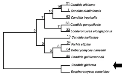
Figure 1: Phylogenetic tree of Candida species representation. (adapted from Fitzpatrick et al, 2010)
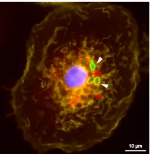
Figure 2: Fluorescent Microscopy Images show Candida glabrata replicating inside macrophages Yeast cells stained with fluorescein isothiocyanate (FITC, shown in green) prior to phagocytosis. The dye is not transferred to daughter cells allowing differentiation between mother cells (white1022 arrows) and newly formed intracellular daughter cells (red arrows) within macrophages after 8 h of co-incubation. Image reported in Kasper et al, 2015.
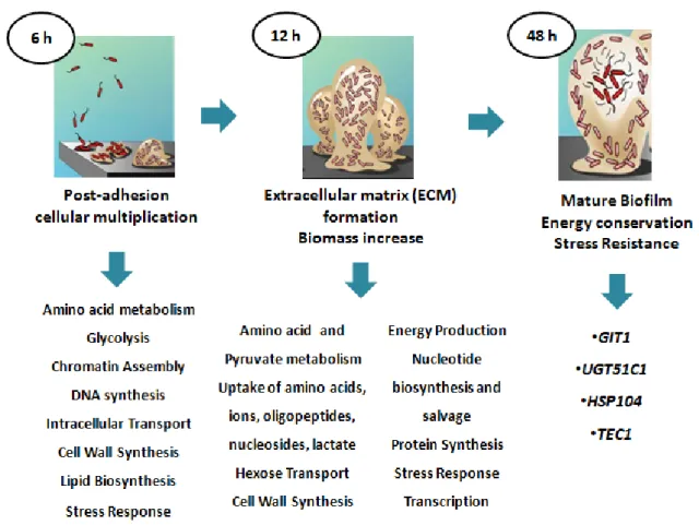
Figure 3: Scheme representing the three major phases for biofilm development. Resumed phases and main factors involved in biofilm formation are presented. Phases from adhesion to mature biofilm development are specified taking into account participating molecules and main mechanisms involved. (adapted from Yeater et al, 2007)
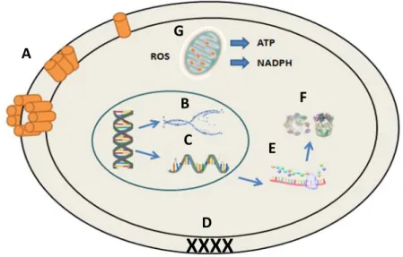
Figure 4: Mechanisms modes of action for antimicrobial peptides in microbial cells. (A) Disruption of cell membrane integrity: starting with random insertion into the membrane, followed by alignment of hydrophobic sequences and removal of membrane sections and pore formation. (B) Inhibition of DNA synthesis. (C) Blocking of RNA synthesis. (D) Inhibition of enzymes necessary for linking of cell wall structural proteins. (E) Inhibition of ribosomal function and protein synthesis. (F) Blocking of chaperone proteins necessary for proper protein folding. (G) Targeting of mitochondria: starting with inhibition of cellular respiration and induction of ROS formation and disruption of mitochondrial cell membrane integrity and efflux of ATP and NADH. (adapted from Peters et al, 2010). Figure 5- Figure 20: Confidential content present in Confidential Appendix I
xi
Table Index
Table 1: C. glabrata strains used to assess stress resistant responses and host-pathogen interactions Table 2 - Test Compounds considered for Susceptibility Assays
xii
Abbreviations
ABC – ATP Binding Cassette AMPs – Antimicrobial peptides ATP – Adenosine triphosphate
C. albicans – Candida albicans C. glabrata – Candida glabrata C. krusei - Candida krusei
CFU – colony forming unit DHA – Drug:H+ antiporter
EDTA - Ethylenediaminetetraacetic acid EEA - Early Endosomal Antigen EPA - Epithelial Adhesin Family
G. mellonella – Galleria mellonella
GM-CSF - Granulocyte-Macrophage Colony-Stimulating Factor GOF – Gain of Fuction
HPLC – High Pressure Liquid Chromatography hst5 – Histatin-5
IC – Invasive Candidiasis IL - Interleukin
INFγ - Interferon gamma
LAMP - Lysosomal-associated Membrane Protein MFS – Major Facilitator Superfamily
MMB – Minimal Medium Broth
MOPS – 3-morpholinopropane- 1-sulfonic acid NCAC – Non-Candida albicans Candida OD – Optical density
ORF – Open Reading Frame
PAMPs - Pathogen Associated Patterns PBS – Phosphate Buffered Saline PCR - Polymerase Chain Reaction PRRs - Pattern Recognition receptors
xiii
ROS – Reactive oxygen species rpm – Rotations per minute
RPMI - Roswell Park Memorial Institute medium
S. cerevisiae – Saccharomyces cerevisiae
SDB – Sabouraud’s Dextrose Broth SEM – Scanning Electron Microscopy Syk - Spleen tyrosine kinase
TNF-α - Tumor necrosis factor alpha
1
1. Thesis Outline
Candida glabrata is one of the most frequently isolated non- albicans pathogenic Candida
species, accounting for nearly 20% of the infections registered in both Europe and North America [6,7,8].
Its success in causing infection is mainly due to high intrinsic stress tolerance that enables this yeast to resist confrontation with the immune system and withstand prolonged harmful conditions such as nutrient starvation and oxidative stress [14,16,9]. Transporters of the Drug:H+ antiporter family, belonging
to the Major Facilitator Superfamily (MFS), well characterized in the model yeast Saccharomyces
cerevisiae, are known to take part in multi-drug resistance as well as in other important physiological
stress response mechanisms, including weak acid and polyamine resistance and transport [58,61,62]. At
present, not much is known about these transporters and their role in stress responses and host-pathogen interaction in C. glabrata. But for some years, the Biological Sciences Research Group has been characterizing the MFS transporters in both S. cerevisiae and, more recently, in Candida
glabrata [60,61,62,63,68,70]. This work, thus, intends to contribute to the functional characterization of two
drug:H+ antiporters, CgDtr1 and CgTpo4, particularly focusing their role in the tolerance to different stresses related to host-pathogen interactions.
The CgDtr1 and CgTpo4 transporters were preliminarily studied by Miguel Cacho Teixeira’s team (Santos, R. unpublished results), within the BSRG group, reaching the conclusion that they were both involved in early biofilm development and in the virulence outcome when interacting with the
Galleria mellonella infection model. In this work, we planned to further evaluate the molecular basis of
these observations. The knowledge gathered for the CgDtr1 and CgTpo4 homologs in S. cerevisiae
[70,71] was further used to complete the functional analysis of their C. glabrata counterparts.
This thesis is organized into five chapters. The first is an introduction to the Candida glabrata epidemiology and pathology, virulence traits, drug-and stress-resistance.
The second part is composed of the materials and method used for the development of this work, followed by the results, that compose chapter 3.
In chapter 4, the integration of the obtained results is performed, within the framework of current knowledge on the subject.
Finally, the fifth chapter is composed by the future perspectives where the unanswered questions raised by the results are highlighted. In this section proposals for future work are brought to light, with the main purposed of further trying to understand the complex mechanisms underlying the action of the studied drug:H+ antiporters that were found to stand in the cross-road of drug resistance and pathogenesis in C. glabrata.
3
2. Introduction
2.1. Candida glabrata as an Opportunistic Pathogen
The haploid yeast Candida glabrata (H.W.Anderson) S.A.Mey. & Yarrow, previously known as
Torulopsis glabrata (H.W.Anderson) Lodder & N.F.de Vries[1], is considered to be part of the Candida
genus, even though it is phylogenetically closer to Saccharomyces cerevisiae (E.C. Hans) Meyen (Figure 1) [2]. Although most S. cerevisiae genes have orthologs in C. glabrata and similar
chromosomal structure [3], C. glabrata seems to have lost some genes during the evolutionary
process, like those involved in galactose, and some related to phospate, nitrogen and sulfur metabolism, most likely due to its adaptation to the mammalian host [4].
The frequency of Candida infections has been increasing over the years, largely due to the increasing size of the at-risk populations, which includes transplant recipients, cancer patients and other patients undergoing immuno-suppressive therapy [5]. Candida species are among the top 10
most frequently isolated nosocomial bloodstream pathogens, with annual incidence rates of 1.9 up to 4.8 cases, in Europe, and 6.0 to 13.3 cases, in the United States, per 100 000 inhabitants [5].
C. albicans (Robin) Berkhout is the predominant cause of invasive candidiasis, representing
50% to 70% of all cases, however the epidemiology of Candida infections has evolved, with longitudinal studies showing that a higher proportion of patients are now infected with non-albicans
Candida species [6]. These include C. glabrata, among other examples, which is now accounting for 15
to 20% of the diagnosed cases of candidiasis in Europe [7] and for 20% in North America [8]. This
dramatic change has been partly related to the selection of less susceptible Candida strains by the widespread use of antifungal drugs [5].
Despite being incapable of switching to true hyphal growth, such as observed in C. albicans, C.
glabrata is a successful pathogen [9] . C. glabrata is able to attach to host cells and form biofilms,
function that is partly mediated by a large family of epithelial adhesins (Epa proteins), but also by other adhesin-like cell wall-anchored proteins. Adhesins can be divided in sugar-sensitive (lectin-like) or
4
sugar-insensitive, and they mediate the adherence either to the epithelium or to abiotic surfaces [10]. In
C. glabrata, only CgEpa1, CgEpa6 and CgEpa7 have been fully characterized [11].
Secretion of extracellular enzymes, such as proteases, lipases, phospholipases, esterases and hemolysins, is considered a virulence factor in Candida species. They enable pathogens to penetrate tissues and obtain nutrients [12]. They also play a role in the evasion of the immune system
by cleaving immune regulatory proteins or deregulating the cascade-activated homeostatic host systems, such as blood coagulation or antibodies formation pathways [13]. Hemolysins, for instance,
induce rupture of erythrocytes, the most common blood cell. Besides the obvious damage this rupture causes, it also releases iron, which is needed for the growth and pathogenicity of yeasts [13].
C. glabrata also appears to undergo chromosomal alterations, including chromosome loss,
rearrangements, translocations, chromosome fusions and interchromosomal duplications leading to distinct karyotypes [1]. It is even capable of de novo chromosomal generation [15]. All these properties
lead to wide population diversity. The genomic plasticity may serve as compensatory mechanism to enable fast adaptation to a changing host environment and compensate for the absence of a sexual cycle [8].
2.2. Immune Evasion Strategies and Macrophage Recognition
C. glabrata has a high intrinsic stress tolerance, which allows her to sustain prolonged
starvation periods and to withstand oxidative stress [14]. In vitro, this pathogen can tolerate the
confrontation with host immune cells, especially macrophages. Indeed, a significant fraction of cells is not only able to survive but also replicate inside human macrophages [9].
Macrophages are professional phagocytes of the monocytic lineage, which can be found in almost all tissues and are abundant in mucosal surfaces. The first contact between a phagocytic cell and a microbe is mediated by host receptors, including pattern recognition receptors (PRRs) which detect conserved basic molecular components of microorganisms, the pathogen associated patterns (PAMPs), and opsonic receptors which recognize opsonized microbes [17]. Recognition of microbial
ligands by macrophage receptors activates a series of intracellular signalling pathways that lead to phagocytosis. Engulfed microorganisms are trapped in a plasma membrane-derived vacuole, the phagosome. This premature compartment lacks the ability to break down particles or kill pathogens. During the maturation process, a series of fusion events with compartments of the endosomal pathway takes place, and allows the phagosomes to become more acidic and hydrolytically active. Nutritional limitation and oxidative and non-oxidative antimicrobial mechanisms lead to death or growth restriction of engulfed microorganisms [9].
Since the host defence mechanisms, such as the innate immune phagocytic cells, are multi-facetted, a number of different evasion strategies have evolved. These include avoidance of contact with macrophages, rapid escape from host immune cells, ability to withstand macrophage antimicrobial activities and, most importantly, use of macrophages as an intracellular niche for protection from other immune cells [9]. A frequent strategy is to mask the immune stimulatory
5
components of the cell wall to avoid recognition and activation by macrophages [18;19]. The cell wall of
yeasts is composed, in addition to other elements such as chitin, by β-glucans and proteins protected by an external layer (namely, linear polymers of mannose). β-1,3-glucan has an important role in the recognition by the immune system since it is recognized by the dectin-1 receptor on macrophages [20].
Intact C. albicans cells have less interaction with this receptor, since they are protected by the mannan layer. But, when the cell wall suffers any damage, β-1,3-glucan is exposed. When that happens, the dectin-1 receptor quickly recognizes and binds to it, allowing the activation of macrophages and the production of inflammatory cytokines such as TNF-α [21].
Candida glabrata displays a similar response, however, binding to macrophages is also partly
mediated by adhesin Epa1 [22]. Therefore, mutants with defects in protein glycosylation are killed more
efficiently by macrophages [23,24]. Recently, dectin-2 receptor was regarded as important in host
defense against this pathogenic yeast. Knock-out mice for this receptor were more susceptible to infection by C. glabrata and phagocytosis decreased in its absence [25]. In addition to recognition of
pathogens, another important feature of the immune system is its ability to effectively create an inflammatory response through chemical mediators, especially proinflammatory cytokines such as TNF-α. In C. glabrata, TNF-α or other cytokines such as IL1, IL6, IL8 and IFNγ are weakly induced upon infection. The only cytokine that appears to be induced upon infection is GM-CSF [26]. This
cytokine functions as a macrophage activator and induces their recruitment. Since C. glabrata has the ability to survive and replicate inside macrophages (Figure 2), it is reasonable to believe that attracting more macrophages to the site of infection is an invasion strategy [26].
Figure 2: Fluorescent Microscopy Images show Candida glabrata replicating inside macrophages
Yeast cells stained with fluorescein isothiocyanate (FITC, shown in green) prior to phagocytosis. The dye is not transferred to daughter cells allowing differentiation between mother cells (white1022 arrows) and newly formed intracellular daughter cells (r ed arrows) within macrophages after 8 h of co-incubation. Image reported in Kasper et al, 2015.
6
2.3. Candida glabrata Survival within the Macrophage
After phagocytosis, microorganisms are internalized in the phagolysosome, the central compartment for the antimicrobial activity of these first line defence cells.
C. glabrata strategy lies in preventing the maturation and acidification of this compartment. So,
this yeast resides in a modified phagosomal compartment that acquire early and late endosomal stage markers (EEA 1, early endosomal antigen 1, and LAMP-1, lysosomal-associated membrane protein 1, respectively), but not phagolysosomal markers (for instance, cathepsin D or signal of lysosomal fusion). Therefore, phagosomes that internalize viable yeasts are only weakly acidified [9,27]. If,
however, we considered heat-killed yeasts and their exposure to macrophages, they are internalized in a compartment with all phagolysosomal properties. This is indicative that for the modification from phagosome to phagolysosome either a heat-labile surface factor is required or the metabolic activity of the yeast is needed [9,27].
In addition, it was recently observed for C. albicans, that the metabolization of amino acids as an alternative carbon source, with consequent ammonia extrusion, is implicated in the alkalinization and neutralization of the phagosome environment [9]. Candida glabrata is also able to alkalinize its
environment in vitro when resorting to the use of amino acids in the absence of glucose. The Mnn10 and Mnn11 mannosyltransferases are important for this environment alkalinization and prevention of phagosome acidification, suggesting that they are included in the strategy to raise the environmental pH [9].
Another possible strategy lies within the dectin-1 mediated Syk signaling. C. glabrata viable cells can induce a less pronounced Syk activation, implying that a reduced signalling could underlie the delivery of viable yeasts to non-matured phagosomes [9]. Still, it is not entirely clear or accepted
that this yeast needs to prevent phagosome maturation or modify its pH to survive. The truth is that, in
vitro, this yeast can grow unaffected when exposed to acidic pH, as low as 2.0 [28]. Even so, the
combination of pH with an arsenal of antimicrobial properties within the macrophage may restrict their survival. That is, it is more likely that the observed effects result from a combination of factors.
The production of reactive oxygen species (ROS), along with a low pH value, represent a central aspect of the antimicrobial response in macrophages. Its production is driven by the NADPH oxidase complex that generates superoxide, hydroxyl anions and radicals [29]. However, C. glabrata
has a high resistance to oxidative stress. This resistance is partly mediated by Cta1 catalase expression [30]. Nevertheless, in vitro response to oxidative stress also includes thioredoxin
peroxidases (Tsa1 and Tsa2) thioredoxin reductases (Trr1 and Trr2), thioredoxin cofactor (Trx2), glutathione peroxidase (Gpx2) and superoxide dismutases (Sod1 and Sod2) [31]. During in vitro
experiences of C. glabrata interaction with macrophages, Sod1 was found to confer resistance to killing in the absence of Yap1 mediated signalling. In addition, the CTA1-reporter gene was found to be induced after phagocytosis, indicating that macrophage interaction leads to oxidative stress sensing by C. glabrata and to the subsequent activation of Cta1 [9]. Studies on human and murine
macrophages have shown that C. glabrata suppresses ROS production by phagocytes. Even so, NADPH oxidase is activated by recognition and phagocytosis of microorganisms, even before the
7
phagosome is sealed [9]. Thus, it is not expected that incomplete phagosome maturation cause a
decrease in ROS induction. ROS suppression is currently viewed as an active down-regulation of macrophage ROS production by this pathogenic yeast, as an immune evasion strategy [27,9]. While a
correlation between phagocytosis-associated oxidative metabolism and killing has been described for
C. albicans, the role of ROS in C. glabrata killing is still unclear. The high intrinsic resistance to
oxidative stress along with the fact that inhibition of ROS production in macrophages did not increase yeast survival [32], suggests that ROS may play a secondary role in direct killing of C. glabrata.
To be able to survive and replicate in the phagosome, C. glabrata has to adapt to the surrounding environment , not only low pH values and oxidative stress but also nutritional restrictions. This includes adjustment to the use of alternative carbon sources, as well as nitrogen deprivation [9].
The transcriptional response of C. glabrata to phagocytosis reflects up-regulation of genes encoding for enzymes of the glyoxylate cycle, gluconeogenesis, and β-oxidation of fatty acids, as well as down-regulation of glycolysis. These responses suggest the triggering of alternative carbon source metabolism [33,34]. Also, down-regulation of protein synthesis and up-regulation of amino acid
bio-synthetic pathways, and amino acid and ammonium transport genes, suggests that the yeast undergoes deprivation of nitrogen. In addition, methylcitrate cycle genes, important for the degradation of fatty acid chains which enables the use of lipids as alternative carbon source, was found to be up-regulated in these conditions [33,34].
Aside from carbon and nitrogen, trace elements, such as iron, are important for C. glabrata growth. Iron is an essential transition metal which serves as a cofactor for many enzymes and is required for numerous biochemical processes, including cell respiration and metabolism, oxygen transport and DNA synthesis [35]. Thus, it is not surprising that the host sequesters iron from
extracellular spaces, to challenge organisms with micronutrient limitations, a process known as nutritional immunity [36]. C. glabrata does not use ferritin or transferrin as iron sources, and is not able
to use heme or hemoglobin. However, it expresses one siderophore-iron transporter called Sit1. Sit1 mediated binding increases fitness and survival during exposure to macrophages. Iron availability and acquisition are important determinants of intracellular survival and replication in macrophages. Recent studies showed that 11 out of 23 mutants identified for reduced survival within macrophages showed defects in growth under iron limitation [32].
Depending on the organism there are a number of possible strategies to evade the host, and that also includes possible exit strategies from macrophages. These strategies include non-lytic exocytosis, observed for C. albicans and C. neoformans (San Felice) Vuillemin (assexual morph of Filobasidiella neoformans Kwon-Chung), induction of host cell death by apoptosis or pyroptosis or caspase-dependent early lytic pro-inflammatory cell death [9]. After phagocytosis, Candida albicans
rapid hypha formation is activated. Its triggering is important for initiation of inflammatory responses and also mechanical damage and escape from the macrophage [9]. C. glabrata grows predominantly in
the yeast form and only forms pseudohypha under certain conditions, particularly nitrogen deprivation. However, microevolution experiments with permanent exposure of C. glabrata to macrophages for several months selected populations with genetic changes that caused a higher pseudohypha-like growth, greater mechanical damage of macrophages and faster escape [37].
8
During the interaction with human macrophages derived from monocytes, C. glabrata remains intracellular for 2-3 days without causing obvious damage to macrophages or inducing cell death
[9].The replicating yeast are still surrounded by membrane and are not released in to the cytoplasm,
as indicated by detection of LAMP-1 (lysosomal-associated membrane protein) around the ingested yeast cells after 24 hours. After 2-3 days, macrophages with increased fungal burden were observed to lyse and release yeasts into the surrounding medium [27]. Even so, the time of macrophage burst is
dependent on the initial yeast-to-macrophage ratio and the macrophage cell type [9].
2.4. Virulence traits for the establishment of infection: biofilm formation and antimicrobial peptide resistance
Besides phagocytosis, in the event of Candida infection, the adherence to host surfaces is required for colonization and infection establishment [38].Adherence contributes to the organism
persistence inside the host, which is crucial for the disease development strategy. Candida species are well known for its adherence capacities and biofilm formation, not only within the host, but also attached to medical devices [38,39].
Cell surface proteins, like adhesins encoded by the EPA gene family, are known to promote specific adherence. But the initial attachment of Candida cells to the host is not only promoted by cell division, proliferation and adherence, but is also dependent on biofilm development [40]. Biofilms are
surface-associated communities of microorganisms involved by an extracellular matrix, and they present the most prevalent growth form amoung different microorganisms [38,41]. Since this structure
has been known to confer resistance to antifungal drugs, by limiting drug penetration, and protection from the host immune system, biofilm formation is considered one of the most important virulence factors used by pathogenic yeast cells, such as C. glabrata [42,43]. Furthermore, they have also been
associated with higher morbidity and mortality rates [44].
C. glabrata is known to produce lower quantities of biofilm mass when compared with other
NCAC (non-C. albicans Candida) species but, is has been showed that higher biofilm biomass is produced, by this yeast, on artificial surfaces, such as silicon in the presence of urine, then in SDB medium when compared with C. parapsilosis (Ashford) Langeron & Talice, 1932 or C. tropicalis Berkhout, 1923 [41,45]. C. glabrata biofilm matrices were already characterized has having high levels of
both proteins and carbohydrates, concordant with what has been seen for C. albicans whose biofilm matrix which is composed mainly by carbohydrates, proteins, phosphorus and hexosamines [41]. It is
also important to highlight that C. albicans biofilm is formed by a mixed population of budding-yeast and hyphae, but no filamentation has yet been observed in C. glabrata biofilms [46,47].
In order to fully establish a functional biofilm, the first step is the adherence of yeast cells to host surfaces or abiotic medical devices. After adherence, formation of colonies, secretion of extracellular polymeric substances, maturation in a three-dimensional structure and cell dispersion have to occur. In the following scheme (Figure 3) the various steps behind biofilm formation are detailed.
9
Besides biofilm development, another important virulence trait is the ability to resist the action of antimicrobial peptides. Antimicrobial peptides are small, positively charged, amphipathic molecules that may be composed by a variety of amino acids and have various lengths (from 6 to 100 amino acids) [48]. In humans, the most prominent innate antimicrobial peptides are the cathelicidins and
defensins, produced by immune system cells, and the histatins produced and secreted into the saliva by the parotid, mandibular and submandibular salivary glands [48].
Most antimicrobial peptides work directly against microbes through a mechanism that involves membrane disruption and pore formation, that eventually leads to the efflux of essential ions and nutrients [49]. The mechanism behind the association with and permeabilizing off the microbial cell
membranes is not entirely clear, but these peptides are proposed to bind to the cytoplasic membrane, creating micelle-like aggregates, leading to a disruptive effect (Figure 4) [48]. Nonetheless, studies
have been indicating the presence of additional or complementary mechanisms such as intracellular targeting of cytoplasmic components crucial to cell physiology functions [48]. Thus, the initial interaction
between peptides and microbial cell membranes would allow them to penetrate inside the cells to bind to intracellular molecules, resulting in the inhibition of cell wall biosynthesis, and processes involving DNA, RNA and protein synthesis and function [50,51] . These modes of action, combined with a broad
Figure 3: Scheme representing the three major phases for biofilm development. Resumed phases and main
factors involved in biofilm formation are presented. Phases from adhesion to mature biofilm development are specified taking into account participating molecules and main mechanisms involved. (adapted from Yeater et al, 2007)
10
range of lethal activity in a short period of time, corroborate the hypothesis that antimicrobial peptides are excellent candidates for the development of novel therapeutic agents [48]. Therefore, the
acquisition of resistance against their killing activity is crucial for yeasts, and other pathogenic microorganisms, to establish a successful infection.
2.5. Infection Models to Study Virulence of Candida glabrata
Historically, there has been little interest in developing animal models for C. glabrata infections, despite the emergence of both systemic and mucosal infections [52]. Even so, different mouse models
of oral, vaginal, gastrointestinal, intraperitoneal and intravenous bloodstream infections have been developed [53]. During systemic mouse infection, fungal dissemination spreads to different organs,
such as brains, kidneys, lungs and even heart, which shows that Candida glabrata is highly adaptable and able to colonize many niches [52,53]. Grating this, different mouse models have been applied to
study the virulence of C. glabrata gene deletion mutants. These studies analyze fungal burden and not host mortality, due to low mortality of mice after infection, since even high fungal burden still causes low tissue damage and low inflammation responses (Kasper et al; 2015). That being established, the respective genes should be termed fitness factors rather than virulence factors of C.
glabrata [9].
Additionally, non-vertebrate models, such as Drosophila melanogaster, Caenorhabditis
elegans and Galleria mellonella have also been established for this yeast. These offer a number of
advantages over mammalian vertebrate models due to economy of size, ease of handling and ethical issues [54,55].
Galleria mellonella larvae, for instance, bears several advantages: they are cheap to acquire
and easy to keep, their size simplifies infection and the inoculation process via syringe allows control
Figure 4: Mechanisms modes of action for antimicrobial peptides in microbial cells. (A) Disruption of cell
membrane integrity: starting with random insertion into the membrane, followed by alignment of hydrophobic sequences and removal of membrane sections and pore formation. (B) Inhibition of DNA synthesis. (C) Blocking of RNA synthesis. (D) Inhibition of enzymes necessary for linking of cell wall structural proteins. (E) Inhibition of ribosomal function and protein synthesis. (F) Blocking of chaperone proteins necessary for proper protein folding. (G) Targeting of mitochondria: starting with inhibition of cellular respiration and induction of ROS formation and
disruption of mitochondrial cell membrane integrity and efflux of ATP and NADH. (adapted fromPeters et al, 2010).
A
B
C
XXXX
D
E
F
G
11
of the amount of pathogen applied. Plus, the larvae can be incubated at a range of various temperatures which may go up to 37ᵒC [55].
The larval immune system has high similarity to the mammalian innate immune system. The haemocytes, phagocytic cellular components similar to neutrophils, are able to phagocyte and kill pathogens by producing reactive oxygen species and lytic enzymes [56]. Furthermore, the humoral
response to infecting microbes involves antimicrobial peptides which were shown to be induced by fungal pathogens, and differ from those induced by bacterial pathogens [55]. This feature is very
important because it allows comparison of data obtained in Galleria virulence studies and murine models. Moreover, experiments with G. mellonella grant fast data acquisition, usually with results within one or two weeks [55]. Notwithstanding, there are also disadvantages of this model. After all, the
genome of G. mellonella has not been fully sequenced and there are no mutant strains available to functionally study host response. Plus, they do not have adaptive immune systems, which disable further immune response studies. Even so, Galleria mellonella is a useful tool and potent alternative infection model to study fungal pathogens.
2.6. Antifungal Drug Resistance
During the past three decades antifungal drug resistance became more common and a serious concern to the medical community [1]. The proportion of azole resistance in clinical isolates
across several countries has been shown to increase in the period from 2001 to 2007 [56]. Furthermore,
resistance to echinocandins of fluconazole-resistance C. glabrata isolates was shown to have increase from no cases between 2001-2004 to a 9,3% frequency in the 2006-2010 time period [57].
This supports the idea that drug resistance in C. glabrata is developing fast.
Classes of antifungals are categorized based on their mechanisms of action. Azoles inhibit the cytochrome P450-dependent enzyme lanosterol-demethylase which is crucial for the biosynthesis of ergosterol. If ergosterol synthesis is affected, the resulting cell membrane dysfunction impairs signalling and transport processes [1]. Polyene antifungals, such as amphotericin B, interact with
ergosterol in the cell membrane creating pores within the cell membrane causing small molecules to leak, resulting in cell death [47]. 5-Flucytosine is an antifungal that interferes with protein synthesis.
Upon entering the cell through a cytosine permease, this drug is converted into a nucleotide analogue, 5-flurouracil, which is incorporated into RNA, subsequently interfering with the synthesis of proteins [38].
Finally, the more recently introduced antifungals, echinocandins, have become the first line therapy for invasive candidiasis. They are able to inhibit β-(1,3)-D-glucan synthase activity, preventing the synthesis of β-(1,3)-D-glucan, essential for the composition of the fungal cell wall [47].
Understanding the mechanisms of antifungal resistance is the key to unravel new therapeutic options. Resistance may be classified as: primary resistance, when strains are unaffected by the antifungal without prior exposure; or secondary resistance, when strains acquire resistance after exposure to the drug. Secondary resistance is, by far, the most commonly occurring phenomenon among Candida species [1].
12
C. glabrata, when compared to other Candida species, is the least susceptible to azoles. The
mechanisms behind azole resistance in C. glabrata include changes in P-450 lanosterol demethylase enzyme, encoded by ERG11, which results in loss of the drug affinity and/or overexpression of ERG11, or activation of drug extrusion catalysed by membrane transport proteins of the ATP binding cassette (ABC) superfamily or the Major Facilitator Superfamily (MFS) [1]. Azoles enter the cell by facilitated
diffusion and bind to the iron present in the heme group of the Erg11 enzyme [58,59]. This N-Fe
connection restrains the activation of oxygen, which is necessary for the demethylation of lanosterol, causing a blockage in the ergosterol production. Consequently, a toxic sterol, known as 14-α-methyl-3,6-diol, accumulates, causing severe stress in the plasma membrane. This results in a fungistatic behaviour in the yeast cells [58].
CDR1 and CDR2 genes, encoding drug efllux pumps of the ABC superfamily, are
up-regulated in response to azole exposure. The transcription factor, Pdr1, that regulates the expression these genes, is activated through direct binding to the azole drug molecule, promoting the expression of the drug efflux pump encoding genes, being a key azole resistance determinant. Disruption and/or removal of the PDR1 gene was found to increase susceptibility to azoles [56]. It was also found that
Gain Of Function (GOF) mutations in PDR1, identified in azole resistant clinical isolates, enhance azole resistance and even virulence in mice models. More recently, a few multidrug transporters of the Major Facilitator Superfamily, suggested to act as Drug:H+ Antiporters (DHA), have also been shown to confer resistance to azole drugs, especially imidazoles [60,61,62,63].
Little is known about the exact mechanism for resistance to polyene antifungal drugs in C.
glabrata. A clinical isolate susceptible to polyene treatment was observed to have severe changes in
the sterol composition of its membrane [64]. This isolate had accumulated numerous sterol
intermediates that were still able to maintain membrane viability. Further studies showed also that nonsense mutations, as opposed to missense, in ERG6 gene generated altered sterol composition in the cell membrane [65].
Unlike azole antifungals, secondary resistance to echinocandins has conclusively been
unrelated to the drug-efflux mechanism as they prove to be poor substrates for most multidrug efflux transporters [66]. Instead, echinocandin resistance is caused by interference between the drug and the
β-1,3-D glucan synthase. The target subunits for this enzyme are encoded by three genes: FSK1,
FSK2 and FSK3 [67]. Point mutation in the FSK1 and FSK2 are documented as being responsible for
echinocandin resistance in Candida glabrata. Mutations in "hot spots" of these genes cause a high prevalence of amino acid substitutions conferring echinocandin resistance [64].
13
2.7. The putative Candida glabrata Multidrug Resistance Transporters CgDtr1 and CgTpo4
In this study, the uncharacterized drug:H+ antiporters Dtr1 and Tpo4, encoded by ORF
CAGL0M06281g and CAGL0L10912g, respectively, were studied in the context of their role in C. glabrata virulence.
First studied in S. cerevisiae, MFS transporters related to mutidrug resistance were grouped into two families: Drug:H+ antiporter family (DHA1), including 12 transporters, and Drug:H+ antiporter family 2 (DHA2), including 10 transporters. The difference between the two groups is established based on the number of transmembrane domains exhibited by each family, 12 for the DHA1 family members and 14 for the DHA2 [68]. In Candida glabrata we find 10 predicted members of the DHA1
and 5 of the DHA2 families. From these, a total of 6 seem to play a role in drug resistance and detoxification of the cell, but none of the DHA2 is, up to date, described as being involved in this phenomenon [61,62].
Studies in the S. cerevisiae homologs of Dtr1 and Tpo4 were used to guide the functional analysis of their C. glabrata counterparts. S. cerevisiae Dtr1 is synthesized in the endoplasmic reticulum and then transits to the Golgi complex before reaching the prospore membrane, it's final location. Dtr1 is formed by 572 amino acids with 12 predicted transmembrane domains and a large cytoplasmic loop in the middle [69). It is encoded by the open reading frame YBR180W, and is
described as being a membrane dityrosine transporter [70). This role is related to the translocation of
bisformil-dityrosine from the cytoplasm of the prospore to the maturing spore wall during the wall formation process. According to Ferder and colleagues, in the absence of DTR1, the amount of D-L dityrosine (present in the spore surface) is reduced to 65% and the racio between this surface macromolecule and the soluble spore precursors (DL/LL racio) changes from 0,6 to 0,2. The change of this value does not seem to have a major consequence in the sporulation process. Even so, it was observed, by the same authors, that the Dtr1 transporter is essential for bisformyl dityrosine molecule being able to pass through the plasmatic membrane, on its way to the prospore membrane [70).
Besides dityrosine, Dtr1 was also shown to play a role in the export of weak acid conjugated bases, its expression conferring resistance to this class of compounds [70).
In the case of Tpo4, it was identified in S. cerevisiae as a determinant of resistance to polyamines [71). The polyamine content present in the cytoplasm of yeast cells appears to be tightly
regulated by Tpo4, but also paralogous transporters including Tpo1, Tpo2 and Tpo3, that guarantee an optimal intracellular polyamine content [71).
14
3. Materials and Methods 3.1. Strains and Growth Media
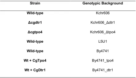
Considering the thesis objectives, several strains of Candida glabrata where used and are described in Table 1.
Table 1: C. glabrata strains used to assess stress resistant responses and host-pathogen interactions
Strain Genotypic Background
Wild-type Kchr606 Δcgdtr1 Kchr606_Δdtr1 Δcgtpo4 Kchr606_Δtpo4 Wild-type L5U1 Wild-type By4741 Wt + CgTpo4 By4741_tpo4 Wt + CgDtr1 By4741_dtr1
Cells of C. glabrata were cultured in rich Yeast extract Peptone Dextrose (YPD) medium, containing per litre: 20g D(+)-glucose (Merck), 20g of bacterial-peptone (LioChem, Inc.) and 10g of yeast extract (Difco) and also in Minimal growth medium (MMB) medium, containing per litre: 20g D(+)-glucose (Merck), 2,7g ammonium sulphate (Difco) and 1,7g of Yeast-Nitrogen-Base without amino acids (Difco). When needed, solid media was obtained by supplementation with 20g/L of agar (NzyTech). In what concerns the L5U1 strains, growth media was supplemented with leucine 20x concentrated and 1mM of copper solution. As for BY4741, solid and liquid media, was supplemented with an amino acid solution containing, per liter, 20 mg methionine, 20 mg histidine, and 60 mg leucine. Furthermore, when using this strain in order to express differences between mutated strains, the sugar composition of MMB changed from 20g/L D(+)-glucose (Merk) to 1g/L D(+)-glucose (Merk) plus 10g/L D(+)-galactose (Sigma).
Roswell Park Memorial Institute medium (RPMI) 1640 and Sabouraud Dextrose Broth (SDB) media were used for biofilm assays. Such media preparation took 10,4 g of RPMI 1640 (Sigma-Aldrich), 34,5g of MOPS (Sigma) and 18g of D(+)-glucose (Merk) dissolved in 1L of deionised water, adjusted to pH 4.0 and 40g of D(+)-glucose (Merk) and 10g of peptone (LioChem) per litre of deionised water, adjusted to pH 5.6, respectively.
3.2. Cloninf of the C. glabrata CgDTR1 and CgTPO4 genes
3.2.1. Cloning of the C. glabrata CgDTR1 gene (ORF CAGL0M06281g)
The pGREG576 plasmid from the Drag & Drop collection [73] was used to clone and express the C.
glabrata ORF CAGL0M06281g in S. cerevisiae, as described before for other heterologous genes [74].
15
yeast selectable marker URA3 and the GFP gene, encoding a Green Fluorescent Protein (GFPS65T), which allows monitoring of the expression and subcellular localization of the cloned fusion protein. CAGL0M06281g DNA was generated by PCR, using genomic DNA extracted from the sequenced CBS138 C. glabrata strain, and the following specific primers: 3’ – GAATTCGATATCAAGCTTATCGATACCGTCGACAATGAGCACCTCCAGCAACAC - 5’ and 3’ – GCGTGACATAACTAATTACATGACTCGAGGTCGACTCAGAAACTGTCTTTAACCC - 5’. The designed primers contain, besides a region with homology to the first and last 22 nucleotides of the
CAGL0M06281g coding region (italic), nucleotide sequences with homology to the cloning site
flanking regions of the pGREG576 vector (underlined). The amplified fragment was co-transformed into the parental S. cerevisiae strain BY4741 with the pGREG576 vector, previously cut with the restriction enzyme SalI, to obtain the pGREG576_CgDTR1 plasmid. Since the GAL1 promoter only allows a slight expression of downstream genes in C. glabrata, to visualize by fluorescence microscopy the sub-cellular localization of the CgDTR1 gene in C. glabrata, a new construct was obtained. The GAL1 promoter present in the pGREG576_CgDTR1 plasmid was replaced by the copper-induced MTI C. glabrata promoter, giving rise to the pGREG576_MTI_CgDTR1 plasmid. The MTI promoter DNA was generated by PCR, using genomic DNA extracted from the sequenced CBS138 C. glabrata strain, and the following specific primers: 3’ - TTAACCCTCACTAAAGGGAACAAAAGCTGGAGCTCTGTACGACACGCATCATGTGGCAATC - 5’ and 3’ - GAAAAGTTCTTCTCCTTTACTCATACTAGTGCGGCTGTGTTTGTTTTTGTATGTGTTTGTTG - 5’. The designed primers contain, besides a region with homology to the first and last 19 nucleotides of the first 1000 bp of the MTI promoter region (italic), nucleotide sequences with homology to the cloning site flanking regions of the pGREG576 vector (underlined). The amplified fragment was co-transformed into the parental strain BY4741 with the pGREG576_CgDTR1 plasmid, previously cut with SacI and NotI restriction enzymes to remove the GAL1 promoter, to generate the pGREG576_MTI_CgDTR1 plasmid. The recombinant plasmids pGREG576_CgDTR1 and pGREG576_MTI_CgDTR1 were obtained through homologous recombination in S. cerevisiae and verified by DNA sequencing.
3.2.2. Cloning of the C. glabrata CgTpo4 gene (ORF CAGL0L10912g)
The pGREG576 plasmid from the Drag & Drop collection [73] was used to clone and express the C.
glabrata ORF CAGL0L10912g in S. cerevisiae, as described before for other heterologous genes [74].
pGREG576 was acquired from Euroscarf and contains a galactose inducible promoter (GAL1), the yeast selectable marker URA3 and the GFP gene, encoding a Green Fluorescent Protein (GFPS65T), which allows monitoring of the expression and subcellular localization of the cloned fusion protein. CAGL0L10912g DNA was generated by PCR, using genomic DNA extracted from the sequenced CBS138 C. glabrata strain, and the following specific primers: 3’ – GAATTCGATATCAAGCTTATCGATACCGTCGACAATGGCCGGTACAAATCAAG- 5’ and 3’ – GCGTGACATAACTAATTACATGACTCGAGGTCGACCTATACCATTCTAGAGGAG - 5’. The designed primers contain, besides a region with homology to the first and last 22 nucleotides of the
CAGL0L10912g coding region, nucleotide sequences with homology to the cloning site flanking
regions of the pGREG576 vector (underlined). The amplified fragment was co-transformed into the parental S. cerevisiae strain BY4741 with the pGREG576 vector, previously cut with the restriction
16
enzyme SalI, to obtain the pGREG576_CgTPO4 plasmid. Since the GAL1 promoter only allows a slight expression of downstream genes in C. glabrata, to visualize by fluorescence microscopy the sub-cellular localization of the CgTPO4 gene in C. glabrata, a new construct was obtained. The GAL1 promoter present in the pGREG576_CgTPO4 plasmid was replaced by the copper-induced MTI C.
glabrata promoter, giving rise to the pGREG576_MTI_Cgtpo4 plasmid. The MTI promoter DNA was
generated by PCR, using genomic DNA extracted from the sequenced CBS138 C. glabrata strain, and
the following specific primers: 3’ -
TTAACCCTCACTAAAGGGAACAAAAGCTGGAGCTCTGTACGACACGCATCATGTGGCAATC - 5’ and 3’ - GAAAAGTTCTTCTCCTTTACTCATACTAGTGCGGCTGTGTTTGTTTTTGTATGTGTTTGTTG - 5’. The designed primers contain, besides a region with homology to the first and last 19 nucleotides of the first 1000 bp of the MTI promoter region (italic), nucleotide sequences with homology to the cloning site flanking regions of the pGREG576 vector (underlined). The amplified fragment was co-transformed into the parental strain BY4741 with the pGREG576_CgTpo4 plasmid, previously cut with SacI and NotI restriction enzymes to remove the GAL1 promoter, to generate the pGREG576_MTI_CgTpo4 plasmid. The recombinant plasmids pGREG576_CgDTR1 and pGREG576_MTI_CgTpo4 were obtained through homologous recombination in S. cerevisiae and verified by DNA sequencing.
3.3. Virulence assessment using the Galleria mellonella infection model 3.3.1. Survival and Proliferation Assessment Assays
Galleria mellonella larvae were reared on a pollen grain diet at 25ᵒC in darkness. Larvae
weighting 250 ± 25 mg were used in survival assays, where the larvae infection was performed as described previously [116]. C. glabrata strains were cultured in YPD up to stationary phase and
harvested by centrifugation in order to achieve an yeast suspension. Each caterpillar was injected with 3.5 μL of yeast suspension (with approximately 5x10⁷ cells per injection) via the last left proleg.
Following injection, 10 and 30 larvae, for survival and proliferation assays, respectively, were placed in Petri dishes and maintained in the dark at 37oC over a period of 72 hours. Control larvae were injected
with PBS (pH 7.4). Regarding the survival assays, caterpillars were considered dead when they displayed no movement in response to touch. For proliferation assays, hemolymph was recovered with a microsyringe after 1, 24 and 48 hours and plated in YPD medium for CFU counting. Larvae injections and hemolymph recoveries were performed in collaboration with Dra. Dalila Mil-Homens, Biological Sciences Research Group of the Institute for Bioengineering and Biosciences.
3.3.2. Haemocyte Interaction Assays
3.3.2.1. In vitro cultivation of haemocytes of G. mellonella.
To isolate G. mellonella haemocytes, hemolymph was collected from larvae previously anesthetized on ice and surface sterilized with ethanol by puncturing the larval abdomen with a sterile needle.The outflowing hemolymph was immediately transferred into a sterile microtube containing anticoagulant buffer (98 mM NaOH, 145 mM NaCl, 17 mM EDTA, 41 mM citric acid [pH 4.5]) in a 1:1 proportion. The hemolymph was centrifuged at 250 × g for 10 min at 4°C to pellet haemocytes. The supernatant was
