ORIENTADOR
Attur Shanmugam Arun, B.V.Sc., M.V.Sc. CO-ORIENTADOR
João Nestor das Chagas e Silva, PhD CONSTITUIÇÃO DO JÚRI
Doutor Fernando Jorge Silvano Boinas Doutor Virgílio da Silva Almeida
Doutor João Nestor das Chagas e Silva
UNIVERSIDADE DE LISBOA
Faculdade de Medicina Veterinária
TUBERCULOSIS INFECTION IN CAPTIVE SLOTH BEARS (MELURSUS URSINUS) - A PILOT STUDY ON DIAGNOSTIC STRATEGIES
MARIA ISABEL RIBEIRO PEREIRA
2016 LISBOA
ORIENTADOR
Attur Shanmugam Arun, B.V.Sc., M.V.Sc. CO-ORIENTADOR
João Nestor das Chagas e Silva, PhD CONSTITUIÇÃO DO JÚRI
Doutor Fernando Jorge Silvano Boinas Doutor Virgílio da Silva Almeida
Doutor João Nestor das Chagas e Silva
UNIVERSIDADE DE LISBOA
Faculdade de Medicina Veterinária
TUBERCULOSIS INFECTION IN CAPTIVE SLOTH BEARS (MELURSUS URSINUS) - A PILOT STUDY ON DIAGNOSTIC STRATEGIES
MARIA ISABEL RIBEIRO PEREIRA
DISSERTAÇÃO DE MESTRADO INTEGRADO EM MEDICINA VETERINÁRIA
2016 LISBOA
“Be the change you wish to see in the world”
i
Acknowledgements
To my parents, who have been the most essential foundation through this whole journey, and for never, ever, finding that my dreams could not go beyond that. To my sister Ana Maria, for simply being the best person that I could have ever asked for to have by my side, since I remember existing. This victory belongs to us four.
To Dr. Arun, for everything you taught me, showed me and entrusted me, for all the missions you let me engage in, all the precious research materials and pictures you provided me, and the long distance effort you endured for me to have this work done. I could not have done this without your help and support.
To Professor Nestor for all the amazing support, for always having a kind word to say, for allow me to go along on your field trips, and, overall, for your never-ending enthusiasm with my Indian wild adventures. You were exactly the person I needed with me in this.
To Professor Telmo Nunes, for your availability in helping me with the study analysis chapter of this dissertation, your great advice and the cheerful words.
To Professor Virgílio Almeida, for impersonation in the best way possible what it really means to be a teacher.
To Wildlife SOS for the externship opportunity and the chance to meet and work with such an inspiring team.
To the Bannerghatta Biological Park Forest Department, for giving permission for me to work at BBRC for so long.
To my nana Celeste and my grandpa Joaquim, though they were not able to watch me finish this mark, will always be a part of the person that chose this path, the one that is now finishing it, and the bigger one that I will become.
To my grandma Helena, my grandpa Carlos, and to the rest of my large ‘Ribeiro’ family, which always gave me the greatest comfort and joy to be part of. A special acknowledgement to my big cousin Diogo, much more of a friend than a colleague, and much more of a brother than a cousin. Veterinary Medicine will remember us.
To my “since ever” friends, Joana, Mirco, Filipa, Marta and Rita, for continuing to collect along with me, more and more years of growth and friendship, and for it to have survived distance, countless finals seasons and too many weeks of “saudade”.
To the friends FMV granted me, Patrícia, Bárbara, Esmeralda, Sara, Gomes, João, Maria and the rest of the “E” class, for making the last six years the best ones yet. You are the true meaning of “second family”.
To VETuna, for keeping something so significant as music present in my live, and, overall, for putting in my way people that, otherwise, might have never crossed it. Gonçalo, Joana, Sandra, Madeira, Menino, Pecho, Filipe, Inês and Raquel: we have come a long way together and it was the biggest pleasure to perform by your side.
ii
To the 2014 FAUNA board, for showing me the true meaning of effort, team work and unmeasured dedication for a common goal.
To all BBRC staff, from the keepers to the cook, for everything I experienced with you all. A special thank you to Sridhar and Imam for making me feel like part of the family and for having my back through our journeys across India.
To Susan, my everyday vet partner, for everything you taught me, for all the help, friendship, culinary experiences, and company.
To Catherine, my favorite vet nurse and dear friend, for everything we lived in India, for being an amazing teacher, for your patience, and for showing me how much someone can be passionate about animal’s welfare. Sincerest thanks for the grammar revision of this dissertation.
To the BBP Zoo Doctors, Nirupama and Vaseem, for always being so available, professional, friendly, kind and for giving me the precious opportunity of learning and engage in so many procedures at the Zoo.
To everyone at Hospital Veterinário Arco do Cego, Sheila, Joana, Rita, Marta, Mariana, Ivo, Teresa, Mari, Liliana and Catarina, for playing such a huge role in the formation and consolidation of the future ‘vet me’, and for making the times I spent (and keep spending) in your company, happy ones. A special thanks to Susana, for being the best and most loyal partner in my 2016 veterinary adventure.
To Dr. Lapão from Lisbon Zoo, for allowing me to use and consult the Zoo’s Wildlife Medicine precious books. They were very useful.
To Mr. Nuno Silva from Pc Medics, for saving my thesis materials when an informatics tragedy fell upon my pen drive.
Finally, to whoever might read this dissertation in the future with a passionate heart about wildlife and conservation medicine. Hopefully, you will not be disappointed.
iii
Abstract
Tuberculosis infection in captive sloth bears (Melursus ursinus) - A pilot study on diagnostic strategies
Tuberculosis (TB) is a cause of significant morbidity and mortality in both domestic and wild animals, and in humans, remaining a major global public health issue, especially in developing countries as India, the one with the highest TB burden in the world.
Infection by Mycobacterium tuberculosis in rescued sloth bears represents a typical case of spillover infection resulting from a prolonged and close cohabitation with infected humans, after being forcibly and illegally poached from the wild as cubs and trained to behave as entertainers in the streets of India. The stress, traumas and hardship that the animals endure, also play a role in the development of the disease, as their immune system is usually compromised. As in the case of many other wild species, there is critical lack of accredited tests for TB screening in sloth bears. This way, it is of major importance to identify the diagnostic assays that have the highest sensitivity and specificity, in order to achieve a reliable diagnosis and implement a standard methodology.
Various diagnostic methods were used to examine 15 presumable positive animals at Bannerghatta Bear Rescue Centre, in the Karnataka state, India, and their sensitivity was calculated, based on the results. M. tuberculosis infection was strongly suspected ante-mortem, based on the animals’ background as “dancing bears” and the revelation of several positive diagnostic results during their lives. Fourteen out of these 15 bears died between 2008 and 2013 and their death certificate reported post-mortem tuberculosis confirmation. Considering the previous statement, it was not possible to calculate the specificity and, thus, obtain a global and underlying insight about the tests in question.
Post-mortem methods present, in general, the highest sensitivity results. According to this study, the ante-mortem methods with the most promising results were the ones belonging to the indirect assay category, which are based upon the animal’s immune response (both cellular and humoral) instead of the organism detection (as in culture, PCR and microscopy).
A sensitivity increment was achieved when two or three tests from the three major test categories (direct, indirect based on cellular immunity and indirect based on humoral immunity) were used in parallel testing. The highest sensitivity achieved by multiple testing (93.3%) was the same for both double and triple parallel combinations, showing, in this case, no advantages in using combinations of three tests, instead of two, in terms of sensitivity increment.
iv
Resumo
Infeção por tuberculose em ursos-beiçudos (Melursus ursinus) em cativeiro - Um estudo piloto em estratégias diagnósticas
A Tuberculose (TB) provoca significativos índices de morbilidade e mortalidade em animais domésticos e selvagens, e em seres humanos, representando um considerável problema de saúde pública, sobretudo em países em vias de desenvolvimento, como a Índia, neste momento aquele com a incidência mais elevada de tuberculose em todo o mundo.
A infeção pelo Mycobacterium tuberculosis em ursos-beiçudos em cativeiro, traduz a típica ocorrência de infeção acidental, resultando da prolongada e próxima co-existência com seres humanos infectados, após serem forçada e ilegalmente capturados do seu habitat natural, enquanto crias, e treinados para se comportarem como animadores nas ruas da Índia. O stress, traumas e adversidades passados por estes animais, são fatores no desenvolvimento desta doença, uma vez que o seu sistema imunitário se encontra normalmente comprometido. Como em muitas outras espécies selvagens, existe uma enorme lacuna no que diz respeito a métodos de testagem de tuberculose em ursos-beiçudos. Deste modo, é da maior importância definir os testes que possuem a sensibilidade e especificidade mais elevadas, potenciando a obtenção de um diagnóstico fiável e de uma metodologia padronizada.
Diversos métodos diagnósticos foram aplicados em 15 animais presumivelmente positivos e residentes no Bannerghatta Bear Rescue Centre, em Karnataka, na Índia, e a sua sensibilidade foi calculada, com base nos resultados obtidos. A infeção por M. tuberculosis era fortemente suspeitada ante-mortem, com base na proveniência e passado dos animais como “dancing bears” e em vários resultados diagnósticos positivos durante a sua vida. Catorze destes 15 ursos morreram, entre 2008 e 2013, e o seu relatório de óbito reportou a confirmação post-mortem de tuberculose. Desta forma, não foi possível calcular a sua especificidade e, assim, obter um conhecimento global e aprofundado dos testes em questão.
Os testes post-mortem apresentam, em geral, as sensibilidades mais elevadas. Segundo este estudo, os testes ante-mortem com os resultados mais promissores pertencem à categoria de métodos indiretos, baseados na deteção da resposta imunitária do próprio animal (tanto celular como humoral), ao invés da deteção do organismo(como em cultura, PCR e microscopia). Um aumento da sensibilidade foi conseguido quando dois ou três testes das três principais categorias exploratórias específicas (diretos, indiretos baseados em imunidade celular e indiretos baseados em imunidade humoral) foram usados em testagem paralela. A sensibilidade mais elevada obtida por uma combinação de testes (93.3%) foi a mesma para combinações paralelas duplas e tripla, demonstrando, neste caso, que não existem vantagens em combinar três testes, em vez de dois, no que toca ao aumento de sensibilidade.
Palavras-chave: urso-beiçudo, Mycobacterium tuberculosis, epidemiologia, diagnóstico, vida-selvagem, zoonose.
v Table of Contents Acknowledgements ... i Abstract ... iii Resumo ... iv Table of Contents ... v
List of Figures ... viii
List of Tables ... x
List of Graphics ... x
List of Abbreviations and Symbols ... xi
Chapter I- Introduction... 1
1. Sloth Bears ... 2
2. Kalandars and “Dancing bears” ... 4
3. Rescue Centres ... 5
4. Content Notice ……….. 7
Chapter II- Training Period Activities ... 8
Chapter III- Literature review ... 15
1. Etiology... 15
1.1. Taxonomy and Description of the Genus ... 15
1.2. Life Cycle... 16
1.3. Transmission ... 17
2. Epidemiology ... 17
2.1. Frequency and Prevalence ……….………. 18
2.2. Mycobacterium tuberculosis complex and reported hosts ... 18
2.3. The Carnivore Host ... 19
2.4. Susceptibility to Infection ... 20
2.4.1. Species Susceptibility ... 20
2.4.2. Immune System Status... 21
2.4.3. Stress ... 22
2.4.4. Human-Animal interface ... 23
2.4.5. Prevalence of TB infection within human populations ... 24
2.5. Zoonotic Risk ... 25 3. Pathogenesis ... 26 3.1. Generalities ... 26 3.2. Lesions ... 26 3.2.1. Lesions Distribution ... 27 3.2.2. Macroscopic lesions ... 28 3.2.3. Microscopic lesions ... 30 3.3. Clinical Signs ... 31 4. Diagnosis ... 32 4.1. Unspecific Diagnosis ... 32
vi
4.1.1. Clinical signs ... 32
4.1.2. Imaging ... 33
4.1.3. Clinical Pathology (Hemathology and Serum Chemistry) ... 34
4.1.4. Gross Pathology ... 34
4.1.5. Histopathology ... 34
4.2. Direct assays ... 35
4.2.1. Culture ... 35
4.2.2. Acid-Fast Staining Microscopy ... 37
4.2.3. Molecular Methods ... 38
4.2.3.1. Polymerase Chain Reaction (PCR)... 38
4.2.3.2. Amplified Mycobacterium tuberculosis Direct Test ... 39
4.2.3.3. Restriction Fragment Length Polymorphism (RFLP) ... 39
4.2.3.4. Spoligotyping ...40
4.3. Indirect assays... 40
4.3.1. Cell-Mediated Immunologic (CMI) Tests ... 41
4.3.1.1. In vivo Cell-Mediated Tests ... 41
4.3.1.2. In vitro Cell-Mediated Tests ... 42
4.3.1.2.1. Lymphocyte Transformation Test ... 43
4.3.1.2.2. IFN-ү Release Assays ... 43
4.3.1.2.3. Other assays ... 44
4.3.2. Serological Tests ... 44
4.3.2.1. Rapid Lateral Flow Technology Test ... 45
4.3.2.2. MAPIA and ELISA ... 47
4.3.2.3. Other assays ... 47
4.4. Current screening recommendations ... 48
4.5. Sample Collection ... 48
4.5.1. Ante-mortem ... 48
4.5.2. Post-mortem ... 50
5. Management Considerations for Positive or Suspected Animals ... 53
5.1. Treatment ... 53
5.1.1. Treatment Protocols ... 54
5.1.2. Dosages and Administration Routes ... 54
5.1.3. Side Effects ... 55 5.1.4. Mechanism of Action ... 56 5.1.4.1. Rifampicin ... 56 5.1.4.2. Isoniazid ... 56 5.1.4.3. Pyrazinamide ... 56 5.1.4.4. Ethambutol ... 56 5.1.4.5. Streptomycin ... 56 6. Prophylaxis ... 56 6.1. Drug Protocols ... 57
vii
6.2. Vaccination ... 57
6.3. Other Preventive Measures ... 57
7. Differential Diagnosis ... 58
Chapter IV- Study ...60
1. Aims of the Study...60
2. Material and Methods ... 60
2.1. Inclusion Criteria ... 60 2.1.1. Specimen Information ... 60 2.1.2. Testing Considerations ... 61 2.1.3. Treatment Considerations ... 63 2.2. Data Analysis ... 63 3. Results...65 3.1. Ante-mortem Tests ... 65 3.1.1. Unspecific Tests ... 65 3.1.2. Direct Tests ... 66 3.1.3. Indirect Tests ... 67 3.2. Post-mortem Tests ... 68
3.3. Ante-mortem Tests Combination ... 69
3.3.1. Combination of two different tests ... 69
3.3.1.1. Inclusion Criteria ... 69
3.3.1.2. Results ... 69
3.3.2. Combination of Three Different Tests ... 70
3.3.2.1. Inclusion Criteria ... 70
3.3.2.2. Results ... 70
3.4. Graphical Analysis ... 71
4. Discussion ... 72
4.1. General Considerations ... 73
4.2. Unspecific Tests (Ante-mortem and Post-mortem) ... 74
4.3. Direct Tests (Ante-mortem and Post-mortem) ... 75
4.4. Indirect Tests ... 76
4.5. Ante-mortem Tests Combination ... 77
4.6. Ante-mortem Testing Strategies ... 78
5. Conclusions ... 81
References ... 83 73
viii
List of Figures
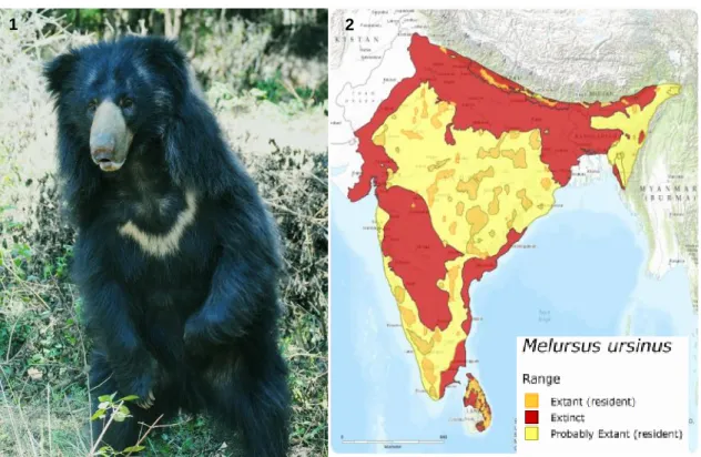
Figure 1- Indian sloth bear (Melursus ursinus ursinus) at Bannerghatta Biological Park ... 3
Figure 2- Sloth bear distribution across India, Sri Lanka, Nepal and Bhutan. ... 3
Figures 3, 4 & 5- “Dancing bears” of India and the Kalandar community. ... 4
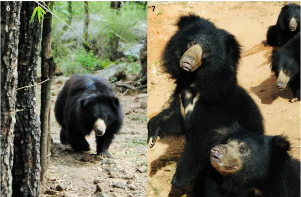
Figures 6 & 7- Rescued sloth bears at BBRC safari at Bannerghatta Biological Park ... 9
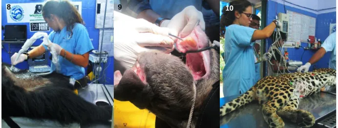
Figure 8- Ultrasonography examination on a sedated sloth bear ... 11
Figure 9- Dental surgery on a sedated sloth bear... 11
Figure 10- Electrocardiography (ECG) performed on a leopard from the BBP Zoo ... 11
Figure 11- Laboratorial area with microscope and centrifuge for basic lab work ... 12
Figure 12- Maggot wound on a tiger’s forelimb ... 12
Figure 13- Wild sloth bear with injuries caused by a tree fall and human attacks ... 12
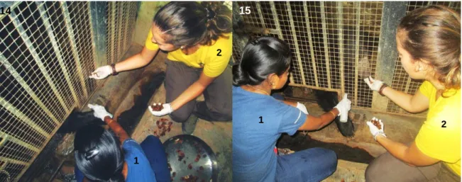
Figures 14 & 15- Blood drawing attempt without the use of sedation. While individual nr.1 executes the trimming and disinfection of the animal’s limb, individual nr.2 provides treats to the animal, as trained in many previous sessions. ... 12
Figures 16 & 17- Construction of suspensive enrichment structure based on animals’ “treat hunting” behaviour and posterior animals’ reaction ... 13
Figures 18 & 19- Sloth bears enjoying environmental enrichment structures ... 13
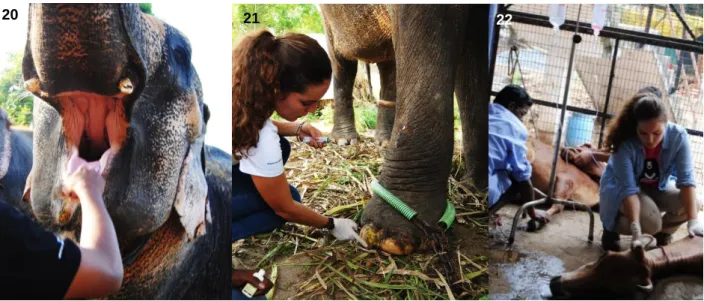
Figure 20- Indian elephant oral inspection ... 14
Figure 21- Treatment of nail fissures on an Indian elephant ... 14
Figure 22- Ruminant treatment durning the Chennai flooding at an Animal Rescue Centre ... 14
Figure 23- Scanning electron micrograph of Mycobacterium tuberculosis organism ... 15
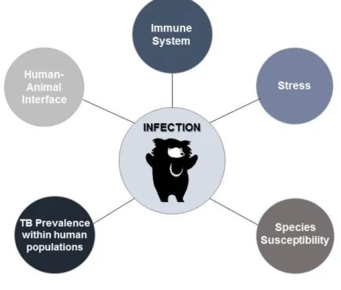
Figure 24- Schematic representation of the risk factors associated with tuberculosis infection in sloth bears in the India subcontinent ... 20
Figure 25- Schematic representation of TB transmission between captive sloth bears and humans. ... 22
Figure 26- Lists of countries belonging to the major three TB-related problematic categories and their areas of overlap ... 24
Figure 27- Estimated TB incidence: top-ten countries in 2014. The range shows the lower and upper bounds of the 95% uncertainty interval and the bullet marks the best estimate ... 25
Figure 28- Schematic representation of granuloma formation. ... 27
Figure 29- Emaciation of diseased TB positive sloth bear ... 28
Figure 30- Enlarged TB positive sloth bear’s lung. Rib cage impressions (black arrows), tuberculous nodules (white circle) and areas of congestion (white arrow) can be seen ... 29
Figure 31- TB positive sloth bear’s lungs with areas of congestion (black circle), presence of hydrothorax and icteric liver (black arrow) ... 29
Figures 32 & 33- 17-year-old male TB positive sloth bear’s lungs. Absence of normal parenchyma and alveolar structure. Several nodules (white circles) and purulent thick material (black arrows) are present. ... 29
Figure 34- TB positive sloth bear thoracic X-ray. Some radiopaque areas (black arrows) indicate a possible presence of calcified lesions in the lungs ... 33
Figure 35- Lateral thoracic radiograph of a dog with disseminated Mycobacterium tuberculosis infection. Note diffuse radiopaque images (arrows) in dorsal and caudal lung lobes, suggesting lung consolidation and granuloma formation.. ... 33
Figure 36- Granuloma (hematoxylin and eosin stain) in human lung. The red outline highlights the distinct structure of the granuloma, caused by immune cells bunching together to surround the pathogenic organism The arrow shows a multinucleated giant cell formed by multiple macrophages that have fused together ... 35
Figure 37- Photomicrograph of a lung from a dog infected by Mycobacterium tuberculosis, showing granulomatous reaction with central necrosis, presence of epithelioid cells and fibroblasts in the middle zone (little arrow). Note proximity of granuloma (large arrow) with bronchial tree (*) indicating active tuberculosis (TB) infection ... 35
ix
Figure 38- Mycobacterium tuberculosis colonies in Löwestein-Jensen medium from a human
sputum sample. ... 37
Figure 39- Mycobacterium tuberculosis colonies in Löwestein-Jensen medium from a sloth bear sputum sample. ... 37
Figure 40- Ziehl-Neelsen staining of Mycobacterium tuberculosis organisms (black arrows) in a human’s sputum smear. ... 38
Figure 41- Ziehl-Neelsen staining of Mycobacterium tuberculosis organisms (black arrows) in a sloth bear’s tracheal smear ... 38
Figure 42- Injection of PPD on a sloth bear’s ear lobe ... 42
Figure 43- TST reaction ... 42
Figure 44- Measurement of TST reaction ... 42
Figure 45- Overview of the QuantiFERON-TB Gold® in tube assay technology ... 44
Figure 46- ElephantTB STAT-PAK® test function.. ... 46
Figure 47- Positive DPP VetTB® test (pink arrow) from a sloth bear ... 46
Figure 48- Positive ElephantTB STAT-PAK® (blue arrow) from a sloth bear. ... 46
Figure 49- Rapid test execution (Wild TB alert kit®) from a wild sloth bear blood sample... 47
Figure 50- Positive immunological tests should trigger exploration of the direct exam category, schematic representation ... 48
Figures 51 & 52- Sloth bear intubation for sputum collection through BAL ... 50
Figures 53 & 54- After de-intubation it is o possible to obtain a swab from the tube for culture/PCR/smear or a direct smear for ZN staining from the exterior of the tube ... 50
Figure 55- Nasal sputum smear showing Mycobacterium organisms (cylindrical structures stained pink)... 50
Figure 56- Lung impression smear showing Mycobacterium organisms (cylindrical structures stained pink)... 51
Figure 57- Post-mortem tracheal swab collection ... 52
Figure 58- Lung section for sample collection ... 52
Figure 59- Potassium permanganate footbaths, located at the entrance of every bear enclosure ... 58
Figure 60- When dealing closely with bears or biological materials, the use of gloves, masks and caps is imperative. ... 58
Figure 61- All animals in this study were rescued “dancing bears” that were hosted at Bannerghatta Bear Rescue Centre, Wildlife SOS ... 61
x
List of Tables
Table 1- Biologic Information of Bears, Order Carnivora, Family Ursidae. ... 2
Table 2- Status of “dancing (sloth) bears” in India (1996–2010). ... 6
Table 3- Hours spent in each activity during the training period. ... 9
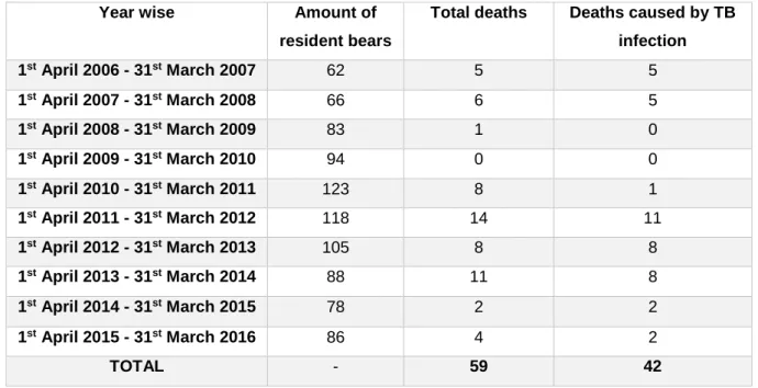
Table 4- Animal mortality at BBRC from 2006 to 2016. ... 18
Table 5- Tuberculosis Complex Mycobacteria and their reported hosts. ... 19
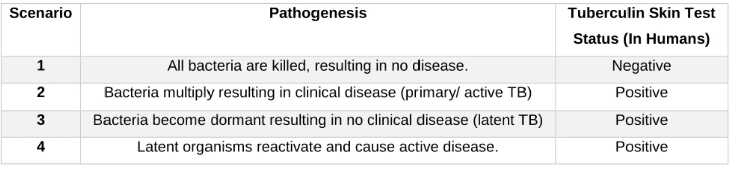
Table 6- Possible scenarios after exposure to active tuberculosis (TB) ... 21
Table 7- Organ specific lesions found in sloth bears post-mortem examination at BBRC. ... 30
Table 8- Sample materials possible to collect ante-mortem and diagnostic methods applicable to each of them. ... 49
Table 9- Sample materials possible to collect post-mortem and diagnostic methods applicable to each of them. ... 52
Table 10- Dosages and routes of administration of first line anti-TB drugs for treatment of Tuberculosis infection in sloth bears at Wildlife SOS. ... 55
Table 11- Specimen information. ... 61
Table 12- Results for the unspecific ante-mortem tests applied to all 15 bears and calculated sensitivity. ... 65
Table 13- Results for the direct ante-mortem tests applied to all 15 bears and calculated sensitivity ... 66
Table 14- Results for the indirect ante-mortem tests applied to all 15 bears and calculated sensitivity. ... 67
Table 15- Results for the post-mortem tests applied to the 14 diseased bears and calculated sensitivity. ... 68
Table 16- Sensitivity comparison for each potential combination of two TB screening methods in sloth bears (Melursus ursinus). ... 69
Table 17- Calculated sensitivity of the combination of the three categories of tests. ... 70
List of Graphics Graphic 1- Sensitivity comparison for all the tests applied to the specimen. ... 71
xi
List of Abbreviations and Symbols
ºC – Degrees Celsius µg - Microgram
AAZV - American Association of Zoo Veterinarians ABRF - Agra Bear Rescue Facility
AFB - Acid-fast bacillus am- Ante-meridian
AMTD - Amplified Mycobacterium tuberculosis direct test BAL - Broncho-Alveolar Lavage
BBRC - Bannerghatta Bear Rescue Centre BBP - Bannerghatta Biological Park
BCG - Bacillus Calmette-Guérin CBC - Complete Blood Count CI - Confidence Interval
CITES - Convention on International Trade in Endangered Species of Wild Fauna and Flora CMI - Cellular-mediated immunity/immunologic
CRP - C-reactive protein CT - Computed tomography DCs - Dendritic cells
DNA - Deoxyribonucleic acid DPP- Dual-path platform assay
EAZWV - European Association of Zoo and Wildlife Veterinarians ELIZA - Enzyme-linked immunosorbent assay
EMB - Ethambutol EN - Endangered
FVM - UL – Faculty of Veterinary Medicine – University of Lisbon HE- Hematoxylin and eosin
HIV - Human immunodeficiency virus HPA - Hypothalamic-pituitary-adrenal IFN-ү - Interferon-gamma
IGRAS -Interferon-gamma release assays IM- Intra-Muscular
INH - Isoniazid
IUCN- International Union for Conservation of Nature LC - Least Concern
LJ - Löwenstein-Jensen
xii LTT - Lymphocyte transformation test
MAPIA- Multi-antigen print immunoassay MDR - Multi-drug resistance
mg/Kg- Milligram per kilogram
MGITs- Mycobacterial growth indicator tube mL- Milliliter
MRI - Magnetic resonance imagery PCR- Polymerase Chain Reaction PET - Positron emission tomography pm – Post-meridian
PZA- Pyrazinamide
RFLP – Restriction fragment length polymorphism RIF- Rifampin/ Rifampicin
RNA - Ribonucleic acid
RNAr- Ribosomal ribonucleic acid sbsp. - Subspecies
SC - Subcutaneous SM - Streptomycin
SNS - Sympathetic nervous system spp. - Species
TB- Tuberculosis
Th- T Helper Lymphocytes TNF- Tumor Necrosis Factor TST- Tuberculin skin test
USAHA- United States Animals Health Association VU - Vulnerable
WHO - World Health Organization WSOS - Wildlife SOS
1 Chapter I- Introduction
Conservation medicine enables us to rethink the ties between human, animal and environmental health (Verma-Kumar, Abraham, Dendukuri, Cheeran, Sukumar & Balaji, 2012). The recognition of this crucial interdependence has led to the multi-disciplinary concept of “One World, One Health, One Medicine”. Pathogens that are transmitted between wildlife, livestock and humans represent major challenges for human and animal health, as well as for wildlife conservation (Maas, Michel & Rutten, 2013).
Tuberculosis (TB) is a cause of significant morbidity and mortality in both domestic and wild animals worldwide, having a wide host range, which includes non-human primates, elephants and other exotic ungulates, carnivores, marsupials, marine mammals, rodents, amphibians, and birds (Montali, Mikota & Cheng, 2001; Miller, 2008; Fefar et al., 2012). It is also considered an important emerging disease in humans, remaining a major global public health issue (Nolte & Metchock, 1995; Ernst & Wolf, 2006; Olsen, Barletta & Thoen, 2010), with one-third of the human population currently infected (World Health Organization [WHO], 2014) and being the leading cause of death in adults due to a single infectious agent (Alexander, Pleydell, Williams, Lane, Nyange & Michel, 2002; Pfyffer, 2007; Lécu & Ball, 2011; WHO, 2016). Of those deaths, about 95% occurred in countries where resources are more limited, with a majority of cases appearing in India and China (WHO, 2014; Fogel, 2015). The best estimate is that, in 2015, there were 1.4 million TB deaths and an additional 0.4 million deaths resulting from TB disease among HIV-positive people. Also, there were 10.4 million new TB cases (including 1.2 million among HIV-positive people) (WHO, 2016).
Serious and vigorous action plans are presently applied to fight the disease in humans in developing countries, but these are still the places facing notable incidences of TB within the animal populations (Rishikesava, Arun, Chandranaik, Basavarajappa, Giridhar & Renukaprasad, 2008; Lécu & Ball, 2011). Wildlife populations that suffer from tuberculosis infection, most often live in close contact with humans or domestic animals, with the highest prevalence rates occurring in captive animals in zoological collections and the lowest rates in animals that rarely come in contact with human populations (Michel, Venter, Espie & Coetzee, 2003).
Awareness of the importance of tuberculosis in wildlife has been increasing, not only for being a potential reservoir of infection for domestic animals, but also a threat to the health and integrity of valuable and rare wildlife species (de Lisle, Bengis, Schmitt & O’Brien, 2002; Isaza, 2003; Michel et al., 2003). The zoonotic potential of these organisms presents an additional concern for animal handlers, like veterinarians and keepers, and, of course, for public health. Therefore, rapid, accurate diagnosis in wildlife species is imperative (Miller, 2008; Arun et al., 2014).
Although a wide variety of mycobacteria are pathogenic, “tuberculosis” refers to infection with specific organisms that belong to the Mycobacterium tuberculosis complex (Backues, 2008; Miller, 2008; Lécu & Ball, 2014). The majority of the mycobacteria from the “tuberculosis complex”
2
have the ability to infect wild animals whereas the susceptibility, pathogeny and immune responses towards mycobacterial infection vary widely between mycobacteria and host-animal species (de Lisle et al., 2002). The predictability of infection outcome is still hard to presume and, therefore, the eradication of TB is potentially linked to the capacity of early diagnosis in domestic and wild host species (Miller, 2008; Lécu & Ball, 2011).
In India, illegally held sloth bears that were confiscated by wildlife authorities and then moved to rehabilitation centres have been reported to have died from tuberculosis infection (Arun et al., 2014; Kemmerer, 2015). A presumptive diagnosis of mycobacterial infection was initially made based on gross findings. Histopathology and microbiology of caseated nodules within the lungs confirmed a diagnosis of Mycobacterium tuberculosis infection (Mehrotra, Bhargava, Choudhary & Mathur, 1999; Arun et al., 2014; Kemmerer, 2015; A.S. Arun, personal communication, April 20th, 2016). Symptoms like progressive weight loss, anorexia, gastritis, enteritis, nasal discharge,
cough and weakness were observed prior to death in some of these bears (Renukaprasad et al., 2013; Collins, 2014). Zoonotic transmission from human handlers was speculated to be the origin of the infection (Renukaprasad et al., 2013; Arun et al., 2014; Kemmerer, 2015).
1. Sloth Bears
Bears are mammals belonging to the order Carnivora of the Ursidae family and are geographically widespread within North and South Americas, Europe, and Asia. Within Ursidae, eight species of bears exist in total (Table 1), divided by three different subfamilies: Ursinae1, Tremarctinae2, and
Ailuropodinae3 (Yoganadn, Rice & Johnsinhg, 2013; Collins, 2014).
Table 1- Biologic Information of Bears, Order Carnivora, Family Ursidae (Adapted from Collins, 2014) and their classification according to the IUCN Red List of Threatened Species (http://www.iucnredlist.org).
LC – Least Concern; VU – Vulnerable; EN – Endangered1.
1 The IUCN divides species into 7 different categories regarding their conservation status: Least Concern (LC), Near Threatened (NT),
Vulnerable (VU), Endangered (EN), Critically Endangered (CE), Extint in the Wild (EW) and Extint (EX). The 2012 “IUCN Red List Categories and Criteria” is available for consult at http://www.iucnredlist.org/technical-documents/categories-and-criteria.
Scientific Name Common Name Geographic Distribution
1Ursus americanus (LC) American black bear North America
1Ursus thibetanus (VU) Asiatic black bear Central, Eastern, and Southeastern Asia 1Ursus arctos (LC) Brown or grizzly bear Europe, Asia, and North America
1Ursus maritimus (VU) Polar bear Artic regions of Eastern Asia and North America 1Helarctos malayanus (VU) Sun bear Southeast Asia
1Melursus ursinus (VU) Sloth bear Southeast Asia
2Tremarctos ornatus (VU) Spectacle bear Andes mountains of South America 3Aiuropoda melanoleuca (EN) Giant panda Central China
3
Sloth Bears (Melursus ursinus) are a small, usually black with a long shaggy coat, bear species that range across India, Nepal, Sri Lanka, and Bhutan (Figure 2) (Arun, Kajal, Selvaraj & Selvaraj, 2008; Garshelis, Ratnayeke & Chauhan, 2008). In these regions, Pocock (1933) distinguished two distinct races: Melursus ursinus ursinus (Figure 1), occurring in continental India and the shorthaired and relatively smaller Melursus ursinus inornatus, found only in Sri Lanka. Adult males weigh between 79 and 140 kg, while female sloth bears weigh between 54 and 109 kg (Garshelis, et al., 2008). Bears usually have a longevity of 20-30 years in captivity (von Hohendorff & Giacomini, 2014).
Sloth bears are reported to exist in 174 Protected Areas in India, which include 46 National Parks and 128 Wildlife Sanctuaries (Chauhan, 2006). Although no truly reliable large-scale population estimates exist for sloth bears, best guesstimates indicate a reasonable possibility of there being ~20.000 or fewer animals, and thus <10.000 adult animals (Garshelis et al., 2008).
These animals subsist primarily on termites, ants, and fruits and are the only bears with specific adaptations for myrmecophagy (ant and termite-eating) (Arun et al., 2008; Garshelis et al., 2008; Yoganand et al., 2013). In captivity, sloth bears are typically fed omnivorous diets, similar to the ones consumed by other bear species (Collins, 2014). Sloth bears are almost nocturnal in their habits due to the human geographic pressure inside protected forest areas and sanctuaries (Seshamani & Satyanarayan, 1997).
Figure 1- Indian sloth bear (Melursus ursinus ursinus) at Bannerghatta Biological Park (Original);
Figure 2- Sloth bear distribution across India, Sri Lanka, Nepal and Bhutan (Source: The IUCN Red List of Threatened Species 2008, http://dx.doi.org/10.2305/IUCN.UK.2008.RLTS.T13143A3413440.en).
4
Sloth bears are listed as “Vulnerable” by the International Union for Conservation of Nature (IUCN), listed under Appendix I of Convention on International Trade in Endangered Species of Wild Fauna and Flora (CITES), meaning that all trade and exportations are banned and protected under Schedule I of the Indian Wildlife (Protection) Act of 1972 (Arun et al., 2008; Garshelis et al., 2008; Arun et al., 2014). Existing sloth bear populations face direct threats from habitat loss, poaching and capture of cubs (Arun et al., 2008; Garshelis et al., 2008; D'Cruze et al., 2011; Yoganand et al., 2013).
Poaching occurs mainly for the commercial trade of bear parts in the black market, particularly gall bladders, male reproductive organs, bones, teeth, claws and even fat (Arun et al., 2008; Garshelis, et al., 2008; Renukaprasad et al., 2013). Capture of live cubs for use as "dancing bears" by the Kalandar community, represented a major and significant threat in the past (Seshamani & Satyanarayan 1997; Arun et al., 2008; Renukaprasad et al., 2013; Kemmerer, 2015). Evidence suggests that sloth bear populations have declined by 30–49% over the past 30 years as a result of all these threats (Garshelis et al., 2008; D'Cruze et al., 2011).
2. Kalandars and “Dancing bears”
The Kalandars are a widely dispersed, endogamous ethnic group of nomadic entertainers, originally composed by Muslim gypsies, found throughout South Asia (Seshamani & Satyanarayan, 1997; D'Cruze et al., 2011). They make their living using a wide number of performing animals like monkeys, bears, fighting roosters and pigeons and keeping others as pets, such as civet cats, owls, falcons, and partridges to display to their audience (Seshamani & Satyanarayan, 1997).
For over four hundred years, “dancing bears” were a very common sight in India (Figure 3, 4 & 5), being part of an old tradition, where captured bear cubs were initially trained and forced to perform for emperors (Seshamani & Satyanarayan, 1997; D'Cruze et al., 2011; Renukaprasad et al., 2013). For this purpose, cubs were poached from the wild and their mothers killed, most of the times, since they instinctively try to protect their young (Seshamani & Satyanarayan, 1997; Renukaprasad et al., 2013; Kemmerer, 2015).
Figures 3, 4 & 5- “Dancing bears” of India and the Kalandar community (Courtesy of Wildlife SOS, India).
5
Many times, the bear cubs reached the villages traumatized and dehydrated and, after a couple of weeks, were tied by a rope, to a bamboo pole. The cubs’ muzzles were pierced with a large iron needle and a coarse rope was passed through the wound, giving Kalandars the power to easily manipulate the animals, since the wound would never properly heal (Seshamani & Satyanarayan, 1997; Renukaprasad et al., 2013; Kemmerer, 2015).
Cubs’ permanent dentition appears when they are around 8-10 months old, which made them more difficult to handle by the Kalandars. For this reason, the canines were removed and young males castrated without the use of anesthesia in either of the procedures (Renukaprasad et al., 2013; Seshamani & Satyanarayan, 1997). The mortality rates were very high at this point (Seshamani & Satyanarayan, 1997).
The major animal suffering and abuse does not lie particularly in the actual “dancing” routine, but in all the other aspects involving the trade: the capturing, transporting and training processes; stressors as heat, dust, and noise; and the urban environment in general, very different from their natural one (Seshamani & Satyanarayan, 1997).
Cubs’ feeding routines vary through India, with animals being fed with milk and wheat porridge in North India and Ragi porridge and milk in the South. When the cubs are around six months old, they start being fed with Roti bread2 mixed in milk in North India and steamed Ragi balls3 and milk
in the South (Seshamani & Satyanarayan, 1997).
This practice was made illegal in 1972 in India but continued for, at least, another 40 years (Seshamani & Satyanarayan, 1997; Kemmerer, 2015).
3. Rescue Centres
Since the late 1990s, national and international wildlife institutions have focused their attention on this issue in India. Their efforts to increase law enforcement, provide lifetime care for confiscated bears, increase public awareness about the issue and provide sustainable alternative livelihoods for bear-owning Kalandars, have resulted in a significant positive effect on the conservation and welfare of sloth bears, as the number of “dancing bears” known to be performing throughout India seems to have declined dramatically over the last two decades, from approximately 1,000–1,200 animals in 1996 to approximately 28 animals in 2010 (Table 2) (D'Cruze et al., 2011; Renukaprasad et al., 2013). Today, there is no record of active “dancing bears” in the streets of India, as stated by Wildlife SOS (A.S. Arun, personal communication, April 20th, 2016).
To this day, hundreds of performing bears have been rescued and given a permanent home and lifetime care in Wildlife SOS rehabilitation centres throughout India (Seshamani & Satyanarayan, 1997).
2Flat bread originating from the Indian subcontinent, made from stoneground wholemeal flour, traditionally known as atta, and water. 3 Wholesome meal in the state of Karnataka and the Rayalaseema region in Andhra Pradesh made with ragi flour and water.
6
Almost every rescued “dancing (sloth) bear” suffers from an enormous variety of disorders. Deficiency of maternal immunity, physical and psychological stress, poor nutrition, poor living conditions, impaired circadian rhythms and lack of veterinary care created a host of predictable medical problems such as external and internal parasites, rabies, leptospirosis, canine adenovirus, tetanus, degenerative joint disease, maggot wounds, severe gum disease and ocular and dental diseases, namely rotting tooth stumps where canines had been purposefully and brutally broken/removed (Arun, 2013; Kemmerer, 2015; A.S. Arun, personal communication, April 20th, 2016).
Their immune systems are frequently compromised and it soon became notorious that a significant number of animals were found to have severe gross lesions of tuberculosis at post-mortem examination. It is presumed that the acquaintanceship between Kalandars and bears, similar to the one observed between elephants and mahouts4 (Maslown & Mikota, 2015), may
have resulted in the animals’ infection, making them spillover hosts (Arun, 2013; Arun et al., 2014; Kemmerer, 2015).
Table 2- Status of “dancing (sloth) bears” in India (1996–2010) (D'Cruze et al., 2011).
The year 2005 if often heralded as the date that TB “emerged” as a disease of concern for sloth bears, after several deaths and post-mortem diagnosis in bear rescue centres (A.S. Arun, personal communication, April 20th, 2016).
4 Elephant rider, trainer, or keeper. A mahout starts as a boy in the “family profession” when he is assigned an elephant early in its
life.
State or union territory
Number of dancing bears recorder (year)
1996 2001 2005 2010 Andhra Pradesh 50 11 72 2 Bihar 86 31 27 5 Chhattisgarh 30 27 21 0 Delhi 36 36 36 0 Haryana 116 8 5 0 Jharkhand 20 20 11 3 Karnataka 88 96 29 2 Madhya Pradesh 29 23 8 1 Maharashtra 36 36 1 2 Rajasthan 126 90 10 0 Uttar Pradesh 430 258 89 13 West Bengal 51 32 37 0 Total 1098 668 346 28
7 4. Content Notice
Considering the lack of published data regarding tuberculosis in sloth bears, this dissertation references TB in humans, elephants, non-human primates and dogs, as these hosts most closely provide models to improve the understanding of the disease as it is observed in sloth bears. For instance, elephants and sloth bears are both treated for TB the same way humans are, which is uncommon in animal species (United States Animals Health Association [USAHA], 2012); preventive measures are very similar between elephants and sloth bears (European Association of Zoo and Wildlife Veterinarians [EAZWV], Tuberculosis Working Group, 2010; A.S. Arun, personal communication, April 20th, 2016); and dogs display many similarities in disease
progression, lesions and clinical manifestations (Martinho et al., 2013), possibly for being phylogenetically related to bears.
This dissertation will focus on infection by Mycobacterium tuberculosis as it is the predominant disease-causing agent in sloth bears (Arun et al., 2014; A.S. Arun, personal communication, April 20th, 2016), with suitable mentions to M. bovis cases mentioned in literature (Collins, 2014).
8 Chapter II- Training Period Activities
The author’s 6th year externship of the Integrated Masters in Veterinary Medicine took place at
the Bannerghatta Bear Rescue Centre (BBRC) in the Karnataka state, India. This externship started on September 14th, 2015 and was completed by December 13th, 2015, having the length
of 13 weeks.
This training had the supervision of Dr. A. Sha. Arun from Wildlife SOS, BBRC and the co-supervision of Dr. João Nestor das Chagas e Silva, from FMV-ULisboa.
The Bannerghatta Bear Rescue Centre was established in 2005 and is one of two major sloth bear sanctuaries in India, the other one being the Agra Bear Rescue Facility, both managed by Wildlife SOS. It is placed inside the Bannerghatta Biological Park (BBP), with many other free-ranging animals occupying the surrounding areas. The Bannerghatta Biological Park area, besides the Bear Rescue Centre, and the main safari area, also contains a Butterfly Park, a feline Rescue Facility and a Zoological Garden within its premises, with a veterinary team that frequently works along with BBRC veterinarians in both places.
Wildlife SOS was established in 1995 and works actively to protect Indian wildlife, mostly through the rehabilitation of rescued wildlife, rehabilitation of the Kalandar community by providing alternative livelihoods5, assisting State Forest Departments to prevent poaching from the wild and
the rescue of conflict animals6. Although Wildlife SOS is mainly known for its work with the
“dancing bears” of India, it also has active projects to help leopards, elephants, reptiles and other animals.
BBRC houses 86 sloth bears (Figures 6 & 7) at the present moment that are distributed among five enclosures.
At BBRC, the author followed the routine activities performed by the veterinary staff that included feeding inspections, updating records, deworming interventions and parasitological evaluations, laboratory work, literature research for eventual projects and Master’s dissertation purposes, environmental enrichment work, animal conditioning, blowpipe training, animal behavior analysis and surveillance, other routine veterinary treatments, wound dressings and clinical interventions/examinations, using anesthesia, some of which happened at BBP Zoo, and different kinds of surgical procedures.
5 As an example, Wildlife SOS animal keepers are mostly previous “dancing bear” owners.
6 In areas where deforestation, usually caused by human population increment, reduced animals’ natural habitat, there is a higher
change for animals and humans to cross paths, creating the so called “human-animal conflict”. For this reason, and despite sloth bears being extremely calm animals, are the bear species responsible for the highest number of human deaths, normally after direct human attacks or threats (von Hohendorff & Giacomini, 2014).
9
The amount of hours spent in each activity is stated in Table 3.
Table 3- Hours spent in each activity during the training period.
Activity Amount of hours spent
1. Routine Rounds & Prophylactic Measures 217 hours
2. Medical Interventions 97 hours
3. Laboratory Work 52 hours
4. Daily Medical Treatments 85 hours
5. Training/Conditioning 57 hours
6. Environmental Enrichment 23 hours
7. Necropsies 12 hours
8. Rescue Missions 81 hours
9. Individual WSOS Assignments 85 hours
10. Oral Presentations 21 hours
11. Research Work 49 hours
12. Blowpipe Training 15 hours
13. Instruction Day 9 hours
Total 803 hours
Figures 6 & 7- Rescued sloth bears at BBRC safari at Bannerghatta Biological Park (Original).
7 6
10
Regarding the activities performed, the author had the opportunity to participate in the following:
1. Routine Rounds and Prophylactic Measures: The bears at BBRC are fed twice a day
(around 11am and 4pm) with porridge (i.e. semisolid food that consists of a mixture of powdered ragi, jowar, soy bean along with honey, vegetables, milk, eggs and salt). The veterinary staff is responsible for the feedings inspection in every enclosure, to register which animals consumed the porridge or not, and perform the hygiene and cleanness inspection of the enclosures. As the symptomatic phase of TB begins with simple gastritis and skipping food, this observation and registry is highly important. Also, the bears are in free ranging areas during day time which makes the individual animal observation highly difficult. Hence, any abnormality or behavioral changes are best noticed during feeding time.The porridge is prepared in the kitchen which is also a target of daily inspection, as well as the fruits that are fed to the animals. Other activities performed included deworming of the animals, administration of medication and vitamins, weighing the bears, preparation of the Operation Theater (OT) before every medical procedure and its cleanup afterwards, and taking part in the daily fruit feeding safari, where enrichment fruit (mostly watermelon) is given to all the bears that have access to the BBP safari area.
2. Medical Interventions: The preparation of all the materials needed for the anesthetic
intervention, like the darts with the right amount of xylazine (2 mg/kg) and ketamine (5 mg/kg), according to the animal’s last registered weight, is one of the veterinarians’ responsibilities. Shortly after the animal is sedated, with the use of the blowpipe (IM route), the animal is moved from its enclosure to the operation theater, where the veterinarians open an IV route, providing fluids to the animal, collect blood from the jugular, and perform a general health examination, otoscopic and ophthalmologic examination, routine ultrasound (US) examination (Figure 8), dental X-ray, if required and collection of other body samples (hair, urine, tracheal/vaginal/rectal swabs) for further analysis either within BBRC (e.g. rapid tests, urine strip) or in a private laboratory (e.g. culture, PCR, interferon-gamma release assays). Clipping of the claws, cleansing of eyes and ears, recording of body measurements, microchipping of the young cubs are some examples of other procedures that usually take place at that moment. If a particular case requires surgery (e.g. bullet wound exploration, dental surgery, etc.), the same is fulfilled at the time. Dental treatments (Figure 9) are the most common ones since the majority of rescue bears had their canines broke or deficiently pulled out by the Kalandars, resulting in posterior infection or necrosis of the root canal.
During all procedures, the animal must have an open intra-venous (IV) route and the anesthesia must be closely monitored as well as the animal’s temperature7.
7Bears are able to use body fat to provide thermal insulation. These fatty deposits cause two issues when there is a need to sedate
11
Besides bears, the author had the chance to be involved in other medical procedures with other animal species as leopards (Figure 10), lions, tigers, elephants and a python.
3. Laboratory Work: At a small laboratorial space (Figure 11) the veterinary staff was able to
fulfill several simple procedures, as parasitological analysis of fecal samples and blood smears; centrifugation of blood samples, in order to obtain serum for the routine rapid tests for TB screening and microscopic analysis of lymph nodes aspirates. In order to carry out fecal parasitological analysis, the keepers are asked to keep the animals inside the enclosures, where they come to eat, after the morning feeding period (11am), and only to release them to the exterior after defecation. Fecal samples are collected to individual plastic jars, which are labeled and, in the same afternoon, the microscopic detection of parasite eggs is fulfilled (e.g. Toxocara canis, Hymenolepis spp.). Some animals excrete adult parasitic forms, which are macroscopically detected (e.g. round worm, tape worm). Although deworming medication is provided routinely in every enclosure, routine parasitological check-ups are very useful to keep a track of deworming drugs performance, of animal status, and, of course, to infer the parasite species most commonly found in these animals.
4. Daily Medical Treatments: The wound dressing of injured bears and tigers8 was performed
on a daily basis. In these animals, wounds are mostly caused by bullets, wire traps, and maggots. The four most routinely “patients” were a tiger with a paw maggot wound (Figure 12), a tiger with a torso injury, a bear with an infected gunshot wound and a wild bear that had fallen down a tree, after being pursued, threated and attacked by humans, and had several abrasions, cuts, a gunshot wound and a maggot wound (Figure 13).
8 BBRC Veterinarians are also responsible for the care of 4 tigers that live in a different area. Figure 8- Ultrasonography examination on a sedated sloth bear (Original). Figure 9- Dental surgery on a sedated sloth bear with a rotten root (Original).
Figure 10- Electrocardiography (ECG) performed on a leopard from the BBP Zoo (Original).
12
5. Training/Conditioning: This activity was performed by the author on a daily basis using a
clicker9, having worked with 8 different bears. The use of positive reinforcement training (in this
case, rewarding the animals with dates or honey) may enable many non-painful and minimally painful veterinary procedures such as inspection of teeth and feet, cleaning of wounds, injections, and blood drawing (Figures 14 & 15), without the need for anesthesia or physical restrain.
6. Environmental Enrichment Work: At least twice a week, besides the daily enrichment fruit
distribution, some kind of enrichment project was put in action within the animals’ enclosures (Figures 16 & 17). The author was strongly encouraged to participate in the same, either as a project designer, part of the construction team and/or as an observer and evaluator of the animal’s behavior afterwards (Figures 18 & 19). This principle seeks to enhance the quality of captive animal care by identifying and providing the environmental stimuli necessary for optimal
9 Clicker training is a type of training technique that uses a clicker device (little toy-like device that emits a “click” sound). Whenever
the animal performs the trainers’ requests correctly, the clicker will sound before the treat/reward is given. This way, the animal will know that it did not performed the order correctly if it does not hear that specific sound.
1 1 1 1 1 1 1 2 1 2 1
Figure 13- Maggot wound on a tiger’s forelimb extremity (Original).
Figure 11- Laboratorial area with microscope and centrifuge for basic lab work and sample process (Original).
Figure 12- Wild sloth bear with injuries caused by a tree fall and human attacks (Original).
Figures 14 & 15 - Blood drawing attempt without the use of sedation. While individual nr.1 executes the trimming and disinfection of the animal’s limb, individual nr.2 provides treats to the animal, as trained in many previous sessions (Original).
11 12 13
13
psychological and physiological well-being, preventing the onset of abnormal behaviors, also called stereotypic behaviors10 which bears are very susceptible to develop. Some examples of
this actions in bears are: head spinning, constantly walking from one side of the room to the other (pacing) and body and head rocking.
7. Necropsies: Fortunately, not a single sloth bear died during the author’s internship length.
None the less, the author had the opportunity to perform several post-mortem examinations to deceased BBP Zoo animals (e.g. python and lion), wild birds and unfortunate stray animals from outside the reserve, victims of India’s typical heavy traffic.
8. Rescue Missions: The author had the chance to accompany the head Veterinarian on two
rescue missions: the “Circus’ elephants rescue mission” (Figures 20 & 21) in October, where a rescue team from BBRC travelled to Tamil Nadu to inspect the health and living status of three circus’ Indian elephants, in order to attempt a rescue later to Wildlife SOS Agra Rescue Facility; and the “Flood relief mission” (Figure 22) at the Chennai flooding in December, where the BBRC
10Repetitive, invariant behavior patterns with no obvious goal or function (von Hohendorff & Giacomini, 2014).
Figures 16 & 17- Construction of suspensive enrichment structure based on animals’ “treat hunting” behaviour and posterior animals’ reaction (Original);
Figures 18 & 19- Sloth bears enjoying environmental enrichment structures (Original).
16 17
14
team went to Chennai for a one week period, after the flooding that occurred in the area from November to December, in order to rescue all kinds of animals in need and treat the injured ones, either at a rescue facility or directly in the streets of the city.
9. Individual Wildlife SOS (WSOS) Assignments: The author had several assignments given
by the head veterinarian during her stay at BBRC, namely the development of a “Hygiene and Organization Check List”, with all the necessary check points regarding the cleanness of the operation theatre before and after every clinical intervention; a “Processing Bear Bones Protocol”, indicating the most proper way to process dead animal’s bones, step by step, for learning and investigation purposes, creation of a “Conditioning Bear Evaluation” document, on which all trained bears had a proper file regarding its behavior and training achievements; improvement of the already existing “Bear Body Condition Score”, development of aging scores based on dental X-ray evaluation and creation of awareness brochures regarding animal conditioning activities and environmental enrichment projects.
10. Oral Presentations: Frequently, the author, as well as other members of the veterinary staff,
was encouraged to prepare oral presentations about any subject that interested her. The author prepared and presented several assays, namely: “Renal lesions in non-domestic felines”, “Low cardiac output as physiological phenomenon in hibernating, free-ranging brown bears”, “Portugal Wildlife”, “Veterinary Medicine in Portugal”, “Dates and its storage: the dangers for bears”11 and
“The C-reactive protein12 (CRP) test in TB diagnosis in sloth bears”.
11As the author noticed some stored bags of dates either with signs of rat bites (indicating its presence) or with rotten dates inside,
she presented a small lecture regarding the dangers of leptospirosis and fungus in animals’ food.
12The CRP is a substance produced by the liver in response to inflammation. High levels in the blood is a marker of any condition that
causes inflammation. In this small study, 12 TB positive bears were analyzed for this protein but no changes were detected in the CRP levels.
Figure 20- Indian elephant oral inspection (Original).
Figure 21- Treatment of nail fissures on an Indian elephant (Original).
Figure 22- Ruminant treatment durning the Chennai flooding at an Animal Rescue Centre (Original).
15
Figure 23- Scanning electron micrograph of Mycobacterium tuberculosis organism (Source: National Institute of Allergy and Infectious Diseases [NIAID]. Licensed under Creative Commons Attribution 4.0:
https://creativecommons.org/licenses/by/4.0/ via Flickr:
https://www.flickr.com/photos/niaid/5149398656/?ytcheck=1). Chapter III- Literature review
1. Etiology
1.1. Taxonomy and Description of the Genus
Mycobacteria belong to the genus Mycobacterium, which is the single genus within the family of Mycobacteriaceae, in the order Actinomycetales (Rastogi, Legrand & Sola, 2001; Isaza, 2003, Pfyffer, 2007; Olsen et al., 2010). Mycobacteria are aerobic, nonspore forming, nonmotile, slightly curved, slender, rod-shaped organisms and are 0.6-1.0 × 1.0-10 μm in size (Figure 23). Their cell walls high lipid content, that includes characteristic mycolic acids, excludes standard aniline dyes, so that once stained with special staining techniques, mycobacteria are resistant to discoloration. This property is termed acid fastness, so that mycobacteria are commonly referred to as acid-fast bacilli. In contrast, these microorganisms are not readily stained with the Gram method and are considered weakly gram – positive (Nolte & Metchock, 1995; Isaza, 2003; Pfyffer, 2007; Olsen et al., 2010; Grange, 2014).
The content of the cell wall enables the bacteria to be resistant to many antimicrobial agents, acidic and alkaline compounds and dehydration (Isaza, 2003; Miller, 2008). It also allows it to grow slowly inside macrophages and to be protected from phagocytosis, being an important virulence factor (Ernst, 2012).
Mycobacterial classification has relied on organisms’ biochemical and phenotypic characteristics (Nolte & Metchock, 1995; Rastogi et al., 2001; Miller, 2008). Colony morphology varies among species, ranging from smooth to rough and from nonpigmented to pigmented (Pfyffer, 2007; Grange, 2014). Strains have also been identified within species using restriction fragment length
16
polymorphism (RFLP), spoligotyping, and DNA sequencing (Rastogi et al., 2001; Miller, 2008). Also, natural division occurs between slowly (>7 days to form visible colonies on solid medium) and relatively rapidly (≤7 days) growing species of mycobacteria (Pfyffer, 2007; Olsen et al., 2010; Grange, 2014).
Overall, Mycobacteria can be divided into three different groups: Mycobacterium tuberculosis complex, Mycobacterium avium-intracellulare complex and other mycobacteria species (Isaza, 2003; Backues, 2008; Olsen et al., 2010).
The term “tuberculosis”, by convention, refers to infection with specific organisms belonging to the Mycobacterium tuberculosis complex, which includes M. tuberculosis, M. bovis, M. caprae, M. africanum, M. microti, M. canetti and M. pinnipedii, with M. tuberculosis and M. bovis being of the most importance (Montali et al., 2001; Pfyffer, 2007; Miller, 2008; Olsen et al., 2010).
Mycobacterium tuberculosis is the predominant disease-causing agent in captive sloth bears, as detected by culture and PCR (Arun et al., 2014; A.S. Arun, personal communication, April 20th,
2016).
1.2. Life Cycle
Tuberculosis by M. tuberculosis is, predominantly, an infection of the lower respiratory tract, mainly transmitted by the airborne route, in the form of droplet nuclei, that causes progressive pulmonary disease in mammals (Montali et al., 2001; Isaza, 2003; Maas et al., 2013; Fogel, 2015), though it can affect virtually every organ in the body (Ernst & Wolf, 2006).
After inhalation, the droplet nuclei reach the lung alveoli, where they are rapidly phagocytosed by alveolar macrophages (Schluger & Rom, 1998; Ernst & Wolf, 2006; Lin, Plessner, Voitenok & Flynn, 2007; Ernst, 2012; Sakamoto, 2012). After phagocytosis, mycobacteria organisms multiply within macrophages, leading to the death of the infected cells (Olsen et al., 2010; Ernst, 2012), and the expanding population of bacteria spreads to newly recruited macrophages and dendritic cells (DCs) (Ernst & Wolf, 2006; Lin et al., 2007). These macrophages are stimulated to produce pro-inflammatory cytokines (TNF, IL-12, IL-1, IL-6) and chemokines, driving the recruitment of more leukocytes to the site of infection, like neutrophils and monocytes, and begin to form the early granuloma (Lin et al., 2007; Ernst, 2012; Sakamoto, 2012). A subset of the bacteria also migrates to the local draining lymph node, transported by DCs, where they likely initiate an adaptive response by antigen presentation to naive lymphocytes that will proliferate, differentiate (to CD4+ and CD8+ T cells) and migrate back to the lungs to participate in the granuloma formation (Cooper & Flynn, 1995; Ernst & Wolf, 2006; Lin et al., 2007; Ernst, 2012; Sakamoto, 2012). CD4+ and CD8+ T cells are important in the immune response to M. tuberculosis, and function by secreting cytokines (including those that can activate macrophages, such as IFN-γ and TNF) (Lin et al., 2007). In addition, part of the bacterial population is believed to disseminate by the bloodstream to other peripheral organs, causing extra-pulmonary tuberculosis (Ernst & Wolf, 2006).
17
After the onset of adaptive immune responses, growth of tuberculosis in the lung is restricted, the progression of the disease is interrupted and most hosts become asymptomatic, not shedding bacteria, and are considered to have latent TB infection (Ernst & Wolf, 2006; Ernst, 2012). It has been suggested that the pathogenic bacteria lie latent in phagosomes inside macrophages, waiting for an immunocompromised condition of the host to grow quickly (Itagaki & Cho, 2013; Dietrich et al., 2015). This latency may also occur in the extra-pulmonary tuberculosis scenario (Ernst & Wolf, 2006).
In the majority of cases, the host adaptive immune response is sufficient to prevent active disease throughout life (Ernst & Wolf, 2006; Fogel, 2015). The animals that develop active disease will be the ones expelling infectious droplet nuclei to start the infection life cycle once more (Ernst & Wolf, 2006).
1.3. Transmission
An obligate step in all infectious diseases is transmission to new hosts (Ernst, 2012). TB transmission routes between animals are directly associated with the granulomas location in infected individuals. Pulmonary lesions generally lead to airborne transmission, in which bacteria are expelled (usually by coughing) from an individual with active disease and then inhaled by susceptible hosts, whereas mesenteric lymph-node lesions may lead to intestinal excretion of mycobacteria, that may be ingested by the next host (de Lisle et al., 2002; Backues, 2008; Mikota, 2008; Lécu & Ball, 2011; Ernst, 2012). Feces, urine, genital discharges, milk, and feed or water may contain contaminated droplets (Mikota, 2008). Horizontal transmission is the most significant and important mean of contamination (respiratory and alimentary routes) but pseudo-vertical transmission can potentially occur during nursing and grooming (Begins, 1999), and possibly vertical transmission through placental or umbilical infection (Kaneene & Pfeiffer, 2006).
As in many other infectious diseases, the transmission of TB is not uniform, and certain individuals cause far more secondary cases than do others. In particular, individuals with a form of TB termed cavitary TB are especially infectious ((Helke, Mankowskia & Manabe, 2006; Ernst, 2012).
2. Epidemiology
There are many reasons that can justify why sloth bears seem to be so susceptible to tuberculosis infection, the most important being obviously the close contact with infected humans, and the high prevalence of tuberculosis in the Indian subcontinent. The lack of positive TB findings in free ranging sloth bears, corroborates this theory (Renukaprasad et al., 2013; Arun et al., 2014; Kemmerer, 2015).
Next, some factors related with the epidemiology of tuberculosis infection in sloth bears are discussed (Figure 24).
![Figure 23- Scanning electron micrograph of Mycobacterium tuberculosis organism (Source: National Institute of Allergy and Infectious Diseases [NIAID]](https://thumb-eu.123doks.com/thumbv2/123dok_br/15464575.1032073/32.892.261.609.741.1042/scanning-micrograph-mycobacterium-tuberculosis-national-institute-infectious-diseases.webp)