RESUMO:O objetivo deste estudo foi a comparação entre diferentes testes de diagnóstico para tuberculose bovina. Foram realizados o isola‑ mento bacteriano, a caracterização histopatológica, a identificação de baci‑ los álcool‑ácido resistentes e a detecção do DNA de M. bovis pela reação em cadeia da polimerase, em bovinos adultos abatidos em matadouros frigoríficos sob o Serviço de Inspeção Federal, valendo‑se de amostras de linfonodos com lesões macroscópicas sugestivas de tuberculose, identifica‑ das e coletadas durante o abate. O isolamento bacteriano foi realizado pelo cultivo em meios de cultura sólidos; a caracterização histopatológica, pela coloração com hematoxilina‑eosina; e a identificação de bacilos álcool‑ácido resistentes foi feita pela coloração de Ziehl‑Neelsen. A detecção de DNA foi realizada em amostra extraída das lesões sugestivas de tuberculose pela reação em cadeia da polimerase, seguida da nested reação em cadeia da polimerase e por meio das colônias isoladas para identificação do M. bovis, utilizando‑se também da reação em cadeia da polimerase . Os resultados obtidos permitiram concluir que os testes histopatológicos, o isolamento bacteriano e a identificação de bacilos álcool‑ácido resistentes são aconse‑ lháveis para o diagnóstico da tuberculose bovina. Além disso, ensaios de reação em cadeia da polimerase utilizando amostras de lesões sugestivas de tuberculose são um modo mais rápido e promissor para diagnosticar a enfermidade, no entanto não deve ser utilizado sozinho, em virtude da baixa sensibilidade apresentada neste estudo.
PALAVRAS‑CHAVE: doenças infecciosas; Mycobacterium bovis; tuberculose; zoonose.
ABSTRACT:Our goal for this article is to compare several different diagnosis tests for bovine tuberculosis identification. We have performed bacterial isolation, histopathological characterization, acid‑fast bacilli (AFB) identification and M. bovis DNA detection. Lesions suggestive of Tuberculosis were sampled from bovine lymph nodes during slaughtering of bovines at an abattoir that operates under federal inspection. The bacterial isolation was performed in solid culture mediums, the histopathological characterization was made by Hematoxylin‑eosinstaining, and AFB identification by Ziehl‑Neelsen staining. Bacterial DNA detection was performed by Polymerase Chain Reaction (PCR) using DNA from two different sources, directly collected from the tuberculosis‑like lesions (PCR followed by nested PCR) and from isolated bacteria. We have concluded that the multi‑step approach, including histopathological characterization, bacterial isolation and AFB identification, is strongly recommended to diagnose tuberculosis in bovines. Furthermore, PCR assays using specimens of lesions suggestive of tuberculosis are a faster and more promising way to diagnose the disease. However, it should not be used alone due to the low sensitivity shown in this study.
KEYWORDS: infectious diseases; Mycobacterium bovis; tuberculosis; zoonosis.
Comparison between tests for tuberculosis
diagnosis in slaughtered bovines
Comparação entre testes de diagnóstico para tuberculose em bovinos abatidos
David Attuy Vey da Silva1*, Márcio Junio Lima Siconelli2, Karina Paes Bürger3, Lara Borges Keid1
1Universidade de Sao Paulo – São Paulo (SP), Brazil
2Universidade de Sao Paulo, Campus de Ribeirão Preto – Ribeirão Preto (SP), Brazil 3Universidade Estadual Paulista “Julio de Mesquita Filho” – Jaboticabal (SP), Brazil *Corresponding author: [email protected]
INTRODUCTION
Tuberculosis is one of the main causes of death world‑ wide, affecting a variety of species. Mycobacteria from the
Mycobacterium tuberculosis complex are known as the tuber‑ culosis causative agent (COSTA et al., 2013). The occurrence of nodular granulomatous lesions are typical of this infirmity (FURLANETTO et al., 2012). Despite being found in almost all tissues of infected animals, the lesions are more frequently observed in lymph nodes, the lungs and the liver (CARDOSO et al., 2009; ROXO, 1997).
Post‑mortem sanitary inspection has a great importance for tuberculosis epidemiological surveillance programs in countries where the disease is considered endemic, since it can consider‑ ably reduce its prevalence when allied to eradication programs (DE LA RUA‑DOMENECH, 2006). PINTO (2003) states that among the foodborne zoonoses detected in the sanitary inspection, tuberculosis has the largest impact in economy and public health.
In addition to the post mortem sanitary inspection, the
Mycobacterium spp. isolation, known as the gold‑standard test for diagnosis, is a useful tool to diagnose tuberculosis, allowing the accurate identification of the mycobacteria. The main disadvantages of bacterial isolation are that it is a time‑consuming method. It took 24 to 40 days until col‑ onies become macroscopically visible. Moreover, dealing with live mycobacteria requires adequate sample handling (KONEMAN et al., 2001).
There are other complementary tests such as histopatho‑ logical analysis by Hematoxylin‑eosin staining and acid fast bacilli (AFB) identification using Ziehl‑Neelsen staining (BRAZIL, 2008). Hematoxylin‑Eosin staining allows the identification of granulomas, which are microscopic tubercu‑ losis‑like changes. The Ziehl‑Neelsen staining focus on iden‑ tifying the presence of AFB (BRAZIL, 2008).
PINTO et al. (2002) proved the importance of bacterial isolation allied with other tests to diagnose bovine tubercu‑ losis, contributing with veterinarians’ choices. ALZAMORA FILHO et al. (2014) demonstrated the need of anatomo‑ pathological identification associated with microbiological and molecular diagnosis to foment the epidemiological inves‑ tigation of bovine tuberculosis by health surveillance agencies to control its incidence.
The PCR analysis is a viable option for a faster Detection of Mycobacterium spp. in clinical samples. This type of molecu‑ lar detection on tissue samples had been broadly used to diag‑ nose tuberculosis in some countries (FURLANETTO et al., 2012; TAYLOR et al., 2001; 2007), although in developing countries such as Brazil, it is not well known yet and as such might not cause much impact (ARAÚJO et al., 2014).
The increased interest in molecular diagnostic methods stems from difficulties faced to diagnose the disease in animals, mostly because of several limitations related to sensitivity and
specificity of the tuberculin test and the time consumed with bacterial isolation (RORING et al., 2000). FURLANETTO et al. (2012) found five times more positive results in tuber‑ culosis‑like lesions than in bacterial isolation when PCR was the diagnostic method. The same authors emphasized that a multi‑step approach to diagnose tuberculosis, with DNA detection and post‑mortem inspection, could make surveil‑ lance actions easier and contribute with the success of the Brazilian National Program of Bovine Tuberculosis Control and Eradication. TAYLOR et al. (2007) also compared bac‑ teriological tests with molecular assay to detect tuberculosis and highlighted the necessity to use both tests in association for a reliable diagnosis.
Therefore, this article has focused on identifying tuber‑ culosis‑like lesions from an abattoir under federal inspection, classify them and compare the results of different diagnostic methods aiming to improve reliability of bovine tuberculo‑ sis identification.
MATERIALS AND METHODS
Anatomopathological analysis
and sample characterization
Fifty lymph nodes with tuberculosis‑like lesions were sam‑ pled according to the Manual of Tuberculosis Bacteriology (BRAZIL, 1994), during the slaughter inspection procedures in slaughterhouses under federal inspection. The samples were collected, classified, identified, and stored in duplicate. Afterwards, half of the samples were frozen in order to per‑ form bacteria isolation and DNA detection, the other half were kept in 10% formalin solution, in a volume proportion of 1:10 (sample/solution) and stored at room temperature for further histopathological diagnosis.
Bacteria isolation
We have followed a modified protocol previously described by FRANCO et al. (2013). The samples were thawed, and then macerated in a laminar flow cabinet. Approximately 1g of the macerated material was placed inside 10 mL assay tubes added by 2 mL of physiological solution. Decontamination was done by Petroff technique (BALIAN et al., 2002). The Stonebrink‑Leslie and Lowenstein‑Jensen were the selec‑ tive mediums used.
Histopathological diagnosis
The presumptive (by Hematoxylin‑eosin staining) and con‑ firmatory histopathological diagnosis (AFB identification by Ziehl‑Neelsen staining) were done by fixating the samples in paraffin. Afterwards, fragments of 5 µm of width were cut and used to prepare blade slides that were then analyzed with an optical microscope (BEHMER et al., 1976).
M. bovis
DNA detection by PCR
DNA extraction
DNA was extracted from lesions and AFB colonies were pre‑ pared at the Laboratory of Molecular Epidemiology of FCAV/ UNESP/Jaboticabal/SP, using a modified chemical extraction protocol previously described by KURAMAE‑IZIOKA (1997), in order to achieve the best possible quality and integrity of bacterial DNA.
The quality of DNA was evaluated by relative absorbance spectra of each sample using wave lengths of 260 nm and 280 nm, adopting values between 1.8 and 2.0 as a desirable value (SAMBROOK; RUSSELL, 2001), with a NanoDrop‑1000 (Thermo Scientific) spectrophotometer. In order to check DNA integrity, agarose gel electrophoresis (1% weight:volume pro‑ portion) was performed and then visually analyzed.
PCR of the DNA extracted from the tuberculosis-like lesions
A pair of SCAR (Sequenced Characterized Amplified Region Marker) to detect the presence of M. bovis in tis‑ sue samples has been chosen. Markers selected were JB21 (5’ TCGTCCGCTGATGCAAGTGC 3’) and JB22 (5’ CGTCCGCTGACCTCAAGAAG 3’), to amplify a spe‑ cific 500 bp region (RODRIGUEZ et al. 1999).
For PCR, we used a buffer containing 2 mM de MgCl2, 0.2 mM of dNTP’s, 1 U of Taq DNA polymerase, 5 pmol of each SCAR marker, 1,2 µL of total genomic DNA and 20 µL of pure sterile water q.s.. A Veriti®
thermocycler was operated as per the program: one cycle at 95°C for 3 minutes, 45 cycles at 94°C for 60 seconds, 60°C for 40 seconds and 72°C for 1 minute, finishing with one cycle of 10 minutes at 72°C. The PCR product was subjected to 1% agarose gel (w:v) electrophoresis, stained with ethidium bromide (0.5 µg/ mL), with a 1kb Plus DNA Ladder®
as standard molecular size. The gel was read under UV light inside the photo‑doc‑ umentation equipment GEL DOC XR®
.
Because the samples are paucibacillary (previously known), a modification was applied to PCR to increase the sensitivity of bacterial DNA detection. The procedure is called Nested PCR and is commonly used with samples where bacterial DNA is found in low quantity (REBOLLO et al., 2006). The same PCR protocol was applied to Nested PCR analysis; however, the PCR product was added, rather than the DNA extracted from esions suggestive of tuberculosis.
M. bovis DNA detection from bacterial colonies
To assure presence of M. bovis in AFB colonies, the same pair of SCAR markers used in PCR from the tuberculo‑ sis‑like lesions were used. The concentration and quanti‑ ties of reagents were the same as well. The reaction was performed in a Veriti®thermocycler programmed for one cycle at 94°C for 5 minutes, 40 cycles at 94°C for 1 min‑ ute, 68°C for 1 minute and 72°C for 1 minute and, finally, one cycle of 10 minutes at 72°C. The PCR product was subjected to 1% agarose gel (w:v) electrophoresis, stained with ethidium bromide (0.5 µg/mL), with 1 kb Plus DNA Ladder® as standard molecular size. The gel was also read under UV light inside the equipment of photo‑documen‑ tation GEL DOC XR®.
Data analysis
The database and the tables were assembled in the 2007 ver‑ sion of Microsoft Excel. Sensitivity, specificity and confidence intervals (CI) were calculated according to Thrusfield’s method (2004). The agreement between results was calculated by the
kappa coefficient (TAYLOR, 1992) and results were interpreted according to LANDIS; KOCH (1977). The mycobiological culture was considered the gold‑standard test (true positive test). All calculations were made with the EpiR tool, in the statistical software R.
RESULTS
All the sampled tuberculosis‑like lesions were classified as case‑ ous (50/50), all of which had been collected from lymph nodes of various locations (Table 1), 50% from the retropharyngeal lymph node, the most affected one, followed by mediastinal, pre‑scapular, pre‑pectoral, tracheobronchial, atloidian, sciatic, inguinal and hepatic lymph nodes.
Table 1. Lymph nodes of slaughtered cattle presenting tuberculosis‑like lesions, from an abattoir under federal inspection.
Lymph nodes RLa MLb PLc PPLd TLe AFf SLg INLh HLi
Positive Samples 25/50 11/50 4/50 3/50 2/50 1/50 1/50 1/50 1/50
50% 22% 8% 6% 4% 2% 2% 2% 2%
There was no bacterial growth in Lowenstein‑Jensen medium, while 56% (28/50) of the samples cultured in Stonebrink‑Leslie medium showed bacterial growth typical of
M. bovis (Fig. 1), suggesting that 100% of them were M. bovis. Among histopathological slides, when stained by Hematoxylin‑eosin (Fig. 2A), 64% were positive, as microscopi‑ cal tuberculosis‑like lesions had been detected. When stained by Ziehl‑Neelsen (Fig. 2B), AFB could not be identified in only 12% of the samples, which tested positive for Hematoxylin‑ eosin staining (Table 2).
The quality and integrity of bacterial DNA was desir‑ able in the 50 tissue samples and in the 28 M. bovis isolates. The amplification conditions were checked previously and a 500 bp PCR product indicates the presence of M. bovis. Bands with the same molecular size were amplified from a M. bovis
reference strain (IB2‑M. bovis AN5). Out of 50 tissue samples subjected to PCR, only 20% (10/50) were positive (Fig. 3) and out of 28 isolates, 100% (28/28) had M. bovis DNA detected.
The strongest agreement between tests was bacterial cul‑ ture and DNA detection, followed by histopathological diag‑ nosis by Hematoxylin‑eosin staining, AFB identification by Ziehl‑Neelsen staining and DNA detection (PCR and nested PCR) from the tuberculosis‑like lesions.
The best association between sensitivity and specificity, shown by the Youden index, was found in the DNA detec‑ tion of isolates. Histopathological Hematoxylin‑eosin stain‑ ing, AFB identification by Ziehl‑Neelsen staining and the DNA detection (PCR and nested PCR) in tuberculosis‑like lesions had lower values.
DISCUSSION
Most of the affected lymph nodes were found in animals’ head region. The same was reported by JORGE (2010) and by REZENDE‑LAGO et al. (2011), who found the highest quantities of tuberculosis‑like lesions in the head and tho‑ rax, allowing them to infer that the animals got the infection by breath. ALZAMORA‑FILHO et al. (2014) analyzed 180 bovine carcasses and classified 100% of lesions as caseous. However, SILVA et al. (2014) observed 100 bovine carcasses
Figure 1. Sample 1ITVLRBM presenting typical growth of M. bovis colonies in solid. Stonebrink‑Leslie medium, example of the gold‑standard diagnostic test for tuberculosis.
Figure 2. Agarose gel showing bands corresponding to M. bovis. The amplification was performed using DNA extracted from tuberculosis‑like lesions. PM: molecular size pattern 1kb Plus DNA Ladder (Invitrogen). CP: positive control. CN: negative control.
Pb
PM 1BM 21BM 24BM 25BM 27OD 40BM CP CN
12000
2000 1000 500 300 100
Figure 3. (A) Typical tuberculosis granuloma with central area of dystrophic calcification, necrosis, surrounded by inflammatory infiltrate composed mainly of macrophages. HE staining, 4‑fold increase. (B) Histological section showing AFB (circled) inside the cytoplasm of a Langerhans type cell. ZN staining, 100‑fold increase.
A
B
Table 2. Histopathological diagnosis of lymph nodes with tuberculosis‑like lesions from cattle slaughtered in an abattoir under federal inspection.
Staining
Hematoxylin‑Eosin Ziehl‑Neelsen
Positive Negative Positive Negative
64.0% (32/50)
36.0% (18/50)
52.0% (26/50)
with tuberculosis‑like injuries and classified 85% of them as caseous and 15% as calcified. Despite the chronic course of tuberculosis, these data allow us to infer that the animals were slaughtered during the recent course of infection due to the type of lesions found (mostly caseous). The high occurrence of acute tuberculosis infections in slaughtered cattle shows that the disease is present in Brazilian herds e therefore poses threats to humans, who can consume meat and milk from infected animals. Furthermore, infected bovines are an infec‑ tion source to those who work in food production industry, exposing them to the etiological agent while handling infected animals or products from infected animals. Therefore, tuber‑ culosis can be considered a high‑risk foodborne disease.
Despite all lesions having characteristics of tuberculo‑ sis lesions, the bacterial isolation was not possible in all of them due to some restrictions such as: low quantity of AFB in samples or even difficulties related to the high level of nat‑ ural contamination. However, M. bovis isolation is the gold standard method for tuberculosis diagnosis, with specificity close to 100% (FRÁGUAS et al., 2008).
SA`IDU et al. (2015) reported 29.16% of positivity using tissue samples from 120 bovines with tuberculosis‑like lesions. VARELLO et al. (2008) analyzed 173 suspected tissue samples from bovines and identified 117 of them (67.63%) as tuberculosis using Hematoxylin‑eosin staining, but when Ziehl‑Neelsen staining was used, AFB was identified in only 31 samples (17.91%). This data underlines the need to asso‑ ciate histopathological characterization and AFB identifica‑ tion, since the latter can be limited due to low quantity of bacteria in lesions.
Bacterial DNA detection was possible in 7% (14/198) of tissue samples (FURNALETTO et al., 2012) and 5.9% (2/34) of nasal swabs samples (FIGUEIREDO et al., 2010). However, CARDOSO et al. (2009) reported different data; the authors detected M. bovis DNA in54.5% (18/33) of all samples. These authors reported that the DNA concentra‑ tion could interfere in PCR sensitivity, since they found 15% more positive results after doubling or triplicating the DNA concentration in PCR analysis. The same was found in our study: 100% of our samples had M. bovis DNA detected when extracted from isolates.
AYELE et al. (2004) emphasize the need to standardize PCR when DNA from tuberculosis‑like lesions is used, since the presence of natural inhibitors such as metal ions or con‑ taminating microorganisms are considered critical and could alter the results of PCR analysis. The standardization would be helpful in PCR analysis in clinical samples, which usually present low quantities of bacteria. The process enables a faster and more reliable diagnosis, helping in actions aimed at pub‑ lic health and animal health surveillance.
The samples in which M. bovis DNA was not detected were then submitted to nested PCR analysis, which increased the number of positive samples increased from 20% (10/50) to
38% (19/50). This was actually expected, because the reampli‑ fication of initial PCR products prevents false negative results. Other researchers who made similar use of nested PCR analysis reported similar (COSTA et al., 2013) or even higher increase in detection of positive samples (ARAÚJO et al., 2014).
Only 12% (6/50) of the samples were positive in all diag‑ nosis tests used in this study. M. bovis DNA from tubercu‑ losis‑like lesions was less detected (19/50) by PCR than by other tests, such as bacterial isolation (28/50), histopatho‑ logical characterization in Hematoxylin‑eosinstained slides (32/50) and AFB identification by Ziehl‑Neelsen staining (26/50). That is why a multi‑step approach involving micro‑ biological and genetic tests requires a reliable method of M. bovis detection.
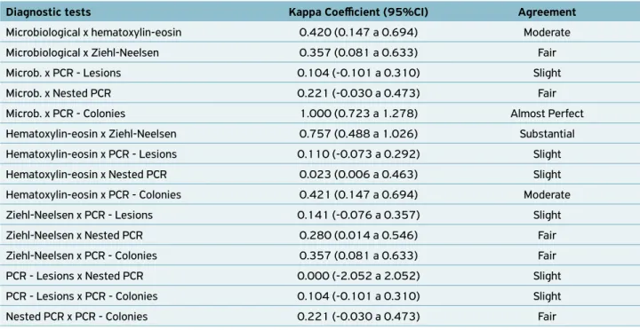
The perfect agreement between the tests was observed only between bacterial isolation, the gold standard test for bovine tuberculosis diagnosis, and M. bovis DNA detection in isolates by PCR (Table 3). This result was also expected, because M. bovis DNA detection in isolates is a genetic confir‑ mation of the isolated bacteria. A slight agreement was found when bacterial isolation was compared to DNA detected from tuberculosis‑like lesions by PCR. Fair concordance was seen when DNA detection by nested PCR and bacterial isolation were compared. Comparisons between histopathological tests resulted in slight agreement as well. LIMA et al. (2008) reported better agreement results when comparing bacte‑ rial isolation and DNA detection by PCR in human sputum samples collected from patients with suspected tuberculosis, which makes possible to infer that the molecular identifica‑ tion of Mycobacterium spp. is a viable alternative for diagnosis. The histopathological characterization with Hematoxylin‑ eosinstained slides showed substantial agreement with AFB identification by Ziehl‑Neelsen staining, while there was a moderate/fair concordance compared with bacterial isolation; FRÁGUAS et al. (2008) reported the same results. It is notice‑ able that AFB identification by Ziehl‑Neelsen staining had similar agreement with DNA detection in tuberculosis‑like lesions by PCR when both were compared to bacterial isola‑ tion. Therefore, AFB identification is the most feasible alter‑ native to diagnose tuberculosis.
Table 3. Agreement between diagnostic tests expressed by the Kappa coefficient.
Diagnostic tests Kappa Coefficient (95%CI) Agreement
Microbiological x hematoxylin‑eosin 0.420 (0.147 a 0.694) Moderate
Microbiological x Ziehl‑Neelsen 0.357 (0.081 a 0.633) Fair
Microb. x PCR ‑ Lesions 0.104 (‑0.101 a 0.310) Slight
Microb. x Nested PCR 0.221 (‑0.030 a 0.473) Fair
Microb. x PCR ‑ Colonies 1.000 (0.723 a 1.278) Almost Perfect
Hematoxylin‑eosin x Ziehl‑Neelsen 0.757 (0.488 a 1.026) Substantial
Hematoxylin‑eosin x PCR ‑ Lesions 0.110 (‑0.073 a 0.292) Slight
Hematoxylin‑eosin x Nested PCR 0.023 (0.006 a 0.463) Slight
Hematoxylin‑eosin x PCR ‑ Colonies 0.421 (0.147 a 0.694) Moderate
Ziehl‑Neelsen x PCR ‑ Lesions 0.141 (‑0.076 a 0.357) Slight
Ziehl‑Neelsen x Nested PCR 0.280 (0.014 a 0.546) Fair
Ziehl‑Neelsen x PCR ‑ Colonies 0.357 (0.081 a 0.633) Fair
PCR ‑ Lesions x Nested PCR 0.000 (‑2.052 a 2.052) Slight
PCR ‑ Lesions x PCR ‑ Colonies 0.104 (‑0.101 a 0.310) Slight
Nested PCR x PCR ‑ Colonies 0.221 (‑0.030 a 0.473) Fair
95%CI: 95% Confidence interval.
Table 4. Sensitivity, specificity and Youden coefficient of diagnostic tests analyzed versus bacterial isolation.
Diagnostic tests Sensitivity (%) Specificity (%) Sensitivity + specificity Youden coefficient (%)
Hematoxylin‑eosin 82.14 59.09 141.23 41.23
Ziehl‑Neelsen 67.86 68.18 136.04 36.04
PCR ‑ Lesions 25.00 86.36 111.36 11.36
Nested PCR 33.33 89.47 122.80 22.80
PCR ‑ Colonies 100.00 100.00 200.00 100.00
and Youden’s coefficient of the diagnostic tests analyzed in this study (Table 4) are in agreement with the concordance test, since the best concordances were shown between DNA detection of the isolates by PCR, followed for the histopath‑ ological diagnosis and the detection of bacterial DNA in the lesions suggestive of tuberculosis by PCR, when compared to the microbiological isolation (gold standard, considered true positive for sensitivity and specificity tests).
The Youden coefficient was used to identify which test hadthe smallest proportion of false diagnoses (the smallest amount of wrong diagnoses, false negative plus false posi‑ tives). The best coefficient values were found for DNA detec‑ tion by PCR, followed by histopathological diagnosis using Hematoxylin‑eosinstained slides, AFB identification by Ziehl‑Neelsen staining, the lowest coefficient being that of DNA detection from bovine tuberculosis‑like lesions by PCR.
Important to highlight that the type of sample used to diagnose tuberculosis is of utter importance once the results are directly influenced by the quantity of bacilli present in the sample collected. The low sensitivity values shown in
this study for DNA detection by PCR from tuberculosis‑like lesions (from 25.00 to 33.33%) were due to the low quan‑ tity of bacilli in the samples, mainly when compared to bac‑ terial isolation. Therefore, when molecular tests are applied in tuberculosis‑like lesions, high percentages of false nega‑ tive results could be found, impairing the quality of results.
CONCLUSIONS
REFERENCES
ALZAMORA‑FILHO, F.; VASCONCELLOS, S.E.G.; GOMES, H.M.; CAVALCANTE, M.P.; SUFFYS, P.N.; COSTA, J.N. Múltiplas estirpes de isolados de Mycobacterium bovis identificados por tipagem molecular em bovinos abatidos em matadouros‑frigoríficos.
Pesquisa Veterinária Brasileira, v.34, n.2, p.103‑108, 2014.
ARAÚJO, C.P.; OSÓRIO, A.L.R.; JORGE, K.S.G.; RAMOS, C.A.N.; SILVA FILHO, A.F.; VIDAL, C.E.S.; ROXO, E.; NISHIBE, C.; ALMEIDA, N.F.; FONSECA JÚNIOR, A.A.; SILVA, M.R.; BARBOSA NETO, J.D.; CERQUEIRA, V.D.; ZUMÁRRAGA, M.J.; ARAÚJO, F.R. Detection of Mycobacterium bovis in bovine and bubaline tissues using Nested‑PCR for TBD1. PLoS One, v.9, n.3, 2014. https://doi. org/10.1371/journal.pone.0091023
AYELE, W.Y.; NEILL, S.D.; ZINSSTAG, J.; WEISS, M.G.; PAVLIK, I. Bovine tuberculosis: an old disease but a new threat to Africa.
The International Journal of Tuberculosis and Lung Diseases, v.8, n.8, p.924‑937, 2004.
BALIAN, S.C.; PINHEIRO, S.R.; GUERRA, J.L.; MORAIS, Z.M.; FERREIRA, F.; FERREIRA NETO, J.S. Estudo comparativo de dois métodos de descontaminação na pesquisa de micobactérias.
Arquivos do Instituto Biológico, v.69, n.2, p.11‑14, 2002.
BEHMER, O.A.; TOLOSA, E.M.C.; FREITAS NETO, A.G. Manual de técnicas para histologia normal e patológica. São Paulo: EDART, Editora da Universidade de São Paulo, 241p, 1976.
CARDOSO, M.A.; CARDOSO, R.F.; HIRATA, R.D.C.; HIRATA, M.H.; LEITE, C.Q.F.; SANTOS, A.C.B.; SIQUEIRA, V.L.D.; OKANO, W.; ROCHA, N.S.; LONARDONI, M.V.C. Direct Detection of Mycobacterium bovis in bovine lymph nodes by PCR.
Zoonoses Public Health, v.56, p.465‑470, 2009. https://doi. org/10.1111/j.1863‑2378.2008.01199.x
COSTA, P.; FERREIRA, A.S.; AMARO, A.; ALBUQUERQUE, T.; BOTELHO, A.; COUTO, I.; CUNHA, M.V.; VIVEIROS, M.; INÁCIO, J. Enhanced detection of tuberculous mycobacteria in animal tissues using a Semi‑Nested Probe‑Based Real‑Time PCR. PLoS
One, v.8, n.11, e81337, 2013. https://doi.org/10.1371/journal.
pone.0081337
DE LA RUA‑DOMENECH, R. Human Mycobacterium bovis infection in the United Kingdom: incidence, risks, control measures and review of the zoonotic aspects of bovine tuberculosis. Tuberculosis, v.86, p.77‑109, 2006.
FIGUEIREDO, E.E.S.; CARVALHO, R.C.T.; SILVESTRE, F.G.; LILEMBAUM, W.; FONSECA, L.S.; SILVA, J.T.; PASCHOALIN,
V.M.S. Detection of Mycobacterium bovis DNA in nasal swabs
from tuberculous cattle by a multiplex PCR. Brazilian Journal
of Microbiology, v.41, n.2, p.386‑390, 2010. http://dx.doi. org/10.1590/S1517‑83822010000200020
FRÁGUAS, S.A.; CUNHA‑ABREU, M.S.; FERREIRA, A.M.R.; MARASSI, C.D.; OELEMANN, W.; FONSECA L.S.; FERREIRA, R.; LILENBAUM, W. Estudo comparativo de métodos complementares para o diagnóstico da tuberculose bovina em animais reagentes à tuberculinização. Revista Brasileira de Ciência Veterinária, v.15, n.3, p.117‑121, 2008. https://doi.org/10.22409/rbcv.v15i3.383
FRANCO, M.M.G.; PAES, A.C.; RIBEIRO, M.G.; PANTOJA, J.C.F.; SANTOS, A.C.B.; MIYATA, M.; LEITE, C.Q.F.; MOTTA, R.G.; LISTONI, F.J.P. Occurrence of mycobacteria in bovine milk samples from both individual and collective bulk tanks at farms and informal markets in the southeast region of Sao Paulo,
Brazil. BMC Veterinary Research, v.9, n.85, 2013. https://doi.
org/10.1186/1746‑6148‑9‑85
FURLANETTO, L.V.; FIGUEIREDO, E.E.S.; CONTE JÚNIOR, C.A.; CARVALHO, R.C.T.; SILVA, F.G.S.; SILVA, J.T.; LILENBAUM, W.; PASCHOALIN, V.M.S. Uso de métodos complementares
na inspeção post mortem de carcaças com suspeita de
tuberculose bovina. Pesquisa Veterinária Brasileira, v.32,
n.11, p.1138‑1144, 2012. http://dx.doi.org/10.1590/ S0100‑736X2012001100011
JORGE, K.S.G. Identificação de Mycobacterium bovis em bovinos e
sua importância na ocorrência de tuberculose zoonótica. Tese de Doutorado, Universidade Federal de Mato Grosso do Sul, Campo Grande‑MS, 2010. 88p.
KONEMAN, E.W.; ALLEN, S.D.; JANDA, W.M.; SCHRECKENBERGER, P.C.; WINN JUNIOR, W.C. Diagnóstico Microbiológico: Texto e atlas colorido. Rio de Janeiro: Medsi, 2001. p. 903‑946,
KURAMAE‑IZIOKA, E.E. A rapid, easy and high yield protocol for total genomic DNA isolation of Colletotrichum gloesporioides and
Fusarium oxysporum. Revista Unimar, v.19, p.683‑689, 1997.
LANDIS, R.J.; KOCH, G.G. The measurement of observer agreement
for categorical data. Biometrics, v.33, p.159‑174, 1977.
LIMA, S.S.S.; CLEMENTE, W.T.; PALACI, M.; ROSA, R.V.; ANTUNES, C.M.F.; SERUFO, J.C. Métodos convencionais e moleculares para o diagnóstico da tuberculose pulmonar: um
estudo comparativo. Jornal Brasileiro de Pneumologia, v.34,
n.12, p.1056‑1062, 2008. http://dx.doi.org/10.1590/ S1806‑37132008001200011
PINTO, P.S.A. Atualização em controle da tuberculose no
contexto da inspeção de carnes. Bioscience Journal, v.19, n.1,
p.115‑121, 2003.
PINTO, P.S.A.; FARIA, J.E.; VILORIA, M.I.V.; BEVILACQUA, P.D. Exame microbiológico da tuberculose como subsídio à
inspeção post‑mortem de bovinos. Revista Brasileira de Saúde
e Produção Animal, v.3, n.1, p.10‑15, 2002. http://dx.doi. org/10.1590/1808‑1657000592014
REBOLLO, M.J.; SAN JUAN GARRIDO, R.; FOLGUEIRA, D.; PALENQUE, E.; DÍAZ‑PEDROCHE, C.; LUMBRERAS C. Blood and urine samples as useful sources for the direct detection of
tuberculosis by polymerase chain reaction. Diagnostic Microbiology
and Infectious Disease, v.56, n.2, p.141‑146, 2006. http://dx.doi. org/10.1016/j.diagmicrobio.2006.03.018
REZENDE‑LAGO, N.C.M.; REIS, L.S.; MARCHI, P.G.F. Levantamento epidemiológico da cisticercose e tuberculose em bovinos abatidos
sob inspeção federal no município de Sertãozinho, SP. Revista
RODRIGUEZ, J.G.; FISSANOTO, J.C.; PORTILLO, P.D.; PATARROYO, M.E.; ROMANO, M.I.; CATALDI, A. Amplification of a 500
base‑pair fragment from cultured isolates of Mycobacterium
bovis. Journal of Clinical Microbiology, v.37, n.7, p.2330– 2332, 1999.
RORING, S.; HUGHES, M.S.; SKUCE, R.A.; NEILL, S.D. Simultaneous detection and strain differentiation of Mycobacterium bovis directly
from bovine tissue specimens by spoligotyping. Veterinary
Microbiology, v.74, n.3, p.227‑236, 2000.
ROXO, E. Mycobacterium bovis como causa de zoonose. Revista
Brasileira de Ciências Farmacêuticas, v.18, p.101‑108, 1997.
SAMBROOK, J.; RUSSEL, D.W. Molecular cloning: a laboratory manual. 3ed. Londres: CSHL Press, 1448 p., 2001.
SILVA, D.A.V.; BÜRGER, K.P.; MARTINS, A.M.C.V.; PROVIDELLO, A. Identificação de lesões macroscópicas sugestivas
de tuberculose bovina. Revista Brasileira de Higiene e
Sanidade Animal, v.8, n.2, p.149‑160, 2014. http://dx.doi. org/10.5935/1981‑2965.20140026
TAYLOR, G.M.; WORTH, D.R.; PALMER, S.; JAHANS, K.; HEWINSON, R.G. Rapid detection of Mycobacterium bovis DNA in cattle lymph
nodes with visible lesions using PCR. B.M.C. Veterinary Research,
v.3, n.12, 2007. http://dx.doi.org/10.1186/1746‑6148‑3‑12
TAYLOR, M.J.; HUGHES, M.S.; SKUCE, R.A.; NEILL, S.D. Detection of
Mycobacterium bovis in bovine clinical specimens using real‑time fluorescence and fluorescence resonance energy transfer probe rapid‑cycle PCR. Jounal of Clinical Microbiology, v.39, n.4, p.1272‑1278, 2001. http://dx.doi.org/10.1128/JCM.39.4.1272‑1278.2001
TAYLOR, R.N. Measurement of variation and significance in serological
test. [S.l.]: Academic Press, 1992. 29 p.
THRUSFIELD, M. Epidemiologia veterinária. 2 ed. São Paulo:
Roca, 2004. 556 p.
VARELLO, K.; PEZZOLATO, M.; MASCARINO, D.; INGRAVALLE, F.; CARAMELLI, M.; BOZZETTA, E. Comparison of histologic techniques for the diagnosis of bovine tuberculosis in the framework of eradication programs. Journal of Veterinary Diagnostic Investigation, v.20, n.2, p.164‑169, 2008.