Clinical Case Study
GE Port J Gastroenterol
Perforated Meckel’s Diverticulum in an
Adult
Rita Camelo
aPaula Santos
a, bRui Mateus Marques
a, baServiço de Imagiologia, Hospital de São José, Centro Hospitalar Lisboa Central, Lisbon, Portugal; bDepartamento de Radiologia, NOVA Medical School, Faculdade de Ciências Médicas da Universidade
Nova de Lisboa, Lisbon, Portugal
Received: June 27, 2018
Accepted after revision: September 2, 2018 Published online: October 31, 2018
Rita Camelo
Serviço de Imagiologia, Hospital de São José, Centro Hospitalar Lisboa Central R. José António Serrano
PT–1150-199 Lisbon (Portugal) © 2018 Sociedade Portuguesa de Gastrenterologia
Published by S. Karger AG, Basel E-Mail [email protected]
DOI: 10.1159/000493439
Keywords
Gastrointestinal symptoms · Surgery · Gastrointestinal tract · Diagnosis · Computed tomography
Abstract
Meckel’s diverticulum is the commonest congenital anoma-ly of the gastrointestinal tract. Its complications have an tensive variety of clinical and imaging manifestations, ex-tending from benign and indolent findings to acute life-threatening conditions. Complicated Meckel’s diverticulum often constitutes a challenging diagnosis for both the clini-cian and the radiologist. Therefore, imaging techniques play an important role in this condition in evaluating its compli-cations, determining decision making. We describe a case of a 49-year-old man suffering from right abdominal pain with fever and constipation, during the past 5 days. Laboratory data revealed C-reactive protein of 306 mg/L and leukocyto-sis. Contrast-enhanced CT features were highly suggestive of perforated Meckel’s diverticulum. The purpose of this ar-ticle is to emphasize that besides its rarity, Meckel’s diver-ticulum complications can occur in adult patients.
© 2018 Sociedade Portuguesa de Gastrenterologia Published by S. Karger AG, Basel
Perfuração de divertículo de Meckel num adulto
Palavras Chave
Sintomatologia gastrointestinal · Cirurgia · Tracto gastrointestinal · Diagnóstico · Tomografia computorizada
Resumo
O divertículo de Meckel é a anomalia congénita mais co-mum do tracto gastrointestinal. As suas complicações variam num amplo espectro, desde achados benignos e indolentes até condições potencialmente graves, e con-stituem frequentemente um desafio diagnóstico tanto para o clinico como para o médico radiologista. Neste sen-tido, os métodos de diagnóstico por imagem desempen-ham um papel importante na avaliação e extensão das suas complicações, determinando muitas vezes a tomada de decisões. Descrevemos um caso de um paciente de 49 anos, que apresenta dor abdominal lateralizada à direita, febre e obstipação, durante um período de 5 dias. Analiti-camente apresenta valores de proteína C reactiva de 306 mg/L e leucocitose. O estudo TC contrastado foi muito sugestivo de divertículo de Meckel perfurado. O objectivo deste artigo é enfatizar que, apesar da sua raridade, as complicações do divertículo de Meckel podem ocorrer em pacientes em idade adulta.
© 2018 Sociedade Portuguesa de Gastrenterologia Publicado por S. Karger AG, Basel
Introduction
Meckel’s diverticulum (MD) is the commonest con-genital anomaly of the gastrointestinal tract. It is a true diverticulum having all the layers of the intestinal wall [1, 2] and is usually localized in the pelvic or periumbilical region or right iliac fossa. The referred locations of MD are related to its complications. The preferred location is at the anti-mesenteric border of the ileum, 2 ft (60 cm) proximal to the ileocecal valve. The well-known “rules of 2” state that the MD occurs in about 2% of the population, it is about 2 inches in length, is usually located within 2 ft of the ileocaecal valve, and usually presents before 2 years of age [3]. MD results from the incomplete obliteration of the omphalomesenteric or vitelline duct during the 5th week of gestation [3].
MD manifests more commonly in children. However, complications can occur in adults and it may give rise to bleeding (11.8%), intestinal obstruction (36.5%), inflam-mation (12.7%), intussusceptions (13.7%), and neoplasm (3.2%). Perforation is very rarely seen and, in a review, was reported as being responsible for 0.5% of symptom-atic diverticulum [4–6]. In adult patients, intestinal ob-struction is the most common complication, with inci-dence rates varying from 22 to 50% [7].
MD complications become clinically apparent in up to 16–20% patients with this condition [1, 8]. Symptomatic diverticula are more common in males than in females, despite the fact that there is no known gender predisposi-tion of asymptomatic MD [3], and the incidence of com-plications decreases with age, with the majority occurring in the pediatric population [9].
Case Report
A 49-year-old Caucasian male attended the emergency depart-ment because of right abdominal pain, fever, and constipation dur-ing the past 5 days. He also reported increasdur-ing symptoms durdur-ing the last day. The patient had no significant medical history.
Physical examination detected significant tenderness on palpa-tion of his right iliac fossa, with signs of localized peritonitis, in-cluding involuntary guarding and rebound tenderness. Auricular
temperature was 37.5 ° C. Laboratory data revealed C-reactive
pro-tein serum level of 306 mg/L (normal range, <5.0 mg/L) and
leu-kocytosis (24,000/µL [normal range, 4,500–11,000/µL]).
Abdominal X-ray and abdominal ultrasound showed no sig-nificant alterations, with no signs of acute appendicitis. In this case of acute abdomen, without an evident cause, an abdominal/pelvic computed tomography (CT) scan with intravenous contrast was performed, and a tubular blind-ending structure arising from the terminal ileum, measuring 65 × 25 mm (longitudinal and trans-verse diameter, respectively), was demonstrated. It was associated
with a thickened and hypercaptant distal ileum, with intramural bowel gas as well as foci of adjacent extraluminal gas. A surround-ing fat strandsurround-ing was also detected (Fig. 1). No significant lymph-adenopathy could be appreciated. The rest of the abdomen, includ-ing the appendix, was unremarkable. In conclusion, contrast-en-hanced CT features were highly suggestive of a perforated MD.
Based on imaging findings in association with worsening gen-eral condition, an emergency laparotomy intervention was per-formed and perforated Meckel’s diverticulitis was confirmed, along with an extensive inflammatory mass (abscess) with ileal, ascending colon, and abdominal wall involvement. Therefore, the patient was submitted to a right hemicolectomy and segmental enterectomy, with an ileocolostomy. Histological examination re-vealed an MD, measuring about 80 × 35 mm, with perforation of its distal portion with contiguous abscess. There was no evidence of ectopic tissues in the MD (Fig. 2). The patient recovered well after surgery and completed 8 days of antibiotic therapy.
Discussion
The majority of MD cases remain clinically silent dur-ing the entire lifetime, and their presence may be discov-ered incidentally during surgery, autopsy, or when per-forming small gastrointestinal studies [10, 11]. On CT, MD may appear as a fluid- or air-filled blind-ending pouch that arises from the antimesenteric side of the dis-tal ileum. However, CT has a low sensitivity for the detec-tion of uncomplicated MD because its appearance mim-ics that of a normal bowel loop. Complicated MD repre-sents an important cause of acute abdominal pain [12, 13], and most cases with inflamed MD may be visualized on CT.
The diverticulum is considered complicated if it is the site of bowel obstruction or if it presents surrounding fea-tures of infiltration or inflammation, signs of perforation, adjacent fluid collection, or active bleeding. Depending on the type of complication, the diverticulum may have surrounding mesenteric inflammatory changes or may look like a localized fluid or air-fluid collection, with nor-mal aspect of the snor-mall bowel proxinor-mal and distal to the diverticular inflammation [3]. However, less than 10% of symptomatic MD is diagnosed preoperatively [5].
The most common presentation associated with symp-tomatic or complicated MD is bleeding, followed by in-testinal obstruction, diverticulitis, intussusceptions, and neoplasm [14]. Perforation is noted to be an occasional consequence of acute Meckel’s diverticulitis, but the exact rate of this has not been reported [15].
There are a few studies described in this particular complication in adults, and they are not consensual: Ac-cording to a study by Kusumoto et al. [16], in a review
of 776 Japanese patients, it has been reported that per-foration accounted for 10.8% (84 of 776) of complica-tions associated with the MD. According to Chae et al. [7], perforation is rarely seen and was reported as being responsible for 0.5% of symptomatic diverticulum. An-other study (composed of 12 men and 11 women, with a mean age of 42.6 years, over a 15-year period) con-cluded that diverticulum-related complications demon-strated by surgery were inflammation in 14 patients, bleeding in 5 patients, intestinal obstruction in 3
pa-tients, and penetrating foreign body causing perforation in 1 patient [3].
MD perforation is a serious and often life-threatening complication, usually secondary to diverticulitis, gan-grene, or peptic ulceration due to ectopic gastric mucosa. Other various pathologies leading to perforation are Lit-tre’s hernia and tumors such as leiomyosarcoma, lym-phatic sarcoma, and poorly differentiated stromal tumor [17]. Perforation of MD by foreign bodies is extremely rare, and in a review, the indication rate for a resection
b a
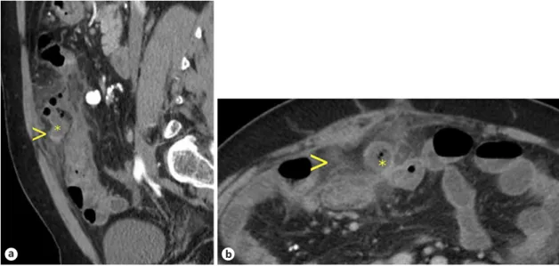
Fig. 1.a Sagittal enhanced CT images showing a tubular blind-ending structure (asterisk) measuring 65 mm
(longitudinal diameter), with a thickened and hypercaptant wall (arrowhead). b Axial contrast-enhanced CT
showing a rounded structure (asterisk). There is also densification of contiguous fat planes along with free fluid suggesting inflammatory process (arrowhead).
b a
Fig. 2. Surgical resection specimen. a, b MD with intestinal mucosa without ectopic tissue evidence. Perforation of its distal portion with contiguous abscess was observed (H&E, ×20).
due to perforation by foreign body was reported to be 8% of all complicated diverticula [18].
However, a symptomatic or complicated MD diagno-sis is difficult to confirm on the badiagno-sis of history, physical examination, laboratory findings, and imaging, because a variety of conditions can mimic the MD both clinically and radiologically (such as appendicitis, ileal/colonic di-verticulitis, or regional enteritis/colitis) [19].
Traditionally, clinicians, when confronted with a pa-tient with complicated MD, relied on conventional gas-trointestinal contrast studies, angiography, or scintigra-phy. However, these methods have been progressively re-placed by CT scan, which is now routinely used as the first-line imaging tool in the diagnostic workup of the acute abdomen [20]. The sensitivity of diagnosing the MD on CT scan has increased owing to the availability of higher spatial resolution and multiplanar isotropic re-construction ability of the latest MDCT scanners, which allow visualization of the small bowel in various planes [21].
Other techniques, including conventional radiograph-ic examination, are of limited value and usually unreveal-ing. However, it may show enteroliths, findings of bowel obstruction, and the presence of gas or a gas-fluid level in the diverticulum [22]. Although of limited value, sonog-raphy has been used for the investigation of MD [23]. High-resolution sonography usually shows a fluid-filled structure in the right lower quadrant having the appear-ance of a blind-ending, thick-walled loop of bowel, with the typical gut signature and a clear connection to a peri-staltic, normal small-bowel loop [21]. Scintigraphy with 99mTc-Na-pertechnetate has only minor diagnostic val-ue and a limited sensitivity of 60% in diagnosing MD [24]. However, it aids in the diagnosis of diverticula with ecto-pic gastric mucosa. Pertechnetate is taken up by mucin-secreting cells of the gastric mucosa and ectopic gastric tissue. Higher sensitivity in pediatric (85–90%) than in adult (60%) patients is noticed [24]. This could be due to earlier symptoms (such as hemorrhage) in patients with ectopic gastric mucosa.
There is no consensus in the literature on the manage-ment of MD complications. In one study, the authors sug-gested four features associated with symptomatic diver-ticula – age <50 years, male sex, diverticulum length >2 cm, and the presence of ectopic or abnormal features within the diverticulum – and recommended that the presence of any of these four criteria should warrant re-moval of the MD [1]. Surgical rere-moval of an MD is the current management of choice in a patient symptomatic for an MD or for any of its complications. However, the
controversy surrounding surgical removal of an inciden-tally detected MD still continues today [1]. This article highlights the importance of CT in the evaluation of MD complications because in the case of diverticulitis and perforation, inflammatory changes and extraluminal air may be present and can be easily identified on CT scan.
In conclusion, if an inflammatory process is visualized on CT in the lower abdomen or pelvis, particularly at midline, or if there is evidence of distal small-bowel ob-struction, one should carefully search for the presence of a complicated diverticulum. If a normal appendix is iden-tified, the likelihood of this diagnosis increases [25]. MD complications can present with a wide range of clinical and imaging manifestations, from benign indolent find-ings to acute life-threatening conditions [21]. CT findfind-ings of complicated MD are very polymorphic and should be remembered in the evaluation of adult patients with acute abdomen [8].
Acknowledgement
The authors would like to thank Dr. Joana Santos, Pathology Department, Centro Hospitalar Lisboa Central, Hospital de São José, for slides review and helpful discussion.
Statement of Ethics
The authors have no ethical conflicts to disclose.
Disclosure Statement
The authors have no conflicts of interest to declare.
Funding Sources
No subsidies or grants contributed to this work.
References 1 Park JJ, Wolff BG, Tollefson MK, Walsh EE, Larson DR: Meckel diverticulum: the Mayo clinic experience with 1,476 patients (1950– 2002). Ann Surg 2005;241:529–533. 2 Matsagas MI, Fatouros M, Koulouras B, Gian-
noukas AD: Incidence, complications, and management of Meckel’s diverticulum. Arch Surg 1995;130:143–146.
3 Platon A, Gervaz P, Becker CD, Morel P, Po-letti PA: Computed tomography of compli-cated Meckel’s diverticulum in adults: a picto-rial review. Insights Imaging 2010;1:53–61.
4 Groebli Y, Bertin D, Morel P: Meckel’s diver-ticulum in adults: retrospective analysis of 119 cases and historical review. Eur J Surg 2001;167:518–524.
5 Ymaguchi M, Takeuchi S, Awazu S: Meckel’s diverticulum. Investigation of 600 patients in Japanese literature. Am J Surg 1978;136:247– 249.
6 Leijonmarck CE, Bonman-Sandelin K, Frisell J, Raf L: Meckel’s diverticulum in the adult. Br J Surg 1986;73:146–149.
7 Chae HD: Perforation of Meckel’s diverticu-lum by a chicken bone; preoperatively pre-senting as bowel perforation. J Korean Surg Soc 2011;80:234–237.
8 Mackey WC, Dineen P: A fifty year experi-ence with Meckel’s diverticulum. Surg Gyne-col Obstet 1983;156:56–64.
9 Kloss BT, Broton CE, Sullivan AM: Perforat-ed Meckel diverticulum, Int J Emerg MPerforat-ed 2010;3:455–457.
10 Levy AD, Hobbs CM: From the archives of the AFIP. Meckel diverticulum: radiologic fea-tures with pathologic correlation. Radio-graphics 2004;24:565–587.
11 Rossi P, Gourtsoyiannis N, Bezzi M, Rapto-poulos V, et al: Meckel’s diverticulum: imag-ing diagnosis. AJR Am J Roentgenol 1996; 166:567–573.
12 Sancar S, Demirci H, Sayan A, Arıkan A, Candar A: Meckel’s diverticulum: ten years’ experience. Ulus Cerrahi Derg 2015;31:65– 67.
13 Yahchouchy EK, Marano AF, Etienne JC, Fin-gerhut AL: Meckel’s diverticulum. J Am Coll Surg 2001;192:658–662.
14 Dimitriou I, Evaggelou N, Tavaki E, Chatzi-theoklytos E: Perforation of Meckel’s diver-ticulum by a fish bone presenting as acute ap-pendicitis: a case report. J Med Case Rep 2013; 7:231.
15 Ferguson H, Soumian S, Dmitrewski J: Perfo-ration of Meckel’s diverticulum secondary to a large faecolith. BMJ Case Rep 2010;2010. pii: bcr09.2009.2308.
16 Kusumoto H, Yoshida M, Takahashi I, Anai H, Maehara Y, Sugimachi K: Compli-cations and diagnosis of Meckel’s divertic-ulum in 776 patients. Am J Surg 1992;164: 382–383.
17 Hager M, Maier H, Eberwein M: Perforated Meckel’s diverticulum presenting as a gastro-intestinal stromal tumor: a case report. J Gas-trointest Surg 2005;9:809–811.
18 Yagcı G, Cetiner S, Tufan T: Perforation of Meckel’s diverticulum by a chicken bone, a rare complication: report of a case. Surg To-day 2004;34:606–608.
19 Khandelwal A, Virmani V, Ryan J, Kielar A, Fraser-Hill M, Sheikh A: Solving the mystery of Meckel diverticulum. AJR Am J Roentgen-ol 2012;198:E166.
20 Leschka S, Alkadhi H, Wildermuth S, Marincek B: Multidetector computed tomog-raphy of acute abdomen. Eur Radiol 2005;15: 2435–2447.
21 Kotha K, et al: Radiologist’s perspective for the Meckel’s diverticulum and its complica-tions Br J Radiol 2014;87:20130743.
22 Elsayes KM, Menias CO, Harvin HJ, Francis IR: Imaging manifestations of Meckel’s diver-ticulum. AJR Am J Roentgenol 2007;189:81– 88.
23 Mostbeck GH, Liskutin J, Dorffner R, et al: Ultrasonographic diagnosis of a bleeding Meckel’s diverticulum. Pediatr Radiol 2000; 30:382.
24 Poulsen KA, Qvist N: Sodium pertechnetate scintigraphy in detection of Meckel’s diver-ticulum: is it usable? Eur J Pediatr Surg 2000; 10:228–231.
25 Bennett GL, Birnbaum BA, Balthazar EJ: CT of Meckel’s diverticulitis in 11 patients. AJR Am J Roentgenol 2004;182:625–629.