Gasserian ganglion neurosarcoidosis
mimicking trigeminal schwannoma
Neurosarcoidose do gânglio de gasser simulando um schwannoma trigeminal
Tatiana Goyanna Lyra
1, Hae Won Lee
1, Eduardo de Arnaldo Silva Vellutini
1,2,
Maria da Graça Moraes Martin
1,3, Ana Paula Torres Cardoso
1, Luis Filipe de Souza Godoy
1,
Giovanni Guido Cerri
1,4, Claudia da Costa Leite
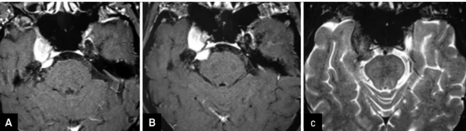
4,5,6A MRI of a 59-year-old male with right hemifacial
hypoesthe-sia showed a low signal T2-weighted expansive mass in the right
Meckel
’
s cave. After failure of initial conservative treatment
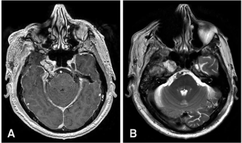
(Figure 1), surgery was done with partial lesion resection
(Figure 2). The pathology and chest CT were consistent with
granulomatous disease: neurosarcoidosis. On follow-up the
lesion increased in size but after corticosteroids it reversed
(Figure 3). The involvement of the trigeminal nerve is very rare
with only few cases described in literature. Although rare,
sar-coid infiltration of the Gasserian ganglion must be considered
in the differential diagnosis of an isolated mass at Meckel
’
s cave,
especially if it has T2 hypointensity signal.
1Hospital Sírio Libanês, Sao Paulo SP, Brazil;
2DFV Neuro, Sao Paulo SP, Brazil;
3Universidade de São Paulo, Faculdade de Medicina, Hospital das Clínicas, Sao Paulo SP, Brazil;
4Universidade de São Paulo, Faculdade de Medicina, Departamento de Radiologia, Sao Paulo SP, Brazil;
5Hospital Sírio Libanês, Instituto de Ensino e Pesquisa, Sao Paulo SP, Brazil;
6The University of North Carolina at Chapel Hill, North Carolina, USA.
Correspondence: Tatiana Goyanna Lyra; Rua Barata Ribeiro, 323 / ap. 51; 01308-000, São Paulo SP, Brasil; E-mail: [email protected] Conflict of interest:There is no conflict of interest to declare.
Received 19 September 2014; Received in final form 02 October 2014; Accepted 22 October 2014.
Figure 1.
Initial pre-operative images and follow up one month later.
DOI:10.1590/0004-282X20140209
IMAGES IN NEUROLOGY
References
1. Shah R, Roberson GH, Curé JK. Correlation of MR imaging findings and clinical manifestations in neurosarcoidosis. Am J Neuroradiol. 2009;30(5):953-61. http://dx.doi.org/10.3174/ajnr.A1470
2. Quinones-Hinojosa A, Chang EF, Khan SA, McDermott MW. Isolated trigeminal nerve sarcoid granuloma mimicking trigeminal schwan-noma: case report. Neurosurgery. 2003;52(3):700-5.
3. Amin A, Balderacchi JL. Trigeminal neurosarcoidosis: case report and literature review. Ear Nose Throat J. 2010;89(7):320-2.
4. Arias M, Iglesias A, Vila O, Brasa J, Conde C. MR imaging findings of neurosarcoidosis of the gasserian ganglion: an unusual presentation. Eur Radiol. 2002;12(11):2723-5. http://dx.doi.org/10.1007/s00330-001-1287-9
5. Ahn JY, Kwon So, Shin MS, Joo JY, Kim TS. Chronic granulomatous neuritis in idiopathic trigeminal sensory neuropathy: report of two cases. J Neurosurg. 2002;96(3):585-8.