Campylobacter spp. as a foodborne pathogen: a review
Joana Silva, Daniela Leite, Mariana Fernandes, Cristina Mena, Paul Anthony Gibbs and Paula Teixeira* CBQF/Escola Superior de Biotecnologia, Universidade Católica Portuguesa, Porto, Portugal
Edited by:
Kostas Koutsoumanis, Aristotle University, Greece
Reviewed by:
Sandra Torriani, Università degli Studi di Verona, Italy
Alexandra Lianou, Aristotle University of Thessaloniki, Greece
*Correspondence:
Paula Teixeira, CBQF/Escola Superior de Biotecnologia, Universidade Católica Portuguesa, R. Dr. António Bernardino de Almeida, 4200-072 Porto, Portugal
e-mail: [email protected]
Campylobacter is well recognized as the leading cause of bacterial foodborne diarrheal disease worldwide. Symptoms can range from mild to serious infections of the children and the elderly and permanent neurological symptoms. The organism is a cytochrome oxidase positive, microaerophilic, curved Gram-negative rod exhibiting corkscrew motil-ity and is carried in the intestine of many wild and domestic animals, particularly avian species including poultry. Intestinal colonization results in healthy animals as carriers. In contrast with the most recent published reviews that cover specific aspects of Campy-lobacter /campyCampy-lobacteriosis, this broad review aims at elucidating and discussing the (i) genus Campylobacter, growth and survival characteristics; (ii) detection, isolation and con-firmation of Campylobacter ; (iii) campylobacteriosis and presence of virulence factors; and (iv) colonization of poultry and control strategies.
Keywords: Campylobacter spp., foodborne pathogens, virulence factors, antimicrobial susceptibility, control measures
THE GENUS CAMPYLOBACTER
It is believed that the first report concerning Campylobacter was back in 1886 by Theodore Escherich who observed and described non-culturable spiral-shaped bacteria (Vandamme, 2000; King and Adams, 2008; Vandamme et al., 2010). After this, Campy-lobacter was identified for the first time in 1906 when two British veterinarians reported the presence of “large numbers of a pecu-liar organism” in the uterine mucus of a pregnant sheep (Skirrow, 2006;Zilbauer et al., 2008). In 1913, McFadyean and Stockman isolated these microorganisms from aborted bovine fetuses. Later in 1927, Smith and Orcutt named a group of bacteria, isolated from the feces of cattle with diarrhea, as Vibrio jejuni. Seven-teen years later, in 1944, Doyle isolated a different vibrio from feces of pigs with diarrhea and classified them as Vibrio coli ( Van-damme, 2000;Vandamme et al., 2010). Due to their low DNA base composition, their non-fermentative metabolism and their microaerophilic growth requirements, the genus Campylobacter was first proposed in 1963 by Sebald and Véron, distinguishing them from the “true” Vibrio spp. (On, 2001). After that, the study of Butzler et al. (1973)raised the interest in Campylobacter by noting their high incidence in human diarrhea (On, 2001). Since its inception, the taxonomic structure of the genus Campylobacter has experienced extensive changes and even some parts of the cur-rent genus taxonomy remain a matter of controversy and require further investigation (On, 2001;Debruyne et al., 2005). According to these latter authors,Debruyne et al. (2005), there are 14 validly described Campylobacter species. More recently,Fernández et al. (2008)stated that the genus comprises 20 species and subspecies. However, other authors have stated that there are 16 species with a further six subspecies within the genus Campylobacter (On, 2001; Foster et al., 2004).
Campylobacters have been known to be the cause of diseases in animals since 1909, but they have been generally recognized as a cause of human disease, only since about 1980.
The family Campylobacteraceae consists of two genera, Campy-lobacter and Arcobacter and occur primarily as commensals in
humans and domestic animals (Vandamme, 2000). The genus Campylobacter contains small (0.2–0.8μm × 0.5–5 μm) Gram-negative, slender spirally curved rods. When two or more bacterial cells are grouped together, they form an “S” or a “V” shape of gull-wing. The majority of the species have a corkscrew-like motion by means of a single polar unsheathed flagellum at one or both ends of the cell. The only exceptions are Campylobacter gracilis which is non-motile and Campylobacter showae which has multiple flagella (seeDebruyne et al., 2005for a comprehensive description of the taxonomy of Campylobacteraceae). Oxidase activity is present in all species except for C. gracilis. They neither ferment nor oxidize carbohydrates; instead they obtain energy from amino acids, or tricarboxylic acid cycle intermediates (Vandamme, 2000). Campy-lobacter jejuni hydrolyzes hippurate, indoxyl acetate and reduces nitrate. Most strains are resistant to cephalothin, and also resis-tance to fluoroquinolones, a category of antibiotics normally used to treat animal and human illness, has been reported (Koenraad et al., 1995).
Under unfavorable growth conditions, these microorganisms have the ability to form viable but non-culturable cells (VBNC; Portner et al., 2007).Cappelier (1997), observed under labora-tory conditions, that Campylobacter strains, isolated from the soil around the broiler house, may have been transformed into viable but non-cultivable forms and might have become cultivable after passing through the intestinal tract of chickens. Many ques-tions have been raised on whether non-culturability equates to non-viability (McKay, 1992), whether it is possible to convert the VBNC form to a culturable form (Jones et al., 1991;Beumer et al., 1992;Stern et al., 1994), and whether, indeed, a VBNC form of Campylobacter actually exists (ACMSF, 2004).
GROWTH AND SURVIVAL CHARACTERISTICS
Thermophilic Campylobacter species are able to grow between 37 and 42˚C, but incapable of growth below 30˚C (absence of cold shock protein genes which play a role in low-temperature adaptation), with an optimum temperature of 41.5˚C. Levin
(2007) suggested that these organisms should be referred to as “thermotolerant” since they do not exhibit true thermophily (growth at 55˚C or above). However, a study byDe Cesare et al. (2003)revealed that C. jejuni survived for more than 4 h at 27˚C and 60–62% relative humidity on some common clean or soiled food contact surfaces. These characteristics, reduce the ability of campylobacters to multiply (i) outside of an animal host and (ii) in food during their processing and storage (Park, 2002). Growth does not occur in environments with water activity (aw) lower
than 0.987 (sensitive to concentrations of sodium chloride (NaCl) greater than 2%w/v), while optimal growth occurs at aw= 0.997
(approximately 0.5% w/v NaCl).
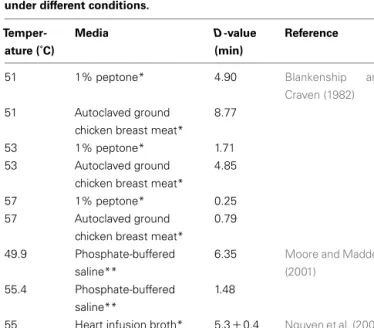
Campylobacter spp. are easily inactivated by heat treatments with their D-value being less than 1 min (Table 1).
Freezing–thawing also reduces the population of Campylobac-ter spp. (Stern and Kazmi, 1989). In pure cultures, Campylobacter spp. are normally inactivated by frozen storage at −15˚C in as few as 3 days (Stern and Kotula, 1982); however, freezing does not eliminate the pathogen from contaminated foods (Lee et al., 1998). Hazeleger et al. (1995)revealed that aged C. jejuni cells survived the longest at 4˚C. Campylobacter spp. will not survive below a pH of 4.9 and above pH 9.0 and grow optimally at pH 6.5–7.5. These non-spore-forming and fastidious bacteria are essentially microaerophilic, growing best in an atmosphere with low oxy-gen tension (5% O2, 10% CO2, and 85% N2; Garénaux et al., 2008).
DETECTION, ISOLATION AND CONFIRMATION
The sensitivity of Campylobacter spp. to oxygen and oxidizing radicals has led to the development of several selective media con-taining one or more oxygen scavengers, such as blood, ferrous iron,
Table 1 | Reported D-values for Campylobacter jejuni and C. coli under different conditions.
Temper-ature (˚C)
Media D-value
(min)
Reference
51 1% peptone* 4.90 Blankenship and
Craven (1982)
51 Autoclaved ground
chicken breast meat*
8.77
53 1% peptone* 1.71
53 Autoclaved ground
chicken breast meat*
4.85
57 1% peptone* 0.25
57 Autoclaved ground
chicken breast meat*
0.79
49.9 Phosphate-buffered saline**
6.35 Moore and Madden (2001)
55.4 Phosphate-buffered saline**
1.48
55 Heart infusion broth* 5.3± 0.4 Nguyen et al. (2006) 55 Heart infusion broth** 6.6± 0.5
*Campylobacter jejuni **Campylobacter coli
pyruvate, etc., and selective agents, particularly antibiotics. Most methods involve a pre-enrichment in a liquid medium, before plating on agar. The developments of methods for Campylobacter have been well-described byCorry et al. (1995).
In some protocols, in order to ameliorate the inhibitory effects of the selective agents on potentially damaged cells, initial suspen-sion of samples is made into a basal broth without selective agents, with the latter being gradually added after a short period of incuba-tion. In order to permit recovery of damaged cells, the incubation temperature may also be gradually increased from 37˚C to the final incubation temperature of 41.5˚C. This methodology is the basis for one of the ISO standard methods (ISO, 1995, 2006a). However, for chicken samples, such a protocol was not necessary as maxi-mal numbers were obtained by using a selective broth followed by plating on selective agars (Mason et al., 1999).
Several of the selective broths, e.g., Bolton broth (BB), Campy-lobacter enrichment broth (CEB), and Preston broth (PB), have been compared for their efficacy (Baylis et al., 2000). The incor-poration of the enzyme Oxyrase in selective broths is particularly effective in reducing the levels of oxygen and improving the isola-tion of Campylobacter spp. from naturally contaminated samples (Abeyta et al., 1997). However, a blood free enrichment broth not requiring the addition of Oxyrase, nor special atmospheres has been tested and found to perform well against other more complex isolation methods (Tran, 1998).
Several selective agars have been formulated and tested for their efficacy in isolating campylobacters. For example, Preston, char-choal cefoperazone deoxycholate (CCDA) and Butzler agars have been found to be equally effective. The use of CCDA and incuba-tion at 42˚C rather than 37˚C is usually the methodology of choice since it allows for the isolation of more Campylobacter strains (Zanetti et al., 1996).
Corry and Atabay (1997)developed CAT agar from modified CCDA by altering the levels of the antibiotics to permit growth of a wider range of strains of Campylobacter spp., notably Campy-lobacter upsaliensis. A later comparison byFederighi et al. (1999), of Karmali, Butzler, and Skirrow isolation agars after enrichment of a large number and wide range of samples in Preston or Park and Sanders broths, showed that Park and Sanders broth followed by isolation on Karmali agar was the more effective combination. The most recent standard method (ISO, 2006a) for detection and isolation, and a direct plating method for enumeration of campy-lobacters (ISO, 2006b), both use mCCDA as the selective agar. Bolton broth is used for the enrichment step and the suspension is incubated at 37˚C in a microaerophilic atmosphere for 4–6 h, fol-lowed by 41.5˚C for 40–48 h and plating on mCCDA and another agar medium of the operator’s own choice. However, methods for Campylobacter spp. are not commonly used in routine laborato-ries as the organisms are difficult to cultivate and to keep reference cultures.
Several alternative and rapid methods have been developed for detecting and confirming Campylobacter spp. e. g. those that include fluorescence in situ hybridization (FISH;Lehtola et al., 2006), latex agglutination (commercially available; e.g., Wilma et al., 1992; Microscreen® Campylobacter kit), and a physical enrichment method (filtration) that permits the separation of Campylobacter from other organisms present in the food matrix
(Baggerman and Koster, 1992). Perhaps the most effective confir-mation methods are those based on the polymerase chain reaction (PCR) reaction, since the phenotypic reactions are often atypical and difficult to read, e.g., the hippurate hydrolysis test for dif-ferentiating Campylobacter coli from C. jejuni. The PCR reaction has been combined with immuno-separation with some success (e.g.,Docherty et al., 1996; Waller and Ogata, 2000) in detect-ing low numbers of the organism in only about 6 h. However, some components of both food samples and selective broths can be inhibitory to the PCR reaction. More recently real-time PCR methods have been developed that show the potential of detecting as few as 1 cfu in chicken samples, and in less than 2 h (Debretsion et al., 2007). Epidemiological studies (e.g., outbreak investigations) have been benefited from the use of molecular typing techniques such as PCR, random amplification of polymorphic DNA (RAPD) and pulsed field gel electrophoresis (PFGE).
CAMPYLOBACTERIOSIS IN HUMANS: OCCURRENCE, SEVERITY AND COSTS
Based on the Community Zoonoses Reports of the European Food Safety Authority (EFSA) and the European Centre for Disease Prevention and Control (ECDC) in their Community Zoonoses Reports, in the last 5 years, campylobacteriosis has been the most commonly reported zoonosis in the EU followed by salmonellosis and yersiniosis (EFSA, 2007, 2010c). In 2008, campylobacterio-sis was the principal cause of zoonotic disease in humans with 190,566 reported confirmed cases (EFSA, 2010c). In 2007, more than 200,000 confirmed cases of human campylobacteriosis were reported by the 24 member states (MS), with an EU notification rate of 45.2 cases per 100,000 inhabitants. Rates varied depending on the MS. When compared to data for 2006, an increase of 14.2% was observed in the number of reported cases (EFSA, 2009). The food vehicle associated with the majority of the reported in 2007 campylobacteriosis infections was contaminated poultry meat. The Foodborne Diseases Active Surveillance Network (FoodNet) of the Centers for Disease Control and Prevention (CDC) in the USA estimates that in 2009, the number of reported infections and incidence per 100,000 population by Campylobacter was 6,033 and 13.02, respectively (Anonymous, 2010). In fact, more than 10,000 cases of campylobacteriosis are reported each year to the CDC (approximately six cases for each 100,000 persons in the population). However, many more cases remain undiagnosed or unreported (Anonymous, 2010). Estimates are that Campylobacter
causes more than two million illnesses (or 1% of the population), 13,000 hospitalizations, and over 100 deaths each year in the USA (Anonymous, 2007).
According toEFSA (2010a), clinical cases of campylobacterio-sis is under-reported in the EU (27 MS): “there may be not less than 2 million and possibly as high as 20 million cases of clinical campylobacteriosis per year in the EU 27 MS.”
In the EU, the reported foodborne outbreaks of campylobacte-riosis are limited, constituting 2, 1, and 0.12% of the total reported campylobacteriosis cases in 2004/2005, 2006 and 2007, respectively (EFSA, 2006, 2007, 2009). In fact, most cases of human campy-lobacteriosis are sporadic. However, the Campylobacter Sentinel Surveillance Scheme Collaborators (2003) suggested that out-breaks of campylobacteriosis may be more common than previ-ously suspected. In fact, the number of Campylobacter outbreaks reported in the United Kingdom (UK) in 2009 was similar to the sum of those reported for 2005–2008 (ACMSF, 2010). Table 2 reports examples of outbreaks that occurred worldwide in the recent years, mainly resulting from consumption of contaminated drinking water, raw milk, and chicken products. Further infor-mation on Campylobacter outbreaks in the USA can be found in the internet12 (accessed in October 2010). The outbreak inves-tigations suggested that in over 25% of the cases, chicken was identified as the source of the outbreak; in 33% of the cases, the source was unknown (EFSA, 2010a). From 2010 until 2015, the UK Government increased the priority of “innovation strategy for Campylobacter.” In the UK, Campylobacter is actually consid-ered the most common cause of food poisoning, responsible for 321,000 estimated cases in England and Wales in 2008, with more than 15,000 hospitalizations and 76 deaths3.
Handling, preparation and consumption of broiler meat may account for 20–30% of human cases of campylobacteriosis, while 50–80% may be attributed to the chicken reservoir as a whole (EFSA, 2010a). According toEFSA (2010b), and with regard to the factors that may contribute to Campylobacter spread in live chickens and chicken carcasses, the above along with the fact that the levels of the pathogen found on single carcasses may also vary greatly, indicate that some slaughterhouses are more capa-ble of controlling this organism than others. Other factors that 1http://www.pritzkerlaw.com/campylobacter-outbreaks/
2http://www.about-campylobacter.com/campylobacter_outbreaks 3http://www.food.gov.uk/multimedia/pdfs/campylobacterstrategy.pdf
Table 2 | Food products implicated in campylobacteriosis outbreaks occurring worldwide.
Year Location Total no. of cases Food implicated Reference
2010 Northumberland, UK 24 Chicken liver parfait Inns et al. (2010)
2009 Crete, Greece 37 Tap water Karagiannis et al. (2010)
2008 Washington, USA 5 Raw milk Anonymous (2008)
2007 British Columbia, Canada 225 Ingestion of mud during a mountain bike race Stuart et al. (2010)
2007 Røros, Norway 105 Untreated tap water Jakopanec et al. (2008)
2005 Madrid, Spain 81 Custard Anonymous (2005)
2005 Australian Capital Territory 11 Several chicken-containing dishes Black et al. (2006)
2005 Copenhagen, Denmark 79 Chicken salad Mazick et al. (2006)
might also influence the risk of contamination with Campylobac-ter include the age of the slaughCampylobac-tered chickens, and the period of the year and the time of the day when carcasses are processed. On the other hand, depopulation of chicken flocks also increases the likelihood of infection since some of the chickens are retained from slaughter to continue growing. In fact, it is believed that during these practices, humans or other vectors may introduce Campylobacter and infect the remaining chickens (EFSA, 2010b). Recently, EFSA described the factors influencing campylobac-teriosis infections, namely the age (higher occurrence rates in children under 5 years old), the season (a higher number of campy-lobacteriosis cases is reported during the summer months), the strain variation (certain strains are less pathogenic than others), host immunity, travel and the demographic factors (i.e., the social economic status). Foodborne zoonoses are an important cause of morbidity and mortality worldwide; the World Health Orga-nization (WHO) estimates that over two million people die each year from diarrheal diseases mainly caused by the ingestion of contaminated foods (WHO, 2005;EFSA, 2007).
European Food Safety Authority has emphasized the impor-tance and recommended the establishment of an active surveil-lance of campylobacteriosis in all MS, including efforts to deter-mine the uncertain and unreported campylobacteriosis cases. In addition, storage and genotyping of human and putative reservoirs of isolates in all MS have also been recommended.
Thereafter, it would be important to identify the Campylobacter properties of virulence, survival characteristics and ecology (EFSA, 2011).
The high numbers of Campylobacter cases are frequently asso-ciated with very large on-costs, i.e., medical expenses, lost wages, product recalls, legal costs, and other indirect expenses. Estimates are that each case of campylobacteriosis costs $920 mainly due to medical and productivity expenses with an annual total cost of almost $1 billion (CAST, 1994). As a result of campylobacteriosis, substantial worldwide losses are accumulated annually (Forsythe, 2000).Havelaar et al. (2005), estimated that in the Netherlands (with approximately 80,000 cases of gastroenteritis per year), the costs of illness caused by campylobacteriosis are about 21 million Euros per year.
PATHOGENESIS OF CAMPYLOBACTER
VIRULENCE FACTORS
Specific virulence mechanisms have not yet been clearly elucidated for Campylobacter spp. probably due to the lack of pathogenesis similarity between campylobacters and other pathogens (Guerry, 2007; Dastia et al., 2010). Flagella-mediated motility, bacterial adherence to intestinal mucosa, invasive capability and the abil-ity to produce toxins have been identified as virulence factors (van Vliet and Ketley, 2001;Asakura et al., 2007; Dastia et al., 2010). Despite the limited knowledge of the modus operandi of this pathogen, it is known that flagella are required for the colonization of the small intestine; after that it moves to the target organ, which is the colon (van Vliet and Ketley, 2001;Poly and Guerry, 2008). Invasion, which causes cellular inflammation, is probably resulting from the production of cytotoxins, and is followed by the reduc-tion of the absorptive capacity of the intestine (Van Deun et al., 2007). It is thought that the ability of this pathogen to reach the
intestinal tract is, in part, due to resistance to gastric acids and also to bile salts (Van Deun et al., 2007), even though the disease severity may depend on the virulence of the strain as well as on the host’s immune condition (Zilbauer et al., 2008).
FLAGELLA
Motility, which increases under highly viscous conditions, is essen-tial for colonization of the small intestine (Jagannathan and Penn, 2005;Guerry, 2007). Moreover, the role of flagella under differ-ent chemotactic conditions is essdiffer-ential for bacterial survival in the various ecological niches encountered in the gastrointestinal tract (Jagannathan and Penn, 2005).
The C. coli flagellum is composed of two highly homologous flagellins, FlaA which is the major one, and FlaB the minor one (Guerry, 2007). These are encoded by two flagellin genes arranged in tandem. The flaA gene is regulated by promoterσ28while flaB gene is regulated by the dependent promoterσ54(Jagannathan and Penn, 2005). The flaA gene seems to be essential for the invasion of epithelial cells, since it has been reported that a mutation in this gene leads to a truncated flagellar filament composed of flaB with a severe reduction in its motility (Guerry, 2007). However, a mutation in flaB appears to have no significance compared with a structurally normal flagellum (Guerry, 2007). The flaA gene is responsible for the expression of adherence, colonization of the gastrointestinal tract and invasion of the host cells (Jain et al., 2008), consequently arresting the immune response. In fact, it is believed that flagella possess another characteristic which is the ability to secrete non-flagellar proteins that may be associated with the virulence phenomenon itself (Poly and Guerry, 2008). C. jejuni possesses a polar flagellum that is composed of O-linked glycosy-lated flagellin; a two-component system comprised of the sensor FlgS and the response regulator FlgR is central for the regulation of the Campylobacter flagellum (Dastia et al., 2010).
CYTOLETHAL DISTENDING TOXIN
Cytolethal distending toxin (CDT) is widely distributed among Gram-negative bacteria (Ceelen et al., 2006;Ge et al., 2008) and is the best characterized of the toxins produced by Campylobac-ter spp. It has been described as an important virulence factor of this pathogen (Asakura et al., 2008). CDT holotoxin, composed of three subunits encoded by the cdt A, cdt B and cdt C genes, causes eukaryotic cells to arrest in the G2/M phase of the cell cycle, preventing them from entering mitosis and consequently lead-ing to cell death (Yamasaki et al., 2006;Ge et al., 2008;Zilbauer et al., 2008). In contrast to CdtB, the roles of CdtA and CdtC are still rather unclear and require further investigation ( Cee-len et al., 2006), partly because these proteins tend to combine with the bacterial outer membrane which probably causes cross-contamination (Lara-Tejero and Galan, 2001). Despite this, CdtA and CdtC are thought to be essential for CdtB delivery into the host cell (Lara-Tejero and Galan, 2001), being responsible for binding the CDT holotoxin to the cell membrane (Lara-Tejero and Galan, 2001;Ge et al., 2008). After that, the CdtB active subunit, which has DNaseI-like activity, induces host DNA damage by breaking its double strand (Ge et al., 2008).
In fact, to be functionally active, all three cdt gene products must be present (Lara-Tejero and Galan, 2001; Asakura et al.,
2008). Cdt genes have already been cloned and/or sequenced for C. jejuni (Pickett et al., 1996;Bang et al., 2001) and more recently for C. coli and Campylobacter fetus (Asakura et al., 2007, 2008). According to some authors, the cdt gene clusters are ubiquitously distributed in C. jejuni, C. coli, and C. fetus in a species-specific manner (Eyigor et al., 1999;Bang et al., 2001;Asakura et al., 2007, 2008;Samosornsuk et al., 2007).
ANTIMICROBIAL SUSCEPTIBILITY OF CAMPYLOBACTER
Antibiotic resistance in Campylobacter is emerging globally and has already been described by several authors and recognized by the WHO, as a problem of public health importance (Greig, 2003; Takkinen et al., 2003;McDermott et al., 2005;Moore et al., 2006). Most patients infected with Campylobacter spp. will recover without any specific treatment other than replacing lost fluids and electrolytes. Antibiotics, generally macrolides, tetracycline and (fluoro)quinolones, are reserved for more severe cases. However, the increasing resistance to (fluoro)quinolones, tetracycline and erythromycin of C. coli and C. jejuni strains, might compromise the efficacy of this treatment (Aarestrup and Engberg, 2001; Eng-berg et al., 2001;Gibreel and Taylor, 2006;Alfredson and Korolik, 2007). Gentamycin is the only alternative to fluoroquinolones and macrolides, for systemic infections caused by Campylobacter spp. (Aarestrup and Engberg, 2001).
Although comparable standardized procedures for susceptibil-ity testing are available for a wide range of organisms, based on the guidelines defined by the National Committee for Clinical Laboratory Standards (NCCLS), no internationally accepted cri-teria are available for susceptibility testing of Campylobacter spp. Specifically, in the European Community there is still a lack of stan-dardization among the monitoring programs available in each MS (EFSA, 2008). As a result, there is a debate in the literature regard-ing the interpretation of the results of antimicrobial resistance (Ge et al., 2003; Moore et al., 2006). Nevertheless, those stud-ies comparing the evolution of antibiotic resistance patterns with time, observed rapid developments for both clinical (Endtz, 1991; Rautelin et al., 1991;Sánchez et al., 1994;Lucey et al., 2002;Gallay et al., 2007;Mazi et al., 2008) and food isolates (Endtz, 1991;Lucey et al., 2002;Gallay et al., 2007;Mazi et al., 2008) and an increase in multiple resistant strains in food production environments is recognized (Smole Možina et al., 2009).
Antibiotics have been indiscriminately used in animal produc-tion for decades in order to control, prevent and treat infecproduc-tions, and enhance animal growth (Rožynek et al., 2007;EFSA, 2008; Igimi et al., 2008). There is strong evidence that supports the hypothesis that the unregulated use of antimicrobial agents in food animal production has led to the emergence and spread of antibiotic resistance among Campylobacter spp. The approval and use of fluoroquinolones in poultry in Europe and the USA were followed by increases in fluoroquinolone resistance in Campy-lobacter spp. isolated from animals and human patients (Takkinen et al., 2003;Smith and Fratamico, 2010). A higher prevalence of (multi)resistant strains has been reported for animal and meat iso-lates than for human isoiso-lates (EFSA, 2009). In countries were use of antibiotics in broiler production is uncommon, the prevalence of resistant strains is very low (Norström et al., 2007). Campylobacter strains isolated from broiler chickens raised in conventional farms
are significantly more resistant to antibiotics than those isolated from animals grown in organic regimes, since the use of antibiotics in the rearing of organic chickens is prohibited (Luangtongkum et al., 2006).
For reasons that remain unclear, C. coli strains isolated from poultry and other animals appear to harbor resistance to mul-tiple antibiotics, such as macrolides and fluoroquinolones, more commonly than C. jejuni strains (D’lima et al., 2007;Kim et al., 2008).
Studies by Gallay et al. (2007) and Han et al. (2009) have demonstrated that the regulation policy of limiting the use of antimicrobial drugs in food animals has resulted in a reduction in the resistance to fluoroquinolones. However, other studies have suggested that resistance may persist for long time periods (Nelson et al., 2007;Price et al., 2007).
PREVALENCE OF CAMPYLOBACTER SPP. IN FOODS AND SOURCES OF INFECTION
Campylobacter spp. are commensal organisms routinely found in
cattle, sheep, swine, and avian species. The avian species are the most common hosts for Campylobacter spp. probably because of their higher body temperature (Skirrow, 1977). Although all commercial poultry species can carry Campylobacter spp. the risk is greater from chicken because of the large quantities consumed (Humphrey et al., 2007).
CAMPYLOBACTER SPP. IN CHICKENS
Chicken meat comprises a substantial source of a high quality protein in most countries. Chicken meat is rich in essential amino acids along with vitamins and minerals. Lean chicken contains more protein than the same amount of lean roasted beef and the prices of chicken meat are lower than those of beef or pork. Additionally, chicken by-products are eaten widely due to their low price, special taste, and the short time required for prepara-tion. The consumption of chicken and chicken products, however, has been implicated over the recent years in a large number of outbreaks of acute campylobacteriosis in human populations worldwide, in both industrialized and developing countries, and especially in children, the elderly and immuno-suppressed patients (Tauxe, 1992;Skirrow, 1998;Corry and Atabay, 2001). Most cases are associated with handling raw poultry, eating raw or under-cooked poultry meat or cross-contamination of raw to under-cooked foods (Butzler and Oosterom, 1991;Tauxe et al., 1997;Corry and Atabay, 2001;Nadeau et al., 2002).El-Shibiny et al. (2005) sug-gested that the dominant species change to C. coli when the type of production system used is organic and free-range chickens. In a recent surveillance study in England and Wales, C. jejuni was reported to be responsible for more than 12 times the number of cases of human campylobacteriosis compared to C. coli ( Fried-man et al., 2000). However, C. coli is still known to be a significant cause of campylobacteriosis even if the risk factors associated with it may be different.
The intestinal tract of chicken, especially the cecum and colon, can harbor a large number of Campylobacter spp.; during pro-cessing, the intestinal tract may leak or rupture and the contents are transferred to the skin of the carcass (Berrang et al., 2001). Campylobacter spp. remain in a liquid film on the skin and
become entrapped in its cervices and channels (Chantarapanont et al., 2003) which provides a favorable environment for cross-contamination (McMeekin et al., 1984). Persistence and survival of Campylobacter spp. are fostered by a suitable microenviron-ment of the skin (Chantarapanont et al., 2003) and even under frozen conditions or storage at 4˚C, Campylobacter spp. are able to persist in the carcass (Simmons and Gibbs, 1979). Previous stud-ies reported that growth on skin stored at room temperature in a controlled atmosphere package is possible, increasing the risk for consumers if contaminated chicken is not adequately stored or handled (Lee et al., 1998;Scherer et al., 2006).
A study was carried out to investigate the effect of environmen-tal temperatures over different seasons on the survival of C. jejuni in poultry.Wills and Murray (1997)demonstrated that Campy-lobacter spp. was a present concern in poultry especially during the warmer months (May through October). During these months 87–97% of the samples tested were positive for C. jejuni. The low-est number of positive samples were obtained in December (7%) and January (33%). It was also reported that there was substantial variability in the intestinal colonization of C. jejuni across different broiler flocks at different ages in the production cycle.
CAMPYLOBACTER SPP. IN OTHER FOODS
Some published comparative information on the incidence of Campylobacter spp. in food animals confirms that these human pathogens are also commonly found in many types of food animals other than chicken, e.g., cattle, pigs, dairy cows, turkeys, duck, or lamb (Humphrey et al., 2007). In fact the digestive tract of healthy cattle has been demonstrated to be a significant reservoir for a number of Campylobacter species (Atabay and Corry, 1998), with prevalence of the enteropathogen in cattle ranging from 0–80%.
The prevalence of Campylobacter spp. in sheep, about 20%, is generally lower than in other animals (Zweifel and Stephan, 2004). The high prevalence of Campylobacter spp. in pigs has been reported in numerous studies and dressed pig carcasses have been shown to be more frequently contaminated than either beef or sheep (Nesbakken et al., 2003). This is most likely attributable to the fact that pig carcasses undergo a communal scalding stage early in the slaughter process combined with the fact that the skin remains on the carcass following all of the dressing procedures (Moore et al., 2005).
Contaminated shellfish have also been implicated as a vehicle in the dissemination of campylobacteriosis. Harvesting shellfish from Campylobacter-contaminated waters would appear to be the most likely cause of infection (Wilson and Moore, 1996).
Consumption of untreated water (Schorr et al., 1994) or rain-water (Eberhart-Phillips et al., 1997) has also been considered as a risk factor for campylobacteriosis. In an ecological study in Swe-den, positive associations were found between the incidence of Campylobacter spp. and the average volume of water consumed per person. There were similar associations with ruminant density. These observations suggest that drinking water and contamina-tion from livestock might also be important factors in explaining at least a proportion of human sporadic campylobacteriosis cases (Nygard et al., 2004).
Raw milk has also been identified as a vehicle of human gas-troenteritis caused by Campylobacter spp. (Blaser et al., 1979;
Robinson et al., 1979;Porter and Reid, 1980;Potter et al., 1983). C. jejuni may be present in milk due to fecal cross-contamination during milking or as a result of udder infection (Doyle and Roman, 1982;Orr et al., 1995).
In addition to risks from food, especially poultry, and water consumption, contact with animals, either domestic pets or farm animals, presents another exposure pathway for human infection (Kapperud et al., 1992;Saeed et al., 1993;Schorr et al., 1994; Stu-dahl and Andersson, 2000). Contamination of the environment by domestic and wild animal feces constitutes an additional risk for human infection via drinking (Duke et al., 1996;Frost et al., 2002; Said et al., 2003) or recreational water use (Adak et al., 1995) for example.
Most infections are believed to result from the ingestion of con-taminated food, although the role of other, non-food exposures in the epidemiology of sporadic campylobacteriosis is still unknown (Brown et al., 2004). Point source outbreaks are thought to be relatively uncommon compared to those by other major enteric pathogens, although there is increasing evidence for localized transmission (Charlett et al., 2003). The primary source of con-tamination is believed to be animal feces. This is consistent with high carriage rates observed in poultry, pigs, and cattle (Kramer et al., 2000).
COLONIZATION OF POULTRY AND TRANSMISSION ROUTES Flock positivity for Campylobacter spp. depends on the type of production system used. Positive flocks are generally more fre-quent among organic and free-range chickens than among inten-sively reared birds, probably due to increased environmental expo-sure (Hendrixson and DiRita, 2004). Consistent with exposure of the chickens to different environmental sources is the find-ing that organic and free-range chickens can be colonized with multiple genotypes of Campylobacter spp. (Newell and Wagenaar, 2000).
Reducing levels of Campylobacter spp. contamination associ-ated with raw poultry requires considerable attention with regard to the application of good agricultural practices (GAP), as well as of good manufacturing practices (GMP) in poultry processing plants (Tauxe, 1992;Allos, 2001).
Before control can be properly applied it is important to identify the sources and routes of infection in housed flocks. Although it is generally agreed that the presence of Campylobacter is restricted to the gastrointestinal tract in broilers, there is still some con-troversy on “How, when, and to what extent Campylobacter is transferred between broiler breeder flocks and their progeny” (Cox et al., 2010).
Horizontal transmission from the environment is considered to be the most likely source of Campylobacter spp. to broilers. It is widely accepted that horizontal transmission within a flock occurs rapidly once individual birds are colonized by Campylobac-ter (Carrillo et al., 2004;Horrocks et al., 2009). Once established, it is very difficult to eliminate. High flock size, environmental water supplies, litter, insects, wild birds, rodents, fecal contact, personnel and other animals, may increase the risk of colonization and dis-semination (Aarts et al., 1995;Line et al., 2001;Adkin et al., 2006; Horrocks et al., 2009). Feed has not been implicated in the spread of Campylobacter spp. although contaminated feed is a potentially
important route of flock infection with Salmonella spp. (White et al., 1997). The ubiquity of Campylobacter spp. in food ani-mals and in the environment, means that raw feed ingredients will often be contaminated with these bacteria by wild bird droppings, for example. However, Campylobacter spp. are very sensitive to dry conditions and have been shown to die quickly when present in poultry feed (Cox et al., 2010). However, it is important to remember that, as with water, feed can act as a vehicle for horizon-tal transmission in a broiler house once Campylobacter spp. have become established (ACMSF, 2004).
There is continuing debate about the relative contribution of vertical transmission of Campylobacter spp. from breeding flocks. Clark and Bueschkens (1985)inoculated fertile chicken eggs with C. jejuni and found that 11% of the resulting chicks at hatch had the inoculated pathogen in their intestinal tract.Lindblom et al. (1986), demonstrated that chickens raised under laboratory conditions without exposure to any farm environment contin-ued to become colonized by C. jejuni. The carrier rate of C. jejuni in the cecal content of newly hatched chicks was found to be as high as 35% suggesting that the chicks were colonized before delivery to the farm (Chuma et al., 1994).Kazwala et al. (1990)andVan de Giessen et al. (1992)suggested that, because it is possible in a minority of flocks to isolate Campylobacter spp. from broiler chicks within 1–2 days after hatching, the bacteria could be acquired vertically. No differences between the types of Campylobacter spp. isolated in the hatcheries and the types of Campylobacter isolated in the subsequent broiler chickens were found byPearson et al. (1996), suggesting that the Campylobacter spp. contamination may have occurred via vertical transmission (EFSA, 2006). Cox et al. (2010) recently demonstrated that C. jejuni can disseminate rapidly to the lymphoid organs of day-old broiler chicks following oral or intra-cloacal inoculation and persist in these sites for an extended time period. Moreover, the presence of Campylobacter in eggs and hatchery fluff may indicate the possibility of vertical transmission.
However,Callicott et al. (2006)did not find any evidence of ver-tical transmission of Campylobacter to the approximately 60,000 progeny parent breeders that were hatched from eggs coming from Campylobacter-positive grandparent flocks. Although this latter possibility was not excluded, it was considered as of little relevance.
CONTROL STRATEGIES
Although Campylobacter spp. have been generally regarded as sen-sitive to the environment exterior to animals, they are in fact more resilient than previously thought (Humphrey et al., 2007). Also, it is now recognized that campylobacters can attain the state of VBNC, that can lead to under-estimation or non-detection of the organism by culture-based techniques, yet cells in this state can still infect susceptible hosts (poultry or humans, e.g.,Saha and Sanyal, 1991). However, campylobacters are sensitive to drying or even low humidities, freezing and freeze–thaw stress, oxygen, etc., so control, in general, should concentrate on these aspects where relevant. Since poultry, especially of chicken as a widely consumed and relatively cheap source of meat, is the main source of human campylobacteriosis, this is the main focus of efforts to reduce human disease.
ON-FARM
As campylobacters are common in wild and domestic animals, and therefore in the environment, it is important to minimize conta-mination of chicken rearing houses from such sources. Installing hygienic barriers between the external and internal environments, such as controlling the entry of farm personnel, strict hygienic rou-tines such as washing and sanitizing of hands, changing boots and coveralls before entering, have been shown to be effective, but these barriers have often been found to be breached. Rearing chickens in a free-range system has a much greater risk of infection compared to conventional production and therefore increased difficulties in control (Humphrey et al., 2007). Minimizing the amounts of or eliminating animal protein in feed, and sanitizing the water sup-ply, have also been effective procedures. Another factor shown to result in spread of infection, is the practice of only partly emp-tying a rearing house, leaving some birds to grow further. The remaining birds show an increased level of infection (Hald et al., 2001).
The use of antibiotics in food animal rearing, is no longer an acceptable approach since this has given rise to antibiotic-resistant strains, severely limiting the efficacy of antibiotics in treating human disease. However, the use of pre- and pro-biotics, i.e., complex polysaccharides and strains of lactic acid bacteria, has shown some promise (Hariharan et al., 2004), and could be exam-ined further. Competitive exclusion (Nurmi principle), generally successful for control of salmonellae, has not always been suc-cessful for controlling campylobacters (Mead, 2002). Application of bacteriocin-producing bacteria (e.g., Paenibacillus polymyxa) or bacteriocins, has shown some promise and deserves further research effort (Stern et al., 2005). Similarly, application of bacte-riophages lytic for C. jejuni to chicks, has resulted in reductions of 0.5–5 log cfu/g of cecal contents during 5 days post-administration (Carrillo et al., 2005).Carvalho et al. (2010)achieved a reduction of 2 log cfu/g in feces after administration of a three-phage lytic cocktail to chickens infected with C. jejuni and C coli, and the effect persisted for the duration of the trials. Although there were phage-resistant strains detected, in one trial (Carrillo et al., 2005) these were limited in their infectivity and were a minor component of the campylobacter flora. However, in the trials byCarvalho et al. (2010)the phage-resistant strains remained infective. Developing an effective phage treatment, by careful selection of lytic phages and use of a cocktail to minimize the appearance of phage-resistant strains, seems a viable means of reducing the level of infection in flocks and individual birds, although it is unlikely to eliminate the organisms. However, reduction in numbers of campylobac-ters on carcasses, can lead to a corresponding reduction in human infections.
IN PROCESSING PLANTS
Segregation of Campylobacter-positive flocks from negative flocks at the slaughter house, and slaughtering of the positive flocks, has proved to be an effective method of reducing spread of contami-nation (Wagenaar et al., 2006;Havelaar et al., 2007) and certified “Campylobacter-free” poultry has been produced by this method in Denmark. The segregation of flocks was achieved by using a rapid testing protocol (a 4-h gel-based PCR technique) to iden-tify positive from negative flocks. Strict cleaning practices after
processing positive poultry were essential to the operation and certification (Krause et al., 2006). Along the processing line, there is a gradual reduction in the levels of campylobacters on the meat as a result of washing, de-feathering, submersion chilling, etc. If there is spread of fecal material from live birds or carcasses by rup-ture of the gut during evisceration, then there will be a local spread and subsequent contamination of later carcasses. Although there are no strict Critical Control Points (i.e., points at which Campy-lobacter can be eliminated in poultry slaughterhouses), application of Good Hygienic Practices reduces the levels of contamination considerably (Mead et al., 1995;White et al., 1997).
IN DOMESTIC AND CATERING KITCHENS
Since Campylobacter spp. are heat sensitive, in domestic and cater-ing settcater-ings cookcater-ing temperatures and times are sufficient to elimi-nate the organisms, as long as this CCP is not compromised by later cross-contamination, e.g., from working surfaces and utensils not properly cleaned and sanitized. Since campylobacters can read-ily transfer and appear to attach to surfaces, cross-contamination
needs to be avoided, and the current recommendation of trans-ferring poultry from wrapping directly to the oven, rather than washing under running water, is a result of this need. Routinely, hot water is used to wash working surfaces and utensils in order to control the presence of Campylobacter spp. in the food pro-cessing environment.Cogan et al. (1999), however, reported that washing with hot water and with the addition of hypochlorite enhances significantly the reduction of contaminated sites. Dip-ping or spraying of carcasses or parts of poultry, with lactic acid, citric acid or hypochlorite, can achieve only maximal reductions of 1.0–1.5 log counts (Ellebroek et al., 2007). Freezing chicken carcasses for up to 3 weeks has been credited with reducing campy-lobacter risks in Norway (Sandberg et al., 2006) although the risk was not entirely eliminated.Humphrey et al. (2007)commented that there is an urgent need to inform consumers and cooks on the best ways to handle chicken in domestic and catering envi-ronments in order to minimize the spread of campylobacters and foodborne infections among the population, especially of children and immunocompromised individuals.
REFERENCES
Aarestrup, F. M., and Engberg, J. (2001). Antimicrobial resistance of ther-mophilic Campylobacter. Vet. Res. 32, 311–321.
Aarts, H. J. M., Van Lith, L. A. J. T., and Jacobs-Reitsma, W. F. (1995). Dis-crepancy between Penner serotyp-ing and polymerase chain reaction fingerprinting of Campylobacter iso-lated from poultry and other ani-mal sources. Lett. Appl. Microbiol. 20, 371–374.
Abeyta, C., Trost, P. A., Bark, D. H., Hunt, J. M., Kaysnet, C. A., and Wekell, M. M. (1997). The use of bacterial membrane fractions for the detection of Campylobacter species in shellfish. J. Rapid Methods Autom.
Microbiol. 5, 223–247.
ACMSF. (2004). Second Report on
Campylobacter. London: Advisory
Committee on the Microbiological Safety of Food.
ACMSF. (2010). Foodborne Outbreaks
of Campylobacter Associated with Consumption of Chicken Liver pâté/parfait. Advisory Committee
on the Microbiological Safety of Food. Available at: www.food. gov.uk/multimedia/pdfs/committee /acm996pate.pdf [accessed November 2010].
Adak, G. K., Cowden, J. M., Nicholas, S., and Evans, H. S. (1995). The Public Health Laboratory Service national case-control study of pri-mary indigenous sporadic cases of
Campylobacter infection. Epidemiol. Infect. 115, 15–22.
Adkin, A., Hartnett, E., Jordan, L., Newell, D., and Davidson, H. (2006). Use of systematic review to assist the development of Campylobacter
control strategies in broilers. J. Appl.
Microbiol. 100, 306–315.
Alfredson, D. A., and Korolik, V. (2007). Antibiotic resistance and resistance mechanisms in
Campy-lobacter jejuni and CampyCampy-lobacter coli. FEMS Microbiol. Lett. 277,
123–132.
Allos, B. M. (2001). Campylobacter
jejuni infections: update on
emerg-ing issues and trends. Clin. Infect.
Dis. 32, 1201–1206.
Anonymous. (1984). Epidemiologic Notes and Reports Campylobacter Outbreak Associated with Certified Raw Milk Products – California.
MMWR. October 05, 1984/33(39);
562.
Anonymous. (2005). An outbreak of
Campylobacter jejuni enteritis in a
school of Madrid, Spain. Euro
Sur-veill. 10, 118–121.
Anonymous. (2007). Emerging infections program. FoodNet News Vol. 1 (1). Available at:
http://cdc.gov/foodnet/news/2007/ October2007_foodnet_news.pdf Anonymous. (2008). Campylobacter
outbreak traced to raw milk in Wash-ington. Food Poison J. Available at: http://www.foodpoisonjournal.com/ 2008/01/articles/foodborne-illness-outbreaks/campylobacter- out-break-traced-to-raw- milk-in-wash-ington/ [accessed October 2010].
Anonymous. (2010). Preliminary FoodNet Data on the incidence of infection with pathogens transmit-ted commonly through food – 10 states, 2009. Weekly MMWR. April 16, 2010/59(14); 418–422. Available at: http://www.foodconsumer. org/newsite/Nutrition/foodborne_ illnesses_on_the_decline_15041008 53.html Asakura, M., Samosornsuk, W., Hinenoya, A., Misawa, N., Nishimura, K., Matsuhisa, A., and Yamasaki, S. (2008). Devel-opment of a cytolethal distending toxin (cdt ) gene-based species-specific multiplex PCR assay for the detection and identification of
Campylobacter jejuni,Campylobacter coli and Campylobacter fetus. FEMS Immunol. Med. Microbiol. 52, 260–266.
Asakura, M., Samosornsuk, W., Taguchi, M., Kobayashi, K., Misawa, N., Kusumoto, M., Nishimura, K., Mat-suhisa, A., and Yamasaki, S. (2007). Comparative analysis of cytolethal distending toxin (cdt ) genes among
Campylobacter jejuni, C. coli and C. fetus strains. Microb. Pathog. 42,
174–183.
Atabay, H. I., and Corry, J. E. (1998). The isolation and prevalence of campy-lobacters from dairy cattle using a variety of methods. J. Appl.
Micro-biol. 84, 733–740.
Baggerman, W. I., and Koster, T. (1992). A comparison of enrichment and membrane filtration methods for the isolation of Campylobacter from fresh and frozen foods. Food
Micro-biol. 9, 87–94.
Bang, D. D., Scheutz, F., Ahrens, P., Pedersen, K., Blom, J., and Madsen, M. (2001). Prevalence of cytolethal distending toxin (cdt ) genes and CDT production in Campylobacter spp. isolated from Danish broilers. J.
Med. Microbiol. 50, 1087–1094.
Baylis, C. L., MacPhee, S. A., Martin, K. W., Humphrey, T. J., and Betts, R. P. (2000). Comparison of three
enrichment media for the isolation of Campylobacter spp. from foods. J.
Appl. Microbiol. 89, 884–891.
Berrang, M. E., Buhr, R. J., Cason, J. A., and Dickens, J. A. (2001). Broiler carcass contamination with
Campylobacter from feces during
defeathering. J. Food Prot. 64, 2063–2066.
Beumer, R. R., de Vries, J., and Rom-bouts, F. M. (1992).
Campylobac-ter jejuni nonculturable coccoid
cells. Int. J. Food Microbiol. 15, 153–163.
Black, A. P., Kirk, M. D., and Mil-lard, G. (2006). Campylobacter out-break due to chicken consumption at an Australian Capital Territory restaurant. Commun. Dis. Intell. 30, 373–377.
Blankenship, L. C., and Craven, S. E. (1982). Campylobacter jejuni sur-vival in chicken meat as a func-tion of temperature. Appl. Environ.
Microbiol. 44, 88–92.
Blaser, M. J., Berkowitz, I. D., La Force, F. M., Cravens, J., Reller, L. B., and Wang, W. L. L. (1979).
Campylobac-ter enCampylobac-teritis: clinical and
epidemio-logical features. Ann. Int. Med. 91, 179–185.
Brown, P. E., Christensen, O. F., Clough, H. E., Diggle, P. J., Hart, C. A., Hazel, S., Kemp, R., Leatherbarrow, A. J. H., Moore, A., Sutherst, J., Turner, J., Williams, N. J., Wright, E. J., and French, N. P. (2004). Frequency and spatial distribution of environmen-tal Campylobacter spp. Appl.
Envi-ron. Microbiol. 70, 6501–6511.
Butzler, J. P., and Oosterom, J. (1991).
Campylobacter: pathogenicity and
significance in foods. Int. J. Food
Butzler, J. P., Dekeyser, P., Detrain, M., and Dehaen, F. (1973). Related vib-rios in stools. J. Pediatrics 82, 493– 495.
Callicott, K. A., Fri dhriksd óttir, V., Reiersen, J., Lowman, R., Bisail-lon, J., Gunnarsson, E., Berndtson, E., Hiett, K. L, Needleman, D. S., and Stern, N. J. (2006). Lack of evidence for vertical transmission of Campylobacter spp. in chick-ens. Appl. Environ. Microbiol. 72, 5794–5798.
Cappelier, J. M. (1997). L’état viable non cultivable chez une bactérie respons-able de toxi-infections alimentaires: Campylobacter jejuni. Ph.D. thesis, Université de Nantes.
Carrillo, C. D., Taboada, E., Nash, J. H. E., Lanthier, P., Kelly, J., Lau, P. C., Verhulp, R., Mykytczuk, O., Sy, J., Findlay, W. A., Amoako, K., Gomis, S., Willson, P., Austin, J. W., Potter, A., Babiuk, L., Allan, B., and Szy-manski, C. M. (2004). Genome-wide expression analyses of
Campylobac-ter jejuni NCTC11168 reveals
coor-dinate regulation of motility and vir-ulence by flhA. J. Biol. Chem. 279, 20327–20338.
Carrillo, C. L., Atterbury, R. J., El-Shibiny, A., Connerton, P. L. Scott, A., and Connerton, I. F. (2005). Bacteriophage therapy to reduce
Campylobacter jejuni colonization
of broiler chickens. Appl. Environ.
Microbiol. 71, 6554–6563.
Carvalho, C. M., Gannon, B. W., Halfhide, D. E., Santos, S. B., Hayes, C. M., Roe, J. M., and Azeredo, J. (2010). The in vivo efficacy of two administration routes of a phage cocktail to reduce numbers of
Campylobacter coli and Campylobac-ter jejuni in chickens. BMC Micro-biol. 10, 232. doi:
10.1186/1471-2180-10-232
CAST. (1994). Foodborne Pathogens:
Risk and Consequences. Task Force
Report No. 122. The Council for Agricultural Science and Tech-nology, Iowa Sate University, Ames, IA.
Ceelen, L., Decostere, A., Ducatelle, R., and Haesebrouck, F. (2006). Cyto-lethal distending toxin generates cell death by inducing a bottleneck in the cell cycle. Microbiol. Res. 161, 109–120.
Chantarapanont, W., Berrang, M., and Frank, J. F. (2003). Direct microscopic observation and viabil-ity determination of Campylobacter
jejuni on chicken skin. J. Food Prot.
66, 2222–2230.
Charlett, A., Cowden, J. M., Frost, J. A., Gillespie, I. A., Millward, J., Neal, K. R., O’Brien, S. J., Painter, M. J., Syed,
Q., and Tompkins, D. (2003). For-eign and domestic travel and the risk of Campylobacter infection: results from a population-based sentinel surveillance scheme. J Travel Med 10,136–138.
Chuma, T., Yamada, T., Yano, K., Okamoto, K., and Yugi, H. (1994). A survey of Campylobacter jejuni in broilers from assignment to slaugh-ter using DNA-DNA hybridization.
J. Vet. Med. Sci. 56, 697–700.
Clark, A. G., and Bueschkens, D. H. (1985). Laboratory infection of chicken eggs with Campylobacter
jejuni by using temperature or
pres-sure differentials. Appl. Environ.
Microbiol. 49, 1467–1471.
Cogan, T. A., Bloomfield, S. F., and Humphrey, T. J. (1999). The effec-tiveness of hygiene procedures for prevention of cross-contamination from chicken carcasses in the domes-tic kitchen. Lett. Appl. Microbiol. 29, 354–358.
Corry, J. E. L., and Atabay, H. I. (1997). Comparison of the produc-tivity of cefoperazone amphotericin teicoplan (CAT) agar and modified charcoal cefoperazone deoxycholate (mCCD) agar for various strains of
Campylobacter, Arcobacter and Heli-cobacter pullorum. Int. J. Food Micro-biol. 38, 201–219.
Corry, J. E. L., and Atabay, H. I. (2001). Poultry as a source of
Campylobac-ter and related organisms. J. Appl. Microbiol. 90, 96S–114S.
Corry, J. E. L., Post, D. E., Colin, P., and Laisney, M. J. (1995). Culture media for the isolation of campylobacters.
Int. J. Food Microbiol. 26, 43–76.
Cox, N. A., Richardson, L. J., Buhr, R. J., and Fedorka-Cray, P. J. (2010).
Campylobacter can remain in various organs – WorldPoultry.net. Available at: http://www.worldpoul try.net/turkeys/management/breedi ng/campylobacter-can-remain-in-va rious-organs-7663.html [accessed October 2010].
Dastia, J. I., Tareena, A. M., Lugerta, R., Zautnera, A. E., and Groß, U. (2010). Campylobacter jejuni: a brief overview on pathogenicity-associated factors and disease-mediating mechanisms. Int. J. Med.
Microbiol. 300, 205–211.
De Cesare, A., Sheldon, B. W., Smith, K. S., and Jykus, L. A. (2003). Sur-vival and persistence of
Campylobac-ter and Salmonella species under
varying organic loads on food con-tact surfaces. J. Food Prot. 66, 1587–1594.
Debretsion, A., Habtemariam, T., Wil-son, S., Ngawa, D., and Yehualaeshet, T. (2007). Real-time PCR assay for
rapid detection and quantification of Campylobacter jejuni on chicken rinses from poultry processing plant.
Mol. Cell. Probes 21, 177–181.
Debruyne, L., Gevers, D., and Van-damme, P. (2005). “Taxonomy of the family Campylobacteraceae,” in
Campylobacter, 3rd Edn, eds I.
Nachamkin and M. J. Blaser (Wash-ington, DC: ASM), 3–27.
D’lima, C. B., Miller, W. G., Mandrell, R. E., Wright, S. L., Siletzky, R. M., Carver, D. K., and Kathariou, S. (2007). Clonal population structure and specific genotypes of multidrug-resistant Campylobacter coli from turkeys. Appl. Environ. Microbiol. 73, 2156–2164.
Docherty, L., Adams, M. R., Patel, P., and McFadden, J. (1996). The magnetic immuno-polymerase chain reaction assay for the detection of
Campy-lobacter in milk and poultry. Lett. Appl. Microbiol. 22, 288–292.
Doyle, M. P., and Roman, D. J. (1982). Recovery of Campylobacter jejuni and Campylobacter coli from inoc-ulated foods by selective enrich-ment. Appl. Environ. Microbiol. 43, 1343–1353.
Duke, L. A., Breathnach, A. S., Jenk-ins, D. R., Harkis, B. A., and Codd, A. W. (1996). A mixed outbreak of
Cryptosporidium and Campylobacter
infection associated with a private water supply. Epidemiol. Infect. 116, 303–308.
Eberhart-Phillips, J., Walker, N., Gar-rett, N., Bell, D., Sinclair, D., Rainger, W., and Bates, M. (1997). Campylobacteriosis in New Zealand: results of a case-control study. J.
Epidemiol. Community Health 51,
686–691.
EFSA. (2006). The community sum-mary report on trends and sources of zoonoses, zoonotic agents, antimi-crobial resistance and foodborne outbreaks in the European union in 2005. EFSA J. 94, 1–62.
EFSA. (2007). The community sum-mary report on trends and sources of zoonoses, zoonotic agents, antimi-crobial resistance and foodborne outbreaks in the European union in 2006. EFSA J. 130, 130–155. EFSA. (2008). Report of the task force
on zoonoses data collection on the analysis of the baseline survey on the prevalence of Salmonella in slaugh-ter pigs, in the EU, 2006-2007, part A: Salmonella prevalence estimates.
EFSA J. 135, 1–111.
EFSA. (2009). The community sum-mary report on trends and sources of zoonoses and zoonotic. Agents in the European union in 2007. EFSA J. 223, 223–440.
EFSA. (2010a). Scientific opinion on quantification of the risk posed by broiler meat to human campylobac-teriosis in the EU. EFSA J. 8, 1437– 1526.
EFSA. (2010b). Analysis of the baseline survey on the prevalence of
Campy-lobacter in broiler batches and of Campylobacter and Salmonella on
broiler carcasses in the EU, 2008.
EFSA J. 8, 1503–1602.
EFSA. (2010c). The community sum-mary report on trends and sources of zoonoses, zoonotic agents and food-borne outbreaks in the European union in 2008. EFSA J. 8, 1496–1906. EFSA. (2011). Scientific opinion on
Campylobacter in broiler meat
pro-duction: controloptions and perfor-mance objectives and/or targets at different stages of the food chain.
EFSA J. 9, 2105–2246.
Ellebroek, L., Lienau, J. A., Alter, T., and Schlichting, D. (2007). Effectiveness of different chemi-cal decontamination methods on the Campylobacter load of poul-try carcasses. Fleischwirtschaft 87, 224–227.
El-Shibiny, A., Connerton, P. L., and Connerton, I. F. (2005). Enumera-tion and diversity of campylobac-ters and bacteriophages isolated dur-ing the reardur-ing cycles of free-range and organic chickens. Appl. Environ.
Microbiol. 71, 1259–1266.
Endtz, H. P. (1991). Quinolone resis-tance in Campylobacter isolated from man and poultry following the introduction of fluoroquinolones in veterinary medicine. J. Antimicrob.
Chemother. 27, 199–208.
Engberg, J., Aarestrup, F. M., Tay-lor, D. E., Gerner-Smidt, P., and Nachamkin, I. (2001). Quinolone and macrolide resistance in
Campy-lobacter jejuni and C. coli: resistance
mechanisms and trends in human isolates. Emerg. Infect. Dis. 7, 24–34. Eyigor, A. Dawson, K.A., Langlois, B. E.,
and Pickett, C. L. (1999). Cytolethal distending toxin genes in
Campy-lobacter jejuni and CampyCampy-lobacter coli isolates: detection and
analy-sis by PCR. J. Clin. Microbiol. 37, 1646–1650.
Federighi, M., Magras, C., Pilet, M. F., Woodward, D., Johnson, W., Jugiau, F., and Jouve, J. L. (1999). Incidence of thermotolerant Campylobacter in foods assessed by NF ISO 10272 standard: results of a two-year study.
Food Microbiol. 16,195–204.
Fernández, H., Vera, F., Villanueva, M. P., and García, A. (2008). Occurrence of Campylobacter species in healthy well-nourished and malnourished children. Braz. J. Microbiol. 39, 1–3.
Forsythe, S. J. (2000). “Food poisoning microorganisms,” in The
Microbiol-ogy of Safe Foods, ed. S. J. Forsythe
(Abingdon: Blackwell Science Pub-lishers), 87–148.
Foster, G., Holmes, B., Steigerwalt, A. G., Lawson, P. A., Thorne, P., Byrer, D. E., Ross, H. M., Xerry, J., Thomp-son, P. M., and Collins, M. D. (2004). Campylobacter insulaenigrae sp. nov., isolated from marine mam-mals. Int. J. Syst. Evol. Micrbiol. 54, 2369–2373.
Friedman, C. R., Neimann, J., Wegener, H. C., and Tauxe, R. V. (2000). “Epi-demiology of Campylobacter jejuni infections in the United States and other industrialized nations,” in Campylobacter, 2nd Edn, eds I. Nachamkin and M. J. Blaser (Wash-ington, DC: American Society for Microbiology), 121–138.
Frost, J. A., Gillespie, I. A., and O’Brien, S. J. (2002). Public health impli-cations of Campylobacter outbreaks in England and Wales, 1995-9: epi-demiological and microbiological investigations. Epidemiol. Infect. 128, 111–118.
Gallay, A., Prouzet-Mauléon, V., Kempf, I., Lehours, P., Labadi, L., Camou, C., Denis, M., de Valk, H., Des-enclos, J. C., and Mégraud, F. (2007). Campylobacter drug resis-tance among Humans, broiler chick-ens, and pigs, France. Emerging
Infect. Dis. 13, 259–266.
Garénaux, A., Jugiau, F., Rama, F., Jonge, R., Denis, M., Federighi, M., and Ritz, M. (2008). Survival of
Campy-lobacter jejuni strains from different
origins under oxidative stress con-ditions: effect of temperature. Curr.
Microbiol. 56, 293–297.
Ge, B., White, D. G., McDermott, P. F., Girard, W., Zhao, S., Susannan Hubert, S., and Meng, J. (2003). Antimicrobial-resistant Campy-lobacter species from retail raw
meats. Appl. Environ. Microbiol. 69, 3005–3007.
Ge, Z., Schauer, D. B., and Foz, J. G. (2008). In vivo virulence properties of bacterial cytolethal-distending toxin. Cell. Microbiol. 10, 1599–1607. Gibreel, A., and Taylor, D. E. (2006). Macrolide resistance in
Campylobac-ter jejuni and CampylobacCampylobac-ter coli. J. Antimicrob. Chemother. 58, 243–255.
Greig, J. R. (2003). Quinolone resis-tance in Campylobacter. J.
Antimi-crob. Chemother. 51, 740–742.
Guerry, P. (2007). Campylobacter fla-gella: not just for motility. Trends
Microbiol. 15, 456–461.
Hald, B., Rattenborg, E., and Madsen, M. (2001). Role of batch depletion of broiler houses on the occurrence
of Campylobacter spp. in chicken flocks. Lett. Appl. Microbiol. 32, 253–256.
Han, F., Lestari, S. I., Pu, S., and Ge, B. (2009). Prevalence and antimicrobial resistance among
Campylobacter spp. in Louisiana
retail chickens after the enrofloxacin ban. Foodborne Pathog. Dis.
6,163–171.
Hariharan, H., Murphy, G. A., and Kempf, I. (2004). Campylobacter
jejuni: public health hazards and
potential control methods in poul-try: a review. Vet. Med. 49, 441–446. Havelaar, A. H., Mangen, M. J., de Koeijer, A. A., Bogaardt, M., Everes, E. G., Jacobs-Reitsma, W. F., van Pelt, W., Wagenaar, J. A., de Wit, G. A., van der Zee, H., and Nauta, M. J. (2007). Effectiveness and effi-ciency of controlling Campylobacter on broiler chicken meat. Risk Anal. 27, 831–844.
Havelaar, A. H., Nauta, M. J., Mangen, M. J. J., de Koeijer, A. G., Bogaardt, M. J., Evers, E. G., Jacobs-Reitsma, W. F., van Pelt, W., Wagenaar, J. A., de Wit, G. A., and van der Zee, H. (2005). Costs and Benefits of
Con-trolling Campylobacter in the Nether-lands; Integrating Risk Analysis, Epi-demiology and Economics. RIVM
report 250911009/2005. Available at: http://www.rivm.nl/bibliotheek/ rapporten/250911009.pdf Hazeleger, W. C., Janse, J. D.,
Koen-raad, P. M., Beumer, R. R., Rom-bouts, F. M., and Abee, T. (1995). Temperature-dependent membrane fatty acid and cell physiology changes in coccoid forms of
Campy-lobacter jejuni. Appl. Environ. Micro-biol. 61, 2713–2719.
Hendrixson, D. R., and DiRita, V. J. (2004). Identification of
Campy-lobacter jejuni genes involved in
commensal colonization of the chick gastrointestinal tract. Mol.
Micro-biol. 52, 471–484.
Horrocks, S. M., Anderson, R. C., Niels-bet, D. J., and Ricke, S. C. (2009). Incidence and ecology of
Campy-lobacter jejuni and coli in animals. Anaerobe 15, 18–25.
Humphrey, T., O’Brien, S., and Mad-sen, M. (2007). Campylobacters as zoonotic pathogens: a food produc-tion perspective. Int. J. Food
Micro-biol. 117, 237–257.
Igimi, S., Okada,Y., Ishiwa, A.,Yamasaki, M., Morisaki, N., Kubo, Y., Asakura, H., and Yamamoto, S. (2008). Antimicrobial resistance of
Campy-lobacter: prevalence and trends in
Japan. Food Addit. Contam. Part
A Chem. Anal. Control Expo. Risk Assess. 25, 1080–1083.
Inns, T., Foster, K., and Gorton, R. (2010). Cohort study of a campy-lobacteriosis outbreak associated with chicken liver parfait, United Kingdom. Euro Surveill. 15, 19704. ISO. (1995). Microbiology of Food and
Animal Feeding Stuffs – Horizon-tal Method for Detection of Ther-motolerant Campylobacter. Geneva: International Organization for Stan-dardization. [ISO 10272: 1995. E]. ISO. (2006a). Microbiology of Food and
Animal Feeding Stuffs – Horizontal Method for Detection and Enumer-ation of Campylobacter spp. Part 1: Detection Method. Geneva: Interna-tional Organization for Standardiza-tion. [ISO 10272-1:2006]. ISO. (2006b). Microbiology of Food
and Animal Feeding Stuffs – Hor-izontal Method for Detection and Enumeration of Campylobacter spp. Part 2: Colony Count Technique. Geneva: International Organization for Standardization. [ISO/TS 10272-2:2006].
Jagannathan, A., and Penn, C. (2005). “Motility,” in Campylobacter.
Molec-ular and CellMolec-ular Biology, eds J. M.
Ketley and M. E. Konkel (Norfolk: Horizon Bioscience), 331–347. Jain, D., Prasad, K. N., Sinha, S.,
and Husain, N. (2008). Differ-ences in virulence attributes between cytolethal distending toxin posi-tive and negaposi-tive Campylobacter
jejuni strains. J. Med. Microbiol. 57,
267–272.
Jakopanec, I., Borgen, K., Vold, L., Lund, H., Forseth, T., Hannula, R., and Nygård, K. (2008). A large water-borne outbreak of campylobacterio-sis in Norway: the need to focus on distribution system safety. BMC
Infect. Dis. 8, 128. doi:
10.1186/1471-2334-8-128
Jones, D. M., Sutcliffe, E. M., and Curry, A. (1991). Recovery of viable but nonculturable Campylobacter jejuni.
J. Gen. Microbiol. 137, 2477–2482.
Kapperud, G., Skjerve, E., Bean, N. H., Ostroff, S. M., and Lassen, J. (1992). Risk factors for sporadic
Campylobacter infections: results for
a case control study in southeast-ern Norway. J. Clin. Microbiol. 30, 3117–3121.
Karagiannis, I., Sideroglou, T., Gkolfinopoulou, K., Tsouri, A., Lampousaki, D., Velonakis, E. N., Scoulica, E. V., Mellou, K., Pana-giotopoulos, T., and Bonovas, S. (2010). A waterborne
Campylobac-ter jejuni outbreak on a Greek
island. Epidemiol. Infect. 138, 1726–1734.
Kazwala, R. R., Collins, J. D., Hannan, R. A., and Crinion, H. O. M. (1990).
Factors responsible for the introduc-tion and the spread of
Campylobac-ter jejuni in commercial poultry
pro-duction. Vet. Rec. 121, 305–306. Kim, J. S., Kim, J. W., and
Kathar-iou, S. (2008). Differential effect of temperature on natural transforma-tion to erythromycin and nalidixic acid resistance in Campylobacter
coli. Appl. Environ. Microbiol. 74,
6121–6125.
King, S., and Adams, M. C. (2008). Inci-dence of Campylobacter in processed poultry: is it a concern for human health? J. Food Saf. 28, 376–388. Koenraad, P. M., Jacobs-Reitsma, W. F.,
van der Laan, T., Beumer, R. R., and Rombouts, F. M. (1995). Antibiotic susceptibility of Campylobacter iso-lates from sewage and poultry abat-toir drain water. Epidemiol. Infect. 115, 475–483.
Kramer, J. M., Frost, J. A., Bolton, F. J., and Wareing, D. R. (2000).
Campy-lobacter contamination of raw meat
and poultry at retail sale: identifica-tion of multiple types and compari-son with isolates from human infec-tion. J. Food Prot. 63, 1654–1659. Krause, M., Josefsen, M. H., Lund, M.,
Jacobsen, N. R., Brorsen, L., Moos, M., Stockmarr, A., and Hoorfar, J. (2006). Comparative, collaborative, and on-site validation of a TaqMan PCR method as a tool for certified production of fresh, Campylobacter-Free Chickens. Appl. Environ.
Micro-biol. 72, 5463–5468.
Lara-Tejero, M., and Galan, J. E. (2001). CdtA, CdtB and CdtC form a tri-partite complex that is required for cytolethal distending toxin activity.
Infect. Immun. 69, 4358–4365.
Lee, A., Smith, S. C., and Coloe, P. J. (1998). Survival and growth of
Campylobacter jejuni after artificial
inoculation onto chicken skin as a function of temperature and pack-aging conditions. J. Food Prot. 61, 1609–1614.
Lehtola, M. J., Pitkanen, T., Miebach, L., and Miettinen, I. T. (2006). Survival of Campylobacter jejuni in potable water biofilms: a comparative study with different detection methods.
Water Sci. Technol. 54, 57–61.
Levin, R. E. (2007).
Campylobac-ter jejuni: a review of its
char-acteristics, pathogenicity, ecology, distribution, subspecies character-ization and molecular methods of detection. Food Biotechnol. 21, 271–347.
Lindblom, G. B., Sjogren, E., and Kai-jser, B. (1986). Natural
Campylobac-ter colonization in chickens raised
under different environmental con-ditions. J. Hyg. 96, 385–391.