Fernando Nogueira de Souza
MASTITIS: ASPECTS OF THE HOST-PATHOGEN INTERACTION
Tese apresentada ao Programa de Pós-Graduação
em Ciência Animal da Escola de Veterinária da
Universidade Federal de Minas Gerais como
requisito parcial para obtenção do título de Doutor
em Ciência Animal
Área de concentração: Medicina Veterinária
Preventiva
Orientadora: Mônica Maria Oliveira Pinho
Cerqueira
Co-orientadores: Marcos Bryan Heinemann
Alice Maria Melville Paiva Della Libera
Belo Horizonte
2
Souza, Fernando Nogueira de, 1982-S729m Mastitis: aspects of the host-pathogen interaction / Fernando Nogueira de Souza. – 2015.
119 p. : il.
Orientadora: Mônica Maria Oliveira Pinho Cerqueira
Co-orientadores: Marcos Bryan Heinemann, Alice Maria Melville Paiva Della Libera Tese (doutorado) – Universidade Federal de Minas Gerais, Escola de Veterinária. Inclui bibliografia
1. Bovino de leite – Doenças – Teses. 2. Mastite – Teses. 3. Resposta imune – Teses. 4. Estafilococos áureos – Teses. I. Cerqueira, Mônica Maria Oliveira Pinho. II. Heinemann, Marcos Bryan. III. Della Libera, Alice Maria Melville Paiva. IV. Universidade Federal de Minas Gerais. Escola de Veterinária. V. Título.
5
DEDICATORY
To my mother Helena Maria Nogueira Venâncio I am sure that it would not be possible without her dedication, care, encouragement, support and trust. I love you!!!
6
ACKNOWLEDGMENTS
This thesis symbolizes the end of an extremely rich phase of exchange of knowledge, experiences and emotions, and many people have contributed to this achievement. I thank everyone that somehow, even with a simple sincere smile, took part in this story of personal and professional growth.
Initially, I thank all my family, my safe haven, which supports me unconditionally in every step of my walk, always believing in my growth as a human being and as a professional. Without you none of this would make sense. I record here my eternal gratitude. I love you !!!
To my promoter, Professor. Dr. Monica Maria Oliveira Pinho Cerqueira. The person who guided me during this period, not strictly in the professional life, but also as important as, in the personal aspect. At this point it is time to thank for trust on me, and for the teachings. "Being master is not only teaching, teaching is not only to convey the curriculum. Being master is to be a supervisor and a friend, to guide, is walking with the student step by step. You forward the secrets of this walk. Being master is to be an example of dedication, giving, personal dignity and love." And with this lesson, I truly hope to transmit it to others.
To my co-promoter, Prof. Dr. Marcos Bryan Heinemann, wonderful person who had the
pleasure to have crossed in my life. ... Essential… a key person who made all efforts in
directing, facilitating and conducting this work. Thanks to his teachings and diligence that turn this work possible. I come here to thank the extreme dedication and tremendous confidence placed in me.
To my co-promoter, Professor. Dr. Maria Alice Melville Paiva Della Libera, counselor, sincere, helpful, and always present in the most joyful and difficult moments of this journey. Thank you to trust on me, and for all efforts to improve my personal and professional growth. Striking icon in building this story. In addition to giving me the honor of sharing his teachings, gives me the greatest of all honors, to have her as a friend.
To Prof. Dr. Sarne De Vliegher and Dr. Sofie Piepers, the Faculty of Veterinary Medicine. - Ghent University - Belgium, for welcoming me in the other side of the ocean. The opportunity and honor to work with you, I am and will always be grateful.
To my dear brother Rodrigo Nogueira de Souza, and his small family - Denia Rocha and the young child Ana Clara, who have always been by my side with your shoulders friends.
To my friends Eduardo Milton Ramos Sanchez and Luiza Campos Reis, who were with me at all times, with words, advice and shoulders friends who helped me to walk this journey. Without your help none of this would be possible!!!
To the dear Maiara Garcia Blagitz, a great friend who became my sister. Always present in all joyful and difficult moments of this journey. Literally, "companion for all hours" ... Without her dedication, this work would not be possible...
7
Ghent University - Belgium. People who welcome me in the other side of the world. Thank you for pleasant living!!!
To Prof. Dr. Evelyne Meyer, Department of Pharmacology, Toxicology and Biochemistry, Faculty of Veterinary Medicine - Ghent University - Belgium, always smiling and in good spirits. Thank you for enable the accomplishment of this work.
To Prof. Dr. Magnus Ake Gidlund, from the Departamento de Imunologia do Instituto de Ciências Biomédicas of Universidade de São Paulo (USP), and Prof. Dr. Hiro Goto, from the Instituto de Medicina Tropical of USP. Reference, vanguard and excellence are their names. People who provided me valuable lessons and with whom I had the great honor of working. Here, I show my deep appreciation.
To PhD students Christiane Yumi Ozaki and Flaviane Alves de Paulo, from the Instituto de Medicina Tropical of USP, for the help and pleasant living.
To Prof. Fernando Ferreira, from the Faculdade de Medicina Veterinária e Zootecnia from USP, for his help with the statistical analysis.
To Prof. Flávio Guimarães da Fonseca and Edel Figueiredo Barbosa Stancioli for sharing knowledge and allow the improvement of this work.
To Prof. Andrey Pereira Lage, your help was of great value, and contributes to the progress of this work. My sincere and heartfelt thanks.
To my friends Prof. Marcos Silva Xavier, Prof. João Paulo Amaral Haddad, and Soraia Araujo Diniz, who marked their participation in this story far beyond collaboration in the statistical analysis, so thank you for the support, care, advice and companionship.
To the Researchers Maria Aparecida Vasconcelos Paiva and José Brito and Renaldi Feitosa and Brito, EMBRAPA - Dairy Cattle, the teachings, provision and care. People that I have always admired. I am grateful and I will always be grateful!!!
To the my friends and researcher Alessandro de Sá Guimarães, Guilherme Nunes Souza, and Letícia Caldas Mendonça EMBRAPA - Dairy Cattle, which undoubtedly contributed immensely to the implementation of this work !!!
To the PhD students Adriano França da Cunha and Dalila Lapinha Silva Oliveira Rosa, always willing to help even the most difficult times and with great skills, dedication and good mood. Make all efforts. I share with you the merits of this achievement.
To the PhD students Denise Freitas Ribeiro and Cristiane Viana Guimarães Ladeira, thank you for the friendship, care and support. Without their companies, everything would have been more difficult.
8
To my friends Rafael Pontara and Wagner Ribeiro, who shared the good moments that made this a less arduous step.
To Claudia Regina Stricagnolo, from the Departmento de Clínica Médica at FMVZ-USP, who became a great friend. Get my sincere thanks for the debt I have with you!!!
To Clara Satsuki Mori, from the Departmento de Clínica Médica at FMVZ-USP. Always willing to help with her skill and good mood. My deepest and sincerest thanks.
The staff of the LabUFMG at EV- UFMG, without exception, always willing to help as best as they can.
To the staff of the Departmento de Tecnologia e Inspeção de Produtos de Origem Animal and the Departmento de Medicina Veterinária Preventiva at EV-UFMG, for all the help with good willing. Thank you a million !!!
To the veterinarian Guilherme Bartholomew, from GEPEC, and farm workers, especially Robson Araújo Rodrigues and Marcus Vinícius. Your help was essential to conclude this work.
To the Conselho Nacional de Desenvolvimento Científico e Tecnológico (CNPq) and the Coordenação de Aperfeiçoamento de Pessoal de Nível Superior (CAPES) for granting scholarships.
To the Conselho Nacional de Desenvolvimento Científico e Tecnológico (CNPq) for the grant of aid for research (Process n. 480102/2011-2 and 481950/2013-3) which enabled the implementation of this work.
To the Fundação de Amparo à Pesquisa do Estado de São Paulo - FAPESP - granting of aid for research (Processes n. 2009/50672-0 and 2012/ 08982-4) that made possible the implementation of this work.
9
“Não sei se estou perto ou longe demais, se peguei o rumo certo ou errado. Sei apenas que sigo em frente, vivendo dias iguais de forma diferente. Já não caminho mais sozinho, levo comigo cada recordação, cada vivência, cada lição. E, mesmo que tudo não ande da forma que eu
gostaria, saber que já não sou a mesma pessoa de ontem me faz perceber que valeu a pena.”
10
"Bom mesmo é ir à luta com determinação, abraçar a vida com paixão, perder com classe e vencer com ousadia, pois o mundo pertence a quem se atreve, e a vida é muito pra ser insignificante"
11
ABSTRACT
Mastitis, generally defined as the inflammation of the mammary gland, is the most costly of the infectious, endemic diseases to affect dairy cows and other dairy species. The greatest obstacle to reduce mastitis is the implementation of effective mastitis control measures in dairy farmers, which mainly depends on a better understanding of the complex interaction between an infecting pathogen and the host immune system. Host-pathogen interaction is a broad, important
area of research encompassing both basic and clinical sciences. Thus, here it was addressed
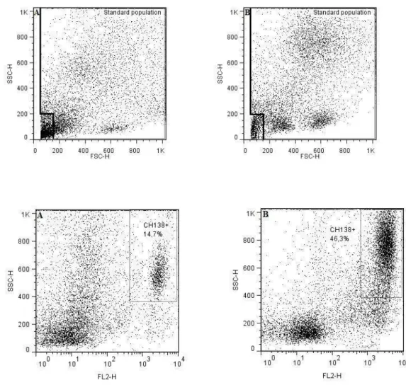
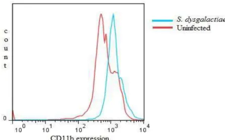
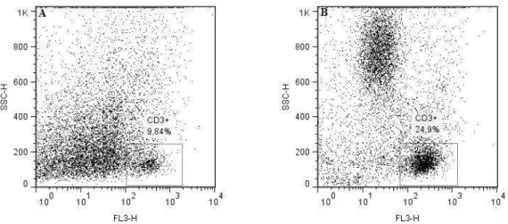
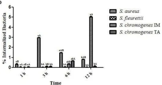
some aspects of the host-pathogen interactions, as follows: 1) Review of the role of pattern-recognition receptors in the innate immunity of bovine mastitis with focuses on the major mastitis pathogens: Escherichia coli and Staphylococcus aureus; 2) Determine a set of rules for classifying the infection status of an udder at quarter (single and duplicate milk samples) and cow (composite milk sample) levels and compare with distincts somatic cell count (SCC) thresholds (at quarter and cow levels); 3) Evaluation of the interdependence of the quarters by evaluating milk neutrophil function and milk lymphocyte profiles in uninfected quarters from infected and uninfected udders by flow cytometry analysis; 4) Function of milk neutrophils and the milk lymphocyte profile in Streptococcus dysgalactiae-infected mammary glands; 5) Capacity of S. aureus, S. fleurettii, and two dissimilar strains of S. chromogenes species to adhere and internalize into bovine mammary epithelial cells; 6) Efficacy of a commercial vaccine under field condition in two dairy herds with high bulk milk somatic cell count (SCC) and a high prevalence of IMIs by S. aureus.
Key words: intramammary infection, Staphylococcus aureus, somatic cell count, immune
12
RESUMO
Mastite, geralmente definida como a inflamação da glândula mamária, é a enfermidade de maior custo para a pecuária leiteira mundial. O maior obstáculo para reduzir da incidência da mastite, e consequentemente as implicações na produção e qualidade do leite, é a implementação de medidas efetivas no controle da mastite nas fazendas leiteiras, que depende principalmente da melhor compreensão da complexa interação patógeno-hospedeiro. A interação patógeno hospedeiro é uma importante e ampla área de pesquisa básica e clínica. Desta forma, o presente trabalho buscou elucidar alguns aspectos da interação patógeno-hospedeiro: 1) revisão sobre a importância dos receptores do tipo Toll na mastite bovina, especialmente considerando os patógenos: Escherichia coli e Staphylococcus aureus; 2) avaliação do diagnóstico da infecção intramamária pelo exame microbiológico do leite de amostras individuais por quarto, em duplicata por quarto e compostas e sua relação com a contagem de células somáticas; 3) avaliação por citometria de fluxo da interdependência dos quartos mamários pela função neutrofílica e perfil linfocítico; 4) estudo da função neutrofílica e perfil linfocítico em quartos mamários infectados por Streptococcus dysgalactiae; 5) determinação a capacidade de aderência e internalização em células epiteliais mamárias por S. aureus, S. fleurettii, e duas cepas de S. chromogenes; e 6) efeito da vacinação sobre a dinâmica da infecção intramamária por S. aureus e estafilococos coagulase-negativo.
13
1 INTRODUCTION
Mastitis, generally defined as the inflammation of the mammary gland, is the most costly of the infectious, endemic diseases to affect dairy cows and other dairy species. The greatest obstacle to reduce mastitis is the implementation of effective mastitis control measures in dairy farmers, which mainly depends on a better understanding of the complex interactions
between an infecting pathogen and the host immune system1.
Host-pathogen interaction is a broad, important area of research encompassing both basic and clinical sciences. From the perspective of the pathogen, the cross-talking between microorganim and the immune cells may also be involved in the selection of microorganisms
well-adapted to the host2, which may be related to the establishment, persistence and severity of
the infection, and the outcome to possible therapeutic and prevention interventions3. On the
other hand, from the perspective of the host, immune response to pathogens is greatly
determined by variable properties of the pathogens, which in turn can affect their lifecycle4.
As milk production and milk quality are mainly affected by mastitis, the economical viability of dairy chain requires effective control measures of this disease which in turn include
a broad comprehension of the defense mechanisms of the mammary gland5, the evasion of the
immune system by pathogens6 and an appropriate use and interpretation of diagnosis measures
and prevention strategies7.
Regarding that, in last years, the Brazilian Normative Instruction and quality based payment program for milk quality established by many Brazilian dairy plants have been proposed as an attempt to increase Brazilian milk quality parameters. Nevertheless, we supposed that without an effective mastitis control program, the Brazilian legislation requirements for milk quality, especially for bulk tank SCC, proposed by Normative
Instructions 51 and 62 (Brazil, 20028; Brazil, 20119) will not be easily met.
1 Chapter I and Appendix I 2 Chapter II
3 Chapter VI
4 Chapters I, III, IV and VI, and Appendix I 5 Chapters I, III and IV, and Appendix I 6 Chapter V
7 Chapters II and VI
8Brasil 2002. Regulamento técnico de produção, identidade e qualidade do leite tipo A, do leite tipo B, do leite tipo C, do leite
pasteurizado e do leite cru refrigerado e o regulamento técnico da coleta de leite cru refrigerado e seu transporte a granel. Instrução Normativa nº 51, de 18 de setembro de 2002. Diário Oficial da União, 18 de setembro de 2002, Ministério da Agricultura, Pecuária e Abastecimento, Brasília, DF.
9 Brasil 2011. Regulamento técnico de produção, identidade e qualidade do leite tipo A, o regulamento técnico de identidade e
14
2
OBJECTIVES
The present study addressed some aspects of the host-pathogen interactions, as follows:
1. Review of the role of pattern-recognition receptors in the innate immunity of
bovine mastitis with focuses on the major mastitis pathogens: Escherichia coli and
Staphylococcus aureus;
2. Determine a set of rules for classifying the infection status of an udder at quarter
(single and duplicate milk samples) and cow (composite milk sample) levels and compare with distincts somatic cell count (SCC) thresholds (at quarter and cow levels);
3. Evaluate the interdependence of the quarters by evaluating milk neutrophil
function and milk lymphocyte profiles in uninfected quarters from infected and uninfected udders using flow cytometry analysis;
4. Study the function of milk neutrophils and the milk lymphocyte profile in
Streptococcus dysgalactiae-infected mammary glands;
5. Determine the capacity of S. aureus, S. fleurettii, and two dissimilar strains of S.
chromogenes species to adhere and internalize into bovine mammary epithelial cells;
6. Evaluate the effect of vaccination on the dynamic of intramammary infection by
15
3C
C
H
H
A
A
P
P
T
T
E
E
R
R
I
I
:
:
T
T
h
h
e
e
i
i
n
n
n
n
a
a
t
t
e
e
i
i
m
m
m
m
u
u
n
n
i
i
t
t
y
y
i
i
n
n
b
b
o
o
v
v
i
i
n
n
e
e
m
m
a
a
s
s
t
t
i
i
t
t
i
i
s
s
:
:
t
t
h
h
e
e
r
r
o
o
l
l
e
e
o
o
f
f
p
p
a
a
t
t
t
t
e
e
r
r
n
n
-
-r
16
American Journal of Immunology, v.8, n.4, p.166-178, 2012Mastitis is the most costly disease for dairy farmers and industry (Hujips et al., 2008; Hogeveen et al., 2011). More than 130 microorganisms can cause mastitis, although this disease is usually caused by some groups of bacteria (Wellenberg et al., 2002; Hillerton and Berry, 2005). Bovine mastitis is defined as an inflammatory condition of the mammary gland in response to injury, which serve to destroy and neutralize infectious agents and promote healing and the return to normal function. In the last few years, antimicrobial resistance has been growing concern worldwide. Thus, in an attempt to reduce the impact of mastitis and decrease the use of antimicrobials on dairy farms, there have been numerous efforts to try to exploit the immune
capacity of the bovine mammary gland to stimulate the animal’s natural defense mechanisms.
These facts can reduce the use of antimicrobials and also minimize the development of resistance of bacterial strains (Wellnitz and Bruckmaier, 2012).
The role of toll-like receptors (TLRs) in innate and adaptive immunity has been subject of many good reviews (Medzhitov, 2001; Takeda et al., 2003; Akira and Takeda. 2004; Iwasaki and Medzhitov, 2004; Takeda and Akira, 2004; Hornung and Latz, 2010; Takeda et al., 2010; Kawai and Akira, 2011; Prince et al., 2011). Therefore, there is a need to summarize the role of these new concepts in bovine mastitis. In this review, we focus on the most recent knowledge about TLRs in bovine mastitis regarding their role in the major mastitis pathogens: Escherichia
coli and Staphylococcus aureus.
Innate Immunity
Bovine mastitis is initiated by the entry of bacteria through the teat canal, and soon after is
characterized by an important inflammatory response. Shortly after entry of the invading pathogen, the resident leukocytes together with epithelial cells initiate the inflammatory response necessary to eliminate the invading bacteria (Paape et al., 2003; Rainard and Riollet, 2006; Aitken et al., 2011). These cells release chemoattractants for the rapid recruitment of polymorphonuclear neutrophil leukocytes to the site of infection and consequently the somatic cell count (SCC) increases, which represents different cells types present in milk, including leukocytes and epithelial cells (Paape et al., 2003; Souza et al., 2012). The marked increase in milk SCC during infection is mainly due to influx of neutrophils from blood to the mammary gland, which neutrophils can represent over 90% of leukocyte population in milk from infected udder quarters in contrast to low numbers of this cell population in uninfected ones (Paape et al., 2003; Pyörälä et al., 2003; Souza et al., 2012).
Neutrophils are essential for innate host defense against invading microorganisms and eliminate pathogens by a process known as phagocytosis. During phagocytosis, neutrophils produce reactive oxygen species, including superoxide, hydrogen peroxide and hypochlorous acid, and release granule compounds into pathogen-containing vacuoles to kill the invading pathogen (Paape et al. 2003; Mehrzad et al., 2005; Rainard e Riollet, 2006; Prince et al., 2011). Thus, the rapid influx of neutrophils with high antimicrobial activity to the foci of infection is the main process that leads to the elimination of infection (Mehrzad et al., 2005). This importance was demonstrated by Mehrzad et al. (2005) who described that SCC in moderate cows increase faster than the colony forming units (CFU) of E. coli bacteria, whereas in severe cows the results were reversed.
17
apoptosis differentiation program facilitates the resolution of neutrophil-mediated inflammation, and it has been suggested that phagocytosis initiates molecular cascade of events that accelerates apoptosis of this leukocyte population. Thus, as neutrophils can accumulate rapidly at sites of infection, and there is a concomitant potential to cause severe tissue destruction should they undergo necrosis lysis and release cytotoxic granule and reactive oxygen species onto host tissues. Thus, apoptosis can be viewed as the terminal stage of PMN-induced inflammation (Kobayashi et al., 2003).
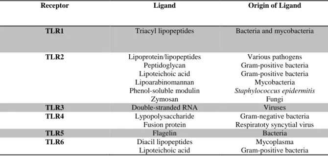
Recognition of microbial pathogens is an essential element for initiation of innate immune responses such as inflammation and is mediated by germline-encoded pattern-recognition receptors (PRRs) that recognize molecular structures that are broadly shared by pathogens, known as pathogen-associated molecular patterns (PAMPs). Upon PAMP recognition, PRRs initiate a serious of signaling programs that execute the first line of host defensive responses necessary for killing infectious microbes (Medzhitov, 2001; Takeda et al., 2003; Akira and Takeda. 2004; Brandley and Medzhitov, 2004; Iwasaki and Medzhitov, 2004; Takeda and Akira, 2004; Takeda et al., 2010; Kawai and Akira, 2011; Prince et al., 2011). TLRs were the first PRRs identified. They are also the best characterized PRRs and recognize a wide range of PAMPs. They are expressed either on the cell surface or associated with intracellular vesicles (Medzhitov, 2001; Takeda and Akira, 2004). To date, 10 functional TLRs have been identified in bovine (Menzies and Ingham, 2006). These 10 TLRs and nucleotide-binding oligomerization domain (NOD) 1 and 2 were detected in tissue from alveolar, ductal, gland cistern and teat canal from infected and healthy quarters, with TLR8 having the least expression in comparison to the other PRRs (Whelehan et al., 2011). Functional analysis of mammalian TLRs has revealed that they recognize specific patterns of microbial components that are conserved among pathogens (Takeda and Akira, 2004). Each TLR detect distinct PAMPs derived from bacteria, viruses, mycobacteria, fungi and parasites. For instance, these include lipoproteins (recognized by TLR1, TLR2, and TLR6), flagellin (TLR5) lipopolysaccharide (LPS) (TLR4) and a 6-base DNA motif consisting of an unmethylated CpG dinucleotide motifs (CpG DNA) that are rarely found in higher vertebrates (TLR9) (Table 1) (Medzhitov, 2001; Takeda et al., 2003; Akira and Takeda. 2004; Iwasaki and Medzhitov, 2004; Takeda and Akira, 2004; Takeda et al., 2010; Kawai and Akira, 2011).
Table 1. Toll-like receptors and their ligands*
Receptor Ligand Origin of Ligand
TLR1 Triacyl lipopeptides Bacteria and mycobacteria
TLR2 Lipoprotein/lipopeptides
Peptidoglycan Lipoteichoic acid Lipoarabinomannan Phenol-soluble modulin
Zymosan
Various pathogens Gram-positive bacteria Gram-positive bacteria
Mycobacteria Staphylococcus epidermitis
Fungi
TLR3 Double-stranded RNA Viruses
TLR4 Lypopolysaccharide
Fusion protein
Gram-negative bacteria Respiratoty syncytial virus
TLR5 Flagelin Bacteria
TLR6 Diacil lipopeptides
Lipoteichoic acid
18
Zymosan Fungi
TLR7 Single stranded RNA Viruses
TLR8 Single stranded RNA Viruses
TLR9 CpG-containing DNA Bacteria and viruses
TLR10 N.D. N.D.
Adapted from Akira and Takeda (2004). N.D.: not determined
*Only ligands that can be related to mastitis pathogens were included
For instance for the role of TLRs in bovine mammary gland, it was found that LPS induced the expression of the chemokines MCP-1, MCP-2, and MCP-3, and slightly increase in CXCL8. Conversely, peptidoglycan combined with lipotechoic acid (LTA) induced the expression of MCP-1 and a slight increase in MCP-3 expression. Indeed, no significant expression for any of the chemokines was observed when induced by CpG-DNA (Mount et al., 2009).
Furthermore, it should be noted that the TLRs can act together with other molecules or other TLRs. For instance, TLR4 requires other molecules in addition to TLR4 to recognize LPS. LPS binds to the LPS-binding protein (LBP) present in serum, and this LPS-LBP complex is subsequently recognized by CD14, which is expressed on monocytes/macrophages and neutrophils. Moreover, LPS stimulation is followed by the increased physical proximity between CD14 and TLR4 in the membrane, suggesting that CD14 and TLR4 may interact in LPS signaling. Indeed, the TLR2 act in cooperation with at least two other TLRs: TLR1 and TLR6. So, the formation of heterodimers between TRL2 and either TLR1 or TLR6 dictates the specificity of ligand recognition (Medzhitov, 2001; Takeda et al., 2003; Akira and Takeda. 2004; Iwasaki and Medzhitov, 2004; Takeda and Akira, 2004; Takeda et al., 2010; Kawai and Akira, 2011). Another factor that can influence the innate immune response in bovine is cell maturation, as demonstrated for the same monocytes subsets - monocytes, macrophages and dentritic cells, which have different responses to the same TLR agonist (Werling et al., 2004).
All TLR signal transduction pathways are known to activate NF-κB factors (Akira and
Takeda, 2004). MyD88 (myeloid differentiation primary-response protein 88) dependent
pathways are associated with early-phase NF-κB response whereas as MyD88 independent
pathways are associated with late-phase NF-κB response. These NF-κB factors subsequently
enter the nucleus and bind to target promoters. A wealth of pro-inflammatory regulated genes
feature NF-κB attachment sites in their promoter region and transcription factor complex act as
a main switch to orchestrate immune defense genes against bacterial infection, as production of several pro-inflammatory cytokines.
Thus, the innate immune system uses various PRRs that are expressed on the cell surface, in intracellular compartments, or secreted into the blood stream and tissue fluids. The principal functions of PRRs include: opsonization, activation of complement and coagulation cascade, phagocytosis, activation and induction of apoptosis (Medzhitov, 2001). For instance, TLR signaling pathway by bacteria regulated phagocytosis at multiple steps, including internalization and phagossome maturation (Blander and Medzhitoz, 2004).
The importance of the innate immunity (TLR2, tumor necrosis factor (TNF)-α,
interleukin (IL-)1 , IL-6, IL-8 complement factor C3, lactoferrin and RANTES) was also
demonstrated by the significantly elevated expression of these innate immune genes in less-susceptible cattle when compared to high susceptibility group by detection of quantitative trait loci (QTL) affecting mastitis (Griesbeck-Zilch et al., 2009).
19
E. coli is among the major mastitis pathogens responsible for clinical mastitis in dairy cows,but the infection is normally cleared by the immune system within a few days. Indeed, in the last few decades, with the improvement of mastitis control programs, which leads to herds with low SCC, the clinical mastitis has become a major problem in many well-managed dairy herds that successfully controlled contagious pathogens (Green et al., 2004). Gram-negative bacteria, such as E. coli, are generally regarded as environmental pathogens, however contagious behavior of these pathogens has been proposed (Burvenich et al., 2003; Dogan et al., 2006; Suojala et al., 2011).
E. coli express a variety of virulence factors, but no coherence between the severity of
disease and specific virulence factors could be defined (Wenz et al., 2006; Suojala et al., 2011; Schukken et al., 2011). However, the ability to grow in mammary secretions and to liberate LPS is crucial in the pathogenesis of E. coli mastitis. The faster bacterial numbers increase in the mammary gland, more LPS is present in the mammary gland and faster inflammatory response and clinical disease may occur (Mehrzad et al., 2008). Sensing the pathogen and initiating an immune response depends on the initial number of bacteria present at the start of the IMI. Increasing the initial challenge dose of E. coli resulted in faster immune response in primiparous cows (Vangroenweghe et al., 2004; Schukken et al., 2011), which the cytokines synthesis, such
as TNF-α and IL-8, in mammary epithelial cells positively correlated with the concentration of
E. coli particles (Güntler et al., 2010). Congruently, expression of IL-8 and interferon
(IFN)-by milk somatic cells was increased in E. coli challenged mammary glands (Lee et al., 2006). Buitenhuis et al. (2011) also described that in the early stages of E. coli mastitis a large number of up-regulated transcripts were associated with immune response functions, mainly those involved in acute phase response, while the down-regulation transcripts were principally involved in fat metabolism, which is consistent with the milk fat content depression commonly observed during mastitis infection, and later the up-regulated transcripts were associated with tissue healing processes, and were independent of E. coli strain and dose and lactation stage and number. Another factor that should be considered is the linkage between lipid metabolism and inflammation, as the nuclear receptors known as peroxisome proliferator-activated receptors (PPARs) and liver X receptors (LXRs) that emerged as key regulators of lipid metabolism and inflammation (Lubick et al., 2006; Bensinger and Tontonoz, 2008; Rios et al., 2008; Aitken et al., 2011; Moyes et al., 2010a; Moyes et al., 2012b), as has been demonstrated in mammary glands infected with Streptococcus uberis (Moyes et al., 2010a).
The innate immune system represents the first line of defense in the host response to infection and is prepared to immediately recognize and respond to the earliest stages of infection. The inherent capability of the innate system to respond to a vast number of pathogens is mediated by its ability to recognize highly conserved motifs shared by diverse pathogens, commonly referred to as PAMPs. It has been shown that a prompt response of the mammary gland after E. coli entry into the lumen of the gland is required to control the infection, which means that early detection of bacteria are of prime of importance (Bannerman et al., 2004; Porcherie et al., 2012).
20
disturbance in neutrophil functions during early lactation stages are accompanied by modulation of TLR4 pathway genes, as diapedesis and migration process (Stevens et al., 2011).
Furthermore, Mehrzad et al. (2005) when classified cows as moderate and severe responders according to clinical symptoms and milk production output, observed an inverse relationship between pre-infection milk neutrophils microbicidal activity and amount of E. coli bacteria in milk, while for moderate cows the pre-infection milk and blood neutrophils
microbicidal activity was about two fold higher than for severe cows. Mammary epithelial
cells challenged by E. coli bacteria must have the capacity to mountain a strong innate immune response in their own right and attract circulating immune effector cells such as neutrophils. The importance of these cells is demonstrated by their role in the production of cytokines (Riollet et al., 2000; Strandberg et al., 2005; Lahoussa et al., 2007; Griesbeck-Zilch et al., 2008; Güntler et al., 2011; Porcherie et al., 2012). The upregulation of cytokine production is a key component of the host innate immune response to infection (Bannerman et al., 2004; Schukken et al., 2011).
Regarding bovine mammary epithelial cells (bMEC), Porcherie et al. (2012) showed that these cells are key players in initiating neutrophil inflammation during E. coli mastitis, as for instance, by producing he chemotractic factor CXCL8 (IL-8). So, recognition of several PAMPs at a time could contribute to the onset of an early response of the cow after infection by
E. coli. These authors showed that a repertoire of potential bacterial agonists can be sensed by
bMEC and udder during E. coli mastitis, as which both bMEC and udder can express domain receptors for NOD1, NOD2, TLR1, TLR2, TLR4 and TLR6, but not hardly TLR5, and can act synergistically. Therefore, LPS upon activation of TLR4 present a central role in the pathogenesis of clinical mastitis caused by this pathogen (Gonen et al., 2007) in a dose-dependent manner (Baumert et al., 2009). The inflammation caused by LPS leads to dose alteration alteration in milk parameters, such as lactose and chloride levels, which are likely caused by tighter junction damage by higher LPS doses (Werner-Misof, 2007). These parameters are also used to evaluate the indicators of inflammation in bovine mastitis, and consequently in their diagnosis (Pyörälä et al., 2003).
Elazar et al. (2010a) demonstrated that neutrophil recruitment to the milk is mediated
through TNF-α, which is produced by alveolar macrophages in response to LPS/TLR4 signaling
and is dependent on IL-8 and IL-1 signaling and regulated by iNOS-derived NO in a murine
mastitis model. The ability to recruit cells into the mammary gland during the bacterial growth phase represent a crucial role since a 1 h delay in recruiting neutrophils can result in an 8-fold increase of E. coli (Hill, 1981). Both the MtD99 dependent and independent pathways in TLR4 signaling were activated in bMEC model (Ibeagha-Awemu et al., 2007). Despite the importance of LPS/TLR4 signaling pathway, Gonen et al. (2007) described that IMI of mice with E. coli P4 resulted in inflammation even in absence of LPS/TLR signaling. This inflammation response points out to additional factors beyond LPS and additional cells beyond alveolar macrophages play a role in the inflammatory response to E. coli. It has been suggested that in the absence of functional TLR4 the infecting E. coli P4 invaded epithelial cells with high efficiency, forming intracellular micro-colonies, since invasion of epithelial cells by E. coli is limited by alveolar macrophages using a process dependent on TLR4 signaling (Gonen et al., 2007; Elazar et al., 2010b; Schukken et al., 2011).
Infections caused by E. coli are more typically, but not exclusively, associated with fast and more dramatic immune response (Lee et al., 2006; Schukken et al., 2011). IMI with E. coli elicited systemic changes, including a febrile response, and induction of acute-phase synthesis
of LBP. In milk, this infection resulted in increased levels of insulin-like growth factor-1, IL-1 ,
21
Petzl et al. (2008) reported that E. coli inoculation in the mammary gland strongly
upregulated the expression of -defensins, TLR2 and TLR4 in the pathogen inoculated udder
quarters as well as in mammary lymph nodes. In constrast, S. aureus did not significantly regulate the expression of these genes during the first 24 h after pathogen inoculation. Only 84 h
after inoculation, the expression of -defensins, but not of TLRs was significantly upregulated (< 20 fold) in S. aureus inoculated mammary glands.
E. coli IMIs induce distinct local and systemic transcriptome responses in the mammary
gland. The local response, only in infected quarters, mainly involved in immune response and inflammation, while the systemic reactions, in both infected and neighboring quarters, comprises antigen processing and presentation, cytokines, protein degradation and apoptosis. Enhanced expression of antimicrobial genes, acute phase genes and indicators of oxidative stress point out to an active defense reaction in infected and neighboring healthy quarters (Mitterhuemer et al., 2010).
In this concern, data support an important sentinel function for teats, as these tissues respond rapidly and intensively, with production of cytokines and antimicrobial peptides. For example, genomic analysis showed that at 12 h post-infection with E. coli the inflammatory response was greatest in teat cistern and gland cistern. Only 24 h post-infection, the lobulo-alveolar region responds, at the time the inflammatory response was greatest of all regions (Rinaldi et al., 2010).
Mastitis caused by Staphylococcus aureus
S. aureus mastitis remains a worldwide problem for the dairy industry and producers, and can
cause both subclinical and clinical mastitis (Barkema et al., 2006), whose severity and outcome of infection depend, in part, on strain-factors (Le Maréchal et al., 2011) and cow factors (Barkema et al., 2006). The cure rate of antimicrobial treatments for this agent is low and, therefore, the disease has not been effectively eliminated and/or controlled in many herds (Barkema et al., 2006). Staphylococcal infections are characterized by an ability to colonize the mammary tissue and bacterial survival inside epithelial cells, macrophages and even neutrophils (Gresham et al., 2000; Hébert et al., 2000; Lowy, 2000).
It is commonly assumed that most IMI are result of cow-to-cow transmission, however other sources of S. aureus bacteria in the environment of dairy cows have been described. Presumably, contagious strains of S. aureus co-exist with a large collection of non-contagious strains (Zadoks et al., 2002). Haveri et al. (2007, 2008) compared bacterial genomics of strains from persistent and transient infections, and found that genetic elements such as clonal type and penicillin resistance were over-represented in S. aureus isolated from persistent IMI. This microorganism is characterized by dynamic fluctuations and cyclic bacterial shedding in milk,
which leads to fluctuations in milk SCC that normally fluctuate depending on organism’s
number and viability (Schukken et al., 2011; Souza et al., 2012).
In contrast to E. coli mastitis, S. aureus mastitis is characterized by a more moderate and delayed SCC increase, due in part, to limited cytokine response (Bannerman et al., 2004).
Riollet et al. (2000) also described no detection of IL-1 , TNF-α, IL-8, bovine serum albumin,
in milk whey from S. aureus infected animals. Indeed, the ability of milk to generate the complement cleavage product C5a in whey samples after addition of zymosan through complement activation was evaluated, and E. coli lead to a huge augment of C5a production (up to 100-fold), in contrast to a lower production in whey milk from S. aureus infected animals. Rainard et al. (2008) also showed that LTA from S. aureus induced an increase in chemokine
22
Although, S. aureus is regarded as a gram-positive bacteria, the expression of TLR2 were correlated with TLR4, in a coordinated regulation of these two PRRs (Goldammer et al., 2004; Ibeagha-Awemu et al., 2008), although the expression of TLR9 was not increased during mastitis (Goldammer et al., 2004).
Cytokine gene expression in mammary epithelial cells induced by S. aureus infection was delayed and less than 5% of the cytokine expression was observed with E. coli infection (Lee et al., 2006; Yang et al., 2008; Güntler et al., 2010). This impaired proinflammatory
activation is simultaneous with a complete lack of NF-κB activation in primary bovine
mammary epithelial cells infected by S. aureus or LTA. In contrast, E. coli and LPS that
activate strongly NF-κB in these cells. A large proportion of this activation is attributed to
TLR-mediated signaling, since dual transdominant negative DN-MyD88-DN-TRIF factor blocks
more than 80% of the pathogen-related NF-κB activation in primary bovine mammary epithelial
cells. These facts may contribute to the well-known ability of this bacterium to establish chronic intramammary infections.
For example, the interleukin (IL)-8 and TNF-α were not detected in milk from quarters
experimentally infected with S. aureus (Riollet et al., 2000; Bannerman et al, 2004), although,
the mRNA expression of TNF-α in mammary cells increases during infection (Alluwaimi et al.,
2003). Expression of IL-8 by milk somatic cells was also increased in S. aureus challenged mammary glands, but in lower magnitude than E. coli challenged mammary glands. However,
the expression of IFN- was not increased in milk somatic cells from S. aureus challenged
quarters (Lee et al., 2006).
Mammary epithelial cells demonstrated in vitro greater mRNA expression of IL-1 , IL
-8 and TNF-α 24 h after infection with E. coli than with S. aureus (Lahouassa et al., 2007).
Wellnitz et al. (2011) also found that infusion of E. coli LPS induced an increased TNF-α in
milk from glands given LPS, but not by S. aureus LTA. The levels of lactate dehydrogenase, an enzyme released by degenerating cells, was greater in milk from glands instilled with LPS than
with LTA. LPS was also a stronger inducer of IL-8 and IL-1 .
Conversely, the ability to induce clinical or subclinical mastitis was dependent on the used dose. LTA strongly induced the secretion of the chemokines CXCL1, CXCL2, CXCL3 and CXCL8, which induced neutrophils recruitment. The complement-derived chemoattractant C5a was generated in milk only with the highest doses of LTA. Furthermore, the
pro-inflammatory cytokine IL-1 was induced in milk, but small amounts of TNF-α, and no IFN
-(Rainard et al., 2008).
The muramyl peptide (MDP), an elementary constituent of the bacterial peptidoglycan, induce a prompt influx of neutrophils mediated by chemoattractants for these leukocytes (CXCL1, CXCL2, CXCL3, CXCL8 and C5a), and the highest concentrations of these chemoattractants were followed after challenge in combination with LTA, whose signal transduction is mediated by TLR2, although they do not significantly contribute to pro-inflammatory cytokines. Thus, TLR2 and NOD2, a major sensor for MDP, pathways could cooperate to trigger an innate immune response to S. aureus mastitis (Bougarn et al., 2010).
Induction of immune functions in mammary epithelial cells is accomplished via the activation of the relevant TLR and their downstream signaling pathways. Induction of these genes by S. aureus is reduced, due to at least in part, the impairment of MyD88 signaling, and the downstream of the trans-membrane TLRs (e.g. TLR2, TLR4). S. aureus apparently prevents the formation of the so-called Myddossome around TIR domain of the TLR forming the structural platform for the attachment of further downstream acting factors (Motshwene et al., 2009; Lin et al., 2010; Schukken et al., 2011). As a consequence, S. aureus elicits an immune
response in these cells mainly by IL-6, while E. coli also activates IL-1 and TNF-α (Güntler et
23
MyD88 independent mechanism (Güntler et al., 2010; Güntler et al., 2011), which, as cited
above, is associated with late-phase NF-κB response.
It has also been suggested that S. aureus impaired NF-κB activation in mammary
epithelial cells resulting in very low cytokine expression (Lara-Zárate et al., 2011). These authors reported that bovine prolactin stimulates S. aureus internalization in bovine mammary
gland by regulating several innate immune elements, which is often modulated by NF-κB. On
the other hand, prolactin induced NF-κB activation in bovine mammary epithelial cells;
however, it was inhibited by S. aureus in presence of this hormone. When, these authors
blocked NF-κB activation with acetylsalicylic acid, an inhibition of S. aureus internalization
was found (48%) in prolactin stimulated cells. The infection of bovine mammary epithelial cells
with S. aureus induced inhibition of NF-κB activation in the presence of prolactin which
correlates with down regulation in prolactin-mediated TNF-α (27%) and nitric oxide production
in mammary epithelial cells.
Curiously, Griesbeck-Zilch et al. (2008) encountered differences in expression of TLR2 and TLR4 by mammary epithelial cells in S. aureus and E. coli infections only after 24 h, when
S. aureus-induced expression was significant lower. After 1 h S. aureus induced a significantly
higher expression level of TNF-α and IL-1 , but after 6 and 24 h the transcription activity in E.
coli treated cells was higher. E. coli induced a significant increase expression of IL-8 after 1h,
but S. aureus caused no alteration in this chemokine. The RANTES (regulated upon activation, normal T-cell expressed and secreted) increased in S. aureus and E. coli treated bovine mammary epithelial cells after 1 h, whereas after 6 and 24 h the expression was significantly higher in E. coli treated cells. Lactoferrin showed a deviating expression pattern to pathogen stimulation, in which at 1 h E. coli induced a higher mRNA expression, whereas the highest level was reached after 24 h of S. aureus stimulation. The complement factor 3 was the only factor that responded equally to both microorganisms.
Genini et al. (2011) described that mastitis induced a prominence of metabolic and stress signals in the early stage and of the immune response and lipid metabolism in the late stage, both mechanisms apparently modulated by few genes. Comparison of E. coli and S.
aureus infections in cattle revealed that affected genes with opposite regulation had the same
altered biological functions and provided evidence that E. coli caused a stronger host response. The majority of genes with opposed regulation were associated with immune response belongs to antigen presentation, inflammatory response, cell-to-cell signaling and interaction network. Both cell death and lipid metabolism were among the most significant molecular functions altered in proteins of cows infected with both E. coli and S. aureus.
TLR1 was significantly expressed in ductal, gland cistern and teat canal after 48 h post-challenge with S. aureus, TLR3 showed a moderate increase in teat canal tissue, TLR6 and TLR7 presented a moderate increased in gland cistern tissue, TLR5 and TLR7 were also significantly increased in alveolar in alveolar tissue. Conversely, the genes encoding TLR4, NOD1 and NOD2 were significantly decreased in teat canal tissue, TLR6 in ductal tissue and TLR8 in gland cistern tissue. TLR2, TLR9 and TLR10 showed no differential expression across tissues of these regions. Chemokine and effector molecule expression was most significantly stimulated in alveolar tissue, in particular the expression of serum amyloid A and haptoglobulin,
two acute phase proteins, and defensins- 4 and 5 (Whelehan et al, 2011).
Thus, S. aureus appears to mostly circumvent the host immune response and IMI typically result in a very moderate host response with minimal observable innate immune response (Bannerman et al., 2004; Petzl et al., 2008; Bannerman et al., 2009; Schukken et al., 2011).
24
CD14 either in membrane or in soluble form (sCD14) is a high-affinity protein for the complex of bacterial LPS and LPS-LBP protein, and thus interact with TLR4 in LPS signaling (Medzhitov, 2001, Takeda et al., 2003; Nemchinov et al., 2006). Cells lacking mCD14, such as endothelial and epithelial cells, utilize sCD14 present in serum and milk to aid in LPS recognition by TLR4 (Aitken et al., 2011). Binding of soluble form of CD14 to LPS, found in the outer of E. coli, enhances the innate immune responses, reduces the severity of mastitis, and facilitates clearance and neutralization of LPS, thus preventing the development of endotoxic mastitis. Thus, Lee et al. (2003) found that the infusion of recombinant bovine sCD14 lead to an increase in SCC, due to more rapid recruitment of neutrophils that was accompanied by a faster
clearance of bacteria, lower concentration of TNF-α and IL-8 in milk, and milder clinical
symptoms. Congruently, Nemchinov et al. (2006) demonstrated that the recombinant bovine CD14 receptor produced in plants reduced the severity of E. coli mastitis, leading to enhancement of LPS-induced neutrophil recruitment, lowing the numbers of viable bacteria in milk and resulting in absence of clinical symptoms.
Kauf et al. (2007) in an attempt to increase the inflammatory response during S. aureus intramammary infection infused LPS in quarters experimentally infected with S. aureus. They found an increase in SCC in quarters between 24 and 72 h post LPS-infusion, as well as, an increase in bovine serum albumin in milk, which reflect alterations in the vascular permeability,
between 4-48 and 480 h post LPS-infusion. There was no detection of TNF-α in S.
aureus-infected quarters administrated PBS at any time during the study, but aureus-infected quarters infused
with LPS showed an increased TNF-α concentrations in milk between 4-8 h post LPS-infusion.
Moreover, a trend toward a lower recovery of viable bacteria from LPS- versus PBS-infused quarters between 4-13 h post LPS-infusion was observed. This trend occurred in the inflammatory responses elicited by LPS. In vitro inoculation of milk obtained from udder quarters infused with LPS or PBS demonstrated that the growth of S. aureus in milk from LPS-infused udder was significantly inhibited, and an overall negative correlation existed between milk SCC and in vitro S. aureus growth in milk inoculated with S. aureus and incubated for 6 or 12 h.
In a mastitis using rat model, the infusion of CpG-DNA in mammary glands stimulated
the secretion of IL-6 and TNF-α at different points, reduced viable S. aureus (Zhu et al., 2007a)
and E. coli (Zhu et al., 2008) in mammary tissues, decreased the activity of NAGase, promoted the expression of TLR9 and induced more rapid infiltration of neutrophils to mammary tissue at initial stages of experimentally induced mastitis induced in rat model (Zhu et al., 2007a; Zhu et al., 2008). In goats, Zhu et al. (2007b) demonstrated that the infusion of CpG-DNA in the mammary glands induced a decrease in viable E. coli, reduced bacteria counts in milk, promoted the expression of TLR9, stimulated the production of IL-6, attenuated the impact of inflammation mediators on cells, and significantly shortened the inflammation course.
The retinoid, a group of derivates of vitamin A, exert various immunomodulatory actions. It has been demonstrated that the administration of retinoid acids protects rats against neutrophil-induced oxidative stress in acute experimental mastitis (Gu et al., 2009a; Gu et al., 2009b). A mechanism by which this protection is conferred is through TLR4. Gu et al. (2010) found that TLR4 gene expression reached its peak earlier in retinoid acid-treated rats, and that
retinoid acid decreased NF-κB DNA binding activity and the level of IL-1 protein expression
in mammary gland. So, retinoid acid leads to attenuation of LPS-induced inflammation response
by repression of TLR4/NF-κB signaling system. Another mechanism that can also be involved
25
Pheromonicin-SA (Ph-SA) is an enginnered multidomain bactericidal peptide (Qiu et al., 2003) that has effect against S. aureus. Zhu et al. (2012) when exposed primary mammary
epithelial cells to Ph-SA found that this compound increases the expression of TLR2, TNF-α,
IL-1 , IL-8 and lactoferrin, and later the expression of TLR4. Thus, Ph-SA may be value as an antimicrobial in promoting innate immune response by S. aureus aureus-infected bovine mammary epithelial cells, especially regarding the inhibition of innate immune response induced by S. aureus which leads to the chronic inflammatory response (Bannerman et al., 2004; Lahouassa et al., 2007; Yang et al., 2008; Motshwene et al., 2009; Güntler et al., 2010; Lin et al., 2010; Lara-Zárate et al., 2011; Schukken et al., 2011; Wellnitz et al., 2012).
TLR signaling induces 25-hydroxyvitamin D3 1α-hydrolase expression in macrophages.
The 25-hydroxyvitamin D3 1α-hydrolase is the primary enzyme that converts
25-hydroxyvitamin D3 to 1,25-dihydroxyvitamin D3, the active vitamin D3 metabolite. It was
shown that the expression 25-hydroxyvitamin D3 1α-hydrolase was significantly increased in
tissue and cells from infected mammary glands, and was predominantly expressed in CD14+
cells (Nelson et al., 2010), which is expressed in both neutrophils and macrophages in milk (Paape et al., 1996; Blagitz et al., 2012). Thus, regarding the importance of innate immunity for mammary gland health (Paape et al., 2003, Rainard and Riollet, 2006; Elazar et al. 2010a,
Elazar et al. 2010b), efforts to find optimal range of 1,25-dihydroxyvitamin D3 concentrations
for proper immune function in cattle has implications for bovine health.
Conclusion
The innate immunity is crucial to maintain mammary gland healthy, which is mediate through recognition of pattern recognition receptors (PRRs). The PRRs recognized specific patterns of microbial components that are conserved among pathogens known as pathogen-associated molecular patterns (PAMPs). The interaction of PRRs and PAMPs mediate the inflammatory response characterized by each mastitis-causing pathogen that can contribute to the development of severe acute or chronic mastitis.
References
Aitken, S.L.; Corl, C.M.; Sordillo, L.M. Immunopathology of mastitis: insights into disease recognition and resolution. Journal of Mammary Gland Biology and Neoplasia, v.16, p.291-304, 2011.
Alluwaimi, A.W.; Leutenegger, C.M.; Farver, T.B.; Rossito, P.V.; Smith, W.L.; Cullor, J.S. The cytokine markers in Staphylococcus aureus mastitis of bovine mammary gland. Journal of
Veterinary Medicine, Series B: Infectious Diseases and Veterinary Public Health, v.50,
p.105-111, 2003.
Bannerman, D.D.; Paape, M.J.; Lee, J-W.; Zhao, X.; Hope, J.C.; Rainard, P. Escherichia coli and Staphylococcus aureus elicit differential innate immune responses following intramammary infection. Clinical and Diagnostic Laboratory Immunology, v.11, n.3, p.463-472, 2004. Barkema, H.W.; Schukken, Y.H.; Zadoks, R.N. Invited review: the role of cow, pathogen, and treatment regimen in the therapeutic success of bovine Staphylococcus aureus mastitis. Journal
of Dairy Science, v.89, p.1877-1895, 2006.
Baumert, A.; Bruckmaier, R.M.; Wellnitz, O. Dose dependent SCC changes after intramammary lypopolysaccharide challenge. Milk Science International, v.64, p.119-121, 2009.
26
and polymorphonuclear leukocytes in bovine milk. In: 51th Annual Meeting National Mastitis
Council, 2012, St. Pete Beach, United States of America, p.185-186, 2012.
Blander, J.M.; Medzhitoz, R. Regulation of phagosome maturation by signals from toll-like receptors. Science, v.304, p.1014-1018, 2004.
Bensinger, S.J.; Tontonoz, P. Integration of metabolism and inflammation by lipid-activated nuclear receptors. Nature, v.454, p.47-477, 2008.
Bougarn, S.; Cunha, P.; Harmache, A.; Fromageau, A.; Gilbert, F.B.; Rainard, P. Muramyl dipeptide synergizes with Staphylococcus aureus lipoteichoic acid to recruit neutrophils in the mammary gland and to stimulate mammary epithelial cells. Clinical and Vaccine
Immunology, v.17, n.11, p.1797-1809, 2010.
Burvenich, C.; Van Merris, V.; Mehrzad, J.; Diez-Fraile, A.; Duchateau, L. Severity of E .coli mastitis is mainly determined by cow factors. Veterinary Research, v.34, p.521-564, 2003. De Schepper, S.; De Ketelaere, A.; Bannerman, D.D.; Paape, M.J.; Peelman, L.; Burvenich, C. The toll-like receptor-4 (TLR-4) pathway and its possible role in the pathogenesis of
Escherichia coli mastitis in dairy cattle. Veterinary Research, 39:05, 2007.
Dogan, B.; Klaessig, S.; Rishniw, M.; Almeida, R.A.; Oliver, S.P.; Simpson, K.; Schukken, Y.H. Adherent and invasive Escherichia coli are associated with persistent bovine mastitis.
Veterinary Microbiology, v.116, p.270-281, 2006.
Elazar, S.; Gone, E.; Livneh-Kol, A.; Rosenshine, I.; Sphpigel, N.Y. Neutrophil recruitment in endotoxin-induced murine mastitis is strictly dependent on mammary alveolar macrophages.
Veterinary Research, 41:10, 2010a.
Elazar, S.; Gone, E.; Livneh-Kol, A.; Rosenshine, I.; Sphpigel, N.Y. Essential role of neutrophils but not mammary alveolar macrophages in a murine model of acute Escherichia coli mastitis. Veterinary Research, 41:53, 2010b.
Genini, S.; Badaoui, B.; Sclep, G.; Bishop, S.C.; Waddington, D.; Van Der Laan, M.-H.P.; Klopp, C.; Cabau, C.; Seyfert, H.-M.; Petzl, W.; Jensen, K.; Glass, E.J.; de Greeff, A.; Smith, H.E.; Smits, M.A.; Olsaker, I.; Boman, G.M.; Pisoni, G.; Moroni, P.; Castiglioni, B.; Cremonesi, P.; Del Corvo, M.; Foulon, E.; Foucras, G.; Rupp, R.; Giuffra, E. Strengthening insights into host responses to mastitis infection in ruminants by combining heterogeneous microarray data sources. BMC Genomics, 12:225, 2011.
Goldammer, T.; Zerbe, H.; Molenaar, A.; Schuberth, H-J.; Brunner, R.M.; Kata, S.R.; Seyfert, H.-M. Mastitis increases mammary mRNA abundance of -defensin 5, toll-like receptor 2 (TLR2), and TLR4 but not TLR9 in cattle. Clinical and Diagnostic Laboratory Immunology, v.11, n.1, p.174-185, 2004.
Gonen, E.; Vallon-eberhard, A.; Elazar, S.; Harmelln, A.; Brenner, O.; Rosenshine, I.; Jung, S.; Shpigel, N.Y. Toll-like receptor 4 is needed to restrict the invasion of Escherichia coli P4 into mammary gland epithelial cells in a murine model of acute mastitis. Cellular Microbiology, v.9, n.12, p.2826-2838, 2007.
Green, M.J.; Green, L.E.; Shukken, Y.H.; Bradley, A.J.; Peeler, E.J.; Barkema, H.W.; de Hass, Y.; Collis, V.J.; Medley, G.F. Somatic cell count distributions during lactation predict clinical
mastitis. Journal of Dairy Science, v.87, p.1256–1264, 2004.
Gresham, H.D.; Lowrance, J.H.; Caver, T.E.; Wilson, B.S.; Cheung, A.L.; Lindberg, F.P. Survival of Staphylococcus aureus inside neutrophils contributes to infection. Journal of
Immunology, v. 164, p. 3713-3722, 2000.
Griesbeck-Zilch, B.; Meyer, H.H.D.; Kühn, C.; Schwein, M.; Wellnitz, O. Staphylococcus
aureus and Escherichia coli cause deviating expression profiles of cytokines and lactoferrina
27
Griesbeck-Zilch, B.; Osman, M.; Kühn, C.; Schwerin, M.; Bruckmaier, R.H.; Pfaffl, M.W.; Hammerle-Fickinger, A.; Meyer, H.H.D.; Wellnitz, O. Analysis of key molecules of the innate immune system in mammary epithelial cells isolated from marker-assisted and conventionally selected cattle. Journal of Dairy Science, v.82, p.4621-4633, 2009.
Gu, B.B.; Miao, J.F.; Zhu, Y.M.; Deng, Y.; Zou, S.X. Protective effect of retinoid against endotoxin-induced mastitis in rats. Inflammation Research, v.58, p.81-88, 2009a.
Gu, B.B.; Zhu, Y.M.; Zhu, W.; Miao, J.F.; Deng, Y.; Zou, S.X. Retinoid protects rats against neutrophil-induced oxidative stress in acute experimentally mastitis. International
Immunopharmacology, v.9, p.223-229, 2009b.
Gu, B.; Miao, J.; Fa, Y.; Lu, J.; Zou, S. Retinoic acid attenuates lipopolysaccharide-induced
inflammatory responses by supressing TLR4/NF-κB expression in rat mammary tissue.
International Pharmacology, v.10, p.799-805, 2010.
Güntler, J.; Liu, S.; Esch, K.; Schuberth, H.J.; Seyfert, H.M. Stimulated expression of TNF-[alpha] and IL-8, but not of lingual antimicrobial peptide reflects the concentration of pathogens contacting bovine mammary epithelial cells. Veterinary Immunology and Immunopathology, v.135, p.152-157, 2010.
Güntler, J.; Esch, K.; Poschadel, N.; Petzl, W.; Zerbe, H.; Mitterhuemer, S.; Blum, H.; Seyfert, H-M. Comparative kinetics of Escherichia coli- and Staphylococcus aureus-specific ctivation of key immune pathways in mammary epithelial cells demonstrates that S. aureus elicits a deleyed response dominated by interleukin-6 (IL-6) but not by IL-1A or tumor necrosis factor alpha.
Infection and Immunity, v.79, n.2, p.695-707, 2011.
Haveri, M.; Roslöf, A.; Rantala, L.; Pyörälä, S. Virulence genes of Staphylococcus aureus from persistent and nonpersistent intramammary infections with different clinical characteristics.
Journal of Applied Microbiology, v. 103, p. 993-1000, 2007.
Haveri, M.; Hovinen, M.; Roslöf, A.; Pyörälä, S. Molecular types and genetic profiles of
Staphylococcus aureus strains isolated from bovine intramammary infections and
extramammary sites. Journal of Clinical Microbiology, v.46, p.3778-3735, 2008.
Hébert, A.; Sayasith, K.; Sénéchal, S.; Dubreuil, P.; Lagacé, J. Demonstration of intracellular
Staphylococcus aureus in bovine mastitis alveolar cells and macrophages isolated from
naturally infected cow milk. FEMS Microbiology Letters, v. 193, p. 57-62, 2000.
Hill, A.W. Factors influencing the outcome of Escherichia coli mastitis in the dairy cow.
Research in Veterinary Science, v.31, p.107-112, 1981.
Hillerton, J.E.; Berry, E.A. Treating mastitis in the cow - a tradition or archaism. Journal of
Applied Microbiology, v.98, p.1250-1255, 2005.
Hogeveen, H.; Hujips, K.; Lam. T.J.G.M. Economic aspect of mastitis: New developments.
New Zealand Veterinary Journal, 2011. Doi: 10.1080/00480169.2011.547165.
Hornung, V.; Latz, E. Intracellular DNA recognition. Nature Reviews Immunology, v.10, p.123-130, 2010.
Hujips, K.; Lam, T.J.G.M.; Hogeveen, H. Costs of mastitis: facts and perception. Journal of
Dairy Research, v.75, p.113-120, 2008.
Ibeagha-Awemu, E.M.; Lee, J-W.; Ibeagha, A.E.; Bannerman, D.D.; Paape, M.J.; Zhao, X. Bacterial lipopolysaccharide induces increased expression of toll-like receptor (TLR)4 and downstream TLR signaling molecules in bovine mammary epithelial cells. Veterinary
Research, v.39:11, 2008.
Iwasaki, A.; Medzhitov, R. Toll-like receptors and control of adaptive immune responses.
Nature Reviews Immunology, v.5, n.10, p.987-995, 2004.
Kauf, A.C.W.; Vinyard, B.T.; Bannerman, D.D. Effect of intramammary infusion of bacterial lipopolysaccharideon experimentally induced Staphylococcus aureus intramammary infection.
28
Kawai, T.; Akira, S. Toll-like receptors and their crosstalk with other innate receptors in infection and immunity. Immunity, v.34, p.637-650, 2011.
Kobayashi, S.D.; Voyich, J.M.; DeLeo, F.R. Regulaion of the neutrophil-mediated inflammatory response to infection. Microbes and Infection, v.5, p.1337-1344, 2003.
Lahoussa, H.; Moussay, E.; Rainard, P.; Riollet, C. Differential cytokine and chemokine responses of bovine mammary gland epithelial cells to Staphylococcus aureus and Escherichia
coli. Cytokine, v.38, p.12-21, 2007.
Lee, J-W.; Paape, M.J.; Elsasser, T.H.; Zhao, X. Recombinat soluble CD14 reduces severity of intramammary infection by Escherichia coli. Infection and Immunity, v.72, n.7, p.4034-4039, 2003.
Lin, S.C.; Lo, Y.C.; Wu, H. Helical assembly in the MyD88-IRAK4-IRAK2 complex in TLR/IL-1R signaling. Nature, v.465, p.885-890, 2010.
Lowy, F.D. Is Staphylococcus aureus an intracellular pathogen? Trends in Microbiology, v. 8, p. 341-343, 2000.
Lubick, K.; Jutila, M.A. LTA recognition by bovine δ T cells involves CD36. Journal of Leukocyte Biology, v.79, p.1278-1270, 2006.
Le Maréchal, C.; Seyffert, N.; Jardin, J.; Hernandez, D.; Jan, G.; Rault, L.; Azevedo, V.; François, P.; Schrenzel, J.; Van de Guchte, M.; Even, S.; Berkova, N.; Thiéry, R.; Fitzgerald, J.R.; Vautor, E.; Le Loir, Y. Molecular basis of virulence in Staphylococcus aureus mastitis.
Plos One, v.6, n.11, e27354, 2011.
Medzhitov, R. Toll-like receptors and innate immunity. Nature Reviews Immunology, v.1, p.135-145, 2001.
Mehrzad, J.; Duchateau, L.; Burvenich, C. High milk neutrophil chemiluminescence limits the severity of bovine coliform mastitis. Veterinary Research, v.36, p.101-116, 2005.
Mehrzad, J.; Janssen, D.; Duchateau, L.; Burvenich, C. Increase in Escherichia coli inoculums
dose accelerates CD8+ T-cell trafficking in primiparous bovine mammary gland. Journal of
Dairy Science, v.91, p.193-201, 2008.
Menzies, M.; Ingham, A. Identification and expression of Toll-like receptors 1-10 in selected bovine and ovine tissues. Veterinary Immunology and Immunopathology, v.109, p.23-30, 2006.
Motshwene, P.G.; Moncrieffe, M.C.; Grossman, J.G.; Kao, C.; Ayaluru, M.; Sandercock, A.M.; Robinson, C.V.; Latz, E.; Gay, N.J. An oligomeric signaling plataform formed by the toll-like receptor signal transducers MyD88 and IRAK-4. Journal of Biological Chemistry, v.284, p.25404-25411, 2009.
Mount, J.A.; Karrow, N.A.; Casweel, J.L.; Boermans, H.J.; Leslie, K.E. Assessment of bovine mammary chemokine gene expression in response to lipopolysaccharide, lipotechoic acid + peptidoglycan, and CpG oligodeoxynucleotide 2135. Canadian Journal of Veterinary
Research, v.73, p.49-57, 2009.
Moyes, K.M; Drackley, J.K.; Morin, D.E.; Bionaz, M.; Rodrigues-Zas, S.L.; Everts, R.E.; Lewin, H.A.; Loor, J.J. Gene network and pathway analysis of bovine mammary tissue challenged with Streptococcus uberis reveals induction of cell proliferation and inhibition of
PPAR signaling as potential mechanism for the negative relationship between immune response and lipid metabolism. BMC Genomics, 10:542, 2010a.
Moyes, K.M.; Drackley, J.K.; Morin, D.E.; Loor, J.J. Greater expression of TLR2, TLR4, and IL-6 due to negative energy balance is associated with lower expression of HLA-DRA and HLA-A in bovine blood neutrophils after intramammary mastitis challenge. Functional