Effects of voluntary physical activity and endurance
training in cardiac mitochondrial function of high-fat
diet-fed rats.
Dissertation submitted to the Faculty of Sports, University of Porto to obtain the 2nd
cycle in Physical Activity and Health, under the decree-law no. 74/2006 of 24 March
Supervisors. Professor Doutor José Magalhães
Professora Doutora Inês Marques Aleixo
Ana Coutinho Nogueira Souto
Souto, A. (2016). Effects of voluntary physical activity and endurance training in cardiac mitochondrial function of high-fat diet-fed rats. Porto: A. Souto. Master thesis presented to the Faculty of Sport, University of Porto.
KEY-WORDS: EXERCISE; HEART; BIOENERGETICS; MITOCONDRIA;
F
UNDINGThe present work was supported by Portuguese Foundation of Science and Technology (FCT) grants to the Research Centre in Physical Activity, Health and Leisure (CIAFEL) I&D UNIT (UID/DTP/00617/2013), to José Magalhães (POCI-01-0145-FEDER-016690 - PTDC/DTP-DES/7087/2014) and to Inês Marques Aleixo (SFRH/BPD/108322/2015).
D
EDICATÓRIAA
GRADECIMENTOSChegando agora ao final de um ano de trabalho gostaria de agradecer a todas as pessoas que me apoiaram e acompanharam ao longo de todo o processo. Sendo assim, agradeço em primeiro ao Prof. Doutor José Magalhães pela confiança depositada logo no início do ano e por todo o apoio, disponibilidade e compreensão que demonstrou.
A Inês Marques Aleixo gostaria de agradecer em primeiro pela disponibilidade, ajuda e paciência durante o ano todo. Agradeço por todos os momentos de amizade, por todas as conversas nas pausas, por me ter motivado e dado confiança em momentos mais difíceis. Muito Obrigada!
Ao centro de Investigação em Actividade Física, Saúde e Lazer (CIAFEL) gostaria de agradecer pelo apoio e colaboração na realização deste trabalho. Gostaria de agradecer a todo o grupo de trabalho. Em especial à Estela por todas as horas perdidas a ajudar-me, pela paciência para os meus disparates no laboratório, por todos os conselhos e conversas, acima de tudo pela amizade. Ao Jorge, o meu companheiro de trabalho e de escalada, muito obrigada pela ajuda e orientação no trabalho mas especialmente pelos momentos de diversão e amizade. Ao João gostaria de agradecer pela tranquilidade e calma que me transmitiu nos momentos mais difíceis e claro por se ter revelado um óptimo amigo. À Sílvia, agradeço por me ter encaminhado para o sítio certo e por todos os momentos de amizade.
Aos meus pais e irmãos um muito obrigada pelo apoio diário e compreensão pelo mau feitio nas piores alturas.
Ao Francisco, por toda ajuda indispensável e apoio incondicional, não só na matemática mas também em tudo o resto. Obrigada por me tranquilizares quando mais ninguém consegue.
Por último, aos meus amigos, obrigada por estarem sempre presentes. Obrigada por todos os momentos de diversão e de “descompressão” desde sempre.
Table of Contents
1. Introduction ... 1
2. State of art ... 3
2.1. Obesity: prevalence and etiology ... 3
2.2. Obesity and Adipose Tissue ... 5
2.2.1. Inflammation and endocrine function of adipocytes: adipocytokines and obesity ... 5
2.2.2. Impact of increased adipose tissue and inflammation on insulin resistance and dyslipidemia ... 8
2.2.2.1. Insulin resistance ... 8
2.2.2.2. Dyslipidemia ... 9
2.3. The relationship between obesity and cardiovascular complications ... 9
2.3.1. Vascular stiffness, endothelial dysfunction and obesity-induced chronic inflammation ... 10
2.3.2. Obesity compromises heart structure and function ... 12
2.3.3. Adipocytokine action on the heart: the particular case of leptin ... 14
2.3.4. The obese myocardium: lipotoxicity and metabolic disarrangements ... 14
2.4 The heart mitochondria ... 16
2.4.1. Mitochondrial structure ... 17
2.4.2. Mitochondrial energy production – oxidative phosphorylation ... 17
2.4.3. Mitochondrial as source of oxidative stress ... 19
2.4.4. Mitochondrial calcium homeostasis and apoptosis ... 23
2.4.5. Mitochondrial quality control ... 25
2.5. The cardiac “obese” mitochondria ... 27
2.5.1. Cardiac energy pathways and sources of acetil-CoA ... 28
2.5.2. Obesity-induced cardiac metabolic inflexibility ... 29
2.5.3. ROS involvement in obese mitochondrial-induced cell death ... 30
2.6. Exercise as non-pharmacologic strategy for obesity-induce cardiac impairment ... 31
2.6.1. Chronic exercise and cardiac mitochondria adaptations ... 33
2.6.1.1. Exercise and mitochondrial bioenergetics ... 33
2.6.1.2. Exercise and mitochondrial biogenesis ... 34
2.6.1.3. Exercise and mitochondrial oxidative stress/ antioxidant capacity ... 35
2.6.1.4. Exercise and mitochondrial calcium resistance and apoptosis ... 37
2.7 Relevance of the present study ... 38
3. Aim of the present study ... 41
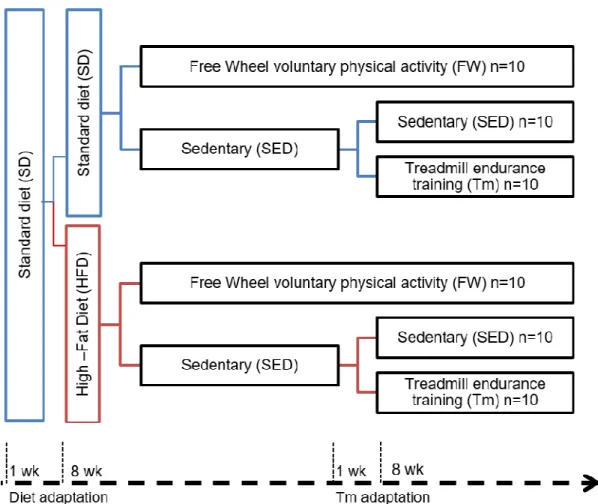
4.1. Animals ... 43
4.2. Exercise and Diet protocol ... 43
4.3. Animal euthanasia and heart extraction ... 45
4.4. Blood sampling and anatomical measurements ... 46
4.5. Isolation of heart mitochondria ... 46
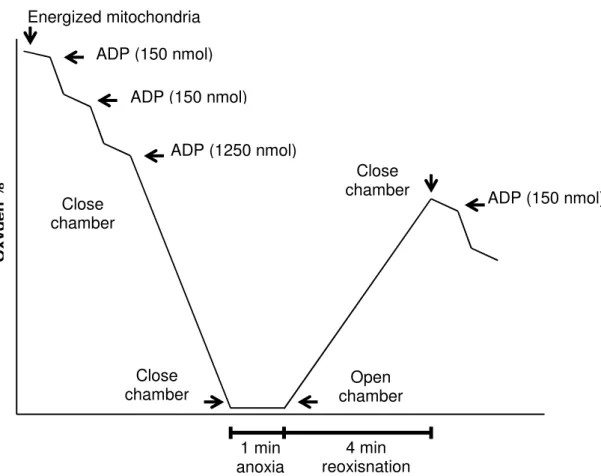
4.6. Mitochondrial respiratory activity and anoxia-reoxygenation procedure ... 47
4.7. Mitochondrial transmembrane electrical potential ... 48
4.8. Western-Blotting... 49
4.9. Lipid peroxidation ... 49
4.10. Gluthatione quantification ... 50
5. Results ... 53
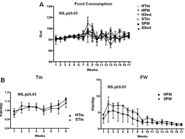
5.1. Food consumption and exercise interventions ... 53
5.2. Animal characterization ... 54
5.3 Heart mitochondria oxygen consumption and transmembrane electric potential ... 58
5.4 Heart mitochondrial oxygen consumption in Anoxia-Reoxygenation... 61
5.5 OXPHOS protein semi- quantification ... 63
5.3 Glutathione and lipid peroxidation measurements ... 65
6. Discussion ... 67
6.1. Diet Protocol ... 67
6.2. Exercise Protocol ... 70
6.3 Animal Characterization ... 72
6.3.1. Serum markers ... 73
6.4 The effects of high-fat diet on cardiac mitochondrial bioenergetics, OXPHOS content and redox state ... 74
6.5 The effects of exercise on cardiac mitochondrial bioenergetics, OXPHOS content and redox state ... 77
7. Conclusions ... 83
Figures
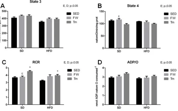
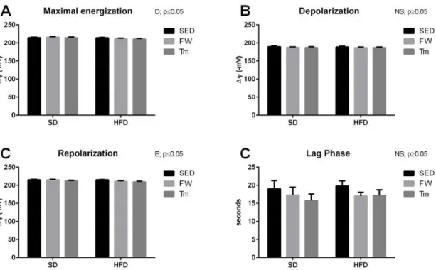
Figure 1. “Proposed mechanisms of vascular stiffness in obesity, insulin resistance, and type 2 diabetes”.. ... 11 Figure 2. “Antioxidant enzyme system” ... 22 Figure 3. Diagram summarizing the design of diet and exercise protocol ... 45 Figure 4. Typical polarographic oxygen traces obtain in Anoxia-reoxygenation model. ... 48 Figure 5. Food consumption (A), running distance in treadmill (Tm) and free wheel (FW) per day (B). ... 53 Figure 6. Effect of diet and exercise treatments on heart mitochondrial oxygen consumption (A) state 3 of mitochondrial respiration, (B) state 4 of mitochondrial respiration, (C) RCR, and (D) ADP/O. ... 59 Figure 7. Effect of diet and exercise treatments on heart mitochondria Δψ fluctuations (A) maximal energization, (B) ADP-induced depolarization, (C) ADP-induced repolarization and (D) ADP phosphorylation lag phase.. ... 60 Figure 8. Effect of diet and exercise treatments on heart mitochondria after Anoxia-Reoxygenation stimulation (A) state 3 of mitochondrial respiration, (B) state 4 of mitochondrial respiration, (C) RCR, and (D) ADP/O.. ... 62 Figure 9. Effect of exercise and diet treatment on heart mitochondria's respiratory complexes (OXPHOS): (A) Complex I; (B) Complex II; (C) Complex IV; (D) Complex V; (E) Protein loading control by Ponseau-S staining; (F) ) Typical immunoblots .... 64 Figure 10. Effect of exercise and diet treatment on heart mitochondria's (A) MDA content and (B) MDA content in Anoxia-Reoxygenation. ... 65 Figure 11. Effect of exercise and diet treatment on heart mitochondria's: (A) Reduced Glutathione content (GSH); (B) Oxidized Glutathione content (GSSG) and (C) Glutathione Ratio: GSH/GSSG.. ... 66
Tables
Table 1. Obesity-induced alterations in Adipocytokine ... 7
Table 2. A brief resume of the mitochondrial respiratory chain complexes ... 19
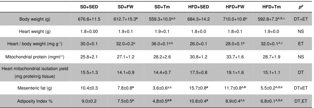
Table 3. Anatomic characterization of animals... 55
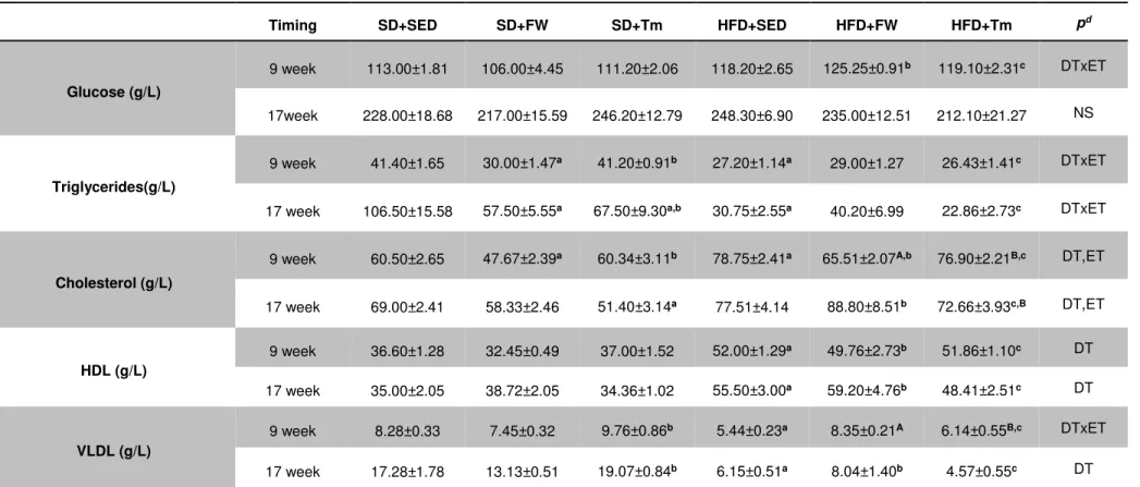
Table 4. Blood analysis after 9 and 17 weeks of treatments ... 57
Table 5. Percentage of the calories provided by carbohydrates; fat and proteins in some typical commercial diets for rodents. ... 69
Resumo
No presente estudo investigamos os efeitos de duas modalidades distintas de exercício (actividade física voluntária em roda livre –FW e treino em tapete rolante – Tm) em animais alimentados com duas dietas isocalóricas diferentes na percentagem de energia proveniente de gordura e de hidratos de carbono: Dieta Standard (SD) e Dieta Gorda (HFD) na bioenergética e stress oxidativo de mitocôndrias cardíacas. Ratos Sprague-Dawley machos foram divididos em quatro grupos: dieta standard sedentário (SD+SED, n=20), dieta standard roda livre (SD+FW, n=10), dieta gorda sedentário (HFD+SED, n=20) e dieta gorda roda livre (HFD+FW, n=10). Passadas 9 semanas, metade (n=10) dos animais dos grupos SD+SED e HFD+SED foram recolocados num novo grupo de treino em passadeira rolante (8 semanas, 5 dias/semana; 60 mins/dia; SD+Tm e HFD+Tm respetivamente). Realizou-se uma avaliação ex vivo de parâmetros de funcionalidade mitocondrial, sob condições de oxigenação normais e em anoxia-reoxigenação, assim como semi-quantificação de subunidades da fosforilação oxidativa e avaliação da peroxidação lipídica e do estado redox da glutationa. 17 semanas de dieta gorda não induziram qualquer efeito na funcionalidade mitocondrial cardíaca nem no estado redox. Contudo, o treino Tm promoveu melhorias na actividade respiratória mitocondrial nas duas dietas, antes e após a anoxia- reoxigenação. Já o treino FW aumentou o conteúdo das subunidades dos complexos IV e V da OXPHOS nas duas dietas, assim como melhorou a capacidade anti oxidativa mitocondrial (GSH/GSSG), particularmente no grupo HFD. Apesar da HFD não ter induzido disfunção na bioenergética mitocondrial, o treino Tm promoveu na generalidade um efeito mais visivel no consumo de oxigénio mitocondrial, enquanto que o exercício FW pareceu modelar de forma positiva a capacidade anti oxidativa mitocondrial. Concluímos assim que o exercício crónico pode constituir uma estratégia eficiente na melhoria da funcionalidade mitocondrial cardíaca num contexto de obesidade e HFD.
PALAVRAS-CHAVE: EXERCÍCIO; CORAÇÃO; BIOENERGÉTICA;
Abstract
We here investigate the effects of two distinct chronic exercise modalities voluntary free wheel training (FW) and the endurance treadmill training (Tm) in animals feed with isocaloric diets different in energy derived from fat and carbohydrates: standard (SD) and high fat diet (HFD) on cardiac mitochondrial bioenergetics and oxidative stress. Male Sprague-Dawley rats were divided into standard-diet sedentary (SD+SED, n=20), standard-diet free wheel (SD+FW, n=10), high-fat diet sedentary (HFD+SED, n=20) and high-fat diet free wheel (HFD+FW, n=10) groups. After 9-weeks, half (n=10) of SD+SED and HFD+SED groups were engaged in a Tm program (8 wks, 5 d/wk, 60 min/day; SD+Tm and HFD+Tm respectively). Ex vivo cardiac mitochondrial function endpoints were assessed under normal oxygenation conditions and anoxia-reoxygenation. Semi-quantification of oxidative phosphorylation subunits, lipid peroxidation and the glutathione redox status were also measured. 17-weeks of HFD treatment, did not affect cardiac mitochondrial function neither the redox state (HFD+SED vs. SD+SED) Importantly, Tm exercise improved mitochondrial respiratory activity in both diet regimens before and after anoxia-reoxygenation. FW running increased OXPHOS complexes IV and V subunits in both diet treatments and improved mitochondrial anti-oxidant capacity (GSH/GSSG), particularly in HFD group. Although 17 weeks of HFD did not induced mitochondria bioenergetics impairments, Tm training had a stronger effect in overall mitochondria oxygen consumption, whereas FW exercise seems to positively modulate the antioxidant machinery. Overall, we conclude that chronic exercise may constitute an effective strategy to increase cardiac mitochondrial functionality in a context of obesity and HFD.
KEY-WORDS: EXERCISE; HEART; BIOENERGETICS; MITOCONDRIA;
Abbreviations and Symbols
CVD Cardiovascular Diseases HFD. High-Fat Diet
ROS Reactive Oxygen Species
AMPK Adenosine Monophosphate-Activated Protein Kinase ANT Adenosine Nucleotide Translocase
ATP Adenosine Triphosphate Ca2+ Calcium Ion CAT Catalase Tm Treadmill FW Free Wheel SED Sedentary SD Standard Diet
DNA Deoxyribonucleic Acid
MRC Mitochondria Respiratory Chain GSH Reduced Glutathione
GSSG Oxidized Glutathione OXPHOS Oxidative Phosphorylation BMI Body Mass Index
GPX Glutathione Peroxidase WC Waist Circumference FA Fatty Acids
H2O2 Hydrogen Peroxide FFA Free Fatty Acid Ang II Angiotensinogen II IL-6 Interleukin-6
TNF- α Tumor Necrosis Factor alpha IL-10 Interleukin-10
IR Insulin Resistance
NEFAs Non-esterified Fatty Acids TGs Triglycerides
LDL Low-Density Lipoprotein HDL High-Density Lipoprotein VLDL Very Low Density Lipoprotein NO Nitric Oxide
PVAT Perivascular Adipose Tissue MnSOD Manganese Superoxide Dismutase
mPTP Mitocohndrial Permeability Transition Pore mtDNA Mitochondrial Deoxyribonucleic Acid EAT Epicardial Adipose Tissue
OMM Outer Mitochondrial Membrane
NADH Reduced Nicotinamide Adenine Dinucleotide
NADPH Reduced Nicotinamide Adenine Dinucleotide Phosphate nmol Nanomol
CPT1 Carnitine Palmitoyltransferase-1 NS Non-Significant
O2 Oxygen
O2-. Superoxide Radical
IMM Inner Mitochondrial Membrane OH. Hydroxyl Radical
PGC-1α Peroxisome Proliferator-Activated Receptor Gamma Coactivator 1-Alpha RCR Respiratory Control Ratio
TOM Translocase of the Outer Membrane ADP Adenosine Diphosphate
FADH2 Flavin Adenine Dinucleotide SEM Standard Error Of The Mean SOD Superoxide Dismutase
∆µH+ Variation in Electrochemical Proton Gradient
H+ Hydron
H2O2 Hydrogen Peroxide
MCU Mitochondrial Calcium Uniporter NCX Sodium Calcium Exchanger TPP+ Tetraphenylphosphonium
VDAC Voltage Dependent Anion Chanel ER/SR Endoplasmic/ Sarcoplasmic reticulum
Δᴪ Variation in Transmembrane Electrical Potential Cyp D Cyclophilin D
Cyp A Cyclophilin A Mfn 1 Mitofusin 1 Mfn 2 Mitofusin 2 OPA 1 Optic Atrophy 1
Drp 1 Dynamin- Related Protein 1 Fis 1 Mitochondria Fission 1 Mff Mitochondria Fission Factor
MID49 Mitochondrial Dynamics Protein 49 MID51 Mitochondrial Dynamics Protein 51 KD Kilodalton
MDV Mitochondria-Derived Vesicles PHD Pyruvate Dehydrogenase PFK-1 Phosphofructokinase-1 PDP 1 Phosphatase 1
PDP 2 Phosphatase 2
mPT mitochondrial Permeability Transition NRF 1 Nuclear Respiratory Factors 1
NRF 2 Nuclear Respiratory Factors 2
Tfam Mitochondrial Transcription Factor A
GST Glutathione S-Transferase
TBARS Thiobarbituric Acid Reactive Substances AI Adiposity Index
g Grams
mg Milligrams
1. Introduction
Obesity is one of the most prevalent diseases worldwide and has been considered a global epidemic (World Health Organization, 2015b). Despite of its complex etiology, obesity is frequently related to unhealthy behavior factors, including rich carbohydrate or fat diet and sedentary life style. The interaction between these two factors leads to a positive energy imbalance that has been believed to be the central cause of obesity (Lau et al., 2015; Ogden et al., 2007). Moreover, the hyperplasia and hypertrophy of adipocytes seen in an obesity condition ultimately culminates in adiposity tissue dysfunction, which is deeply associated to Cardiovascular Diseases (CVD) (Ferranti & Mozaffarian, 2008). Among several obesity-inducer models, a high-fat diet (HFD) consumption has been suggested to be responsible for cardiac and mitochondrial dysfunction as it is associated to increased production of reactive oxygen species (ROS), mitochondria respiratory chain (MRC) uncoupling, decreased oxidative capacity and calcium overload (Essop et al., 2016; Ferreira et al., 2015; Goncalves et al., 2016; Sverdlov et al., 2015; Sverdlov et al., 2016). Importantly, most of the studies are concerned with the effects of a high-fat and high-energy diets. The fact that not only the caloric intake but also the diet composition is responsible for inducing obesity (Estrany et al., 2011; Goyal et al., 2012), justify the need to study the effect of HFD compared with an isocaloric control diet on cardiac mitochondrial function, a topic underexplored.
Regular physical exercise has been considered a potential non-pharmacological strategy to counteract obesity, and also a potent cardio-protective therapeutic justifying its use within a clinical context (Ascensao et al., 2006; Bruun et al., 2006; Golbidi & Laher, 2012; Hafstad et al., 2013; Huang et al., 2015; Pons et al., 2013). Among the different animal exercise models, endurance training performed in treadmill (Tm) and voluntary free wheel running (FW) have been proven to be potential therapeutic and/o preventive strategies against cardiac mitochondrial dysfunction promoted by a variety of physiopatological conditions (Antonio Ascensao et al., 2011; Ascensao et al., 2006; Judge et al., 2005; Lesniewski et al., 2013). In fact, exercise training seems to positively modulate cardiac mitochondrial functionality, increasing antioxidant capacity, improving
membrane integrity and consequently improving oxidative capacity (Alleman et al., 2015; Jacobs & Lundby, 2013; Kavazis et al., 2008). However, there are still questions to be elucidated concerning Tm and FW exercise models. Differences in exercise intensity, volume, power, frequency, among other factors, can culminate in different cardiac mitochondrial adaptations.
Therefore, our aim is to analyze the effects of two isocaloric diets (differing in energy derived from fat and carbohydrates) in cardiac mitochondrial bioenergetics and redox state. Additionally, to our knowledge, no studies so far analyzed if the effects of different exercise modalities with distinct characteristics, including intensity, volume and length (FW and Tm) in cardiac mitochondria are dependent on the percentage of fat consumption. To do so, heart mitochondria respiratory parameters were measured, which included oxygen consumption under normal oxygenation conditions and in anoxia-reoxygenation, and transmembrane electric potential. Oxidative phosphorylation (OXPHOS) subunits content was also determined. Finally, markers of mitochondrial lipid peroxidation (MDA content) along with parameters of antioxidant capacity evaluated by reduced glutathione (GSH), oxidized glutathione (GSSG) were quantified.
2. State of art
2.1. Obesity: prevalence and etiology
Nowadays, obesity is one of the most prevalent diseases in developed countries, and it has started to spread through less developed countries all around the world. Moreover, dramatic increases in obesity have occurred in both children and adults (World Health Organization, 2015b), becoming a global epidemic. According to the World Health Organization (2015b), more than 1.9 billion adults were overweight in 2014. Among these, 600 million where obese, which shows how alarming this public health problem really is.
Currently, the WHO (World Health Organization, 2015b) defines obesity as an "abnormal or excessive fat accumulation that may impair health". The Body Mass Index, obtained by dividing the person’s weight (in Kilograms) by the square of his/her height (in Meters) (Bastien et al., 2014), has been largely used to classify overweight and obesity in adults. Therefore, a person with a BMI higher than 25 is considered to be overweight and a person with a BMI higher than 30 is considered to be obese. However, BMI does not take into consideration the distinction between an elevated BMI due to high level of lean mass or due to high level of fat body mass nor the body fat distribution. Indeed, several studies confirmed that the regional distribution of body fat is much more important than excess adiposity per se regarding the CVD risk associated with a given excess of body weight/fat (for ref see Despres, 2012). Comparatively, markers of absolute and relative accumulation of abdominal fat, such as Waist Circumference (WC) and Waist-To-Hip Ratio have been used to underline the importance of abdominal fat as a serious health risk (Bastien et al., 2014). The use of imaging techniques as computed tomography and magnetic resonance (MRI) represent remarkable advances in the ability to precisely and reliably quantify individual differences in body fat distribution and to selectively distinguish subcutaneous adiposity from visceral abdominal adipose tissue (for ref see Despres, 2012).
Regardless of its complex etiology, obesity is now considered to be the result from the interaction of various factors, such as behavior, environmental and genetic factors. The genetic seem to be the one that has less impact (Lau et al.,
2015), being the stem cause of this problem the energy imbalance, meaning more calories consumed than expended. Determinants responsible for the increased caloric consumption counts increased portion size, consumption of sugar-sweetened beverages, refined carbohydrates, advertising that promotes overconsumption. On the other hand, the reduced daily energy expenditure have been related to sedentary lifestyle, not only working time but also leisure time, both lacking of any sort of physical activity (Ogden et al., 2007). Therefore, the combination of energy-dense food products of poor nutritional value combined with a sedentary lifestyle has contributed to the emergence of obesity and the consequent health problems.
This excess of energy is stored in adipocytes (fat cells), leading to hypertrophy and hyperplasia (Ferranti & Mozaffarian, 2008) and resulting in a dysfunctional adipose tissue, which is associated with an increased prevalence of metabolic disorders, enlarging the risk of diabetes, CVD and mortality (Dario A. Gutierrez et al., 2009). Indeed, HFDs are not only responsible for inducing obesity but also for leading to metabolic alterations in adipose tissue, increasing levels of circulating fatty acids (FA) in the blood, which ultimately contributes for the development of metabolic syndrome and CVD (Betanzos-Cabrera et al., 2012). In accordance, the American Heart Association has published several position statements emphasizing the health hazards of obesity (Cornier et al., 2011; Klein et al., 2004; Poirier et al., 2006).
Although several strategies to control epidemic obesity have emerged, including the engagement with an active life style, it has been proposed that when compared, caloric restriction and dietary fat percentage reduction, the second option can be as effective as the first one in weight reduction and even more effective in improving insulin sensitivity along with others cardiovascular risk factors (Racette et al., 2006).
The following sections will address the effects of obesity on the deregulation of adipose tissue function, as well as the link with CVD.
2.2. Obesity and Adipose Tissue
Adipose tissue is composed by adipocytes and a vascular stromal fraction incorporating macrophages, fibroblasts, endothelial cells and pre-adypocytes. Pre-adypocytes, originated from a multi-potent stem cell, hold ability to generate new fat cells during the entire human life. The main roles of adipose tissue are to store energy in shape of triglycerides, insulate and cushion the body, this means control body temperature and mechanically protect the organs. This energy store results from free fatty acids (FFA) after food intake and it can be released during the fasting state, ensuring sufficient energy status (Hajer et al., 2008).
Adipose tissue can be subdivided in fractions of subcutaneous and visceral adipose tissue with distinct functions. For instances, accumulation of i ntra-abdominal or visceral adipose tissue has been reported to be quite deleterious and associated with a constellation of metabolic abnormalities increasing CVD risk (Despres, 2012). Although the visceral adiposity and liver fat are common key drivers of the cardiometabolic risk associated with overweight/obesity, other ectopic fat depots may also contribute to the risk of various cardiovascular outcomes (Despres, 2012).
Nowadays, adypocytes are also known to generate peptides, hormones and cytokines, termed adipocytokines, with endocrine, autocrine or paracrine repercussion and controlling lipid and glucose metabolism, blood coagulation, inflammatory state, blood pressure and hormonal modulation. Therefore, adipose tissue is now considered to be an endocrine organ (for ref see Moura & Monteiro, 2010) with a crucial relevance on body homeostasis. The consequences of obesity-mediated adipose tissue dysfunction will be addressed below.
2.2.1.
Inflammation and endocrine function of adipocytes: adipocytokines and obesityAs stated before, obesity has been established as a chronic inflammatory disease that leads to adipose tissue dysfunction, culminating in CVD and metabolic diseases (Cancello, Rouault, et al., 2005).
Adipocytokines have an important role in generating feedback action, affecting metabolism and function of organs and tissues, such as muscle, liver, vascular and brain (for ref seeHajer et al., 2008). Some of the most relevant adypocytokines are: leptin, angiotensinogen II, interleukin-6 (IL-6), tumor necrosis factor α (TNF-α), adiponectin and interleukin-10 (IL-10) (Moura & Monteiro, 2010). However, there are, at least, twenty four adipocytokines already identified (Hajer et al., 2008).
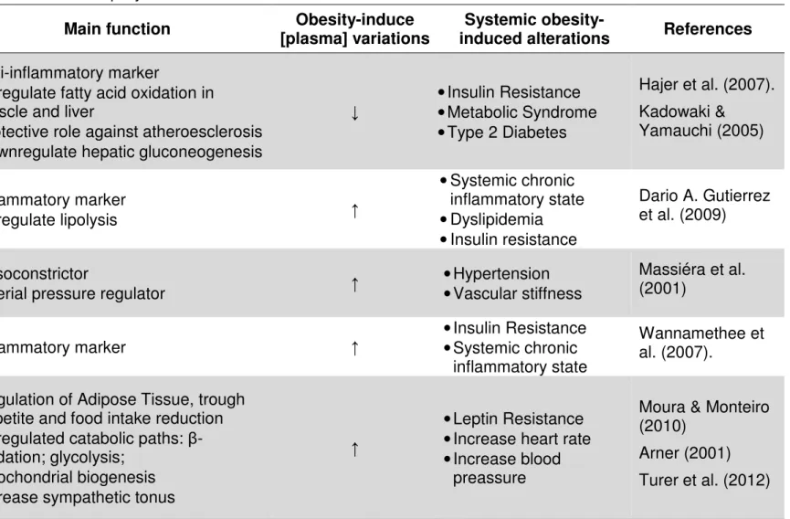
In obesity, adipocytokines production is deregulated, leading to insulin resistance, energetic homeostasis disturbance, coagulation alterations and arterial hypertension (Arner, 2001). The table 1 summarizes the main function associated with a few relevant adipocytokines, obesity-related alterations in their production, reflected in plasma concentration, and its overall consequences at a systemic level.
Table 1. Obesity-induced alterations in Adipocytokine
Adipocytokine Main function [plasma] variations Obesity-induce induced alterations Systemic obesity- References
Adiponectin
• Anti-inflammatory marker
• Upregulate fatty acid oxidation in muscle and liver
• Protective role against atheroesclerosis • Downregulate hepatic gluconeogenesis
↓ • Insulin Resistance • Metabolic Syndrome • Type 2 Diabetes Hajer et al. (2007). Kadowaki & Yamauchi (2005) TNF-α • Inflammatory marker • Upregulate lipolysis ↑ • Systemic chronic inflammatory state • Dyslipidemia • Insulin resistance Dario A. Gutierrez et al. (2009) Angiotensinoge
n • Vasoconstrictor • Arterial pressure regulator ↑ • Hypertension • Vascular stiffness
Massiéra et al. (2001)
IL-6 • Inflammatory marker ↑ • Insulin Resistance • Systemic chronic inflammatory state
Wannamethee et al. (2007).
Leptin*
• Regulation of Adipose Tissue, trough appetite and food intake reduction • Upregulated catabolic paths:
β-oxidation; glycolysis; • Mitochondrial biogenesis • Increase sympathetic tonus
↑
• Leptin Resistance • Increase heart rate • Increase blood
preassure
Moura & Monteiro (2010)
Arner (2001) Turer et al. (2012)
↑- upregulated; ↓- downregulated *Leptin limits the excess calories store and blocks the development of chronic steatosis. In obesity, contrarily of what expected, the leptin production is up-regulated suggesting a possible leptin resistance, along with alteration in its signaling (for ref seeMoura & Monteiro, 2010).
2.2.2. Impact of increased adipose tissue and inflammation on insulin resistance and dyslipidemia
2.2.2.1. Insulin resistance
Insulin Resistance (IR) is characterized by decreased insulin sensitivity, not only in the liver, but also in peripheral tissues. Moreover, it’s considered to be a precursor of Type 2 Diabetes Mellitus (Dario A. Gutierrez et al., 2009). As it was already mentioned, obesity is now established as a chronic inflammatory disease. This inflammatory state is assumed to contribute to IR by reducing insulin sensitivity in adipose tissue and other organs (Apovian et al., 2008).
As a result of adipose tissue dysfunction and inflammatory state, the production of pro-inflammatory adipocytokines, such as TNF-α is upregulated (Table 1). The TNF-α contributes to insulin signaling defects resulting in increased nonesterified fatty acids (NEFAs) blood concentration and their storage in liver, muscle and pancreas, directly inhibiting insulin signaling pathways and culminating in a systemic IR (for ref seeDario A. Gutierrez et al., 2009).
Besides TNF-α, other adipocytokines can also contribute to lipid homeostasis (Table 1). In fact, leptin and adiponectin play an important function. For instance, leptin can upregulate FA oxidation, reducing lipid accumulation in other organs; however, it is thought that obesity can induce leptin resistance, thereby losing its protective role against IR. Adiponectin, which has also a protective role enhancing muscle fat oxidation, is down-regulated in obesity (Unger et al., 1998). In obesity, the increased plasma concentration of NEFAs and TNF-α, the leptin resistance and the down regulated production of adiponectin contribute to lipid accumulation in the liver, muscle and pancreas, ultimately culminating in IR (Dario A. Gutierrez et al., 2009).
2.2.2.2. Dyslipidemia
Dyslipidemia is characterized by elevated plasma NEFAs, triglycerides (TGs), low-density lipoprotein (LDL) and reduced plasma high-density lipoprotein (HDL). Obesity is considered to be a precursor of dyslipidemia, as an increased in adipocyte size can lead to adipocyte saturation and inability to store lipid excess. The uncontrolled fatty acid lipolysis from visceral adipose tissue, results in an increased delivery of fatty acids to the liver to act as substrate for very low density lipoprotein (VLDL) (Dario A. Gutierrez et al., 2009).
Furthermore, as presented in Table 1, obesity is associated with an up-regulation of TNF-α, which is able to upregulate lipolysis, resulting in an increased of circulating NEFAs and, consequently, their delivery to the liver promoting TG synthesis and VLDL secretion. Therefore, it is obvious that the increased secretion of TNFα has a major impact in dyslipidemia (Jovinge et al., 1998). Also, in table 1, is mentioned that adiponectin is down regulated in obesity. Besides having a protective role against dyslipidemia, this adipocytokine is also associated with a lower plasma concentration of TG and VLDL and a higher plasma concentration of HDL. Consequently, the downregulated production of adiponectin increases the risk of dyslipidemia (for ref see Dario A. Gutierrez et al., 2009).
The deregulated production of adipocytokines, a distinct characteristic of adipose tissue dysfunction commonly seen in obesity, is associated with the development of IR, dyslipidemia and chronic inflammation. The next sections will address the mechanisms and the consequences behind obesity-induced cardiovascular complications, including vascular stiffness, hypertension and associated myocardial complications and heart dysfunction.
2.3. The relationship between obesity and cardiovascular complications
The CVD are some of the most deadly and disability-induced diseases worldwide, affecting the heart and the blood vases. It is estimated that 31% of all global deaths are caused by CVD (World Health Organization, 2015a). They include coronary heart disease, cerebrovascular disease, peripheral arterial disease, rheumatic heart disease, congenital heart disease, deep vein thrombosis and
pulmonary embolism. Myocardial infarction and strokes are the ultimate outcome, mostly culminating in death. The main cause is the accumulation of fat within the blood vascular walls, known as atherosclerosis, which can cause a blockage in the blood flow, preventing blood from flowing to the heart and brain (Hobbs, 2015; World Health Organization, 2015a).
Obesity is an important cardiovascular risk factor. It is now established that adiposity not only induces adverse local effects but also has a systemic effect (Ferranti & Mozaffarian, 2008). Moreover, obesity in now considered as an independent risk factor for CVD, suggesting that the increased adiposity indices are the main drivers behind the intermediate risk factors, such as vascular stiffness, hypertension, dyslipidemia, IR, chronic inflammation, diabetes mellitus, endothelial dysfunction and atherosclerosis (Bastien et al., 2014; Strasser et al., 2015).
2.3.1. Vascular stiffness, endothelial dysfunction and obesity-induced chronic inflammation
Obesity and IR are known to interact and impair vascular function and structure, as they are linked to endothelial dysfunction, increased artery intima media thickness and increased vascular stiffness (Herouvi et al., 2013). The endothelium is a complex organ. It is responsible for the regulation of the smoothed muscle cells proliferation, platelet function, vasomotor tone and thrombosis (López-Jiménez & Cortés-Bergoderi, 2011).
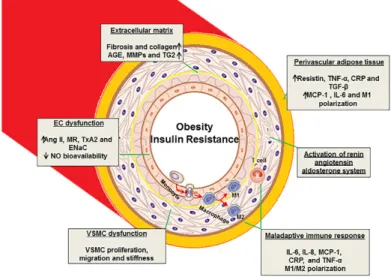
The first event of atherogenesis is the endothelial damage caused by various risk factors, such as arterial hypertension, hyperglycemia and dyslipidemia. The outcome of endothelial damage is actually endothelial dysfunction, which consists in an unbalance production of vasodilators agents and vasoconstrictors agents, with a decreased production of nitric oxide (NO) and an increased production of Ang II, respectively. Moreover, the production of ROS is also reported to be up-regulated (Husain et al., 2015). This contributes to an inflammatory state, along with an increase in intima membrane thickness (Figure 1).
In fact, the main driver of atherosclerosis is the excess of circulatory cholesterol, more precisely of VLDL. As the ROS production is up regulated due to the endothelial dysfunction, the VLDL molecules become oxidized. Thereafter, the oxidized VLDL connects with receptor-1, which is also up regulated in behalf of Ang II, and it is able to get through the endothelium, ending up in intima media. Additionally, the proliferation of VLDL oxidized through endothelium amplifies endothelium dysfunction, resulting in an even bigger decrease of NO production (Allahverdian et al., 2012). Furthermore, oxidized VLDL induces endothelial cells apoptosis and the expression of intracellular adhesion molecule-1 (ICAM-1), vascular cell adhesion molecule-1 (VCAM-1) and selectin-P, which results in endothelial damage as well as in monocyte infiltration in the intima media, boosting the inflammatory state (Apovian et al., 2008; Pasceri et al., 2000) .
Figure 1. “Proposed mechanisms of vascular stiffness in obesity, insulin resistance, and type 2 diabetes”. EC, endothelial cell; VSMC, vascular smooth muscle cell; AGE, advanced glycation end products; MMPs, matrix metalloproteinase; TG2, tissue transglutaminase; Ang II, angiotensin II; MR, mineralocorticoid receptor; TxA2, thromboxane A2; ENaC, epithelial Na+ channel; IL, interleukin; TNF, tumor necrosis factor; NO, nitric oxide; MCP-1, monocyte chemotactic protein-1; CRP, C- reactive protein; TGF-β, transforming growth factor- β” (Jia et al., 2015).
During the immune response and within the intima media, the macrophages (M1, M2) are not capable of fagocitating the VLDL oxidized, forming macrophage foam cells. This maladaptive immune response increases the cytokine production of TNF-α and IL6, resulting in a continuous lipid accumulation along with chronic inflammatory state (Allahverdian et al., 2012). The atheroma plaque growth is the
result of an increased number of macrophage foam cells and lymphocyte, which up regulate the production of hydrolytic enzymes, cytokines and growth factors, leading to fibrosis and local necrosis (for ref see Allahverdian et al., 2012). At this point, atherosclerosis is settled, the blood flow is reduced due to the atheroma plaque along with the diminished endothelial vasodilatation capacity. Over time, the atheroma plaque becomes unstable, meaning an increased risk of rupture and, consequently, espousing its lipid content. This event is highly thrombogenic, ultimately resulting in myocardial infarction or in cerebrovascular accident (Apovian et al., 2008; Jia et al., 2014; Maiolino et al., 2015).
Additionally, perivascular adipose tissue (PVAT) is also a serious risk factor for the development of obesity-induced atherosclerosis (Shimabukuru, 2009). Perivascular fat serves as a structure component present in most arteries and is defined as the accumulation of fat around vascular structures, mostly in the proximity of all blood vessels and around the coronaries and aorta (Shimabukuru, 2009). This tissue is a source of molecules with varied paracrine effects, holding ability to modulate vascular responsiveness to vasoactive agents (Villacorta & Chang, 2015). In obesity, the expression and infiltration of pro-inflammatory immune cells in PVAT is increased along with a reduced expression of anti-inflammatory factors (Aroor et al., 2013), which is associated with endothelial dysfunction. Therefore, obesity is considered an important risk factor for vascular stiffness and atherosclerosis with consequently adverse effects in several organs, including the heart, kidney, liver and brain. (for ref see Jia et al., 2015). Particularly, the compromised heart function and myocardial dysfunction linked with endothelial deregulation are addressed below.
2.3.2. Obesity compromises heart structure and function
Obesity is responsible for inducing structural adaptations in cardiovascular structure and function, in order to maintain whole body homeostasis. Stroke volume augmentation, which is thought to be a compensatory adaptation to increased adipose tissue mass (Szczepaniak et al., 2007), as well as the stiffening of central arteries increase systolic pressure and decrease diastolic pressure, and culminate in increased afterload. Over time, as a response to the higher cardiac workload, the ventricle starts to get thicker (ventricular
hypertrophy) so it can overcome the new demands. Further, the decrease in diastolic pressure is associated with reduced coronary flow during the diastole. These alterations are associated with left ventricular remodeling and fibrosis, leading to left ventricular-diastolic dysfunction and development of coronary artery disease (Jia et al., 2014; Lavie et al., 2009).
Epicardial adipose tissue (EAT) is the visceral fat that is located between the outer layer of the myocardium and visceral pericardium (Shimabukuru, 2009). In a similar way asabdominal adipose tissue, EAT also has considerable secretory activity. It has been reported that the increased EAT volume (commonly seen in obesity) directly compromises myocardial metabolism (Rosito et al., 2008) and is associated, among others, to the overexpression of pro-inflammatory TNF-α and IL-6 (Kremen et al., 2006). Additionally, due to the proximity to coronary arteries, EAT has been linked to increased atherosclerosis burden (Shimabukuro et al., 2013), increasing even more the risk for cardiac structural and functional impairments.
Alongside with vascular alterations that induced cardiac functional and structural disarrangements, obesity also promotes fat infiltration into the myocardium (Powell et al., 2006). Lipid over-accumulation in cardiomyocytes can result from increased FA uptake, decreased FA oxidation or even a combination of both (Szczepaniak et al., 2007). In obesity conditions, when adipocytes reach their maximum storage capacity, plasma FFA concentration increases, resulting in an ectopic accumulation of lipids in non-adipose tissues, including the myocardium (Szczepaniak et al., 2007). Therefore, myocardial fat infiltration is associated with several cardiac complications, including healed myocardial infarction, arrhythmogenic cardiomyopathy, dilated cardiomyopathy and cardiomyopathy with muscular dystrophy (Komatsu et al., 2014). In fact, this link between obesity and the severity of myocardial disease has been demonstrated in various studies. It has been proposed that an increased prevalence of myocardial fibrosis is proportional to the degree of obesity and is associated with cellular degeneration and inflammation (for ref see López-Jiménez & Cortés-Bergoderi, 2011). It is also suggested that the longer duration of morbid obesity, the greater the left ventricular mass and the worse the impairment of left ventricular systolic function and diastolic filling (for ref see Szczepaniak et al., 2007). Also, the
over-accumulation of lipids in the cardiomyocytes induces steatosis of the myocardium, ultimately leading to lipotoxic cardiomyopathy (Szczepaniak et al., 2007).
2.3.3. Adipocytokine action on the heart: the particular case of leptin
Several key metabolic hormones have important central inputs besides the well known peripheral effects. Leptin is one good example of this fact, as it is known to have the ability to regulate sympathetic nervous system (Turer et al., 2012). Although the precise mechanisms by which leptin regulates autonomic nervous system (ANS) are still unclear, it is accepted that leptin plays as important role on cardiovascular regulation and that is a link between excess weight gain, increased sympathetic tone and hypertension (Bassi et al., 2015; Wang et al., 2013).
Circulatory leptin levels provide information regarding energy expenditure and amount of fat stored. This information is centrally processed by the ANS, regulating food intake and energy expenditure. Therefore, if leptin levels are elevated due to obesity, one mechanism able to increase energy expenditure is the augmentation of sympathetic tone (for ref see Barnes & McDougal, 2014). Sympathetic tone stimulation has profound impacts in cardiovascular function, including arterial blood pressure and heart rate (Correia et al., 2000). Additionally, leptin also upregulates blood pressure through increase of renal sympathetic nerve activity (for ref see Barnes & McDougal, 2014).
Additionally, Wang et al. (2013) demonstrated a relation between circulatory leptin levels and endothelial dysfunction, in which leptin enhances the effects of ang II on blood pressure. According to other studies, these effects of leptin are mediated by sympathetic nervous system activation and contribute to vascular stiffness and hypertension in obesity (for ref see Brooks et al., 2015).
2.3.4. The obese myocardium: lipotoxicity and metabolic disarrangements
Lipotoxicity can result from FA accumulation in the heart induced by excessive FA consumption. Fat infiltration in the heart leads to an alteration in substrate
preference and utilization towards FA metabolism (Hall et al., 2015). This means that the heart no longer has the ability to transit between FA metabolism to glucose metabolism, under energetically demanding conditions, which results in energy imbalance, impaired contractility, and post-translational protein modifications. This is known as a central trait of obesity-induced cardiometabolic disease (Griffin et al., 2015). This metabolic alteration is promoted by an increase in the expression of proteins involved in fatty acid oxidation, including carnitine palmitoyltransferase-1 (CPT1). As a result of this FA oxidation alteration, toxic lipids, including ceramide, are formed, contributing for cardiac dysfunction (Park et al., 2008; Slawik & Vidal-Puig, 2006).
Several studies have demonstrated that toxic lipid accumulation in myocardium, such as ceramide, is associated with an increased in the inflammatory marker TNF-α (Sharma et al., 2004) and the production of ROS leading to insulin signaling impairment, which ultimately results in cardiac IR (Murphy & Brown, 2009) and cardiac contractile dysfunction by influencing sarcoplasmic reticular calciumstores, promoting mitochondrial dysfunction and, at last, cardiomyocyte apoptosis(Listenberger et al., 2001; Turer et al., 2012) .
Cardiomyocyte death, the ultimate result from myocardium fat infiltration, is recognized as a critical and direct factor in the development of heart dysfunction. Among the several molecular mechanisms associated with obesity-induced cardiac dysfunction, mitochondrial abnormalities seem to have a central role. As mitochondria integrity preservation is absolutely essential not only for maintaining metabolic homeostasis but also for survival of cardiomyocytes (Murphy & Brown, 2009), these organelles are fundamental to a proper cellular functioning. Moreover, as cardiomiocytes depend entirely on healthy mitochondria for normal heart function, mitochondria are reliable sensors of cellular homeostasis. Indeed, (dys)functional mitochondria correlates with (dys)functional cardiac tissue submitted to a variety of stimuli, including obesity. Heart mitochondrial structure and function and its relation with obesity are addressed in the following section.
2.4 The heart mitochondria
Mitochondria are essential organelles situated in the cytoplasm and associated not only with energy production, but also with ion regulation, osmotic regulation, pH control, calcium homeostasis, redox reactions, cell signaling and regulation of programmed cell death, thus assuming pivotal role in cellular function (Antonio Ascensao et al., 2011). Therefore, in the present thesis, a brief description of mitochondrial-mediated mechanisms associated with cell function will be accomplished before obesity effects on heart mitochondria.
In cardiomyocytes, there are two distinct types of mitochondria with distinct roles in cardiomyocyte physiology. Considering the energy demand of cardiomyocyte for the heart beat maintenance, the energy source is assigned by these two distinct mitochondria, depending on its final use (Pereira, 2012). There are mitochondria closer to the T tubules and to sarcoplasmic reticulum that provides energy (ATP- Adenosine Triphosphate) for the required calcium influx to the sarcoplasmic reticulum. In fact, this energy source is essential for calcium homeostasis, which is critical for the relaxation and contraction cycle. Additionally, there are also cardiac mitochondria pivotal to energy source and responsible for the release of myosin from actine and consequently for the stroke force (for ref see Pereira, 2012).
As a result of the high energy demand of the heart, a refined mitochondrial quality control is needed, this means that non-functional or damage mitochondria are removed by auto(mito)phagy and new healthy mitochondrial are formed by biogenesis. Both situations depend on fission and fusion events, and can occur in certain pathological situations. For example, very small/ fragmented mitochondria can be found in dilated cardiomyopathy and ventricular-associated congenital heart disease, or large and defective mitochondria in aged cardiomyocytes (for ref see Pereira, 2012).
Although heart mitochondria might be slightly different in morphology they both have the basics characteristics of mitochondrial from other tissues.
2.4.1. Mitochondrial structure
Mitochondria have two membranes with distinct function, the outer mitochondrial membrane (OMM) and the inner mitochondrial membrane (IMM), being the space between the two membranes called inter-membrane space. These two membranes separate the cytoplasm from the mitochondrial matrix, where mitochondrial DNA (mDNA) is found. Indeed, mitochondria are the only organelles, besides cell’s nucleus, that have their own DNA (Pocock & Richards, 2006).
The OMM is highly permeable due to the presence of transmembrane channels, such as porins and protein complex known as translocase of the outer membrane (TOM), which allow the passage of proteins to the inter membrane space. (Pocock & Richards, 2006). The structure of the IMM is characterized by its folded aspect due to the presence of invaginations, formally known as cristae. These invaginations significantly increase membrane surface. In contrast to the OMM, the IMM is less permeable, which means that it is selective permeable to certain molecules depending on specific carriers (Pocock & Richards, 2006). Importantly, inserted in the inner membrane it is located the MRC pivotal for the mitochondrial energy production.
2.4.2. Mitochondrial energy production – oxidative phosphorylation
The OXPHOS is a metabolic pathway in which energy produced by oxidation/reduction reactions is used for the phosphorylation of adenosine diphosphate (ADP) to ATP (Kadenbach, 2003). The process begins with the production of reduced equivalents, reduced nicotinamide adenine dinucleotide (NADH) and flavin adenine dinucleotide (FADH2) in the Krebs cycle and
beta-oxidation. The main role of these two elements is to give way electrons to the MRC (Johannsen & Ravussin, 2009).
The MRC is inserted in the inner membrane and constituted by five protein complexes (I-V) (Table 2). Some of these complexes are responsible for the transportation of protons (H+) from the mitochondrial matrix to the
inter-membrane space, producing an electric potential that is of particular relevance for ATP producton (∆ψ) (Kadenbach, 2003).
Both processes (electron transference and directional proton transportation) are intrinsically bounded. As complexes I, III and IV get oxidized (receive electrons) they pump H+ to the inter-membrane space, complex I pumps four H+, complex
III also pumps four H+ and complex IV pumps two H+, resulting in the
transportation of approximately ten H+ (Kadenbach, 2003). Although, this only
happens if the first electron accepter is complex I (NADH dehidrogenase - as it receives electrons from NADH), originated in Krebs cycle, It is also possible that the first electron receptor is complex II (succinate-dehidrogenase: ubiquinone reductase - as it receives the electron pair from FADH2) excluding complex I from
the chain. If this occur, it can result in the transportation of approximately six H+
to the inter-membrane space, four H+ less than with complex I as the first point of
electron entry in MRC (Johannsen & Ravussin, 2009).
The transference of electrons (oxidation/reduction – redox reactions) along the complexes (I-IV) is conducted by the decrease in their redox potential, until they reach their final acceptor, oxygen (O2), in complex IV, forming water (H2O)
(Johannsen & Ravussin, 2009). Therefore, mitochondria produce ATP in an aerobic way, meaning in the presence of O2.
At the end of the MRC, implicating that the electrons have already encountered O2, the electrochemic gradient is amplified, due to the transportation of H+ to the
inter-membrane space. This results in a returning tendency of H+ from the
inter-membrane space to the mitochondrial matrix. Complex V (ATP- syntase) allows the protons to return to the mitochondrial matrix, originating a protomotriz force, which activates ATP synthesis (ADP+Pi) (Johannsen & Ravussin, 2009). For each four H+ reentering the mitochondrial matrix, one ATP will be phosphorylated,
this means, in averaged, that for every electron pair provided from NADH, which results in 10 H+ pumped to the cytoplasm, 2,5 ATP are formed, and if the pair of
electrons are provided from FADH2 it will be formed 1,5 ATP (Hinkle, 2005).
As ATP is formed, it has to be transported to the inter-membrane space through adenine nucleotide translocase (ANT), which is also responsible for the reentry of ADP needed for ATP re-synthesis. Furthermore, the ability to ATP reach cell’s cytoplasm through the OMM is dependent of the presence of non-selective channels designated as voltage-dependent anion channel (VDAC) (Johannsen & Ravussin, 2009).
Table 2. A brief resume of the mitochondrial respiratory chain complexes
Complexes Function References
I – NADH dehidrogenase: Ubiquinone/Coenzyme Q (CoQ) Reductase
First point of entry of electrons into MRC;
Oxidizes NADH and reduces ubiquinol to ubiquinone; Pumps 4 H+ to the
inter-membrane space. Johannsen & Ravussin (2009) II – Succinate Dehidrogenase: Ubiquinone Reductase
First or second point of entry of electrons into MRC;
Converses succinate to fumarate in Krebs Cycle.
Johannsen & Ravussin (2009)
III – Ubiquinol: cytochrome-c reducytochrome-ctase
Catalysis the transference of electrons from
ubiquinol(oxidation) to cytochorme-c (reduction); Pumps 4 H+ to the
inter-membrane space. Johannsen & Ravussin (2009) IV – Cytochrome-c: Oxygen oxidoreductase
Catalysis the transference of electrons from cytochrome-c (oxidation) to O2 (reduction),
forming H2O;
Pumps 2 H+ to the
inter-membrane space.
Johannsen & Ravussin (2009)
V – F0F1 - ATPase Uses the amplified proton
gradient (protomotriz force) to synthesize ATP
Johannsen & Ravussin (2009)
MRC- mitochondrial respiratory chain; NADH- nicotinamide adenine dinucleotide; O2 - oxygen
;H2O- water; ATP- adenosine triphosphate
2.4.3. Mitochondrial as source of oxidative stress
The OXPHOS in MRC is one of the major redox system in the cell and responsible for a significant amount of free radicals production (Ray et al., 2012). Free radicals are defined as any molecule with one or more unpaired electron and capable of freely exist and highly reactive in a way that they are able to “attack” any biomolecule or biological structure (Kehrer & Klotz, 2015). Free radicals are
a result of normal cellular aerobic metabolism and represent a physiological consequence of O2 use. However, there are situation where this compound
formation is deregulated, meaning that they can exist in a free form and interact with various biological structures, ultimately resulting in injury. Importantly, these molecules trigger important signaling events, both directly and through various redox-regulated transcription factors (Ray et al., 2012).
Kehrer & Klotz (2015) feature radical’s basic reaction characteristics in three categories: Initiation reactions, where the numbers of radicals increase, these reactions involve O2 reduction; propagation reactions, involving hydrogen
abstraction, electron transfer or addiction and where the number of radicals does not change; and termination reactions, resulting from the interaction of two radicals, reducing the number of radicals until they disappear.
The prevalence of oxygen in mitochondria induces oxygen-centered reactive species production, formally known as ROS (reactive oxygen species, abbreviation previously defined), which include superoxide radical (O2.-),
hydrogen peroxide (H2O2), hydroxyl radical (OH.), among others (Kehrer & Klotz,
2015). Besides MRC, there are other redox-related system located in several places of the cell, including endoplasmic reticulum, peroxisomes and cytosol. The damaging effects of ROS have been demonstrated along the years of experiments and investigation. Nowadays, there are also evidences regarding the beneficial effects of a controlled ROS production. Actually, ROS are responsible and imperative for regulating several cellular mechanisms, including mitochondrial biogenesis and apoptosis, which are pivotal processes for cell’s health maintenance, along with others cellular signaling pathways (Cadenas, 2004; Finkel, 2001). However, an additional ROS production can be implicated in several physiological and pathological conditions, including aging, DNA mutagenesis, inflammation and cell death pathways (Kehrer & Klotz, 2015). The deleterious effects of ROS in lipids, proteins and DNA are addressed bellow. Lipid peroxidation has a pivotal effect in terms of ROS mediated injury, justified by lipid’s critical structural and functional role in cell’s membrane (for ref see Kehrer & Klotz, 2015). In addition, the protein oxidation is associated with age-related losses of selected biochemical and physiological functions that may be related to unrepaired damage to other macro molecules, such as DNA (Baraibar
et al., 2012). In fact, the DNA oxidation affects the integrity and regulation of genes, which may be harmful or beneficial to cells. These changes can result from direct modifications to DNA or may be due to changes in transcription factors or enzymes involved in gene expression and repair (for ref seeKehrer & Klotz, 2015). When DNA repair processes are affected, it can culminate in permanent DNA damage as it can modify DNA sequences, also known as mutations (Regulus et al., 2007).
As already mentioned, the main source of ROS in mitochondria is the MRC, more precisely, complex I, II and III (Kornfeld et al., 2015). This complexes promote O2
reduction to O2.- (superoxide anion), a highly reactive specie and a precursor of
other ROS, such as H2O2 (hydrogen peroxide) and OH: (hidroxil radical) (Ray et
al., 2012).. Normally, this happens in a controlled and caged form due to the
presence of antioxidant molecules that are responsible for maintaining ROS production balanced. Nevertheless, there are conditions where the production of pro-oxidant molecules and antioxidant molecules is imbalanced, resulting in what is known for oxidative stress (Kehrer & Klotz, 2015). The more recent definition of oxidative stress is featured in Kehrer & Klotz (2015) work as “an imbalance between oxidants and antioxidants in favor of the oxidants, leading to a disruption of redox signaling and control and/or molecular damage” (for ref see Kehrer & Klotz, 2015).
Antioxidants are in permanent activity as ROS production is also permanent, as a result of aerobic metabolism. There are two different types of antioxidants, enzymatic and non enzymatic. Furthermore, they can also be classified according to their action against free radicals: scavenger antioxidants are able to turn a free radical into another less reactive free radical and quencher antioxidants able to completely neutralize free radicals (Kehrer & Klotz, 2015). Enzymatic antioxidants present in mitochondria will be addressed below.
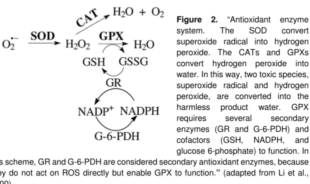
Enzymatic antioxidant defense systems include superoxide dismutase (SOD), catalase (CAT), glutathione peroxidase (GPx) and glutathione reductase, among others (Figure 2):
• The SOD are a family of metalloenzymes that are able to convert two superoxide anions to triplet oxygen and H2O2. Bearing in mind that H2O2 is
less reactive than O2.-, SOD’s role is simply to turn a highly reactive radical into a less reactive one. Mitochondrial SOD is manganese SOD (MnSOD) (Kehrer & Klotz, 2015).
• After superoxide dismutase, both CAT and GPx are able to cope with H2O2.
CAT catalyzes the dismutation of H2O2 to water and molecular oxygen and it
is essentially located in peroxisomes and its action is potentially diminished in the heart, lungs and brain, due to their reduced peroxisome presence (Kornfeld et al., 2015).
• The GPx is located in cytoplasm and in mitochondria. It catalyzes H2O2, using
glutathione (GSH) as substrate. GSH transfers two H+ to H2O2, forming two
water molecules. As GSH gives away electrons, it gets oxidized, becoming glutathione disulfide (GS – GS). In order to reduce GS – GS into GSH de
novo, NADPH/ FADH2, from Krebs cycle, transfer electrons to GS –GS,
forming GSH, this reaction is catalyzed by Glutathione reductase. Bearing in mind GPx action, as it neutralizes completely H2O2, it is acknowledged as a
quencher antioxidant. GPx is present in various organs, including the heart (Herbet et al., 2015).
Figure 2. “Antioxidant enzyme system. The SOD convert superoxide radical into hydrogen peroxide. The CATs and GPXs convert hydrogen peroxide into water. In this way, two toxic species, superoxide radical and hydrogen peroxide, are converted into the harmless product water. GPX requires several secondary enzymes (GR and G-6-PDH) and cofactors (GSH, NADPH, and glucose 6-phosphate) to function. In this scheme, GR and G-6-PDH are considered secondary antioxidant enzymes, because they do not act on ROS directly but enable GPX to function.” (adapted from Li et al., 2000).
2.4.4. Mitochondrial calcium homeostasis and apoptosis
Mitochondria are essential for the maintenance of cytosolic calcium homeostasis, as they are able to accumulate calcium through an electrochemical gradient that favors transport across the IMM (Gustafsson & Gottlieb, 2008). Mitochondrial mechanisms of calcium uptake and release have been studied along the years and include, the mitochondrial calcium uniporter (MCU) and the sodium calcium exchanger (NCX) (Carafoli, 2010). However, other pathways remain to be elucidated, such as the mitochondrial permeability transition pore (mPTP) (Bernardi & Di Lisa, 2015). Calcium homeostasis and mitochondrial permeability transition will be addressed below.
As it is already mentioned above, mitochondria play an important role in calcium homeostasis, constituting a calcium buffering system. This mitochondria role is pivotal for cell’s health since elevated calcium concentrations may be deleterious, justifying the necessity of maintaining an ideal calcium concentration in the cytoplasm. The majority of cellular calcium is stored in the endoplasmic/ sarcoplasmic reticulum (ER/SR) (Gustafsson & Gottlieb, 2008), thus mitochondria positioned near calcium release sites on ER/SR are able to capture a substantial amount of calcium and to prevent the accumulation of calcium in the cytosol (for ref see Gustafsson & Gottlieb, 2008).
Mitochondrial calcium also interferes with energy metabolism. One good example is the activation of dehydrogensases in the mitochondrial matrix, culminating in increase of mitochondrial NADH/NAD ratio and leading to an increase of available energy for mitochondrial physiological processes. Furthermore, calcium present in the matrix has an effect in ATPase, boosting ATP synthesis (Balaban, 2002). Two calcium transporters control mitochondrial calcium concentrations. The MCU is responsible for the calcium influx and NCX for calcium efflux. The first one, works in favor of the electrochemical gradient, while the second works against electrochemical gradient (Gunter et al., 2000). However, if calcium concentration in the matrix increases beyond a certain level, mitochondrial lose the ability to regulate calcium concentration, resulting in calcium overload. This condition can lead to the opening of the mPTP (Bernardi & Di Lisa, 2015; Gunter et al., 2000),
an event that have been associated with cell death, and justifies the release of proapoptotic proteins.
The mPTP is an IMM channel that is thought to be voltage and calcium dependent, cyclosporine A sensitive and conductance channel (for ref see Bernardi & Di Lisa, 2015). Besides mitochondria calcium overload, mPTP opening is facilitated through oxidative stress, mitochondrial depolarization, increased in phosphate concentrations and adenine nucleotide depletion (Bernardi et al., 2006; Rasola & Bernardi, 2011). When IMM loses its permeability (transitory permeability), allows the entrance of molecular solutes, including ions and cofactors resulting in detrimental effects that include the disruption of metabolic and ion gradients and energetic imbalance of ATP/ADP ratio between matrix and cytosol. As a consequence, mitochondrial ∆p drops and ATP hydrolyses takes place (for ref see Halestrap, 2004; Pereira, 2012). Furthermore, mPTP induces membrane depolarization, OXPHOS uncoupling, respiratory inhibition due to the loss of both co-factors and cytochrome c, and increased oxidative stress. If mPTP remains open, differences in protein concentration between cytosol and mitochondrial matrix potentiate the osmotic pressure, culminating in IMM expansion known as mitochondrial swelling, which leads to OMM disruption (Bernardi et al., 2006; Halestrap, 2004). In a situation where OMM gets ruptured, proapoptotic proteins are released, including cytochrome c, endonuclease G and apoptosis-inducing factor(AIF), resulting in cell death (Rasola & Bernardi, 2011).
The molecular nature of mPTP has been a matter of study for a long time. The multi-component composition, also known as classical model of mPTP, suggested that mPTP is formed by sevral proteins, including cyclofiline D (Cyp D), ANT and VDAC (Bernardi & Di Lisa, 2015). This suggests that OMM is also involved in mPTP structure as VDAC are present in the OMM. The Cyp-D is a member of cyclophilins family and has a pivotal role in mPTP regulation. It is located in mitochondrial matrix and during mPTP opening induces alterations in IMM’s channels, leading to an increase in IMM permeability (Broekemeier et al., 1989). Studies have demonstrated the existence of an interaction between Cyp-D and ANT, which can be inhibited by cyclophilin-A (CsA). In fact, CsA is one of the most studied mPTP inhibitor, as it is able to connect to Cyp-D (Galat, 1993).
However, evidences from some studies with ANT and VDAC knockout rats have called into question the truly role of these components in mPTP composition
(Duchen & Szabadkai, 2010). Kokoszka et al. (2004) observed that mitochondria from ANT1 and ANT2 knockout mice still shown mPTP- CsA-sensitive activity, although triggered by an increased in calciumconcentration. This suggests that ANT is not essential for pore formation but play a role in its regulation as mPTP opening is influenced by ATP and ADP concentrations (Kokoszka et al., 2004). Recently, it has been suggested that F0 subunit from ATP synthase is also involved in mPTP opening (Bernardi & Di Lisa, 2015). In fact, several studies shown that Cyp-D binds to the F1F0 ATP synthase (for ref see Halestrap, 2014), this interaction is known to modulate mPTP formation. However, some questions remain to elucidate, which justifies the need for more research in this matter.
2.4.5. Mitochondrial quality control
Mitochondria are dynamic organelles with plastic proprieties, which means that they are capable of modifying their morphology and play important roles in maintaining cell survival, cell death and cellular metabolic homeostasis (Ni et al., 2015). These plastic properties result from a balanced interaction of fusion, fission, auto(mito)phagy and mitochondrial biogenesis events, ensuring damaged mitochondria get removed and proper organization of the mitochondrial network is maintained (Lee et al., 2004). Mitochondrial dynamics is now considered to be a pivotal cell biological process as it allows mitochondrial quality control and is fundamental for cell functioning, mPTP regulation, apoptotic signaling and, at last, for cell survival (Campello & Scorrano, 2010).
Mitochondria are responsible for cell death, including apoptotic and necrotic cell death. For this reason, mitochondria quality needs to be well controlled (Ni et al., 2015). The regulation of mitochondria quality control by several mechanisms will be addressed below with more detail.
Mitochondria’s fission and fusion events constitute one important quality control mechanism. Dysfunctional mitochondria are programmed to lose their fusion capacity by inactivating fusion and activating fission machineries. This process prevents the damaged mitochondria from incorporating back into the healthy