Genomic and proteomic characterization of Escherichia
coli and Enterococcus spp. from food-producing animals
PhD Thesis in Veterinary Science
SÓNIA CATARINA DA SILVA RAMOS
Orientadores: Prof. Dr. Patrícia Alexandra Curado Quintas Dinis Poeta Prof. Dr. Gilberto Paulo Peixoto Igrejas
Prof. Dr. José-Luís Capelo-Martinez
Genomic and proteomic characterization of Escherichia
coli and Enterococcus spp. from food-producing animals
PhD Thesis in Veterinary Sciences
SÓNIA CATARINA DA SILVA RAMOS
SUPERVISORS
Prof. Dr. Patrícia Alexandra Curado Quintas Dinis Poeta Prof. Dr. Gilberto Paulo Peixoto Igrejas
Prof. Dr. José-Luís Capelo-Martinez
JURY COMPOSITION:
Prof. Dr. Vicente de Seixas e Sousa Prof. Dr. Michel Hébraud
Prof. Dr. António Carlos Matias Correia
Prof. Dr. Patrícia Alexandra Curado Quintas Dinis Poeta Prof. Dr. MariaConstança Matias Ferreira Pomba
Prof. Dr. Luís Miguel Martins Lucas Cardoso Prof. Dr. Alexandra Sofia Miguens Fidalgo Esteves
Prof. Dr. José-Luís Capelo-Martinez
Prof. Dr. Maria da Conceição Medeiros Castro Fontes
This thesis was specifically prepared to obtain the PhD degree in Veterinary Sciences.
Financial support provided by FCT (“Fundação para a Ciência e a Tecnologia”) and POPH-QREN/FSE (“Programa Operacional Potencial Humano-Quadro de Referência Estratégico Nacional/Fundo Social Europeu”, PhD Grant SFRH/BD/47706/2008).
The presented doctrines are the exclusive responsibility of the author.
The partial reproduction of this thesis is authorized only for research purposes, by written declaration of the person concerned, who commits to do so.
University of Trás-os-Montes and Alto Douro, _____/_____/_______
To my Sons, José Pedro and Francisco
Always remember, nothing is impossible!
“Despite the ruins and death,
Where illusion always ended,
The strength of my dreams is so strong,
That exaltation is reborn out of everything
And my hands never end up empty.”
Sophia de Mello Breyner Andresen (Poet, 1919 - 2004)
ACKNOWLEDGMENTS
During these final steps, I would like to acknowledge all the persons and institutions that were essential to me and to the consolidation of this project.
Institutionally, through the numerous efforts made to provide the best conditions for the development and realization of this work I would like to recognize Professor António Augusto Fontaínhas Fernandes, Rector of the University de Trás-os-Montes e Alto Douro, the Department of Veterinary Sciences, the Centre of Animal and Veterinary Research, the Department of Genetics and Biotechnology, and the Institute for Biotechnology and Bioengineering - Center of Genomics and Biotechnology.
For the financial support through the attribution of a PhD grant I would like to recognize the “Fundação para a Ciência e a Tecnologia” (FCT), and the “Programa Operacional Potencial Humano/Fundo Social Europeu” (POPH/FSE).
I would like to thank my supervisors, Professor Patrícia Poeta and Professor Gilberto Igrejas, for welcoming me to their group and for the efforts made to provide all the essential working conditions. I appreciate the time they have given me, apart from their extrememly busy schedules. Further, I am deeply grateful for their continuous friendship, and for their intellectual wisdom and guidance.
I am very grateful to Professor José-Luis Capelo-Martinez who accepted to be my co-advisor and received me in the BIOSCOPE Group. Thank you for providing all the working conditions for the protein identification by MALDI-TOF/TOF.
Thank you, Porfessor Manuela Caniça for having received me in the Dr. Ricardo Jorge National Institute of Health, I am grateful for all the kindness I have felt there.
My special gratitude to Professor Michel Hébraud, and to Ingrid, who, in a particularly special time of my life, welcomed me at INRA-Clermont-Ferrand. Thank you for sharing all your knowledge, and especially for your support.
To my laboratory colleagues, you are many and I will not name you all. However, all of you were available to help me whenever I needed it; words are not enough to express my gratitude. But a special thanks is due to Alex and Hajer, “my lab mentors”, who started this journey with me.
Finally, thank you to all my family, and most especially to João Pedro, who gave me unconditional love and encouragement, and to my “Mãe”, my example of courage.
During these past five years I managed to accomplish some personal goals, I won knowledge, new experiences but most importantly, friends and two lovely sons! Thank you all!
GENOMIC AND PROTEOMIC CHARACTERIZATION OF ESCHERICHIA COLI AND ENTEROCOCCUS SPP. FROM FOOD-PRODUCING ANIMALS
ABSTRACT
Nowadays antimicrobial resistance is compromising bacterial infections treatment in human and veterinary medicine. Commensal bacteria, such as Escherichia coli and
Enterococcus spp., may carry transferable resistance determinants, which can be further
transmitted to other pathogenic bacteria. Furthermore, these antimicrobial-resistant bacteria, and their genes of resistance, can easily be transmitted to Humans through the food chain. To address these questions, the main focus of this study was to assess and characterize the prevalence of antimicrobial resistance among commensal strains, isolated from food-producing animals (pigs, cattle and sheep) at slaughter level.
Firstly, the prevalence rate of resistant E. coli and enterococci isolates, in the faecal flora of food-producing animals, was evaluated. Among E. coli isolates, resistance to one or more antimicrobial classes was found at extremely high levels (97%) in pigs, and at very high levels in sheep and cattle (74% to 55%, respectively); resistance to tetracycline alone, or co-resistance to ampicillin, streptomycin, tetracycline and trimethoprim-sulfamethoxazole were the most common phenotypes detected. Concerning enterococci, tetracycline-resistance was detected at very high to high levels in all animals species (95% to 49%), and the association between the tet(M) gene and Tn916/Tn1545-like, or Tn5397-like, transposons was detected in, respectively, 30.8% and 11.2% of the tetracycline-resistant isolates.
Enterococcal strains with intrinsic vancomycin resistance were detected in 9.9%, 3.7% and 2.7% of faecal samples from pigs, cattle and sheep, respectively, whereas Enterococcus
faecium containing the vanA gene, encoding for acquired vancomycin resistance, were
detected in 25.3% and 2.7% of pigs and sheep samples, respectively, but not among cattle samples. Moreover, all vanA-positive isolates from pigs were resistant to tetracycline and erythromycin, and the mobile element Tn916/Tn1545-like transposon was detected in 90.5% of the tetracycline-resistant isolates, supporting the hypothesis of a putative linkage between the persistence of vancomycin-resistant enterococci in food-producing animals with the detection of glycopeptide, macrolide and tetracycline-resistant genes.
The prevalence rate of extended-spectrum ß-lactamses-producing E. coli isolates recovered within the faecal flora of food-producing animals was evaluated, where percentages of 49%, 9.3% and 5.5%, were found, in pigs, cattle and sheep, respectively. The prevalent
ß-lactamase detected was the CTX-M-1 enzyme, followed by CTX-M-9, CTX-M-14, SHV-12 and CTX-M-32 and, for the first time, CTX-M-enzymes where reported from beef cattle and sheep in Portugal. With respect to the pattern of resistance of the ESBL producing E. coli isolates, most of them presented a phenotype of multidrug-resistance. Furthermore, in this study we also observed that 31% to 44% of CTX-M–producing E. coli strains belong to CC10, commonly recovered from clinical samples.
Finally, we used a proteomic approach for comparison antibiotic-resistant E. coli strains with different phenotypes. We detected and confirmed the expression of beta-lactamase proteins in β-lactam-resistant strains. Furthermore, we assessed the differentially expressed proteins on bacterial strains subjected to antibiotic stress. The hydrolase L-asparaginase was found to be overexpressed in the ESBL-producing E. coli strain, stressed with ciprofloxacin, which could lead to a diverse secondary response, by influencing the production of other proteins or directly mediating ciprofloxacin resistance. Lastly, we assessed the response of a VRE strain, cultured with different vancomycin concentrations, concluding that the overall compensatory response was an alteration in the expression of proteins related to antibiotic resistance, cell wall formation and energy metabolism. Moreover, proteins involved in the vancomycin resistance mechanism were upregulated, while metabolism-related proteins were downregulated.
This thesis showed that a high percentage of antibiotic resistance may be found in commensal bacteria of food-producing animals, raising important questions on the potential impact of antibiotic use in animals, and further, on the possible transmission of these resistant bacteria and resistance genes to humans, through the food chain. Special concern should be addressed to the wide dissemination of ESBL-producing E. coli found among pig isolates, and most often associated with co-resistance to other critically important antibiotics used in human therapy. Taken all together, and considering the direct (or indirect) implications on human health, antibiotic resistance in food-producing animals should be faced as a food safety problem.
KEYWORDS: Antimicrobial resistance, Food-producing animals, Escherichia coli,
CARACTERIZAÇÃO GENÉTICA E PROTEICA DE ESCHERICHIA COLI E DE
ENTEROCOCCUS SPP. DE AMOSTRAS FECAIS DE ANIMAIS PARA CONSUMO
RESUMO
Actualmente em medicina humana e veterinária, o tratamento de determinadas infecções está comprometido pela resistência, manifestada por algumas bactérias, a antibióticos. Bactérias comensais, tais como Escherichia coli e Enterococcus spp., podem apresentar determinantes de resistência passíveis de ser transmitidos a outras bactérias mais patogénicas. Além disso, o Homem pode facilmente adquirir estas bactérias resistente ou os seus genes de resistência, através da cadeia alimentar. Nesse sentido, o objectivo principal deste estudo foi avaliar e caracterizar a prevalência de resistência a antibióticos em estirpes bacterianas comensais isoladas de animais abatidos para consumo humano (suínos, bovinos e ovinos).
Inicialmente, foi avaliada a prevalência de resistência a antibióticos em E. coli e
Enterococcus spp.. Nas amostras fecais de suínos, foi detectada uma elevada percentagem
(97%) de isolados de E. coli com resistência a uma ou mais classes de antibióticos. Nas amostras fecais de ovinos e bovinos a percentagem de isolados de E. coli com resistência foi de 74% e 55%, respectivamente. Os fenótipos de resistência mais comuns foram a resistência à tetraciclina e co-resistência à tetraciclina, ampicilina, estreptomicina e sulfametoxazol-trimetoprim. Relativamente aos isolados de Enterococcus spp., e em todas as espécies animais, as percentagens de resistência à tetraciclina variaram entre os 95% e os 49%. A associação entre o gene tet(M) e um dos transposões, Tn916/Tn1545 ou Tn5397, foi encontrada em, respectivamente, 30,8% e 11,2% dos isolados tetraciclina-resistentes.
Enterococcus spp. com mecanismos intrínsecos de resistência à vancomicina foram
detectados em, respectivamente, 9,9%, 3,7% e 2,7% das amostras fecais de suínos, bovinos e ovinos. Mecanismos adquiridos de resistência à vancomicina, foram detectados em, respectivamente, 25,3% e 2,7% de Enterococcus faecium isolados de suínos e ovinos, mas não entre os isolados de bovinos. Todos os isolados de suínos com o gene vanA, eram, simultaneamente, resistentes à tetraciclina e à eritromicina. O elemento móvel, transposão Tn916/Tn1545, foi detectado em 90,5% dos isolados tetraciclina-resistente com o gene
tet(M). Estes dados apoiam a hipótese de uma possível ligação entre a persistência de
enterococci vancomicina-resistentes entre os animais de consumo e a detecção de genes codificadores de resistência aos glicopeptidos, aos macrólidos e à tetraciclina.
Foi avaliada a prevalência de ß-lactamases de amplo espectro em isolados de E. coli, onde, percentagens de 49%, 9,3% e 5,5%, foram encontradas, respectivamente, em suínos, bovinos e ovinos. A enzima beta-lactamase predominante foi a M-1, seguida pela M-9, M-14, SHV-12 e M-32, sendo que, pela primeira vez, enzimas do tipo CTX-M foram reportadas em bovinos e ovinos, em Portugal. No que diz respeito ao padrão de resistência dos isolados de E coli produtores de ESBL, a maioria deles apresentou um fenótipo de multirresistência. Neste estudo observou-se, ainda, que entre 31% e 41% de estirpes de E. coli CTX-M-produtoras pertenciam ao CC10, onde se agrupam frequentemente amostras clínicas.
Por fim, foi efectuada a comparação proteómica entre estirpes de E. coli, com diferentes fenótipos de resistência a antibióticos, onde, nas estirpes com resistência a β-lactâmicos, foi confirmada a expressão de proteínas ß-lactâmicas. Adicionalmente, as diferenças de expressão proteica em estirpes submetidas a stress com antibiótico foram avaliadas. Na presença de ciprofloxacina, a estirpe de E. coli produtora de ESBL apresentou uma sobre-expressão da hidrolase, L-asparaginase, que poderá ter conduzido a uma resposta secundária influenciando a produção de outras proteínas ou, eventualmente, esta poderá estar directamente implicada no mecanismo de resistência à ciprofloxacina. Por último, avaliou-se a resposta de uma estripe de enterococci vancomicina-resistentes sujeita a stress com diferentes concentrações de vancomicina, concluindo que a resposta compensatória observada foi a alteração da expressão de proteínas relacionadas com resistência, com a formação da parede celular e com o metabolismo energético. No geral, proteínas envolvidas nos mecanismos de resistência à vancomicina mostraram um aumento da sua expressão, enquanto proteínas relacionadas com o metabolismo foram sob-expressas.
Com esta tese demostrou-se que em bactérias comensais de animais de consumo podemos encontrar uma alta prevalência de resistência a antibióticos, o que pode levantar importantes questões sobre uso de antibióticos em animais e o seu potencial impacto no desenvolvimento de resistência, e sobre o papel da cadeia alimentar na transmissão de bactérias resistentes, e genes de resistência, ao ser humano. Especial atenção deve ser dada à elevada prevalência de E. coli produtora de ESBL encontrada em suínos, na maioria das vezes associada a co-resistência a outros antibióticos com importância crítica em terapia humana. Logo, e considerando as implicações diretas (ou indiretas) em saúde humana, a resistência a antibióticos em animais de consumo deve ser encarada como um grave problema de segurança alimentar.
PALAVRAS CHAVE: Resistência a antibióticos, animais para consumo, Escherichia coli,
GRAPHICAL ABSTRACT
Antibiotic
Resistance
ESBLs / VREs
Molecular
Research
Proteomic Research
TABLE OF CONTENTS ACKNOWLEDGMENTS ... I ABSTRACT ... III RESUMO ... V GRAPHICAL ABSTRACT... IX TABLE OF CONTENTS ... XI TABLE OF TABLES ... XVII TABLE OF FIGURES ... XIX ABBREVIATIONS ... XXI SECTION I: GENERAL CONSIDERATIONS ... 1 CHAPTER 1: ANTIMICROBIAL RESISTANCE IN FOOD-PRODUCING ANIMALS: A PUBLIC HEALTH PROBLEM ... 3 1. INTRODUCTION ... 3 2. DEVELOPMENT AND DISSEMINATION OF ANTIMICROBIAL RESISTANCE ... 4 3. A PUBLIC HEALTH CONCERN... 7
3.1.THE COMPLEX ECOSYSTEM OF ANTIMICROBIAL RESISTANT BACTERIA ... 7
3.2.ANTIMICROBIAL USE IN FOOD-PRODUCING ANIMALS ... 10
3.3.FROM ANIMALS TO MAN, HOW ANTIBIOTIC USE IN ANIMALS BECAME A PUBLIC HEALTH PROBLEM... 15
4. ANTIMICROBIAL RESISTANCE MECHANISMS ... 18
4.1.ANTIMICROBIALS TARGETING BACTERIAL CELL WALL ... 20
4.1.1. ß-Lactams ... 20 4.1.2. Glycopeptides ... 22
4.2.ANTIMICROBIALS THAT INHIBIT RIBOSOMAL PROTEIN SYNTHESIS ... 24
4.2.1. Aminoglycosides ... 24 4.2.2. Tetracyclines ... 26 4.2.3. Macrolides and related compounds ... 28 4.2.4. Phenicols ... 29
4.3.ANTIMICROBIALS THAT AFFECT THE SYNTHESIS AND CONFORMATION OF NUCLEIC ACIDS ... 31
4.3.1. Quinolones ... 31 4.3.2. Sulfonamides and Trimethoprim ... 34
5. THE USE OF PROTEOMIC TOOLS IN THE STUDY OF ANTIMICROBIAL RESISTANCE
MECHANISMS ... 35
5.1.ANTIMICROBIALS THAT ACT ON CELL WALL ... 37
5.2.ANTIMICROBIALS THAT INHIBIT PROTEIN SYNTHESIS ... 40
5.3.ANTIMICROBIALS THAT AFFECT NUCLEIC ACIDS SYNTHESIS ... 42
6. ESCHERICHIA COLI AS COMMENSAL AND PATHOGENIC BACTERIA ... 45
6.1.ANTIMICROBIAL RESISTANCE TRENDS IN E. COLI ... 48
6.2.THE MAJOR THREAT FROM E. COLI PRODUCING ESBLS ... 49
7. ENTEROCOCCI, FROM HARMLESS BACTERIA TO A PATHOGEN ... 52
7.1.EMERGENCE AND DISSEMINATION TRENDS OF VRESTRAINS ... 55
8. FUTURE PERSPECTIVE ... 60 RESEARCH FOCUS ... 63 OBJECTIVES ... 65 BIBLIOGRAPHY ... 66 SECTION II: MICROBIOLOGY AND MOLECULAR RESEARCH ... 79 CHAPTER 2: HIGH PREVALENCE OF ANTIMICROBIAL-RESISTANT ESCHERICHIA COLI FROM ANIMALS AT SLAUGHTER: A FOOD SAFETY RISK ... 79 ABSTRACT ... 79 1. INTRODUCTION ... 80 2. MATERIALS AND METHODS... 81
2.1.SAMPLES AND BACTERIAL ISOLATES ... 81
2.2.ANTIMICROBIAL SUSCEPTIBILITY TESTING ... 81
2.3.CHARACTERIZATION OF ANTIBIOTIC RESISTANCE GENES ... 82
2.4.DETECTION OF PHYLOGENETIC GROUPS AND VIRULENCE FACTORS ... 84
2.5.STATISTICAL ANALYSIS ... 84
3. RESULTS ... 84
3.1.PERCENTAGES OF ANTIMICROBIAL RESISTANCE AND RESISTANCE PHENOTYPES ... 84
3.2.MECHANISMS OF ANTIBIOTIC RESISTANCE ... 85
3.3.PHYLOGENETIC GROUPS ... 90 3.4.VIRULENCE FACTORS ... 90 4. DISCUSSION ... 92 5. CONCLUSIONS ... 95 ACKNOWLEDGMENTS ... 95 BIBLIOGRAPHY ... 96 CHAPTER 3: CLONAL DIVERSITY OF ESBL-PRODUCING ESCHERICHIA COLI IN PIGS AT SLAUGHTER LEVEL, IN PORTUGAL ... 99 ABSTRACT ... 99 1. INTRODUCTION ... 100 2. MATERIAL AND METHODS ... 100
2.1SAMPLES AND BACTERIAL ISOLATES ... 100
2.2.ANTIMICROBIAL SUSCEPTIBILITY TESTING ... 101
2.3.CHARACTERIZATION OF ANTIBIOTIC RESISTANCE MECHANISMS ... 101
2.4.MLST AND PHYLOGENETIC GROUPS ANALYSIS ... 102
3. RESULTS ... 102
3.1. ß-LACTAMASES DETECTED IN CTX-RESISTANT E. COLI ISOLATES ... 102
3.2.ANTIMICROBIAL RESISTANCE ... 103 3.3.MLST TYPING ... 103
4. DISCUSSION ... 104 5. CONCLUSIONS ... 107 ACKNOWLEDGMENTS ... 108
BIBLIOGRAPHY ... 108 CHAPTER 4: FIRST REPORT OF CTX-M PRODUCING ESCHERICHIA COLI, INCLUDING THE NEW ST2526, ISOLATED FROM BEEF CATTLE AND SHEEP IN PORTUGAL ... 111 ABSTRACT ... 111 1. INTRODUCTION ... 112 2. MATERIAL AND METHODS ... 112
2.1.SAMPLES AND BACTERIAL ISOLATES ... 112
2.2.ANTIMICROBIAL SUSCEPTIBILITY TESTING ... 112
2.3.CHARACTERIZATION OF ANTIBIOTIC RESISTANCE MECHANISMS ... 113
2.4.MLST AND PHYLOGENETIC GROUPS ANALYSIS ... 113
2.5.VIRULENCE FACTORS ... 113
3. RESULTS AND DISCUSSION ... 114 4. CONCLUSIONS ... 117 ACKNOWLEDGMENTS ... 117 BIBLIOGRAPHY ... 117 CHAPTER 5: ANTIBIOTIC RESISTANCE AND MECHANISMS IMPLICATED IN FECAL
ENTEROCOCCI RECOVERED FROM PIGS, CATTLE AND SHEEP IN A PORTUGUESE
SLAUGHTERHOUSE ... 119 ABSTRACT ... 119 1. INTRODUCTION ... 120 2. MATERIAL AND METHODS ... 121
2.1.SAMPLES AND BACTERIAL ISOLATES ... 121
2.2.ANTIMICROBIAL SUSCEPTIBILITY TESTING ... 122
2.3.PCR DETECTION OF ANTIBIOTIC RESISTANCE GENES ... 122
3. RESULTS AND DISCUSSION ... 123 4. CONCLUSION... 131 ACKNOWLEDGMENTS ... 131 BIBLIOGRAPHY ... 132 CHAPTER 6: GENETIC CHARACTERISATION OF ANTIBIOTIC RESISTANCE AND VIRULENCE FACTORS IN VANA-CONTAINING ENTEROCOCCI FROM CATTLE, SHEEP AND PIGS
SUBSEQUENT TO THE DISCONTINUATION OF THE USE OF AVOPARCIN ... 135 ABSTRACT ... 135 1. INTRODUCTION ... 136 2. MATERIALS AND METHODS... 136 3. RESULTS AND DISCUSSION ... 137 CONFLICT OF INTEREST STATEMENT ... 141 ACKNOWLEDGMENT ... 141 BYBLIOGRAPHY ... 141 SECTION III: PROTEOMIC RESEARCH ... 143
CHAPTER 7: PROTEOMIC EVALUATION OF MULTIPLE ANTIBIOTIC-RESISTANT
ESCHERICHIA COLI RECOVERED FROM SLAUGHTERED PIGS ... 145
ABSTRACT ... 145 1. INTRODUCTION ... 146 2. MATERIAL AND METHODS ... 147 3. RESULTS AND DISCUSSION ... 148 ACNOWLEDGEMENTS ... 153 BYBLIOGRAPHY ... 153 CHAPTER 8: COMPARISON OF MULTIDRUG RESISTANT AND CIPROFLOXACIN STRESS PROTEOMES OF EXTENDED-SPECTRUM ß-LACTAMASE PRODUCING ESCHERICHIA COLI FROM SLAUGHTERED PIGS ... 155 ABSTRACT ... 155 1. INTRODUCTION ... 156 2. MATERIAL AND METHODS ... 158
2.1.BACTERIAL STRAINS AND CULTURE CONDITIONS ... 158
2.2.PROTEIN EXTRACTION AND QUANTITATION ... 159
2.3.ONE-DIMENSIONAL ELECTROPHORESIS (1-DE) AND STAINING ... 159
2.4.TWO-DIMENSIONAL GEL ELECTROPHORESIS (2-DE) AND PROTEOME ANALYSIS ... 160
2.5.PROTEIN IDENTIFICATION BY MALDI-TOF/TOF AND DATABASE SEARCH... 161
3. RESULTS AND DISCUSSION ... 162
3.1.MDRE.COLI ISOLATES HAVE DISTINCT 1-DE PROTEIN PROFILES... 162
3.2.FULL PROTEOME ANALYSIS OF FOUR MDRE.COLI ISOLATES ... 164
3.3.METABOLIC PROTEINS ... 167 3.4.TRANSPORT PROTEINS ... 169 3.5.STRESS RESPONSE PROTEINS ... 170 3.6.CIPROFLOXACIN STRESS ... 172
4. CONCLUSION... 174 ACKNOWLEDGMENTS ... 176 BIBLIOGRAPHY ... 176 CHAPTER 9: EFFECT OF VANCOMYCIN ON THE PROTEOME OF THE MULTIRESISTANT
ENTEROCOCCUS FAECIUM SU18 STRAIN ... 181
ABSTRACT ... 181 1. INTRODUCTION ... 182 2. MATERIAL AND METHODS ... 184
2.1.BACTERIAL STRAINS AND CULTURE CONDITIONS ... 184
2.2.PROTEIN EXTRACTION AND QUANTITATION ... 184
2.3.TWO-DIMENSIONAL GEL ELECTROPHORESIS (2-DE) AND STAINING ... 185
2.4.2-DE GEL IMAGING AND STATISTICAL ANALYSIS ... 185
2.5.IDENTIFICATION OF PROTEINS BY LC-MS/MS ... 186
3. RESULTS AND DISCUSSION ... 187
3.2.METABOLIC ENZYMES ARE DOWNREGULATED IN RESPONSE TO VANCOMYCIN ... 192
3.3.EFFECT OF VANCOMYCIN CONCENTRATION ON PROTEIN EXPRESSION ... 194
4. CONCLUSION... 198 ACKNOWLEDGMENTS ... 198 BIBLIOGRAPHY ... 199 SECTION IV: CONCLUSION ... 201 GRAPHICAL RESULTS: ESCHERICHIA COLI AND ESBLS ... 203 GRAPHICAL RESULTS: ENTEROCOCCUS SPP. AND VRES ... 205 GRAPHICAL RESULTS: PROTEOMICS ... 207 CHAPTER 10: DISCUSSION ... 209 BIBLIOGRAPHY ... 221 CONCLUDING REMARKS ... 227 SUPPLEMENTS ... 235 ANNEX A ... 237 BIBLIOGRAPHY ... 239 ANNEX B: MULTILOCUS SEQUENCE TYPING OF VANCOMYCIN-RESISTANT ENTEROCOCCUS
FAECIUM ISOLATED FROM PIG AND SHEEP. ... 240
BIBLIOGRAPHY ... 242 ANNEX C ... 243 BIBLIOGRAPHY ... 277 ANNEX D ... 284
TABLE OF TABLES
CHAPTER 1
TABLE 1.1. Examples of antimicrobial agents used in animals and/or humans, and their specific applications, as
individual or herd treatment, on the different animal species. ... 14
TABLE 1.2. Measures to minimize the selection and dissemination of antibiotic resistant bacteria in
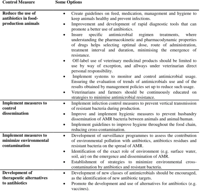
food-producing animals.. ... 61
CHAPTER 2
TABLE 2.1. Primers and annealing temperatures of PCR reactions used to detect antimicrobial resistance genes,
phylogenetic groups and virulence genes. ... 82
TABLE 2.2. Phenotypes of resistance detected among the 192 E. coli isolates recovered from slaughtered
animals. ... 87
TABLE 2.3. Percentages of antimicrobial resistance in the 192 E. coli isolates from faecal samples of
slaughtered animals. ... 88
TABLE 2.4. Genes of antibiotic resistance detected among antimicrobial resistant E. coli isolates of pig, sheep
and cattle origins. ... 89
TABLE 2.5. Phenotypes of resistance and phylogenetic groups detected among the antibiotic-resistant isolates
of E. coli recovered from pigs, sheep and cattle. ... 91
TABLE 2.6. Virulence factors genes detected and its correlation with phylogenetic groups studied in resistant
isolates. ... 91
CHAPTER 3
TABLE 3.1. Characteristics of cefotaxime-resistant Escherichia coli isolates recovered from pigs. ... 105 CHAPTER 4
TABLE 4.1. Characteristics of CTX-resistant E. coli isolates recovered from sheep and beef cattle. ... 115 CHAPTER 5
TABLE 5.1. Primers used in pcr reactions for enterococcal species identification and detection of genes
implicated in antibiotic resistance. ... 122
TABLE 5.2. Enterococcal isolates recovered from the 198 fecal samples of pigs, cattle and sheep analysed in
this study. ... 124
TABLE 5.3. Percentages of antibiotic resistance in 194 enterococci isolated from fecal samples of pigs, cattle
and sheep at slaughter. ... 125
TABLE 5.4. Antibiotic resistance phenotypes detected in the series of 194 enterococci in relation to the species
and origin. ... 128
TABLE 5.5. Antibiotic resistance and resistance genes among enterococcal isolates from pigs, cattle and sheep
at slaughter. ... 129
CHAPTER 6
TABLE 6.1. Characteristics of vancomycin-resistant enterococci isolated from the faeces of beef cattle, sheep
and pigs at slaughter. ... 139
CHAPTER 7
TABLE 7.1. Phenotypic and genotypic characterization of the four stains selected for IEF x SDS-PAGE. ... 149 CHAPTER 8
TABLE 8.1. Number (percentage) of total spots collected, spots identified and spots not identified in strains
TABLE 8.2. Proteins differentially expressed identified by MALDI-TOF/TOF or LC-ESI MS/MS in the MDR
ESBL-producing E. coli, strain SU03, under ciprofloxacin stress. ... 175
CHAPTER 9
TABLE 9.1. Proteins differentially expressed after exposure of VRE Enterococcus faecium to different
vancomycin concentrations, 16 µg/ml (EFVAN16) and 64 µg/ml (EFVAN64). ... 196
SUPPLEMENTS
TABLE A-1. Chromosome- and/or plamid-mediated resistance mechanisms found in different antimicrobial
classes. ... 237
TABLE B.1. Characteristics and MLST results of the vanA-containing enterococci E. faecium strains analyzed. ... 241 TABLE C -1. Proteins identified by MALDI-TOF/TOF in strain SU76. ... 243 TABLE C -2. Proteins identified by MALDI-TOF/TOF in strain SU23. ... 253 TABLE C -3. Proteins identified by MALDI-TOF/TOF in strain SU62. ... 261 TABLE C -4. Proteins identified by MALDI-TOF/TOF in strain SU60. ... 268 TABLE D.1. Number of gels compared in each different condition, without vancomycin (EFVAN0) and with
different vancomycin concentrations, 16 µg/ml (EFVAN16) and 64 µg/ml (EFVAN64). ... 284 TABLE D.2. Spots parameters that showed protein expression variation on progenesis samespots analyses. 284
TABLE OF FIGURES
CHAPTER 1
FIGURE 1.1. Representation of genetic elements involved in the dissemination of antibiotic resistance genes in
response to the selective pressure of antibiotic use. ... 6
FIGURE 1.2. The complex ecosystem of antimicrobial resistant bacteria. ... 8 FIGURE 1.3. Proportion of the total sales of the different veterinary antimicrobial classes, by EU country, for
2011. ... 13
FIGURE 1.4. A - representation of antibiotic mechanisms of action, and B - esquematic representation of
antibiotic resistance mechanisms. ... 19
FIGURE 1.5. Proteomic workflow representing the classical gel-based approach for protein identification. ... 37 CHAPTER 2
FIGURE 2.1. Prevalence of antimicrobial resistance among E. coli isolates... 85 CHAPTER 7
FIGURE 7.1. Distribution of the biological processes related to the protein spots identified found in the 2-DE
gels of the E. coli. strains, respectively, SU76, SU23, SU62 and SU60. ... 149
FIGURE 7.2. monodimensional profile and respective 2-DE gel image obtained in strains. ... 151 CHAPTER 8
GRAPHICAL ABSTRCT ... 155 FIGURE 8.1. SDS-PAGE profiles of proteins from MDR E. coli strains ... 163 FIGURE 8.2. Monodimensional profile and respective 2-DE gel image of proteins from strains SU76, SU23,
SU62 and SU60. ... 164
FIGURE 8.3. Venn diagram highlighting the distribution of identified proteins from strains SU76, SU23, SU62
and SU60. ... 165
FIGURE 8.4. Distribution of the biological processes related to the total protein spots identified in the four E.
coli strains studied. ... 167
FIGURE 8.5. Representative 2-DE protein profile of the total cellular proteins obtained from ESBL
multi-resistant E. coli strain SU03. ... 173
CHAPTER 9
GRAPHICAL ABSTRCT ... 181 FIGURE 9.1. Growth curves obtained from the control culture EFVAN0 and the culture medium contained 16
µg/ml (EFVAN16) or 64 µg/ml (EFVAN64) vancomycin. ... 184 FIGURE 9.2. Representative 2-DE pattern of total cellular E. faecium SU18 strain proteins stained with
coomassie blue. ... 188
FIGURE 9.3. Distribution of biological processes related to functions of identified proteins. ... 189 FIGURE 9.4. Principal component analysis of differentially expressed proteins and full 2-DE protein patterns of
E. faecium SU18 strain. ... 195
GRAPHICAL RESULTS: Escherichia coli AND ESBLS. ... 203 GRAPHICAL RESULTS: Enterococcus SPP. AND VRES. ... 205 GRAPHICAL RESULTS: PROTEOMICS. ... 207 CHAPTER 10
FIGURE 10.1. Percentage of antimicrobial resistance among E. faecium isolates from pigs and cattle, in
different European countries. ... 211
FIGURE 10.2. Percentage of antimicrobial resistance among E. faecallis isolates from pigs and cattle, in
different European countries. ... 211 FIGURE 10.3. Percentages of antimicrobial resistanceamong E. coli isolates from pigs in different countries.
FIGURE 10.4. Percentage of antimicrobial resistance among E. coli isolates from cattle in different countries. ... 213
FIGURE 10.5. Percentages of vancomycin-resistance enterococci reported in Portugal in different settings. . 216
FIGURE 10.6. Percentage ESBL-producing E. coli strains reported in health food-producing animals in
different countries. ... 217
FIGURE 10.7. Global distribution of ESBL enzymes frequently (bolt) detected in E. coli isolates from
food-producing animals. ... 219
SUPPLEMENTS
FIGURE D.1-A Enlarged image of each spot that showed protein expression variation on Progenesis Samespots
analyses. ... 285
FIGURE D.1-B Enlarged image of each spot that showed protein expression variation on Progenesis Samespots
analyses. ... 286
FIGURE D.1-C Enlarged image of each spot that showed protein expression variation on Progenesis Samespots
ABBREVIATIONS
A A - Adenine
AAC - Aminoglycoside Acetyltransferases ACC - Acetyl-CoA carboxylase
ACN - Acetonitrile A.D. - Anno Domini ADI - Arginine Deiminase AK - Amikacin
Ala - Alanine AMP - Ampicillin
AmpC - Cephalosporinases Enzyme AMC - Amoxicillin-Clavulanic Acid AMR - Antimicrobial Resistance ANOVA - Analysis of Variance
ANT - Aminoglycoside
Nucleotidyltransferases
APEC - Avian Pathogenic E. coli APH - Aminoglycoside
Phosphotransferases
API - Analytical Profile Index
AcrAB-TolC - Multidrug efflux system Arg - Arginine
Asn - Asparagine Asp - Aspartic acid ATM - Aztreonam
ATP - Adenosine Triphosphate
ATTC - American Type Culture
Collection
B
bp - base pair BHI - Brain Heart Infusion
C C - Cytosine
C. jejuni - Campylobacter jejuni
CAT - Chloramphenicol
Acetyltransferases
CAZ - Ceftazidime CC - Clonal Complex CFU - Colony-Forming Unit
CHAPS - 3-[(3-cholamidopropyl)
dimethylammonio]-1-propanesulfonate
CHL - Chloramphenicol CK - Carbamate Kinase
CLSI - Clinical and Laboratory Standards
Institute
ClpB - Caseinolytic Peptidase B Protein CIP - Ciprofloxacin
CN - Gentamicin CTX - Cefotaxime
CTX-M - ß-lactamase enzyme type
D Da - Dalton
DAEC - Diffusely Adheren E. coli DANMAP - Danish Integrated
Antimicrobial Resistance Monitoring and Research Programme
Ddl - D-alanine-D-alanine ligase DEC - Diarrhoeagenic E. coli
DGAV - General Food and Veterinary
Directorate (Direção Geral Alimentação Veterinária)
DHFR - Dihydrofolate Reductase DHPS - Dihydropteroate Synthetase DNA - DeoxyriboNucleic Acid DTT - Dithiothreitol
E
E. avium - Enterococcus avium E. casseliflavus- Enterococcus
casseliflavus
E. coli - Escherichia coli
E. durans - Enterococcus durans E. faecalis - Enterococcus faecalis E. faecium - Enterococcus faecium E. gallinarum - Enterococcus gallinarum E. hirae - Enterococcus hirae
E. mundtii - Enterococcus mundtii
EAggEC - EnteroAggregative E. coli EARS-Net - European Antimicrobial
Resistance Surveillance Network
EARSS - European Antimicrobial
Resistance Surveillance System
EC - European Commission Ec-PTZ -
Piperacillin-Tazobactam-Resistant E. coli
EDTA - Ethylenediaminetetraacetic acid EFSA - European Food Safety Authority
EfVAN0 - E. faeciumgrown without the
antibiotic
EfVAN16 - E. faecium grown on 16 µg/ml of
vancomycin
EfVAN64- E. faecium grown on 64 µg/ml of
vancomycin
e.g. - Example
EHEC - EnteroHaemorrhagic E. coli EIEC - EnteroInvasive E. coli Eno - Enolase
ENR - Enrofloxacin
EPEC - EnteroPathogenic E. coli ERY - Erythromycin
ESBL - Extended-Spectrum β-Lactamase ESI - Electrospray Ionization
ESVAC - European Surveillance of
Veterinary Antimicrobial Consumption
EU - European Union
ETEC - EnteroToxigenic E. coli
ExPEC - Extraintestinal Pathogenic E. coli
F FA - Formic Acid
FASP - Filter Aided Sample Preparation FBPA - Fructose-Bisphosphate Aldolase
class 2
FCT - Portuguese Foundation for Science
and Technology (Fundação para a Ciência e a Tecnologia)
FDR - False Positive Rate
FKBPs - FK506-binding Proteins FOX - Cefoxitin G g - Gram g - G-force G - Guanine GABA - Gamma-Aminobutyrate GAD - Glutamate decarboxylase Gap - Glyceraldehyde-3-phosphate
Dehydrogenase
GAPDH-A - Glyceraldehyde-3-Phosphate
Dehydrogenase A
GDP - Guanosine Diphosphate. Glu - Glutamic acid
GMP - Guanine Monophosphate
GRE - Glycopeptide-resistant enterococci GroEL - Molecular Chaperone
H h - hour
Hbp - Hemoglobin-binding protease HCl - Hydrochloric acid
HGT - Horizontal Gene Transfer HiRECC - High-Risk Enterococcal
Complex
His - Histidine
HLR - High-Level Resistance HLR-G - High-Level Resistance for
Gentamicin
HLR-S - High-Level Resistance for
Streptomycin
HLR-K - High-Level Resistance for
Kanamycin
HRA1 - Heat Resistant Agglutinin 1 HPLC - High-Performance Liquid
Chromatography
HtpB - Heat Shock Protein
I ICAT - Isotope-coded Affinity Tags
ID - Identification
IEF - Isoelectric Focusing Ile - Isoleucine
Inc. - Incorporation
IPG - Immobiline™ pH Gradient
IPM - Imipenem IS - Insertion Sequence
iTRAQ - Isobaric Tags for Relative and
Absolute Quantitation
K
K. pneumoniae - Klebsiella pneumoniae
KAN - Kanamycin Kbar - Kilobar
KCl - Potassium Chloride kDa - Kilo Dalton
L L - Litre Lac - Lactate LC - Liquid Chromatography Leu - Leucine Log - Logarithm
LuxS - S-ribosylhomocysteine lyase Lys - Lysine
M m - Minute
M - Molar
mA - Milliampere
MalE - Maltose-binding Periplasmic
Protein
MALDI - Matrix-Assisted Laser
Ionization
MARAN - Monitoring of Antimicrobial
Resistance and Antibiotic use in Animals in the Netherlands
MBLs - Metallo-β-lactamases
MCTES - National Funds of Ministry of
Science and Technology for High Education
MDR - Multidrug Resistance
MFS - Major Facilitator Superfamily mg - Milligram
MgCl2 - Magnesium Chloride
MIC - Minimum Inhibitory Concentration min - Minute
mL - Millilitre
MLS - Macrolides Lincosamides
Streptogramins
MLSB - Macrolides Lincosamides
Streptogramin B
MLST - MultiLocus Sequence Typing mM - Millimolar
mRNA - Messenger RNA
MRSA - Methicillin-Resistant
Staphylococcus aureus
MS - Mass Spectrometry
MS/MS - Tandem Mass Spectrometry MSCRAMM - Microbial Surface
Components Recognizing Adhesive Matrix Molecules
MW - Molecular Weight
N n - Natural numbers
N. meningitides - Neisseria meningitidis
NaCl - Sodium Chloride NA - Nalidixic acid
NADH - Enoyl-[acyl-carrier-protein]
reductase
NCBI - National Center for Biotechnology
Information USA
NDA - Nicotinamide Adenine
Dinucleotide
NDM - New Delhi metallo-β-lactamase ng - Nanogram
NL - Non-Linear
Nl/min - Normal Liters per Minute NMEC - Neonatal Meningitis E. coli nm - Nanometre
No. - Number
O OD - Optical Density
Odp1 - pyruvate dehydrogenase E1
component
OIE - World Organisation for Animal
Health
OM - Outer Membrane
OMPs - Outer Membrane Protein Orf - Open Reading Frame
OTC - Carbamoyltransferase OXA - ß-lactamase Enzyme type
P
P. aeruginosa - Pseudomonas aeruginosa
PAGE - Polyacrylamide Gel
Electrophoresis
PAI - Pathogenicity Islands
PANRUAA – “National action plan for
reducing the use of antibiotics in animals”
PBP - Penicillin-Binding Protein PBS - Phosphate-Buffered Saline PCA - Principal Component Analysis PCR - Polymerase Chain Reaction PFGE - Pulsed-Field Gel Electrophoresis PhD - Doctor of Philosophy
pI - Isoelectric point PK-1 - Pyruvate kinase I
PMF - Peptide Mass Fingerprints PMQR - Plasmid-Mediated Quinolone
Resistance
PMSF - Phenylmethylsulfenylfluoride PPIases - Peptidyl-Prolyl
cis/trans-Isomerases
ppm -Part Per Million
PRPs - Penicillinase-Resistant Penicillins PTC - Peptidyl Transferase Center PTM - Post-Translational Modification
Q QD - Quinupristin-Dalfopristin
qPCR - Quantitative PCR QRDR - Quinolone Resistance
Determining Region
Qnr - Quinolone resistance protein QS - Quorum Sensing
R R&D - Research and Development
RNA - RiboNucleic Acid
RND - Resistance-Nodulation-cell
Division
rRNA - Ribosomal RNA
S s - Second
S. enterica - Salmonella enterica S. aureus - Staphylococcus aureus S. pneumoniae - Streptococcus
pneumoniae
SCX - Strong Cation Exchange SDS - Sodium Dodecyl Sulfate
SDS-PAGE - Sodium Dodecyl Sulfate
Polyacrylamide Gel Electrophoresis
Ser - Serine
SLV - Single Locus Variant SHV - ß-lactamase Enzyme type
siRNA - Small interfering RNA SOD - Superoxide Dismutase Protein SspA - Stringent starvation protein A ST - Sequence Type
STEC - Shiga toxin-producing E. coli STR - Streptomycin
SUL - Sulfonamide
SXT - Sulfamethoxazole-Trimethoprim SU - Pig
SVARM - Swedish Veterinary
Antimicrobial Resistance Monitoring
T T - Thymine
TCA - Trichloroacetic Acid TEC - Teicoplanin
TET - Tetracycline
TEM - ß-lactamase-type Enzymes TFA - Trifluoroacetic Acid
TM
- Trademark TMP - Trimethoprim
Tn - Transposon TOB - Tobramycin
TolC - Outer membrane channel Tris-HCl - Tris-Hydrochloride
Tris - Tris (hydroxymethyl) aminomethane tRNA - Transfer RNA
Trp - Tryptophan Tyr - Tyrosine
U U - Unit
UK - United Kingdom
UMP - Uridine Monophosphate Uniprot - Universal Protein Resource
UPEC - UroPathogenic E. coli U.S. - United States
USA - United States of America UTI - Urinary Tract Infection
V V - Volt
Vh - Volt/Hour v/v - Volume/Volume VAN - Vancomycin
VRE - Vancomycin-Resistant Enterococci VREar - VRE with acquired mechanisms
of resistance
VREir - VRE with intrinsic mechanisms
of resistance
VREfm - Vancomycin Resistant E.
faecium
VTEC - Vero cytotoxin producing E. coli
W
w/v - Weight/Volume WHO - World Health Organization
OTHERS μg - microgram μl - Microlitre μm - Micrometre 1st - First 2nd - Second 3rd - Third 4th - Fourth
2-DGE - Two-Dimensional Gel
Electrophoresis
® - Registered trademark % - Percentage
ºC - Degree Celsius $ - Dollars
GENERAL CONSIDERATIONS
SECTION I
Antimicrobial Resistance in Food-producing Animals: a Public Health Problem.
CHAPTER 1
RESEARCH FOCUS
CHAPTER 1:
ANTIMICROBIAL RESISTANCE IN FOOD-PRODUCING ANIMALS: A PUBLIC HEALTH PROBLEM
1. INTRODUCTION
The discovery and use of antibiotics was a life-saving turning point in human history. The availability of antibiotic chemotherapy deeply changed society by prolonging life expectancy, and allowing rapid population growth through both a reduction in infant mortality, and the ability to treat common infectious diseases (Gonzalez-Zorn and Escudero, 2012). Antibiotics became very important for both human and animal health. For humans the use of antibiotherapy is now critical to the success of complex surgery (such as organ transplants), in intensive care units and for the survival of immunosuppressed patients; while in animals, antibiotics are broadly used in food-production animals (Acar and Moulin, 2012).
Meanwhile, the development of antimicrobial resistance (AMR) was a concomitant fact. Recent evidence strongly supports the idea that many types of resistance mechanisms and resistant bacteria were present long before the production and use of antibiotics (Andersson and Hughes, 2012; Acar and Moulin, 2012). Indeed, resistance mechanisms, could have been originated from pre-therapeutic use of antibiotics, in non-pathogenic environmental bacteria that are antibiotic producers, they always needed to be resistant to their own antibiotic (Huttner et al., 2013). However, it was believed that bacterial mutation frequencies were low enough to allow antimicrobial resistance to be largely ignored for decades.
Notwithstanding this, warnings on AMR are not new. Well before antimicrobials became widely available, Alexander Fleming warned in his Nobel Prize acceptance speech: “it is not difficult to make microbes resistant to penicillin in the laboratory by exposing them to concentrations not sufficient to kill them” (Huttner et al., 2013).
The history of AMR development shows that for every antimicrobial therapeutic compound used, resistance will develop. The scale of AMR changed rapidly, and became an issue with great impact on human life and global economy. Nowadays, the overall picture of infectious diseases treatment is more complicated and dramatically less optimistic. The term “superbug” is now commonly used in reference to microbes with enhanced morbidity and mortality, endowed with high levels of resistance to the antibiotic classes that are specifically recommended for their treatment (Davies and Davies, 2010). Antimicrobial drug resistance is
now considered a major public health problem, not limited to industrialized nations but occurring everywhere in the world, that should be taken seriously by all, veterinary and health sectors, and even the general community.
2. DEVELOPMENT AND DISSEMINATION OF ANTIMICROBIAL RESISTANCE
By definition, antimicrobial resistance enables microrganisms to grow in the presence of a chemical (drug) that would normally kill them or limit their growth. It is a natural phenomenon and a complex issue, linked to the ability of bacteria to adapt quickly to their environment (Acar and Moulin, 2012). As a matter of fact, in the presence of a therapeutic compound bacteria have the remarkable ability to adapt, evolve and survive by developing resistance mechanisms (Acar et al., 2012). Additionally, antimicrobials have been used so widely and for so long that the infectious organisms that they are designed to kill have adapted to them, making these drugs less effective. Currently, the pressure on the use of antimicrobials is considered the key promoter of antimicrobial resistance (Acar and Moulin, 2012; da Costa et al., 2013; Huttner et al., 2013). Moreover, the use and increased consumption of antimicrobial agents in modern animal husbandry, and human medical practices creates special conditions for selection, spread and evolution of resistant strains (da Costa et al., 2013).
Bacteria develop resistance mechanisms either through spontaneous mutations, or through the acquisition of exogenous resistance determinants from other bacterial strains, by horizontal gene transfer (HGT) (da Costa et al., 2013; Martinez and Baquero, 2009). HGT is considered the most important mechanism in the dispersal of antibiotic resistance genes, providing the means by which resistance genes, such as blaCTX-M, spread amongst different
bacterial strains and species (Hawkey and Jones, 2009). There are three fundamental mechanisms by which acquisition of resistance genes occur through HGT: conjugation, transformation and transduction. Nevertheless, conjugation and mobile genetic elements, are considered the major players in HGT (van Hoek et al., 2011).
By HGT, substantial amounts of deoxyribonucleic acid (DNA) can be introduced into or deleted from the chromosome, producing extremely dynamic genomes (Schubert et al., 2009). Some bacteria, but particularly Gram-negative, are adapted to exchange genetic information. In these organisms antibiotic resistance derives from the acquisition of genes from a shared pool (Partridge, 2011). Genes from this pool are not intrinsically mobile and appear to have
been captured from the chromosomes of different bacterial species. Different types of mobile genetic elements can be involved in the capture of such gene: those able to transfer genes between DNA molecules, for example insertion sequences (IS), gene cassettes, integrons and transposons; and those able to transfer genes between cells, for example, conjugative and mobilizable plasmids, and integrative and conjugative elements (ICEs) (Partridge, 2011).
Integrons are not themselves mobile genetic elements, they include components of a site-specific recombination system enabling them to capture and mobilize genes of resistance (Davies and Davies, 2010; van Hoek et al., 2011). In turn, transposons are unique mobile genetic elements with the ability to excising themselves from one genetic locus to another, whether it is within the same bacteria or to bacteria from other taxa (Roe and Pillai, 2003). Plasmid-mediated transmission is by far the most common mechanism of HGT. Plasmids are bacterial extra-chromosomal elements, notorious for their ability to transfer genes, by conjugation, between different bacterial species. In addition, they can carry genes conferring resistance to one or more antibiotic classes, being significantly involved in the emergence and dissemination of multiple drug resistance associated with bacterial infections in humans (Clewell, 2014). Bacteriophages, also play a role in the spread of DNA between bacteria by transduction (van Hoek et al., 2011); however, bacteriophages with antibiotic genes of resistance have been rarely identified in resistant bacteria isolates weither, from the environment or from hospital settings (Davies and Davies, 2010).
The antimicrobial resistance gene pool has never been so accessible, nor its selection pressure so strong (Huttner et al., 2013). All potential sources of DNA encoding antibiotic resistance determinants in the environment (including hospitals, farms and other places where antibiotics are used to control bacterial development) are included in this gene pool. Apart from their incorporation in a gene transfer unit capable to replicate in microbial pathogens, which is fundamental for their dissemination, the gene pool can suffer further diversification as a consequence of strong selective pressure by antibiotic use (Martinez and Baquero, 2009). Figure 1.1. shows a schematic representation of genetic elements involved in the dissemination of antibiotic resistance genes in response to the selective pressure of antibiotic use.
FIGURE 1.1. Representation of genetic elements involved in the dissemination of antibiotic
resistance genes in response to the selective pressure of antibiotic use. (a) The gene pool includes all potential sources of DNA encoding antibiotic resistance determinants in the environment (including hospitals, farms and other places where antibiotics are used to control bacterial development). (b) HGT contributes to the spread of antibiotic resistance gene amongst different bacterial species and strains. Further, mobile genetic elements are involved in the captured and dissemination of resistance genes (c) which can be captured by integrons (d) that can be inserted in a transposon (e), which can be part of a plasmid (f). The plasmid can be transferred to a bacteria, where interactions between the chromosome and the plasmid might occur, or it might be transferred by conjugation between different bacterial species.
The cumulative collection of antibiotic resistance traits within these multimodular structures (integrons, transposons and plasmids), which, by turn, act as single transmissible modules, result in multidrug-resistant strains (Martinez and Baquero, 2009). In the past, and because the acquisition of resistance genes was associated with a fitness cost to the microorganism, it was assumed that these multidrug-resistant bacteria would be unstable (Acar and Moulin, 2012; Davies and Davies, 2010; Martinez and Baquero, 2009). However, recently it was suggested that resistant bacteria can develop compensatory mutations and survive successfully. Moreover, they are able to acquire even more resistance genes and mutations (Acar and Moulin, 2012; Davies and Davies, 2010).
3. A PUBLIC HEALTH CONCERN
3.1. THE COMPLEX ECOSYSTEM OF ANTIMICROBIAL RESISTANT BACTERIA
It stands to reason that antimicrobial usages, and resistance, are a multifactorial problem. After decades of research, from the paucity of new antimicrobials, to the inefficient contingency plans to reduce the use of antimicrobials, numerous difficulties in tackling resistance have emerged (Gonzalez-Zorn and Escudero, 2012).
There is now the overwhelming evidence that antibiotic use in both human and veterinary medicine is a powerful selector for resistance, which can appear not only at the point of origin, but also nearly everywhere else. Nowadays, there is an increasing concern that the pervasive use of antimicrobials in farming and the general widespread antimicrobial contamination of the environment may be indirectly implicated in this phenomenom. Therefore, when we discuss about antimicrobial resistance, two major drivers should be taken into account: firstly, the use of antimicrobials, which exerts an ecological pressure on microorganisms and contributes to the emergence and selection of antimicrobial-resistant microorganisms in populations; and secondly, the spread and cross-transmission of resistant microorganisms, and resistant genes, between humans, animals, and the environment. In figure 1.2. a diagram illustrating different antimicrobial uses, and the possible transmission pathways of antimicrobial resistance among different habitats is represented. To understand how antibiotic use might impact on the emergence and dissemination of antibiotic resistance, it is essential to consider the complex interaction of different elements: the physical environment (e.g., farms, crops, air, soil, and water), social exchanges (e.g., between animals within a herd, farmers and animals, domestic and wild animals, between clinical settings and the community), in food processing chain (e.g., farming activities, food preparation, transportation and storage), and in human use patterns (e.g., meat consumption habits and susceptibility to infection).
The use of antibiotics in human and veterinary medicine may have consequences beyond their intended applications. Antimicrobial molecules are not restricted to treated patients, either animals or humans, but rather circulate throughout the whole ecosystem. From a public health point of view, the concern that bacteria will become resistant during antibiotic treatment can be negligible, compared to the impact of the dissemination of resistant bacteria in the environment (Gonzalez-Zorn and Escudero, 2012). Antibiotic use in food-producing
animals, including nontherapeutic use, is responsible for a significant proportion of antibiotic consumption (Landers et al., 2012), however, their use extends to improve agriculture production and aquaculture (Wang et al., 2012). There are a multitude of routes that can contribute to the pathways by which antibiotic use in food-producing animals, or at agriculture level, can directly or indirectly, pose risks to human health (Landers et al., 2012; Marshall and Levy, 2011).
FIGURE 1.2. The complex ecosystem of antimicrobial resistant bacteria. The diagram illustrates different
antimicrobial applications (the size of the blue arrows is proportional to antimicrobial use) and potential routes by which resistant bacteria, and resistant genes can spread between animals, humans and in the environment.
The potential threat to human health posed by animals harbouring resistant bacteria is significant. The isolation of resistant bacteria from food-producing animals is frequent (Cortes et al., 2010; Drugdová and Kmet, 2013; Jiang et al., 2011; Karczmarczyk et al., 2011; Lay et al., 2012; Ozaki et al., 2011; Ramos et al., 2013; Wasyl et al., 2013). Furthermore, several reports have documented the presence of resistant-bacteria in pets (Costa et al., 2008; Harada et al., 2012; Leonard et al., 2012) which can be a potential source of resistant threats to their human co-habitants (da Costa et al., 2013). However, human-to-animal transmission may be equally as relevant as animal-to-human transmission. In this scenario, farm and
slaughterhouse workers, Veterinarians, and those in close contact with farm work are directly at risk of being colonized or infected with resistant bacteria through close animal contact (Marshall and Levy, 2011). In addition, human-to-human transmission by occupational workers, and their families, provide a conduit for the entry of resistance genes into the community and hospital environments, where further spread ofto pathogens is possible (Marshall and Levy, 2011). Notwithstanding this, where it was once believed that hospital and other health-care facilities were responsible for antibiotic resistance, community-associated resistant strains have also been implicated as a cause of hospital-acquired infections (Park et al., 2009; Qu et al., 2014).
The environment can be considered the melting pot of antimicrobial resistance. Evidence of potential environmental risk pathways shows that antimicrobial resistant bacteria, and resistant genes, are frequently found in the farm environment (Garcia-Migura et al., 2005; Hartmann et al., 2012; Karczmarczyk et al., 2011; Nilsson et al., 2009; Tamang et al., 2013; Watson et al., 2012), including animal manure and sewage (Furtula et al., 2010; Holzel et al., 2010; Hutchison et al., 2005; Klein et al., 2010). The detection of antimicrobial resistant bacteria, and resistance genes, in watercourses, or in manure and slurry, directly applied to arable land as a fertiliser, enhance the cross-contamination of crops grown for human food or for animal feeds. Moreover, effluents from urban areas and animal production units (husbandry and slaughter houses), even when treated in wastewater treatment plants, can discharge resistant bacteria into the receiving surface waters (Araujo et al., 2010; Dungan et al., 2012; Freitas et al., 2009b). Even watercourses not closely associated with the farm environment have been shown to contain potentially resistant bacteria (Abgottspon et al., 2014; Cox et al., 2005; Macedo et al., 2011; Machado et al., 2009; Wooldridge, 2012).In addition, the dispersion of antibiotics into soil and water enhances the risk of breaking natural barriers, and currently it is estimated that 75% to 90% of antibiotics used in humans and food-producing animals are excreted and accumulated in the environment, largely unmetabolized (Andersson and Hughes, 2012; Marshall and Levy, 2011) and some molecules such as fluoroquinolones have long half-lives (Gonzalez-Zorn and Escudero, 2012).
In a wider context, the presence of multidrug-resistant bacteria is frequently reported in wild birds and mammals with no apparent exposure to antimicrobials (Costa et al., 2006; Figueiredo et al., 2009; Goncalves et al., 2013a; Goncalves et al., 2011; Goncalves et al., 2013b; Marinho et al., 2013; Pinto et al., 2010; Poeta et al., 2009; Radhouani et al., 2013a;
Radhouani et al., 2013b; Radhouani et al., 2009; Santos et al., 2013b), indirectly, these may also drive from/into an environmental contamination source.
Ultimately, antibiotic resistance does not respect geographical or biological borders; food animals and foods from animal origin are traded worldwide facilitating the spread of resistance. An extensive trade of animals occurs within the European Union (EU), thus, the occurrence of antimicrobial resistance in one country is a problem for all countries (Ahmed et al., 2009).
3.2. ANTIMICROBIAL USE IN FOOD-PRODUCING ANIMALS
In veterinary medicine, antibiotics are used not only to treat food animals but also to prevent them from developing diseases and to enhance animal growth (Landers et al., 2012; Marshall and Levy, 2011). Today, it is almost impossible to imagine veterinary medicine without the use of antimicrobials; they have had an enormous impact on the health and welfare of animal species. Moreover, the increasing demand for livestock products has prompted to developments in breeding, nutrition and management practices, where frequently large populations of livestock are breed at a single site, and often in the same airspace. Consequently, the use of antimicrobial agents allows the growth of healthier and more productive animals, with lower incidence of disease, reduced morbidity and mortality, leading to the production of abundant quantities of nutritious, high-quality and low-cost food for human consumption (Pagel and Gautier, 2012).
Nowadays, it is well recognised that the selective pressure exerted by antibiotic use has a major role in the emergence and amplification of antimicrobial resistant pathogens (Wang et al., 2012). In several studies, the association between antibiotic use in food-producing animals and the prevalence of antibiotic resistant bacteria isolated from those animals was established. In a study by Cavaco et al (2008), after pigs were treated with amoxicillin, ceftiofur, or cefquinome, higher counts of CTX-resistant coliforms were observed; moreover, ceftiofur and cefquinome exerted larger selective effects than amoxicillin, providing evidence that cephalosporins used in pig production select for CTX-M-producing Escherichia coli strains (Cavaco et al., 2008). Bibbal et al (2009) studied the development of resistance in faecal E.
coli populations during the pigs` treatment with ampicillin, results showed that
ampicillin-resistant isolates, present in the digestive tract before any treatment, were selected and one specific pulsed field gel electrophoresis (PFGE) genotype, encoding resistance to six antibiotics, became predominant (Bibbal et al., 2009). In a randomized, controlled, blinded
clinical trial, oxytetracycline was included in the starter ration or administered subcutaneously to feedlot cattle, concluding that before slaughter, there were significantly more animals with tetracycline-resistant E. coli isolates (Checkley et al., 2010).
The growth promoter effect of antibiotics was discovered in the 1940s, when it was observed that animals fed dried mycelia of Streptomyces aureofaciens containing chlortetracycline residues improved their growth (Castanon, 2007). The use of antibiotics to promote growth implies that they are administered in low doses over long periods to large groups of healthy animals. This practice has continued since the 1950s, while the positive effects stemmed from antibiotics use to promote growth were championed, and the negative consequences went undetected. However, as researchers linked it to antibiotic resistance in humans and animal, this practice became increasingly under scrutiny (Marshall and Levy, 2011). Concerns about antimicrobial resistance development and transference of antibiotic resistance genes from animal to human microbiota, led to bans on the use of antibiotics as growth promoters in the EU, first Sweden (1986), then Denmark (1998) and the United Kingdom (1999) (da Costa et al., 2013). In 1997 the EU banned avoparcin for all uses in agriculture, and in 1999 banned antibiotics used in human medicine from being added as animal feed additives. Thereby imposing a ban on tylosin, spiramycin, virginiamycin, and bacitracin (Cogliani et al., 2011). Further, the Feed Additives Regulation (EC) No 1831/2003 completed these measures with the total ban on antibiotics as growth promoters from 1st January 2006 and, further, prohibited anticoccidial substances, such as antibiotics ionophores, as feed additives by 31 December 2012 (EC, 2003). From 2013, all medical substances for use in food-producing animals will be limited to therapeutic use, and only by Veterinary prescription.
With these measures the EU intended to lead the world in reducing antibiotic use in healthy animals. These strong top-down approaches to regulate nontherapeutic use of antibiotics in animals go towards a reduction of the amount of antibiotics used in animal production, and consequently, a reduction of the resistant gene pool in animals was expected. Some of these bans were closely monitored from the outset, and in countries like Sweden, Denmark and the United Kingdom an overall decline in sales of antibiotics for animals with major reductions in vancomycin-resistant Enterococcus faecium were observed (Cogliani et al., 2011). On the other hand, immediately following the ban, it was also reported an increase in total therapeutic antibiotic consumption to control specific infectious outbreaks (Cogliani et al., 2011). Nevertheless, no lasting negative effects were detected on mortality rates, average