DEVELOPMENT OF AN INJECTABLE MODIFIED
CAL-CIUM PHOSPHATE (BONELIKE)-DEXTRIN HYDROGEL
BIOMATERIAL FOR BONE REGENERATION
ANTERO JESUS DA SILVA OLIVEIRA
DISSERTAÇÃO DE MESTRADO APRESENTADAÀ FACULDADE DE ENGENHARIA DA UNIVERSIDADE DO PORTO EM MESTRADO EM ENGENHARIA BIOMÉDICA
Faculty of Engineering of the University of Porto
Development of an injectable modified calcium
phosphate (Bonelike)-dextrin hydrogel biomaterial
for bone regeneration
Antero Oliveira
Dissertation carried out under the
Master in Biomedical Engineering
Supervisor: Prof. Dr. José Domingos Santos
Co-supervisor: Prof. Dr. Miguel Gama
i
Resumo
O desenvolvimento de biomateriais compósitos pode tirar partido de sinergias entre as proprieda-des benéficas de dois ou mais materiais numa nova e sofisticada matriz, uma vez que poucos biomateriais possuem todas as características necessárias para apresentarem um desempenho ideal. O osso é um exemplo perfeito de um material compósito criado pela natureza.
O principal objetivo deste estudo foi o de produzir um biomaterial compósito para a regeneração do tecido ósseo. O material desenvolvido é composto por uma hidroxiapatite reforçada com biovi-dro (Bonelike), que já demonstrou alta bioactividade e boa osteointegração, e um hibiovi-drogel biode-gradável, feito a partir de dextrino oxidado (hidrogel oDex). O hidrogel irá funcionar como veículo injectável para os grânulos do Bonelike.
Para melhorar as características do injectável, o sistema foi estudado e modificado para actuar como um transportador de fármaco, nomeadamente de simvastatina, que tem reportado a capa-cidade de promover actividade osteoblástica e inibir a actividade osteoclástica. Primeiramente, o Bonelike foi testado como um transportador para a simvastatina e apresentou uma libertação lenta e gradual do fármaco. Em seguida, tirando partido das capacidades da nanotecnologia no campo da administração de fármacos, nanopartículas de dextrino foram utilizadas para melhorar a solu-bilização da simvastatina e controlar o seu perfil de libertação. As nanopartículas carregadas com simvastatina, sob as condições testadas, formam uma solução estável que liberta o fármaco em 24 horas, e revelaram de um modo geral boa biocompatibilidade.
O novo biomaterial injetável compósito para aplicações em regeneração óssea apresenta boas ca-racterísticas de injectabilidade e propriedades promissoras como um veículo injectável para a simvastatina.
ii
iii
Abstract
Given that few biomaterials possess all the required characteristics to perform ideally, the devel-opment of composite biomaterials can synergize the beneficial properties of two or more materials into an improved new matrix. Bone is a perfect example of a composite material designed by nature.
The primary purpose of this study was to produce a composite biomaterial for bone tissue regen-eration. The developed material is composed by a glass reinforced hydroxyapatite (Bonelike), that have already demonstrated high bioactivity plus good osteointegration, and a biodegradable hy-drogel, made from oxidized dextrin (oDex hydrogel). The oDex hydrogel will perform as an inject-able carrier of the Bonelike granules.
To enhance the characteristics of the injectable, the system was studied and modified to act as a drug carrier of simvastatin, a drug that has been reported to promote osteoblastic activity and inhibit osteoclastic activity. At first, Bonelike was tested as a carrier for simvastatin and showed a slow and gradual release of the drug over 2 weeks. Then, taking advantage of nanotechnology in the drug delivery field, dextrin nanoparticles were used for improving simvastatin solubilisation and controlling its release profile. The simvastatin loaded nanoparticles, under the conditions tested, form a stable solution that release its content in a 24 hours time frame and revealed an overall good biocompatibility.
The new injectable composite biomaterial for bone regeneration present good extrusion charac-teristics and promising properties as an injectable carrier of simvastatin.
iv
v
Acknowledgments
I want to start by thanking my supervisors, for their guidance and availability. I also wanna thanks Professor Ana Colette and all Biosckin team, especially Dina.
A very special thanks to all the people in the DEB for all the support: Catarina, Paula, Isabel, Al-berto, Ana Cristina, Tânia, Karol, Ana and Alexandra.
A special thanks to my housemates, Ivana and Mara, and all my friends. Finally, my deepest gratitude to my parents and grandparents.
vi
vii
Contents
Chapter 1
... 1Motivation and main goals
... 1Chapter 2
... 3State of the art
... 32.1 Bone ... 3
2.1.1 Structure ... 3
2.1.2 Extracellular matrix and bone cells ... 4
2.1.3 Bone remodeling and repair ... 5
2.2 Bone grafts ... 6
2.2.1 Introduction ... 6
2.2.2 Bone grafts... 7
2.2.3 Synthetic materials... 8
2.2.3.1 Calcium phosphate-based materials ... 8
2.2.3.2 Bioactive glasses and glass-ceramics ... 8
2.2.3.3 Glass reinforced apatite ... 9
2.2.4 Injectable bone graft substitutes ... 10
2.3 Hydrogels ... 11
2.3.1 Introduction ... 11
2.3.2 Dextrin hydrogel ... 12
2.3.3 Dextrin nanogel ... 14
2.4 Simvastatin in bone regeneration ... 14
Chapter 3
... 19Materials and methods
... 193.1 Glass reinforced hydroxyapatite (Bonelike) preparation ... 19
3.2 Preparation of oDex Hydrogel ... 19
Dextrin Oxidation: ... 19
Preparation of oDex-ADH Hydrogels: ... 20
3.3 Preparation of self-assembled nanoparticles of dextrin substituted with hexadecanethiol ... 20
viii
Synthesis of dexC16: ... 20
Sample preparation: ... 20
Dynamic light scattering:... 20
1H NMR: ... 20
3.4 Preparation of the oDex-Nanogel Hydrogels and Bonelike-oDex-Nanogel Hydrogels ... 20
3.5 Cryo-Scanning Electron Microscopy (Cryo-SEM) Analysis ... 21
3.6 Injectability tests ... 21
3.7 Preparation of simvastatin-loaded Bonelike scaffolds and in vitro assay of simvastatin release ... 21
3.8 Incorporation of simvastatin in the nanogel ... 22
3.9 In vitro assay of simvastatin release from the nanogel ... 22
3.10 Materials sterilization ... 22
3.11 MTT assay ... 22
3.12 In vivo tests ... 23
Chapter 4
... 25Results and Discussion
... 254.1 Glass reinforced hydroxyapatite (Bonelike) preparation ... 25
4.2 Preparation of oDex Hydrogel ... 28
4.3 Preparation of self-assembled nanoparticles of dextrin substituted with hexadecanethiol ... 28
4.4 Preparation of the oDex-Nanogel Hydrogels and Bonelike-oDex-Nanogel Hydrogels ... 30
4.5 Cryo-Scanning Electron Microscopy (Cryo-SEM) Analysis ... 32
4.6 Injectability tests ... 36
... 36
4.7 Preparation of simvastatin-loaded Bonelike scaffolds and in vitro assay of simvastatin release ... 37
4.8 Incorporation of simvastatin in the nanogel ... 39
4.9 In vitro assay of simvastatin release from the nanogel ... 40
4.10 Materials sterilization ... 41
4.11 MTT assay ... 41
4.12 In vivo tests ... 43
Chapter 5
... 44Conclusions and Perspectives
... 44ix
Lists of figures
Figure 2.1 - Cortical bone and trabecular bone. 4
Figure 2.2 - Bone remodeling process. 5
Figure 2.3 - Process of fracture healing: (a) inflammation, (b) soft callus, (c) hard callus and (d)
remodel-ling. 6
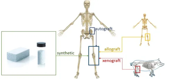
Figure 2.4 - Various types of bone graft sources. 7
Figure 2.5 - Structure of Dextrin. 13
Figure 2.6 - Periodate Oxidation of Dextrin, Yielding Two Aldehyde Groups at Positions C2 and C3 of a
D-Glucose Unit. 13
Figure 2.7 - Structure of simvastatin. 15
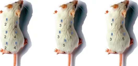
Figure 3.1 - Implant scheme: (1) oDex-nanogel/SIMV hydrogel; (2) oDex-nanogel/SIMV hydrogel + MSCs; (3)
oDex-nanogel hydrogel; (4) oDex-nanogel hydrogel + MSCs; (5) Bonelike/SIMV-oDex hydrogel. 23
Figure 4.1 - X-ray diffraction pattern of Bonelike, which is composed of hydroxyapatite (HAp), α-TCP (α)
and β-TCP (β). 26
Figure 4.2 - Glass reinforced hydroxyapatite (Bonelike) with a particle size from 250 to 500 µM; (A)
macro-scopic aspect of Bonelike granules and (B) SEM image showing the surface morphology. 27
Figure 4.3 - 1H NMR spectra of (A) dextrin and (B) dextrin-VMA (DS
VMA 24%) in D2O at 25ºC. 29
Figure 4.4 - 1H NMR spectra in D
2O of dextrin-VMA reacted with hexadecanethiol with TEA. 30
Figure 4.5 - Macroscopic aspect of (A) Bonelike oDex-nanogel hydrogel, with 10mg/mL of dextrin
nanopar-ticles and (B) Bonelike oDex hydrogel. 31
Figure 4.6 - Bonelike oDex-nanogel hydrogel. 31
Figure 4.7 - (A) Cross-section of oDex hydrogel and (B) detail of the porous structure. 33 Figure 4.8- Cryo-SEM images from cross-section of (A) Bonelike granules completely embedded in the
hy-drogel matrix and (B) detail of the porous structure of the hyhy-drogel inside Bonelike. 34
Figure 4.9 – Cryo-SEM images from (A) dextrin nanoparticles and (B) dextrin nanoparticles inside the oDex
x
Figure 4.10 - Extrusion profiles for the different injectables. 36
Figure 4.11 - Cumulative in vitro release of simvastatin from Bonelike. 38
Figure 4.12 - Simvastatin loading into dextrin nanoparticles of different formulations varying the
simvas-tatin (5 or 10 mg/mL), the nanoparticles (1 or 3 mg/mL) and time (3 or 24 hours). 39
Figure 4.13 - In vitro simvastatin release from dextrin nanoparticles (1mg/mL). 40
Figure 4.14 - Cells metabolic activity for different concentration of nanoparticles (0.75, 1 and 2 mg/mL),
nanoparticles loaded with simvastatin and free simvastatin, assessed by MTT assay. Results presented as average ± SD (n=3). Statistical analysis were performed using a 2-way ANOVA. The comparison between diferent time points and t0 control are represented as n.s.1: non significant, p>0.05; *p<0.05; ***p<0.001.
n.s.2: non significant, p>0.05; # p<0.05; ### p<0.001, represents each condition compared with the control
xi
Lists of tables
Table 2.1 - Injectable bone grafts commercially available. 10
Table 2.2 - Carriers and animal models used for the local application of simvastatin. 16
Table 4.1 - Quantitative chemical analysis of the hydroxyapatite. 25
Table 4.2 - Quantification of crystalline phases by X-ray diffraction of the hydroxyapatite. 25
xii
xiii
Lists of abbreviations
ADH Adipic acid dihydrazide BMP Bone morphogenetic protein DBM Demineralized bone matrix DCP Dicalcium phosphate
DexC16 Hydrophobically modified dextrin Dex-HEMA Dextrinhydroxyethylmethacrylate Dex-VA Dextrin-vinyl acrylate
DLS Dynamic light scattering
DMEM Dulbecco´s modified Eagle´s medium DO Degree of oxidation
DS Degree of substitution
DSC16 Degree of substitution of dextrin with alkyl chains ECM Extracellular matrix
HAp Hydroxyapatite
MSCs Mesenchymal stem cells
MTT 3-(4,5-dimethylthiazol-2-yl)-2,5-diphenyltetrazolium bromide NMR Nuclear magnetic resonance
oDex Oxidized dextrin
PBS Phosphate buffered saline PLGA Poly(lactic-co-glycolic acid) SIM Simvastatin
TCP Tricalcium phosphate TEA Triethylamine
xiv
1
Chapter 1
Motivation and main goals
Biomaterials degree of sophistication has increased significantly; the actual trend is towards biomaterials that are capable of perform active roles, in which they can assist and promote tissues regeneration.
Selecting an appropriate material can lead to an improvement on the quality of the newly formed tissue. The major challenge for the field of tissue engineering is the identification or development of biomaterials capable of promoting the desired outcome. Given that few bio-materials possess all the required characteristics to perform ideally, the development of hybrid or composite biomaterials can synergize the beneficial properties of two or more materials into an improved new matrix.
In this work, the development of a composite biomaterial for bone tissue regeneration was attempted. It is composed by a glass reinforced hydroxyapatite (Bonelike), that has a compo-sition very similar to the bone mineral phase and have already demonstrated high bioactivity plus good osteointegration, and a biodegradable hydrogel, made from dextrin (oDex hydrogel), a glucose polymer with low molecular weight. The oDex hydrogel will perform as an injectable carrier of the Bonelike granules.
To enhance the characteristics of the injectable formulation, different strategies were selected and tested. The capacity of the system to act as a drug carrier of simvastatin (a cholesterol-lowering drug that recently has been reported to promote osteoblastic activity and inhibit os-teoclastic activity) was tested. It is expected that simvastatin will bring osteogenic properties to the bone graft material; however, the current level of evidence regarding regenerative ap-plications of simvastatin has not been established in a systematic way, owing to this line of research still being very recent.
Self-assembled nanoparticles are promising nanotecnhological tools with potencial in the drug delivery field. In this work, dextrin nanoparticles and Bonelike were selected as carriers for the simvastatin and a release study was performed. The cytotoxicity of the simvastatin loaded na-noparticles was also evaluated.
The injectability of the whole system was also assessed, with the view to its application in clinical environment.
2
3
Chapter 2
State of the art
2.1 Bone
2.1.1 Structure
Bone tissue is the major structural and supportive connective tissue of the body. It is a living, highly vascularized, dynamic, mineralized connective tissue that forms the skeleton of the most vertebrates [1], [2].
Bone presents a unique structure and mechanical properties: it is one of the most rigid and resistant tissue of the human body and have relatively light structure, capable of supporting considerable forces and able to remodel and repair itself. Bone is a dense multi-phase material or composite made up of cells embedded in a matrix composed of both organic and inorganic elements [1], [3], [4].
However, its structure and proportion of its components differ widely with age and site, result-ing in different classifications of bone that exhibit various mechanical and functional charac-teristics [1].
The skeleton is designed to protect the vital organs of the body and provide the frame for locomotion of the musculoskeletal system. Furthermore, bone is a reservoir for many essential minerals, such as calcium and phosphate, and plays an important role in the regulation of the ion concentrations in extracellular fluid [5].
Bone marrow contains mesenchymal stem cells (MSCs), which are multipotent cells capable of differentiation into bone, cartilage, tendon, muscle, skin and fat tissue. In this cavity, there are also different kinds of hematopoietic cells that produce the red and white blood cells, that have the function of gas transportation and immune resistance, respectively [5], [6] .
The adult human skeleton is composed of 20% trabecular bone and 80% cortical bone (figure 2.1) [7]. In the body, different bones show different percentages of cortical and trabecular bone [8].
4
Figure 2.1 - Cortical bone and trabecular bone[7].
Cortical bone is also called compact bone or haversian bone and it is typically found in the shafts of long bone and the vertebral endplates [9].Cortical bone has only 10% of porosity, whit a small number of cells and blood vessels. The structural unit of cortical bone is the cylindric shaped osteon, which is composed of concentric layers of bone called lamella [5], [10]. Blood vessels are present along the Haversian canals located at the center of each osteon. The nutri-ent diffusion is further allowed by microscale canals within the bone. Osteons are aligned in the longitudinal direction of bone and therefore, cortical bone is anisotropic [5].
Trabecular bone is also called cancellous bone and is primarily found in the vertebral bodies, pelvis, and distal ends of long bones [9]. Trabecular bone, which may has as much as 50 – 90 % pores, is an interconnected network of small bone trusses (trabecula) aligned in the direction of loading stress. The porous of cancellous bone contains vessels and bone marrow, which pro-vides lower mechanical support compared to cortical bone [5], [9].
2.1.2 Extracellular matrix and bone cells
Bone tissue is essentially constituted of an extracellular matrix and three main cell types. The extracellular matrix is a composite of inorganic (65%) and organic (35%) phases. Calcium-con-taining minerals are the components of the inorganic part. The organic part of the extracellular matrix is composed of collagen type I and numerous noncollagenous proteins, like bone sialo-protein, osteocalcin and osteopontin [11]. This organic matrix is calcified by the deposition of crystals of the mineral phase, which is a highly substituted hydroxyapatite [12]–[14].
Three types of differentiated cells inhabit the organic-inorganic composite structure of bone. These cells are osteoblasts, osteoclasts and osteocytes [5], [15], [16].
The osteoblasts are fully differentiated cells responsible for the production of the extracellular bone matrix and its mineralization and they also manufacture hormones, such as prostaglan-dins, to act on bone itself. They produce alkaline phosphatase, an enzyme that has a role in bone mineralization. Osteoblasts originates from less differentiated precursor cells known as osteoprogenitor or MSCs [16]–[18].
5
Osteocytes are the most abundant cell type in bone. They are responsible for bone matrix maintenance by secreting enzymes and maintaining its mineral content. The osteocytes derive from osteoblasts and they are not on the bone surface but regularly entrapped throughout the extracellular matrix [16], [17].
Osteoclasts are giant multinucleated cells responsible for bone resorption. Unlike osteoblastic cells, osteoclasts are derived from hematopoietic cell lines of macrophage/monocyte linage. Osteoclasts differentiation occurs within the bone microenvironment, where interaction be-tween monocyte precursors and osteoblasts enables the cells to differentiate into osteoclasts [16], [17], [19].
2.1.3 Bone remodeling and repair
Bone is a living organ that undergoes remodelling throughout life. Bone remodeling is an active and dynamic process that relies on the correct balance between bone resorption by osteoclasts and bone deposition by osteoblasts (figure 2.2) [20], [21]. Bone remodeling involves the re-moval of mineralized bone by osteoclasts followed by the formation of bone matrix through the osteoblasts that subsequently become mineralized. The remodeling cycle consists of three con-secutive phases: resorption, during which osteoclasts digest old bone; reversal, when mononu-clear cells appear on the bone surface; and formation, when osteoblasts lay down new bone until the resorbed bone is completely replaced [19]
.
Figure 2.2 - Bone remodeling process[21].
Moreover, these functions must be tightly coupled not only quantitatively, but also in time and space. When the coupling is lost, the correct bone mass could be compromised, leading to several skeletal pathologies [20].
The process of fracture healing can be considered a form of tissue regeneration. However, despite the regenerative capacity of skeletal tissue, this biological process sometimes fails and fractures may heal in unfavourable anatomical positions, show a delay in healing or even de-velop pseudoarthrosis or non-unions [22], [23].
6
Healing occurs in three distinct but overlapping stages: the early inflammatory stage; the repair stage; and the late remodeling stage (figure 2.3) [23], [24].
In the inflammatory stage, a hematoma develops within the fracture site during the first few hours and days. Inflammatory cells (macrophages, monocytes, lymphocytes, and polymorpho-nuclear cells) and fibroblasts infiltrate the bone under prostaglandin mediation. This results in the formation of granulation tissue, ingrowth of vascular tissue, and migration of mesenchymal cells. The primary nutrient and oxygen supply of this early process is provided by the exposed cancellous bone and muscle [25], [26].
During the repair stage, fibroblasts begin to lay down a stroma that helps support vascular ingrowth. With the progress of the vascular ingrowth, a collagen matrix is laid down while osteoid is secreted and subsequently mineralized, which leads to the formation of a soft callus around the repair site. This callus is very weak, in terms of resistance to movement, during the first 4 to 6 weeks of the healing process and usually requires adequate protection in the form of bracing or internal fixation, being these events related to the orthopaedic area. Eventually, the callus ossifies, forming a bridge of woven bone between the fracture fragments. Otherwise, if proper immobilization is not applied, ossification of the callus may not occur, and an unstable fibrous union may develop instead [23], [25]. Fracture healing is completed during the remod-eling stage in which the healing bone is restored to its original shape, structure, and mechanical strength [19], [25].
2.2 Bone grafts
2.2.1 Introduction
The field of biomaterials incorporates a broad spectrum of ideas, sciences and technologies, and it is in constant evolution. The applications of materials to problems in biology and medi-cine requires follow the medical needs and the technological advance and research, always considering the ethical concerns and implications [27].
Advances in engineering - for example nanotechnology - are greatly increasing the sophistica-tion with which biomaterials are designed and have allowed fabricasophistica-tion of materials with in-creasingly complex functions [28].
Figure 2.3 - Process of fracture healing: (a) inflammation, (b) soft callus, (c) hard callus and (d)
7
Biomaterials are widely used to replace and/or restore the function of traumatized or degen-erated tissues or organs, and thus improve the quality of life of the patients. The first and foremost requirement for the choice of the biomaterial is its acceptability by the human body. The most common classes of materials used as biomedical materials are metals, polymers, ce-ramics, and composite. These four classes are used singly and in combination to form most of the implantation devices available today [29].
2.2.2 Bone grafts
Autograft bone is the bone of a patient for use in grafting procedures in their own body. Bone is taken from one part of the body and grafted onto another to replace damaged tissues (figure 2.4). This procedure present a high probability of successful bone fusion, non-rejection and absence of transition of diseases [30]. Despite having osteogenic, osteoinductive and osteocon-ductive properties, autograft procedure involves a second surgery to harvest the bone graft and could result in additional chronic pain at the site where the bone was harvested [31]. Besides that, the quantities of autograft bone are limited and the mortality of the tissue related with the harvesting is around 10% [30].
An allograft is a graft between genetically non-identical members of the same specie. An allo-graft may be obtained from living donors who are having bone removed during surgery or ca-daveric ones. In these cases, there are no limitations with the volume of available tissue, and it is not necessary a second surgery in the patient [32]. It is possible to obtain a different variety of physical forms, like powder, gel, fibers, pastes, etc [33]. The major disadvantage of this graft is the transmission of diseases. Due to processing and sterilization, the graft loses the osteogenic capacity and presents the possibility of rejection [33]–[35].
Xenograft bone substitute has its origin from a species other than human, such as bovine bone and porcine bone and, more recently, coral [36]. However it is necessary several treatments such antigenic, demineralization and deproteinization, resulting in a loss of osteoinductive ca-pacity [37]. The principal advantage is the large amount of available material; however, the risk of viral and bacterial diseases still occurs [38].
8
Although autograft is still perceived as the gold-standard material in bone grafting, the wide array of alternatives available in the market has resulted in a gradual shift towards increased adoption of bone grafts and substitutes, especially in the US and European market. The global bone grafts and substitutes market value will increase steadily over the coming years, rising from almost $2.1 billion in 2013 to approximately $2.7 billion by 2020, at a Compound Annual Growth Rate of 3.8% [39].
2.2.3 Synthetic materials
2.2.3.1 Calcium phosphate-based materials
Due to their abundance in nature and presence in living organisms, calcium apatites and other calcium orthophosphates remain the chemical compounds of a special interest in many fields of science, including geology, chemistry, biology and medicine [40]–[42]. Synthetic materials, such as calcium phosphate ceramics (e.g. hydroxyapatite (Ca10(PO4)6(OH)2, HAp), tricalcium
phosphate (Ca3(PO4)2, TCP), dicalcium phosphate (Ca2P2O7, DCP) and tetracalcium phosphate
(Ca4P2O9, TeCP), etc.) are graft materials used in the bone tissue regenerative process [38],
[43]–[45].
Successful bone grafting has to follow four basic criteria: osteoinduction, osteoconduction, os-teogenesis, and stability [30]. Osteoinduction refers to the stimulation of osteoprogenitor cells to differentiate into osteoblasts that then begin new bone formation. Osteoconduction is the process which provides a structural framework and environment that supports the migration, attachment and growth of osteoblasts and osteoprogenitor cells into the graft. Osteogenesis refers to cellular process of new bone formation by osteoblasts following osteoinduction [46]. Finally, stability, or movement resistance ability at the union site, is crucial to avoid delayed neovascularisation, which could result in an inadequate growth of the newly formed bone over the bone graft leading to pseudoarthrosis [30], [38], [43], [47].
Calcium phosphate-based materials are of great interest for use as bone synthetic graft mate-rials due to their chemical and biological properties, similar to human bone [43], [47]. These graft materials do not possess the risk of disease transmission and eliminate the need for an additional surgical procedure for transplantation, reducing patient pain and recovery time [38], [47]. In regenerative medicine, bone graft to restore skeletal integrity, give mechanical support and enhance bone healing is used in several orthopaedic, dental and maxillofacial proce-dures[48]–[54].
2.2.3.2 Bioactive glasses and glass-ceramics
Bioactive glasses are amorphous materials and glasses ceramics are polycrystalline materials composed of one or more glassy and crystalline phases. For the preparation of glass-ceramic materials there are three main techniques used: casting and controlled crystallization; sintering and crystallization of glass powder; and sol-gel technique [55]–[57].
Almost all of the bioactive glasses and glass-ceramics currently used contain large amounts of silica (SiO2). Specific additives may be incorporated into the base glass composition to induce
the nucleation and growth of particular crystal phases, within the residual vitreous matrix, with specific physicochemical properties in order to obtain glass-ceramics. The characteristics of
9
the final constituent phases and microstructure of the glass-ceramic establish its properties and main applications. The most well-known glass-based materials are Bioglass®, Ceravital® and Cerabone® A/W [58]–[60]. Several clinical applications of bioactive glasses and glass ce-ramics are reported in the literature [61]–[66].
All materials elicit a response from the host when implanted in living tissues. Generally, both tissue and material undergo physical and/or chemical modifications. Based on these modifica-tions, ceramics can be classified as nearly inert ceramics, surface reactive ceramics (bioactive) and bioresorbable ceramics [67]–[69]. The nearly inert ceramics such as alumina and carbons are chemically stable and elicit minimal response within the surrounding tissue, maintaining its characteristics throughout the entire period of implantation in the organism [27], [70], [71]. The surface reactive ceramics are midway between nearly inert and resorbable in behaviour. This kind of ceramic elicits a biological response to facilitate a direct chemical bond between the material surface and the surrounding tissues. The glass-based materials are considered sur-face reactive ceramics and some examples included in this group are bioactive glasses (Bio-glass®) and glass-ceramics (Ceravital®, Cerabone A/W)[27], [58], [70], [72]. When implanted, the materials undergo dissolution and release ions into the surrounding environment with con-sequent local pH changes. The composition of the materials controls their surface reactivity. These kinds of materials do not become encapsulated when implanted, but closely adhere to the surrounding living bone tissues. The glass, glass-ceramics and calcium phosphates that show the ability to bond to bone after implantation became known as bioactive ceramics [73], [74]. Bioresorbable ceramics are designed to degrade progressively with time and be replaced with natural host tissue, without toxicity and rejection. Bioresorbable materials may show some complications in the clinical use, such as maintenance of strength and stability, and matching resorption rates to the repair rates of body tissues. This point is very important, since some materials display precocious resorption or delayed resorption. Since a great concentration of ions or/and particles of a bioresorbable material is released, it is important that it consists only of metabolically tolerable substances, which restricts the material’s compositional design and therefore the mechanical behaviour and eventually its final applications [27], [68], [75], [76].
2.2.3.3 Glass reinforced apatite
A synthetic hydroxyapatite sintered in the presence of CaO-P2O5 based glasses, forms a material
patented as Bonelike®, that was designed to improve the mechanical properties of calcium phosphate ceramics and mimic the inorganic composition of bone tissue [49], [52], [77], [78]. Its composition has the advantage of mimicking the mineral composition of natural bone. In fact, the addition of CaO-P2O5 based glass into the HAp structure leads to the formation of
secondary phases, α- and β-TCP. Their percentage is dependent upon the sintering treatment, content and the composition of the glass added. Due to the presence of α- and β-TCP in the HAp matrix of Bonelike® the mechanical properties of the material are improved. Furthermore, this biodegradable and bioresorbable phases allow a local enrichment in calcium, phosphorous and several ionic species, such as magnesium, sodium and fluoride, that in physiological condi-tions uphold a positive effect in the biomaterial’s behaviour since they promote osteointegra-tion and enhance bone regeneraosteointegra-tion [79], [80].
10
Clinical applications of Bonelike® in maxillofacial surgery indicate perfect bone bonding be-tween new bone formed and Bonelike granules, along with partial surface biodegradation. This quick and effective osteoconductive response from Bonelike reduce the time needed to recon-struct the bone defected area of patients [50], [81] .
The controlled biodegradation of Bonelike® strongly enhances new bone formation and stimu-lates the revascularization of the bone tissue. A clinical report were Bonelike® was implanted in eleven patients, to repairing surgical cystic bone defects, showed that after 48 weeks of implantation all the patients showed high bone regeneration and none of the patients presented any symptoms of rejection or infection [81].
2.2.4 Injectable bone graft substitutes
Injectable bone substitutes that are self-setting in situ can bring substantial benefits in some clinical situations, such as augmentation of osteoporotic fractures, treatment of maxillofacial defects and deformities. In fact, the orthopaedic community is still within the learning curve in many aspects of many of these products since they were introduced to the market not long ago [82], [83].
Most injectable bone substitutes consist of a powder and a liquid or gel that are mixed imme-diately before use [82], [83]. The ability of the surgeon to properly mix and inject the material within the prescribed time is crucial. In addition, the force needed to extrude the material should be taken into consideration.
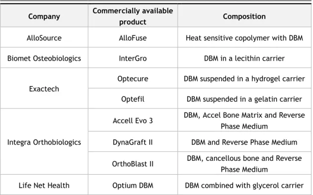
In table 2.1, a research was made to quantify some of the injectable bone graft substitutes that were commercially available in 2014. The Food and Drug Administration (FDA) approved all products presented in the table [83], [84].
Table 2.1 - Injectable bone grafts commercially available
Company Commercially available
product Composition
AlloSource AlloFuse Heat sensitive copolymer with DBM
Biomet Osteobiologics InterGro DBM in a lecithin carrier
Exactech
Optecure DBM suspended in a hydrogel carrier
Optefil DBM suspended in a gelatin carrier
Integra Orthobiologics
Accell Evo 3 DBM, Accel Bone Matrix and Reverse Phase Medium
DynaGraft II DBM and Reverse Phase Medium
OrthoBlast II DBM, cancellous bone and Reverse Phase Medium
11
BioHorizons
Osteofil DBM DBM in porcine gelatin
Progenix Putty DBM in Type-1 bovine collagen and sodium alginate
MTF/Synthes DBX DBM in sodium hyaluronate carrier
Osteotech GRAFTON gel DBM in a syringe
Smith & Nephew VIAGRAF DBM combined with glycerol Wright Medical
Tech-nology
PRO-DENSE injectable Regenerative Graft
75% calcium sulphate and 25% cal-cium phosphate
The polymers used as carriers must have rheological, mechanical and biological properties ap-propriate to its applications and the site of the implant [83], [85], [86]. Possible toxicity of degradation products and their elimination routes also need to be considered [85]. These in-jectable systems should allow minimally invasive implantation, fill a desired shape, and easy incorporation of various therapeutic agents [85].
In fact, one of the most attractive features of injectable bone substitutes, besides providing mechanical support, is their potential use for controlled release of therapeutic or bioactive agents [87].
2.3 Hydrogels
2.3.1 Introduction
Hydrogels are water-swollen polymeric materials, with a stable three-dimensional structure that provide scaffolds for tissue engineering, wound dressings, and drug delivery systems, among other application[88]–[90].
Depending on their method of preparation, ionic charge, or physical structure features, hydro-gels maybe classified in several categories. Based on the method of preparation, they may be homopolymer hydrogels, copolymer hydrogels, multipolymer hydrogels, or interpenetrating polymeric hydrogels[91], [92].
Hydrogels remain as appealing candidates to tissue engineering scaffolds, due to the controlla-ble and reproducicontrolla-ble polymer properties and to the large water uptake, promoting excellent biocompatibility due to low protein adsorption [27]. In addition, hydrogels present mechanical properties and hydrophilicity that resembles those of the extracellular matrix (ECM) of native tissue, tunable viscoelasticity, and high permeability for oxygen and essential nutrients[93]– [97]. Despite from the favorable physico-chemical and mechanical properties, the most im-portant requirement for a hydrogel to be used in medical applications is its biocompatibility and the non-cytoxicity of its degradation products [98], [99].
12
Injectable hydrogels can be maintained in the liquid state before injection and harden after transplantation in vivo. The hydrogel allows the filling of irregular defects, decreases the risk of implant migration and minimizes surgical defect to the size of a needle. In addition, the hydrogel can be incorporated with therapeutic factors and cells[100]. Hydrogels can also be stimuli-sensitive and respond to temperature, pH, electric field, glucose and antigens, among others. This way the hydrogel can have a controlled drug release due to volume changes [101]. Hydrogels present some limitations regarding drug delivery such as the high water content and large pore sizes that frequently result in relatively rapid release. Recently, composite systems where micro or nano hydrogels are incorporated in a bulk hydrogel matrix emerge as a platform for drug delivery [102]–[104]. The micro or nano hydrogel particles can act as a drug reservoir from which release can be triggered by a suitable stimulus or simply released in a diffusion-controlled manner [104]. The major advantage relies on the improvement of the kinetic release profile of the drug, as the nanogel phase provides an additional diffusion barrier moderating or eliminating the initial burst release typically observed in hydrogel or nanogel drug delivery systems [105].
Within the large range of materials used in the development of hydrogels, polysaccharide-based materials have been referred to as promising materials, presenting appealing properties for biomedical applications[27], [92], [106], [107].
Among the numerous macromolecules that can be used for hydrogel formation, polysaccharides are advantageous compared to synthetic polymers since they are coming from renewable sources. Polysaccharides have also frequently economic advantages over synthetic materials and they are usually non-toxic, biocompatible and show a number of convenient physico-chem-ical properties such as viscosity, hydrophilicity and reactive groups [108]. The major disad-vantages of natural polymers, when compared with synthetic ones, are the difficulty in con-trolling their physico-chemical properties, such as molecular weight, strength, degradation time and mechanical properties. However, there are several strategies to overcome these lim-itations, like the combination with other natural (e.g., collagen/glycosaminoglycans) or syn-thetic polymers (e.g., collagen/PLGA). This combinations, may improve the biocompatibility of the ensuing scaffolds, by reducing inflammatory response in vivo and improving initial cell at-tachment and differentiation on the material [109]–[112]. Polysaccharides, such as starch, cel-lulose, chitin/chitosan, alginate, carrageenan, gellan, guar gum, hyaluronic acid, pullulan, among others, have been used in the formulation of several hydrogels [113]–[121].
2.3.2 Dextrin hydrogel
Among starch-based materials, those based on dextrin are widely used in a variety of applica-tions, since adhesives used in food to textile industries [122], peritoneal dialysis solution [123] and the cosmetic industry [124].
Dextrins are a group of low-molecular-weight carbohydrates produced by partial hydrolysis of starch, which can be accomplished by the use of acid, enzymes, or a combination of both. Dextrin is a glucose-containing saccharide polymer linked by α-1,4 D-glucose units, containing few (< 5%) α-1,6 links, having the same general formula as starch, but smaller and less complex (figure 2.5) [107], [123].
13
Figure 2.5 - Structure of Dextrin [125].
Due to its proven clinical tolerability [126] and its efficient absorption due to degradation by amylases [105], [127], [128], dextrin appears as a polymer that might be ideal for development as a drug carrier [123].
Dextrin hydrogels can be obtained by crosslinking following oxidation. The oxidized dextrin is characterized by their oxidation degree, which consists on the quantification of aldehyde groups. The oxidation reaction is characterized by the specific cleavage of the C2-C3 linkage of glucopyranoside rings, yielding two aldehyde groups per glucose unit (figure 2.6).
Figure 2.6 - Periodate Oxidation of Dextrin, Yielding Two Aldehyde Groups at Positions C2 and C3 of a
D-Glucose Unit [105].
The degree of oxidation (DO) can be easily controlled by the relative quantity of sodium perio-date used, yielding free aldehyde reactive groups to create covalent linkages with reticulating molecules, cellular adhesion binding peptides or specific drugs for controlled delivery systems [105].
In recent works, dextrin-hydroxyapatite complex was used as a bone filling material, with good performance [129]. Dextrin hydrogels, namely, dextrin-vinyl acrylate (Dex-VA) and dextrinhy-droxyethylmethacrylate (Dex-HEMA) was shown their noncytotoxicity as well as their appealing diffusivity and degradability profiles for targeted delivery therapeutics [107], [130].
The concentration of dextrin that guarantees the ideal texture is between 30% and 40%, giving rise to an injectable hydrogel; above 40%, the solution is extremely viscous and very difficult
14
to homogenize. A polymer concentration below 25% will originate a viscous fluid instead of a hydrogel. The crosslinking times of the hydrogel is between 5-30 minutes. If used in maxillofa-cial surgery applications, as an adjuvant to osteogenic granular compounds, this time would allow an unhurried handling and implantation [107], [125].
2.3.3 Dextrin nanogel
Polymeric nanogels, also referred to as hydrogel nanoparticles, macromolecular micelles or polymeric nanoparticles, are emerging as promising drug carriers for therapeutic applications [131].
The study of nanogels has intensified during the past two decades due to enormous potential applications in the development and implementation of new environmentally responsive mate-rials, biomimetics, biosensors, artificial muscles and drug delivery systems [132]. Solid nano-particles made from biodegradable polymers for long-term delivery of drugs can potentially provide benefits such as increased therapeutic effect, prolonged bioactivity, controlled release rate, and finally decreased administration frequency, thereby increasing patient compliance [133].
Among the available nanosystems, self-assembled polymeric nanogels, like dextrin nanogel, are particularly attractive, since they are easy to produce, affordable and may effectively incor-porate a variety of drugs [132].
Gonçalves et al. developed and characterized self-assembled nanoparticles of dextrin with great potential for biomedical applications [131], [134]–[138]. In vitro studies with bone mar-row-derived macrophage revealed that the nanoparticles are non-cytotoxic and do not elicit a reactive response when in contact with macrophages [136]. Dextrin nanoparticles served as an effective nanocarrier for the formulation of lipophilic curcumin by increasing its water solubil-ity, improving its stabilsolubil-ity, and controlling its release profile, without compromising the cyto-toxicity in HeLa cell line [138].
2.4 Simvastatin in bone regeneration
Currently, researchers are searching for bone graft materials with the advantages of autologous bone grafts (osteogenesis, osteoinduction and osteoconduction), but without their disad-vantages (donor site morbidity, difficulty of storage and maintenance, unlimited availability, etc.) [139].
In 1980, Urist reported the identification in the rat organic bone matrix of an insoluble protein of low molecular weight called Bone Morphogenetic Protein, BMP [140]. BMPs are multi-func-tional growth factors that belong to the transforming growth factor beta (TGFbeta) superfamily [141]. BMP signaling plays critical roles in heart, neural and cartilage development and play an important role in postnatal bone formation. Preclinical and clinical studies have shown that BMP-2 can be utilized in various therapeutic interventions such as bone defects, non-union fractures, spinal fusion, osteoporosis and root canal surgery [141]. BMP-2 and BMP-7 are oste-oinductive BMPs: they have been demonstrated to potently induce osteoblast differentiation in a variety of cell types [141], [142]. However, the use of BMPs entails some problems such as
15
their short life, storage and handling difficulties, inefficiency in the recognition of target cells, and high cost, which has hindered its popularization in procedures for regeneration of bone tissue [139], [143].
As alternatives of BMPs, some authors have suggested the topical use of drug compounds aimed at upregulating intrinsic bone growth factors. For example, some widely known pharmacologic compounds (such as bisphosphonates or statins) have recently been shown to upregulate bone growth through distinct and complex biochemical pathways [144]–[147].
In 1999, was first reported, by Mundy et al., that lovastatin and simvastatin stimulate bone regeneration when injected subcutaneously in mouse calvaria [148].Statin is a specific inhibitor of 3-hydroxy-3-methyl-glutaryl coenzyme A (HMG-CoA) reductase, rate-limiting enzyme of the cholesterol synthesis pathway [149]. Simvastatin (figure 2.7) [150], a chemical modification of lovastatin, is an inactive lactone drug that, after oral administration, is converted to its active dihydroxy open acid form by the intracellular enzyme cytochorome P450 (3A4 isozyme) in the liver [149]. Simvastatin is not well absorbed, and less than 5% of an oral dose reaches the systemic circulation. Concentrations of statins in bone marrow have not been well established yet, but osteoblasts and osteoclasts may be exposed to very low concentrations of statin with existing oral regimens [151].
Figure 2.7 - Structure of simvastatin [150].
Simvastatin is suggested to support bone morphogenetic protein (BMP)-induced osteoblast dif-ferentiation through antagonizing TNF-α-to-Ras/Rho/mitogen activated protein kinase and aug-menting BMP-Smad signalling [152]. Simvastatin increases alkaline phosphatase activity and mineralization, as well as increases the expression of bone sialoprotein, osteocalcin and type I collagen, and it is shown to have anti-inflammatory effect by decreasing the production of interleukin-6 and interleukin-8 [153].
Simvastatin has been reported to promote osteoblastic activity and inhibit osteoclastic activity. There have been many studies demonstrating the bone promoting effect of local application with different carriers in various animal models [154]–[156].
The use of topical simvastatin for bone regeneration can be seen as a relatively recent research line, since most studies have been carried out in the last 5 years [139].
The successful use of simvastatin to promote bone formation in vivo depends on the local con-centration [154]. Therefore, an appropriate carrier would bring several advantages, including localization and retention of the molecule to the site of application thus reducing the loading dose and providing a matrix for mesenchymal cell infiltration and a substrate for cell growth
16
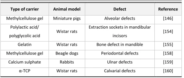
and differentiation [157]. In addition, the optimal carrier should help to define the shape of resulting new bone and should have a degradation rate that does not inhibit bone growth and prevent fibrous tissue formation or fibrous encapsulation of the carrier. There have been many studies demonstrating the osteopromotive effect achieved by the local application of the drug (table 2.2) with different carriers in various animal models [157].
Table 2.2 - Carriers and animal models used for the local application of simvastatin
.
Type of carrier
Animal model
Defect
Reference
Methylcellulose gel
Miniature pigs
Alveolar defects
[146]
Polylactic acid/
polyglycolic acid
Wistar rats
Extraction sockets in mandibular
incisors
[154]
Gelatin
Wistar rats
Bone defect in mandible
[155]
Methylcellulose gel
Beagle dogs
Periodontal defects
[158]
Calcium sulphate
Rabbits
Ulnar defects
[159]
α-TCP
Wistar rats
Calvarial defects
[160]
Pradeep et al. has carried out randomized clinical trials demonstrating that locally- adminis-tered simvastatin, versus placebo, significantly improves the clinical outcomes of scaling and root planning for treating mandibular buccal Class II furcation defects [161] and in patients with chronic periodontitis [162].
Studies focusing on pharmacological development have tested several biodegradable polymeric formulas for the local delivery of simvastatin such as a hydrogel of gelatin [163] or microspheres of PLGA [164].
The large majority of animal studies reported favorable results concerning topical application of simvastatin, either injected alone [165] or in combination with biomaterials [166], or cover-ing acellular scaffolds [167]. Ozeç et al., make a 3 mm diameter defect in the angulus mandib-ular region of Wista albino rats and grafted with simvastatin gelatin sponge producing 240% more new bone than the control group [155] . Nyan et al., create critical-sized bone defects in rat calvaria and treated with calcium sulfate or with combination of 1 mg simvastatin and calcium sulfate. It was reported that the combination of simvastatin and calcium sulfate stim-ulated bone regeneration [156].
Most animal studies performed intraorally have reported good results for topical simvastatin administration in enhancing bone regeneration in rat mandibular defects [168] and periodontal lesions in rats [144], [169], Beagles [158]and minipigs [146].
However, some authors have reported unfavourable results after using simvastatin for bone formation. Lima et al. [170] found a negative impact of combining simvastatin with demineral-ized bovine bone matrix for repairing calvarial defects in rats after 30 to 60 days of healing. On postoperative day 60 the use of simvastatin, regardless of the dose, resulted in lower density than that observed in control and demineralized bovine bone matrix group samples.
17
Anbinder et al. also found that simvastatin administration, either orally or subcutaneously, did not improve bone repair for experimental tibial defects in rats [171].
Regarding the dosing of the simvastatin, caution should be exercised. Some authors have re-ported a dose-dependent inflammatory response [166]. In animal studies, a 0.1-0.5 mg dose of simvastatin would be the optimal dose for stimulating maximum bone regeneration without inducing inflammation [146].
18
19
Chapter 3
Materials and methods
Materials
All reagents used were of laboratory grade and purchased from Sigma-Aldrich, unless stated otherwise. Dextrin - Tackidex was from Roquette. Dextrin-VMA was synthesized by transester-ification of dextrin with vinyl methacrylate (VMA), with few modtransester-ifications from the protocol described by Ferreira et al. [172] for the transesterification of dextran with VA. In this work, dextrin-VMA with 20 acrylate groups per 100 dextrin glucopyranoside residues was used. Re-generated cellulose tubular membranes, were obtained from Membrane Filtration Products. All other chemicals and solvents used in this work were of the highest purity commercially availa-ble.
3.1 Glass reinforced hydroxyapatite (Bonelike) preparation
In the present study, Bonelike granules size ranging from 250 to 500 µm were prepared as follows. First, a P2O5-based glass with the composition of 65P2O5-15CaO-10CaF2-10Na2O, in %mol,
was prepared from reagent grade chemicals using platinum crucible at 1400ºC. The glass was ground and then the particles were sieved to a particle size less than 50 μm, using riddles with descending diameter. Medical grade hydroxyapatite was synthesized by chemical precipitation. After drying during 72 hours at 60ºC the hydroxyapatite was sieved until a particle size less than 75 μm was obtained. Then, the hydroxyapatite was mixed with the glass in a proportion of 2.5wt% [77], [81]. The mixed powders were dried for 24 hours at 60ºC and then sintered at 1300ºC for 1 hour. Finally, using, using standard crushing and sieving techniques the desirable particle range (250-500 µm) was obtained. The obtained hydroxyapatite was submitted to X-ray fluorescence spectrometry and X-X-ray diffraction (PANalytical, X'PERT-PRO model). Bonelike phase identification and quantification was performed using X-ray diffraction and Rietveld anal-ysis.
3.2 Preparation of oDex Hydrogel
Dextrin Oxidation:
Briefly, aqueous solutions of dextrin (2% w/v) were oxidized with a 2 mL sodium m-periodate solution, to yield the theoretical degree of oxidation of 40%, at room temperature, with stirring, and in the dark. After 20 h, the oxidation reaction was stopped by adding dropwise an equimolar
20
amount of diethyleneglycol to reduce any unreacted periodate. The resulting solution was dia-lyzed for 3 days against water using a dialysis membrane with a molecular weight cutoff of 1000 Da and then lyophilized for 10 days. The degree of oxidation (DO) of oxidized dextrin (oDex) is defined as the number of oxidized residues per 100 glucose residues and was quantified using the tert-butylcarbazate method [105].
Preparation of oDex-ADH Hydrogels:
oDex was dissolved in PBS buffer (phosphate-buffered saline) (30% w/v) at room temperature, and an adipic acid dihydrazide (ADH) solution (prepared separately) was added at the concen-tration of 3,76% w/v. The cross-linking reaction was allowed to proceed during 30 minutes. The material was considered as gelified when it stopped slipping along a 90° inclined surface. The hydrogel preparation followed the protocol proposed by Molinos et al. [105].
3.3 Preparation of self-assembled nanoparticles of dextrin substituted with
hexade-canethiol
For the nanoparticles preparation, the protocol proposed by Gonçalves et al. [131] was followed, as described ahead:
Synthesis of dexC16:
Dextrin-VMA and 1-hexadecanethiol were dissolved in dimethyl sulfoxide (equivalent VMA = 0.0413 M). A molar percentage of 1-hexadecanethiol (100% relative to VMA) were added to the reaction mixture in order to obtain the pretended degree of substitution (DS). Triethylamine (TEA) (1 mol equiv to VMA) was added to the reaction mixture. The medium was stirred for 24 h, at 50 ºC. The mixture was dialysed for 48 h against water with frequent water change. After freezing, the mixture was lyophilized and stored.
Sample preparation:
Lyophilized dexC16 was dissolved in water or PBS under stirring at 50 ºC until a clear solution was obtained. The degree of solubility of dexC16 depends on the degree of substitution. In-creasing the degree of substitution reduces the solubility. In the range of DSC16 used, to pre-pare a 1.0 g/dL solution, 3 h of stirring is the maximum time required to dissolve dexC16.
Dynamic light scattering:
The size distribution was determined with a Malvern Zetasizer, Model Nano ZS (Malvern Instru-ments Limited, U.K.). A dispersion of nanoparticles in water (1 mL) was analysed at 25 ºC in a polystyrene cell using a Helium-Neon laser – wavelength of 633 nm and a detector angle of 173º. The dispersion was filtered through a 0.22 μm pore.
1
H NMR:
Lyophilized dexC16 was dispersed in deuterium oxide (1.0 g/dL). Solutions were transferred to 5 mm NMR tubes. 1D 1H NMR measurements were performed with a Varian Unity Plus 300
spec-trometer operating at 299.94 MHz. 1D 1H NMR spectra were measured at 298 K with 80 scans,
a spectral width of 4800 Hz, a relaxation delay of 1 s between scans, and an acquisition time of 3.75 s.
21
oDex, DO 40%, (30% w/v) was dissolved in PBS or in a suspension of nanogel for ∼16 h at room temperature. Then, the oDex suspensions were mixed with 5% (in molar base taking into ac-count the number of glucose residues in the original dextrin) ADH. The cross-linking was allowed to proceed at room temperature for 30 minutes. Ahead, the oxidized dextrin hydrogels are termed as oDex hydrogels, and the oxidized dextrin hydrogels with incorporated dextrin nano-gels are called oDex-nanogel hydronano-gels.
For the Bonelike-oDex-Nanogel Hydrogel, the same protocol was followed, but before adding the ADH, Bonelike (60% or 40% w/v) was added to the samples and stirred. Then, ADH was added to each sample and the cross-linking was allowed to proceed at room temperature for 30 minutes.
3.5 Cryo-Scanning Electron Microscopy (Cryo-SEM) Analysis
The topography and porosity of the injectable bone graft substitutes (30% w/v oDex, 10 mg/mL nanogel and 40 or 60% w/v Bonelike) were studied by Cryo-SEM.
The SEM / EDS analysis was performed using a High Resolution Scanning Electron Microscope with X-Ray Microanalysis and CryoSEM experimental facilities: JEOL JSM 6301F/ Oxford INCA Energy 350/ Gatan Alto 2500. The samples (n=3) were rapidly cooled (plunging it into sub-cooled nitrogen – slush nitrogen) and transferred under vacuum to the cold stage of the prepa-ration chamber. The specimens were fractured, sublimated (‘etched’) for 90 seconds at -90°C, and coated with Au/Pd by sputtering for 45 seconds and then transferred into the SEM chamber where were studied at a temperature of -150°C.
3.6 Injectability tests
The injectability tests were conducted at the Faculty of Pharmacy, University of Porto. The equipment TA.XT2i Texture Analyser was used to evaluate the extrusion force of injectables, in order to determine if they can be applied in a clinical environment. The tests were performed in triplicate per each testing condition. 1mL samples of the Injectable Bonelike-oDex-Nanogel Hydrogel were placed inside 2mL syringes, followed by a 30 minutes period to allow the cross-linking reaction. The syringes were fixed vertically to the bottom plate and the equipment applied the necessary force to move the plunger 1 mm/s in the syringe. The results were ex-pressed as the force required to move the plunger out of the syringe.
3.7 Preparation of simvastatin-loaded Bonelike scaffolds and in vitro assay of
simvas-tatin release
Simvastatin was dissolved in pure ethanol at a concentration of 5mg/mL. For the preparation of simvastatin-loaded Bonelike scaffolds, 100µl of the previous solution was dropped onto Bone-like under sterile conditions, and then allowed to dry completely in a laminar flow hood for 24 h. 2 groups of samples were prepared (n=3), containing 60 mg of Bonelike and 0 mg or 0.5mg simvastatin, respectively.
The scaffolds (60 mg) were placed in 1 ml PBS at 37˚C at 60 rpm. At 24, 48, 72, 96, 168, 192, 216 and 240 hours, the PBS was changed. At every time-point the solution absorbance was measured at a wavelength of 238 nm using an ultraviolet-visible spectrophotometer, while the
22
simvastatin concentration was determined from a standard curve prepared with various amounts of simvastatin.
3.8 Incorporation of simvastatin in the nanogel
Simvastatin (SIM) was loaded into the hydrophobic domains of nanoparticles. The physical en-trapment of simvastatin in the nanoparticles was performed following the nanoparticles for-mation, as described ahead. 2 stock solutions of simvastatin in ethanol with a concentration of 5 mg/mL and 10 mg/mL were prepared. The required volume of simvastatin from these solu-tions was added to the nanoparticles solusolu-tions (final concentration of ethanol < 1%). The effect of polymer/simvastatin ratio on the loading efficiency and stability of the formulation was studied. Different formulations were prepared by varying the concentrations of simvastatin (5 mg/mL and 10 mg/mL) and polymer (1.0 and 3.0 mg/mL). In order to evaluate the entrapment efficiency, the solutions were kept under stirring during predeterminated times (3 and 24 hours). The resultant solutions were centrifuged at 10000 rpm, for 5 min, to remove the insoluble simvastatin. The supernatants were carefully collected and analyzed spectrophotometrically. The quantification was carried out using a calibration plot obtained with different simvastatin concentrations. The entrapment efficiency was calculated by the Equation 3.1:
𝐸𝑛𝑡𝑟𝑎𝑝𝑚𝑒𝑛𝑡 𝑒𝑓𝑓𝑖𝑐𝑖𝑒𝑛𝑐𝑦 (%) =
[𝑆𝑖𝑚𝑣𝑎𝑠𝑡𝑎𝑡𝑖𝑛] 𝑒𝑛𝑐𝑎𝑝𝑠𝑢𝑙𝑎𝑡𝑒𝑑[𝑆𝑖𝑚𝑣𝑎𝑠𝑡𝑎𝑡𝑖𝑛] 𝑎𝑑𝑑𝑒𝑑
× 100
, (equation 3.1)The size distribution of unloaded and loaded dextrin nanoparticles was determined by dynamic light scattering (DLS) using a Malvern Zetasizer, Model Nano ZS. The nanoparticles dispersion was analysed at 25ºC in a polystyrene cell, using a He-Ne laser - wavelength of 633 nm and a detector angle of 173º. The DLS analysis provides the characterization of a sample through the mean value (z-avg) for the diameter, and a width parameter known as the polydispersity index (PdI).
3.9 In vitro assay of simvastatin release from the nanogel
The release of simvastatin from the nanoparticles was studied using sink conditions. The simvas-tatin loaded nanoparticles (5mg/mL of simvassimvas-tatin and 1.0 mg/mL of polymer in an aqueous solution) was placed in a dialysis membrane.
The dialysis membrane (molecular cut off of 2 kDa) was placed in 200 mL of PBS, shaken under 250 rpm at 37ºC. At predefined timeframe (up to 24 h), a sample of 200 µL was withdrawn, centrifuged to guarantee removal of unentrapped simvastatin and the supernatant was ana-lyzed spectrophotometrically.
3.10 Materials sterilization
Sterilization of Bonelike granules was performed by autoclaving the material during 35 minutes, at 121ºC. oDex hydrogel was sterilized by UV light, during 1 or 2 hours. To acess the efficacy of this method, sterilized samples (n=3) were immersed in αMEM during 4 weeks. The dextrin nanoparticles were sterilized by filtrating the dispersion through a 0.22µm pore (Sterile Cellu-lose Acetate Membrane, Frilabo).
23
Mouse embryo fibroblasts 3T3 (ATCC CCL-164) were grown in Dulbecco’s modified Eagle’s media supplemented with 10% newborn calf serum (Invitrogen, U.K.) and 1 μg/mL penicillin/streptav-idin (cDMEM) at 37 °C in a 95% humidified air containing 5% CO2. Before reach confluency, 3T3 fibroblasts were harvested with 0.05% (w/v) trypsin-EDTA and subcultivated in the same me-dium.
The cytotoxicity of the nanoparticles unloaded and loaded with simvastatin was assessed in a 3T3 fibroblasts culture previously incubated for 24 h (2.0 × 10⁴ cells/well, in a 24-well polysty-rene plate, n=3) using 3-[4,5-dimethylthiazol-2-yl]-2,5-diphenyl tetrazolium bromide (MTT) as-say, as described ahed. The nanoparticles were sterilized and dissolved in cDMEM. Increasing concentrations of loaded and unloaded nanoparticles (0.5, 0.75, 1 and 2 mg/mL) dissolved in cDMEM at 20% (v/v), were then added to the fibroblast culture. Free simvastatin, in the same concentration that was incorporated in the nanoparticles, was dissolved in cDMEM at 1% (v/v). After 24h and 48 h of incubation, cell viability was accessed by MTT assay. Morphological eval-uation of 3T3 cells was also made by regular light microscope observations.
3.12 In vivo tests
The objective was to observe ectopic bone formation in subcutaneous implants in rats, due to simvastatin action, during an 8-week experiment. The injectable composite materials were sterilized by the methods previously referred.
100 µl of samples were surgically implanted subcutaneously in the lumbar region of three male rats (Sasco Sprague Dawley, Barcelona, Spain, weighting around 300 g), each one receiving 5 implants with the following scheme:
Previous experimental work already evaluated the biological response of rats using a control with no implant where the suture was performed (Sham group). Two animals were housed per cage (Makrolon type 4, Tecniplast, VA, Italy), in a temperature and humidity controlled room with 12-12h light/dark cycles, and were allowed normal cage activities under standard labora-tory conditions. The animals were fed with standard chow and water ad libitum. Adequate
Figure 3.1 - Implant scheme: (1) oDex-nanogel/SIMV hydrogel; (2) oDex-nanogel/SIMV hydrogel + MSCs;
24
measures were taken to minimize pain and discomfort taking into account human endpoints for animal suffering and distress.
All procedures were performed with the approval of the Veterinarian Authorities of Portugal, and in accordance with the European Communities Council Directive of November 24th 1986 (86/609/EEC). Anaesthesia was achieved with an intraperitoneal injection of a pre-mixed solu-tion consisting in ketamine (Imalgene 1000R), 100 mg/kg body weight, and xylazyne (RompunR), 200 mg/kg body weight. Hair from the dorsal area was clipped and the skin scrubbed in a routine fashion with an iodopovodone 10% solution (BetadineR). Five 1.5-2cm long linear incisions were performed. After blunt dissection towards the ventral aspect of the body, the biomaterials were implanted subcutaneously. Skin and subcutaneous tissues were closed with a simple-in-terrupted suture of a non-absorbable filament (SynthofilR, Ethicon).
25
Chapter 4
Results and Discussion
4.1 Glass reinforced hydroxyapatite (Bonelike) preparation
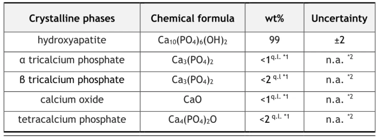
The obtained hydroxyapatite was submitted to X-ray fluorescence spectrometry to perform a chemical and elemental analysis. X-ray diffraction was used to identify and quantify the differ-ent crystalline phases. The values of the tables 4.1 and 4.2 are inside the range of values of the norm ISO/DIS 13779-1 (Implants for surgery — Hydroxyapatite; Part 3: Chemical analysis and characterization of crystallinity and phase purity) [173].
Table 4.1 - Quantitative chemical analysis of the hydroxyapatite1.
Analytical parameter
Obtained value
Uncertainty
Units
Arsenic (As)
<1.0
q.l. *1n.a.
*2mg/Kg
Cadmium (Cd)
<0.5
q.l. *1n.a.
*2mg/Kg
Mercury (Hg)
<0.5
q.l. *1n.a.
*2mg/Kg
Lead (Pb)
<0.5
q.l. *1n.a.
*2mg/Kg
Ca/P
1.68
n.a.
*2Molar ratio
Table 4.2 - Quantification of crystalline phases by X-ray diffraction of the hydroxyapatite.
Crystalline phases
Chemical formula
wt%
Uncertainty
hydroxyapatite
Ca10(PO4)6(OH)2
99
±2
α tricalcium phosphate
Ca3(PO4)2
<1
q.l. *1n.a.
*2β tricalcium phosphate
Ca3(PO4)2
<2
q.l *1n.a.
*2calcium oxide
CaO
<1
q.l. *1n.a.
*2tetracalcium phosphate
Ca4(PO4)2O
<2
q.l. *1n.a.
*226
As table 4.1 shows, the molar ratio was the expected for hydroxyapatite and very similar to the human bone Ca/P molar ratio (1.631) [174].
During the sintering process of Bonelike, the CaO–P2O5 glass (also produced for this work) reacts
with hydroxyapatite, forming β-tricalcium phosphate, which then can transform into α-trical-cium phosphate at higher temperatures. Figure 4.1 display an X-ray diffraction pattern of Bone-like [81]. Rietveld analysis previously reported indicated that BoneBone-like is composed by 68.4% of hydroxyapatite, 24% of α-TCP, and 7.6% of β-TCP [81], [175], [176].
Figure 4.1 - X-ray diffraction pattern of Bonelike, which is composed of hydroxyapatite (HAp), α-TCP (α)
and β-TCP (β) [81].
Bonelike has been reported to be osteoconductive and bioactive, supporting the formation of mechanically and chemically bonded bone directly on its surface [49], [51], [177]–[179]. A bal-ance between the least soluble phase of hydroxyapatite and most soluble phase of tricalcium phosphate determines the bioactivity of Bonelike. Due to the presence of biodegradable α and β-TCP phases in the structure of Bonelike, a local enrichment in Ca and P in the physiological environment occurs, which stimulates new bone formation [175]. For this work, Bonelike with granules size ranging from 250 to 500 mm, as shown in figure 4.2, was obtained using using standard crushing and sieving techniques. A porosimetry of 65% for the Bonelike produced was reported previously, in our lab, using mercury intrusion porosimetry analysis.
27
Figure 4.2 - Glass reinforced hydroxyapatite (Bonelike) with a particle size from 250 to 500 µM; (A)
macroscopic aspect of Bonelike granules and (B) SEM image showing the surface morphology. 500 µm
![Figure 2.1 - Cortical bone and trabecular bone [7].](https://thumb-eu.123doks.com/thumbv2/123dok_br/15714644.1069693/22.892.321.596.109.413/figure-cortical-bone-trabecular-bone.webp)
![Figure 2.2 - Bone remodeling process [21].](https://thumb-eu.123doks.com/thumbv2/123dok_br/15714644.1069693/23.892.150.781.630.878/figure-bone-remodeling-process.webp)
![Figure 2.3 - Process of fracture healing: (a) inflammation, (b) soft callus, (c) hard callus and (d) remod- remod-elling [24]](https://thumb-eu.123doks.com/thumbv2/123dok_br/15714644.1069693/24.892.161.680.577.820/figure-process-fracture-healing-inflammation-callus-callus-elling.webp)
![Figure 2.6 - Periodate Oxidation of Dextrin, Yielding Two Aldehyde Groups at Positions C2 and C3 of a D-Glucose Unit [105]](https://thumb-eu.123doks.com/thumbv2/123dok_br/15714644.1069693/31.892.154.791.684.822/figure-periodate-oxidation-dextrin-yielding-aldehyde-positions-glucose.webp)
![Figure 2.7 - Structure of simvastatin [150].](https://thumb-eu.123doks.com/thumbv2/123dok_br/15714644.1069693/33.892.351.583.545.747/figure-structure-of-simvastatin.webp)