Dedicatória
Gostaria de agradecer principalmente ao Doutor Luís Figueira pelo constante
apoio durante o desenvolvimento desta dissertação tal como à Doutora Joana
Araújo pela sugestão deste tópico.
Gostaria ainda de agradecer a minha namorada, Joana, por me conseguir
manter focado nos momentos chave e distrair em outros tal como aos meus
pais porque sem eles este trabalho simplesmente não existia.
Full title: Epidemiology of Uveitis in a tertiary care centre in Portugal Short title: Epidemiology of Uveitis in a tertiary care centre in Portugal
Authors:
Lukasz Hermann (1), Joana Araújo (1,2), Fernando Falcão-Reis (1,2), Luís
Figueira (1,2,3)
1. Faculty of Medicine of the University of Porto, Porto, Portugal
2. Department of Ophthalmology, Hospital S. João, Porto, Portugal
3. Center for Drug Discovery and Innovative Medicines (MedInUP),
University of Porto, Porto, Portugal
Abstract
Purpose: Evaluate the prevalence and incidence, demographic characteristics,
anatomical classification, concomitant complications and treatment of uveitis, as
well as its association with systemic diseases in the northern Portuguese
population referred to a tertiary care centre (São João Hospital, Porto).
Methods: A retrospective observational study of 545 consecutive uveitis cases
was performed between 12th April 2012 up until the 26th October 2017. Uveitis
was classified according to Standard Uveitis Nomenclature while etiology and
complications were diagnosed by specific clinical and laboratory testing.
Results: Prevalence was calculated to be 12,4 cases per 100,000 people while
incidence was 3.9 cases per 100,000 people annual incidence. Ratio of females
to males was 1.32:1 and mean age at diagnosis was 47,86 years. Anterior
uveitis was the most common classification made up 47.5% followed by
non-infectious posterior uveitis 18,0%, non-infectious posterior uveitis 8,4%, intermediate
uveitis 5,5% and panuveitis 4,2%. Extrauveal entities made up the rest of the
causes (16.4%). In terms of etiology the majority of uveitis were idiopathic (28,4%) followed by axial spondyloarthritis (12.1%), Behçet’s disease (8,8%), sarcoidosis (6,1%), tuberculosis (5,5%), herpes (5,1%), pars planitis (3,7%),
toxoplasmosis (3,1%) among other less frequent causes.
Conclusion: In our referral region, 72% of uveitis cases were correctly identified
surpassing other studies. Intermediate uveitis increased while panuveitis
decreased when compared to previous publications. Extensive monitoring of
the development of uveitis by international multicenter prospective studies could
KEY-WORDS: EPIDEMIOLOGY; ETIOLOGY; INCIDENCE; PREVALENCE;
TERTIARY CARE CENTRE; UVEITIS
Introduction
Uveitis is synonymous with intraocular inflammation and represents a
heterogeneous group of inflammatory intraocular diseases that can cause great
visual damage and even blindness. Uveitis is classified in terms of the
Standardisation of Uveitis Nomenclature into several categories, the major one
being anatomical: anterior (iridocyclitis) (AU), intermediate (vitritis) (IU),
posterior (PU) and panuveitis (PANU) (1). The causes may be infectious and
non-infectious, and the disease may afflict only ocular tissues or be part of a
systemic infectious or autoimmune entity. The rest of the extrauveal entities
were classified as Escleritis, Episcleritis, Optic Neuropathy (ON) and Inflammatory Ocular Superficial Disease (IOSD).
Recent studies suggest that uveitis can cause visual acuity change in 5-10%
and severe loss of visual acuity and blindness in up to 35% (2). The worldwide
incidence is 17-52 per 100,000 person-years and the prevalence is 38-714
cases per 100,000 people (2). The incidence and prevalence in Portugal where
this epidemiological study takes place has been reported as 15 - 60 per
100 000 people and 0,7% respectively. (3) Prevalence and incidence may vary
widely between different geographic regions, due to population and
environmental characteristics, migratory flows, life expectancy, diagnostic
between different studies may be challenging, but useful to uncover the natural history of uveitis and standardize the best treatment possible.
The existence of epidemiological studies in Portugal is very limited with only
one carried out in 1990 (4) but more internationally robust studies have been
conducted in the United States (6,7,8) Southern Europe (5,9), Northern Europe
(10,11) among others (2). Even though there have been many advances in
diagnostic accuracy, many cases of intraocular inflammation continue to defy
expectations and there is no universally accepted approach to the evaluation of
uveitis.
Methods
The target population of this study was the northern Portuguese patients
attending specialised imunophtalmologic consultation at São João Hospital.
According to the 2011 census the Portuguese northern population was
3,689,862 million people with a female to male ratio of 1.09:1 (12). Using this
population, we estimated the prevalence and incidence of uveitis hoping that
this would lead to useful epidemiological conclusions. Demographical
characteristics such as age and gender were collected from the clinical
database in São João Hospital as well anatomical classification under the SUN
criteria, extrauveal entities, laterality of uveitis and systemic disorder.
Alternating uveitis was classified as bilateral. Complete ophthalmologic
examination was performed systematically including visual acuity,
biomicroscopy, applanation tonometry and indirect fundoscopy.Uveitis
fluorescein angiography, and automated perimetry, and its etiology assessed
with further ancillary testing including serology, radiology, microbiology, and
biopsy when appropriate.
Clinical history, physical examination, laboratory testing as well as serologic
tests, lumbar punctures and chest radiography among others were undertaken
to identify the etiology of each uveitis case.Etiologies such as trauma,
endophthalmitis and post-surgical were excluded from this study because such
cases are primarily managed surgically and thus rarely referred to tertiary care
centres.The term idiopathic was applied when a secondary cause for the
uveitis could not be identified. When the presence of two or more concurrent
etiologies was confirmed the most likely etiology was chosen to be cause of the
uveitis based upon the presenting symptoms and signs. Each case was
updated in terms of uveitis classification and overall systemic diagnoses as
dictated by evolution and complementary tests in the study population from 12th
April 2012 up until the 26th October 2017 (~43 months). The endpoint for each
patient included in the study was the stabilization of uveitis and resolution of
symptoms.
Statistical analysis was performed using IBM SPSS program. A descriptive
analysis was performed for the different variables and the results are shown in
frequency tables. Statistical significance was defined as P-value less than 0.05.
Chi-square test and t-test analysis were used to test for differences between
groups, when appropriate.
Treatment of these cases was recorded including the use of topic ocular and
systemic steroids, antihypertensive drops, periocular treatment, antimetabolites,
macular edema, glaucoma, cataracts and presence of ocular hypertension were
also recorded as variables.
This study was approved by the ethical committee of São João Hospital and
followed the tenets of the declaration of Helsinki for epidemiological studies.
Identities of patients were kept confidential and no informed consent was
required.
Results
A total of 545 patients (832 eyes) were included in the study. Prevalence of
uveitis was calculated to be 12,4 cases per 100.000 people while incidence 3.9
cases per 100,000 people annual incidence. 310 patients (56,9%) were female
and 235 (43,1%) were male (female to male ratio of 1.32:1). Mean age of
diagnostic was 47,86 years (IC 95%, 46,32 – 49,40) with age ranging from 10 to 92. Highest proportion of patients were in 51-60 range (21.5%) and the lowest
in the over 81 group (2.6%). Of the 545 patients, 287 (52,7%) had bilateral
disease (either recurrent or alternating), while 258 (47,3%) had unilateral
disease. Unilateral disease was present in herpes (82.1%) and Fuchs
Heterochromic Iridocyclitis (FHC) (72.7%) as well as syphilis and toxoplasmosis
(~70%). In turn bilaterality was more common in Behçet (66.7%), Juvenile Rheumatoid Arthritis (JRA) (75%), sarcoidosis (81.8%) and Vogt–Koyanagi– Harada disease (VKH) (64.3%).
The most frequent anatomic type was AU (47,5%), followed by non-infectious
PU (NIPU) (18,0%), infectious PU (IPU) (8,4%), IU (5,5%) and PANU (4,2%). In
(6,2%) closely followed by IOSD (5,9%) then ON (2,4%) and finally we had
episcleritis as the least identifiable cause (1,8%).
Considering all anatomical classifications, the majority of uveitis were idiopathic (28,4%) followed by axial spondyloarthritis (12.1%), Behçet’s disease (8,8%), sarcoidosis (6,1%), tuberculosis (5,5%), herpes (5,1%), pars planitis (3,7%),
toxoplasmosis (3,1%), JRA (2,9%), VKH disease (2,6%), FHC (2,0%) among
other less frequent causes (20,6%).
There is a clear prevalence of IU under 50 years of age (80%) as well as ON
(84.6%). In terms of etiology there is a higher rate of pars planitis (92.9%), Behçet’s disease (76.6%) and toxoplasmosis (81.3%) under 50 years of age while tuberculosis (71.4%) and sarcoidosis (66.7%) were more prevalent over
50 years of age. JRA had the youngest age of onset (23,81) while herpes had
the oldest (53,11).
In terms of sex, there was a clear higher rate of all types of uveitis in women than in men. Behçet’s disease was found to be more prevalent in men (55.3%) as well as FHC (63.6%). In terms of systemic etiologies, women had more
unidentifiable causes than men (54.8% in men compared to 45.2% in women).
There is also clear prevalence of autoimmune conditions such as JRA, systemic
lupus erythematosus (SLE) and sarcoidosis in the female sex.
In terms of complications ocular hypertension was present in 9,4% of cases
followed by macular edema (8,1%), cataracts (7,9%) and glaucomatous optic
neuropathy (7,5%).
Regarding treatment 84.1% of patients were given topical or systemic therapy
Only 6.6% of patients received periocular treatment. In terms of topical therapy,
corticoids were applied more frequently than nonsteroidal anti-inflammatory
drugs (32,3% compared to 11,7%). The most commonly utilized systemic
treatment was steroids (22,9%) methotrexate (11,9%), anti-TNF agents (6,2%),
azathioprine (4,4%) and cyclosporine (4,2%).
Discussion
The prevalence and incidence of uveitis in this study was much lower than in
other retrospective publications (2,4,5,6,7,8,9,10,11). This could be explained
by the following factors: only potentially challenging cases are referred to our
tertiary care centre (referral bias); plus, there are private clinics which do not
provide epidemiological data, population shifts may have occurred during the
4-year period since the census and general ophthalmologists may not have
sufficient proficiency in identifying many types of uveitis. Comparisons may
nevertheless be justifiable between these different countries
(2,4,5,6,7,8,9,10,11) because of the similarities in the environment, population
genetics and health care organisations.
Average age at presentation in our study (47,86) was in line with previous
studies (2,5,7,8,9,10,13) but not in line with German and Portuguese studies
where it was much lower (~37) (4,11). A possible explanation for this
phenomenon could be the ageing Portuguese population: 19% (12). The
highest proportion of patients in the 51-60 year range was confirmed in previous
publications and even surpassed others (2). 6,8% of cases were in the pediatric
herpes were the most frequent causes among our youngest and oldest patients
respectively and as reported by other studies (2,5). No statistical difference was
found between the mean age of males and females (p=0.293) or between age
and anatomic type (p=0.427).
In terms of sex, our study was in concordance with other authors
(2,4,5,6,7,8,9,10,11,14) showing a slight prevalence of females over males
(ratio of 1.32:1). This may be explained by the superior rate of blindness in
females (64.5%) and autoimmune etiologies such as SLE and JRA as well as
differential effects of sexual hormones, Th2 immune responses and the role of
the gut microbioma among other reasons. (2,14) The severity and ocular
manifestations between sexes due to the same cause are also variable. (2,14)
Idiopathic intraocular inflammation was found to be more common in women as
previous publications suggest (2). There was no association found between sex
and anatomic type (p=0.207) or systemic disease (p=0.328).
The presence of bilateral ocular disease was more frequently noted in our study
(47.3%) than in others (2, 5, 8, 11) except for Jones et al. (13) There is a
statistical association between the laterality of the uveitis and its anatomic type
(p<0.05) especially in IOSD where a clear bilateral prevalence can be observed
(78%) as well as in IU (70%) and NIPU (60.2%). All the different associations
between laterality and systemic diseases (p<0.05) found in this study were
confirmed in previous publications such as Behçet and sarcoidosis (2).
AU was the most prevalent cause of uveitis in our study as customary in all
major tertiary referral centres (28.5-60%) (2,4,5,6,7,8,9,10,11). IU is still
comparably difficult to diagnose but in our study its prevalence has increased
(4.2%) when compared to previous studies (2,4,5,8,9,10,11) and is the least
common type of uveitis. IU is still not as prevalent as in Northern populations
(22,9% in Germany) (11). The high rates of IU and low rates of PANU could be
explained by a more precise and earlier identification of the main component of
ocular inflammation, decreasing misdiagnosis as well as specific genetic and
environmental factors of our population. PU has comparable rates in most other
studies (15-38%) (2,4,5,9,10,11) as do the extrauveal causes (11).
It may be difficult to make a diagnosis other than idiopathic especially in AU and
extrauveal cases (32,8% and 41,6% respectively) because many cases are
benign, non-recurrent, responsive to treatment and are not investigated further
due to cost-effective reasons unlike others such as IU and PU (0% and 22%
respectively) which are more clinically aggressive. (2,5,10) PU is also more
readily identified with a suspected cause because of the appearance of the
ocular fundus (10). Uveitis is associated with systemic disease between 30 to
70% according to previous long-standing studies (2,4,5,6,7,8,9,10,11,13), but
has been increasing in rate in recent years just like in our study (70.5%) This
could be attributed to the development of better diagnostic accuracy and
improved practitioner awareness to the association between uveitis and
systemic disorders.
The most important cause of uveitis identified in our study was axial
spondyloarthritis, in order of frequency: ankylosing spondylitis, psoriatic arthritis,
Reiter syndrome, IBD-associated spondylitis among other less common causes,
both HLA-B27 positive and negative (12.1%) which was confirmed or surpassed
by some previous studies (2,4,5,10,11). The most frequent anatomic type
of other studies (13 to 58,3%) (11,15) AU is characterised by a genetic
predisposition expressed by positive HLA-B27 in more than 50% of cases, more
frequently in Western countries, a feature it shares with spondyloarthritis (15). It
is therefore imperative to refer these patients to a specialized rheumatologic
and ophthalmologic consultation.
The second most identified systemic disease in the study was Behçet’s (8,8%) which does not correlate with previous studies in Europe (9,10,11,16) or North
America (6,7,8) except for Portugal and Spain (~5%) (4,5). The geographic region, from the 30° to the 45° north latitudes, once considered the “silk route”, plays an important role in the prevalence of Behçet’s disease. (16) The
frequency of ocular involvement in patients in tertiary care centres was reported
to be from 0.0009 to 32% depending on the exact latitude and longitude. (2,16)
The age at which the disease is more common as well as sex distribution is
confirmed by our study (second to fourth decade and no male or female
preponderance respectively). (5,16) The most common anatomical sites were
NIPU and PANU as found in previous studies (2,5,11,27).
Unlike some earlier Mediterranean studies (4,5,9) a higher proportion of
sarcoidosis patients have been identified (6,1%) which is closer in accordance
with Northern European and American publications (9.6%-18.6% although
mostly in AU and IU) (2, 6,7,8,10,11). This may be due to different diagnostic
exclusion criteria (CT scan, minor salivary glands biopsy, endoscopic
ultrasound-guided fine-needle aspiration of intrathoracic nodes among others)
and patient characteristics. (17) Around 30-60% of patients with sarcoidosis
develop ophthalmic disease even in the absence of other extra-ocular
vital to the well-being of the patient. (17) There was also a great preponderance
of cases in male patients (84.8%) unlike most other studies (2,17) PANU had
the greatest prevalence of sarcoidosis (17,4%) and NIPU the second (15,3%) which conforms to the pattern observed by previous studies (up to 7–14% for PANU and up to 28% for NIPU) (2,11)
There has been an increase in the cases of latent tuberculosis (5.5% mostly in
escleritis and IPU) in discordance with Palmares et al (4) which could be due to
the increasing elderly population that fought in the Portuguese colonial wars
(endemic African regions) as well as increased immigration from those regions.
Other publications (5,9,10,18) however agree with our study which confirms the
increasing availability of higher diagnostic accuracy tests such as
immune-based rapid blood tests, new forms of presentation of tuberculous uveitis (such
as serpiginous-like choroiditis) have encouraged investigation into a new subset
of patients. It can be considered a differential diagnosis in many uveitic entities
contributing to its high rates (10,18,27)
Herpes disease maintained a similar number of cases in Portugal (5,1%) mainly
in the PIU group (10.6%) which is in the lower end of the spectrum of causative
agents in developed countries (1% - 22% mainly in the AU group)
(2,4,5,8,9,11,20) This similar rate could be explained by be the increased
application of aqueous analysis polymerase chain reaction which detects the
progressive outer retinal necrosis at an earlier stage so that uveitis never has
time to develop (19) as well as better differentiation of clinical features between
other systemic causes (Posner-Schlossman syndrome, Cytomegalovirus) (20).
The importance of toxoplasmosis has diminished (3.1% compared to 9.0%) and
infectious cause like in other developed countries (2). Like in other studies
(4,5,9,11) it is still associated mostly with IPU (34.8%). This lower prevalence
may be due in part to earlier screening, new diagnostic techniques (such as
genetic HLA-B27 associations and intraocular fluid analysis) and specific
prophylaxis in endemic regions. (21,22) Better treatment of water as well as
lower rates of infection in animal reservoirs could also be implicated. (2,22)
FHC diagnosis remained similar (2.0% mostly in AU) like in other Mediterranean
countries (4,5,9) with a massive number of cases in Northern Europe (10,11).
This could be explained by the increased iris pigmentation of eyes in Southern
countries when compared to Northern countries where diagnosis is easier (10)
or other ethnic and environmental factors.
A decrease in syphilis in Portugal (1.8% compared to 2.9%) (4), could be due to
earlier and more widespread use of imaging and treponemal serologic
techniques (as well as Polymerase Chain Reaction), its clinical variability and
optimal treatment (4,10,23). VKH (2.6%) was also on the lower end of the scale
for developed countries (2) mostly due to most of the Portuguese population
being Caucasian who are much less predisposed. It is however still higher than
in the previous Portuguese study (1.6%) (4) which could be explained by a
greater availability and awareness of serologic testing in recent years.
Complications were low or similar in our series of patients when compared to
previous studies (25) with cystoid macular edema being the highest (8,1%)
which is considered the main cause of visual loss in uveitis and is the most
There is an increasing demand for more regular national epidemiological
monitoring to enable health professionals to have better clinical planning when
identifying symptoms and signs of uveitis in different age groups and sexes,
such as the ability to identify an underlying systemic disease that may be
otherwise asymptomatic. This is the first epidemiological study that has been
conducted for 27 years in Portugal but its results amount to a shorter amount of
time (~4 years). Extensive and regular monitoring of the epidemiology of uveitis
by international multicenter prospective studies is required for the future.
Some of the limitations of this study could be the lack of analysis of ethnic origin
although our referral population had a very low racial diversity (great majority
were Caucasian). Another limitation could be the lack of information about
chronicity and overall damage pertained by each eye lesion as well as the final
References
1. Jabs DA, Nussenblatt RB, Rosenbaum JT. Standardisation of Uveitis Nomenclature (SUN) working group. Am J Ophthalmol. 2005;140:509–516. 2. Tsirouki T, Dastiridou A, Symeonidis C, et al. A Focus on the Epidemiology of
Uveitis. Ocul Immunol Inflamm. 2016 Jul 28:1-15.
3. J. N. Murta et al. Rede Nacional de Especialidade Hospitalar e de
Referenciação: Oftalmologia. SNS Nov 2016
4. Palmares J, Coutinho MF, Castro-Correia J. Uveitis in Northern Portugal. Curr
Eye Res. 1990;9 Suppl:31-4.
5. Llorenç V, Mesquida M, Sainz de la Maza M, et al. Epidemiology of uveitis in
a Western urban multiethnic population. The challenge of globalization. Acta
Ophthalmol. 2015 Sep;93(6):561-7.
6. Thorne JE, Suhler E, Skup M, et al. Prevalence of Noninfectious Uveitis in the
United States: A Claims-Based Analysis. JAMA Ophthalmol. 2016 Nov
1;134(11):1237-1245.
7. Engelhard SB, Patel V, Reddy AK. Intermediate uveitis, posterior uveitis, and
panuveitis in the Mid-Atlantic USA. Clin Ophthalmol. 2015 Aug 25;9:1549-55.
8. Bajwa A, Osmanzada D, Osmanzada S, et al. Epidemiology of uveitis in the
mid-Atlantic United States. Clin Ophthalmol. 2015 May 20;9:889-901.
9. Mercanti A, Parolini B, Bonora A, Lequaglie Q, Tomazzoli L. Epidemiology of
endogenous uveitis in north-eastern Italy. Analysis of 655 new cases. Acta
10. Jones NP. The Manchester Uveitis Clinic: the first 3000
patients--epidemiology and casemix. Ocul Immunol Inflamm. 2015 Apr;23(2):118-26.
11. Jakob E, Reuland MS, Mackensen F et al. Uveitis subtypes in a german
interdisciplinary uveitis center--analysis of 1916 patients. J Rheumatol. 2009
Jan;36(1):127-36.
12. 2011 Census. Institituto Nacional de Estatística, Statistics Portugal.
www.censos.ine.pt. Accessed June 11, 2017.
13. Harman LE, Margo CE, Roetzheim RG. Uveitis: the collaborative diagnostic
evaluation. Am Fam Physician. 2014 Nov 15;90(10):711-6.
14. Ian YL Yeung, Nicholas A Popp, Chi-Chao Chan. The Role of Gender in
Uveitis and Ocular Inflammation. Int Ophthalmol Clin. 2015 Summer; 55(3): 111–131
15. Cantini F, Nannini C, Cassarà E, Kaloudi O, Niccoli L. Uveitis in
Spondyloarthritis: An Overview. J Rheumatol Suppl. 2015 Nov;93:27-9.
16. Khairallah M, Accorinti M, Muccioli C, Kahloun R, Kempen JH.
Epidemiology of Behçet disease. Ocul Immunol Inflamm. 2015 2012
Oct;20(5):324-35.
17. Jamilloux Y, Kodjikian L, Broussolle C, Sève P. Sarcoidosis and uveitis.
Autoimmun Rev. 2014 Aug;13(8):840-9.
18. Shakarchi FI. Ocular tuberculosis: current perspectives. Clin Ophthalmol.
2015 Nov 26;9:2223-7.
19. Mandelcorn ED. Infectious causes of posterior uveitis. Can J Ophthalmol.
20. Jap A, Chee SP. Viral anterior uveitis. Curr Opin Ophthalmol. 2011
Nov;22(6):483-8.
21. Kijlstra A, Petersen E. Epidemiology, pathophysiology, and the future of
ocular toxoplasmosis. Ocul Immunol Inflamm. 2014 Apr;22(2):138-47.
22. A. Calderaro, S. Peruzzi, G. Piccolo, et al. Laboratory diagnosis of Toxoplasma gondii infection. Int J Med Sci. 2009; 6(3): 135–136.
23. Davis JL. Ocular syphilis. Curr Opin Ophthalmol. 2014 Nov;25(6):513-8.
24. Tomkins-Netzer O, Ismetova F, Bar A, Seguin-Greenstein S, Kramer M,
Lightman S. Functional outcome of macular edema in different retinal disorders.
Prog Retin Eye Res. 2015 Sep;48:119-36.
25. Jones NP. The Manchester Uveitis Clinic: The first 3000 patients, 2: Uveitis
Manifestations, Complications, Medical and Surgical Management. Ocul
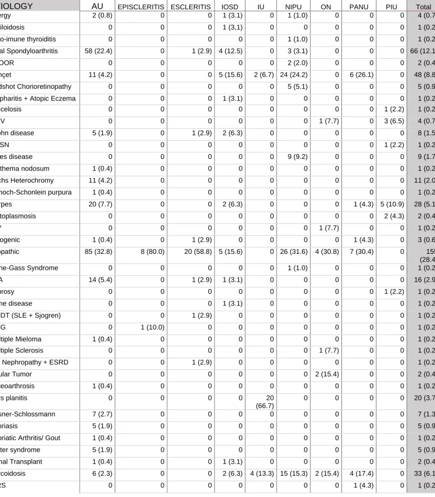
Table I: Etiology organized by uveitis anatomical classification in a tertiary referral centre in Porto, Portugal from 2012 to 2017
ETIOLOGY
Anatomical Classification (SUN) N (%)
Total
AU EPISCLERITIS ESCLERITIS IOSD IU NIPU ON PANU PIU
Allergy 2 (0.8) 0 0 1 (3.1) 0 1 (1.0) 0 0 0 4 (0.7) Amiloidosis 0 0 0 1 (3,1) 0 0 0 0 0 1 (0.2) Auto-imune thyroiditis 0 0 0 0 0 1 (1.0) 0 0 0 1 (0.2) Axial Spondyloarthritis 58 (22.4) 0 1 (2.9) 4 (12.5) 0 3 (3.1) 0 0 0 66 (12.1) AZOOR 0 0 0 0 0 2 (2.0) 0 0 0 2 (0.4) Behçet 11 (4.2) 0 0 5 (15.6) 2 (6.7) 24 (24.2) 0 6 (26.1) 0 48 (8.8) Birdshot Chorioretinopathy 0 0 0 0 0 5 (5.1) 0 0 0 5 (0.9)
Blepharitis + Atopic Eczema 0 0 0 1 (3.1) 0 0 0 0 0 1 (0.2)
Brucelosis 0 0 0 0 0 0 0 0 1 (2.2) 1 (0.2) CMV 0 0 0 0 0 0 1 (7.7) 0 3 (6.5) 4 (0.7) Crohn disease 5 (1.9) 0 1 (2.9) 2 (6.3) 0 0 0 0 0 8 (1.5) DUSN 0 0 0 0 0 0 0 0 1 (2.2) 1 (0.2) Eales disease 0 0 0 0 0 9 (9.2) 0 0 0 9 (1.7) Erythema nodosum 1 (0.4) 0 0 0 0 0 0 0 0 1 (0.2) Fuchs Heterochromy 11 (4.2) 0 0 0 0 0 0 0 0 11 (2.0) Henoch-Schonlein purpura 1 (0.4) 0 0 0 0 0 0 0 0 1 (0.2) Herpes 20 (7.7) 0 0 2 (6.3) 0 0 0 1 (4.3) 5 (10.9) 28 (5.1) Histoplasmosis 0 0 0 0 0 0 0 0 2 (4.3) 2 (0.4) HIV 0 0 0 0 0 0 1 (7.7) 0 0 1 (0.2) Iatrogenic 1 (0.4) 0 1 (2.9) 0 0 0 0 1 (4.3) 0 3 (0.6) Idiopathic 85 (32.8) 8 (80.0) 20 (58.8) 5 (15.6) 0 26 (31.6) 4 (30.8) 7 (30.4) 0 155 (28.4) Irvine-Gass Syndrome 0 0 0 0 0 1 (1.0) 0 0 0 1 (0.2) JRA 14 (5.4) 0 1 (2.9) 1 (3.1) 0 0 0 0 0 16 (2.9) Leprosy 0 0 0 0 0 0 0 0 1 (2.2) 1 (0.2) Lyme disease 0 0 0 1 (3.1) 0 0 0 0 0 1 (0.2) MCDT (SLE + Sjogren) 0 0 1 (2.9) 0 0 0 0 0 0 1 (0.2) MNG 0 1 (10.0) 0 0 0 0 0 0 0 1 (0.2) Multiple Mieloma 1 (0.4) 0 0 0 0 0 0 0 0 1 (0.2) Multiple Sclerosis 0 0 0 0 0 0 1 (7.7) 0 0 1 (0.2)
IgA Nephropathy + ESRD 0 0 1 (2.9) 0 0 0 0 0 0 1 (0.2)
Ocular Tumor 0 0 0 0 0 0 2 (15.4) 0 0 2 (0.4) Osteoarthrosis 1 (0.4) 0 0 0 0 0 0 0 0 1 (0.2) Pars planitis 0 0 0 0 20 (66.7) 0 0 0 0 20 (3.7) Posner-Schlossmann 7 (2.7) 0 0 0 0 0 0 0 0 7 (1.3) Psoriasis 5 (1.9) 0 0 0 0 0 0 0 0 5 (0.9)
Psoriatic Arthritis/ Gout 1 (0.4) 0 0 0 0 0 0 0 0 1 (0.2)
Reiter syndrome 5 (1.9) 0 0 0 0 0 0 0 0 5 (0.9)
Renal Transplant 1 (0.4) 0 0 1 (3.1) 0 0 0 0 0 2 (0.4)
Sarcoidosis 6 (2.3) 0 0 2 (6.3) 4 (13.3) 15 (15.3) 2 (15.4) 4 (17.4) 0 33 (6.1)
SLE 0 0 1 (2.9) 3 (9.4) 0 0 0 0 0 4 (0.7) Sjogren 0 1 (10.0) 0 2 (6.3) 0 0 0 1 (4.3) 0 4 (0.7) Syphylis 1 (0.4) 0 0 0 1 (3.3) 0 0 0 8 (17.4) 10 (1.8) Systemic sclerosis 1 (0.4) 0 0 0 0 0 0 0 0 1 (0.2) TINU 3 (1.2) 0 0 0 0 0 0 0 0 3 (0.6) Toxicity to etambutol 0 0 0 0 0 0 1 (7.7) 0 0 1 (0.2) Toxocariasis 0 0 0 0 0 0 0 0 2 (4.3) 2 (0.4) Toxoplasmosis 0 0 0 0 1 (3.3) 0 0 0 16 (34.8) 17 (3.1) Tuberculosis 14 (5.4) 0 6 (17.6) 1 (3.1) 1 (3.3) 0 0 2 (8.7) 6 (13.0) 30 (5.5) Ulcerative Colitis 1 (0.4) 0 0 0 1 (3.3) 0 0 0 0 2 (0.4) Urticaria 0 0 0 0 0 1 (1.0) 0 0 0 1 (0.2) VKH disease 2 (0.8) 0 0 0 0 11 (11.2) 1 (7.7) 0 0 14 (2.6) Wegener Granulomatosis 0 0 1 (2.9) 0 0 0 0 0 0 1 (0.2) TOTAL 259 (47.5) 10 (1,8) 34 (6.2) 32 (5,9) 30 (5.5) 98 (18.0) 13 (2.4) 23 (4.2) 46 (8.4) 545 Table I: Continued
AU – Anterior Uveitis, AZOOR - Acute Zonal Occult Outer Retinopathy, CMV - Citomegalovirus, DUSN - Diffuse Unilateral Subacute Neuroretinitis, ESRD - End-stage Renal Disease, HIV - Human
Immunodeficiency Virus, IOSD – Inflammatory Ocular Superficial Disease, IPU – Infectious Posterior Uveitis, IU – Intermediate Uveitis, JRA - Juvenile Rheumatoid Arthritis, MCDT - Mixed Connective Tissue Disease, MNG - Multi-Nodular Goiter, NIPU – Non-Infectious Posterior Uveitis, ON – Optic Neuropathy, PANU – Pan-Uveitis, SIRS - Systemic Inflammatory Response Syndrome, SLE - Systemic Lupus Erythematosus, SUN - Standardisation of Uveitis Nomenclature, TINU - Tubulointerstitial nephritis and uveitis syndrome, VKH - Vogt-Koyanagi-Harada
Table II: Uveitis classified according to sex, age, complications and laterality Anatomical Classification (SUN) N (%)
AU EPISCLERITIS ESCLERITIS IOSD IU NIPU ON PANU PIU
Sex Female 132 (51.0) 8 (80.0) 23 (67.6) 22 (68.8) 20 (66.7) 56 (57.1) 8 (61.5) 13 (56.5) 28 (60.9) Male 127 (49.0) 2 (20.0) 11 (32.4) 10 (31.3) 10 (33.3) 42 (42.9) 5 (38.5) 10 (43.5) 18 (39.1) Age Interval (years) Under 20 24 (9.3) 0 2 (5.9) 2 (6.3) 4 (13.3) 2 (2.0) 1 (7.7) 2 (8.7) 0 21-30 34 (13.1) 1 (10.0) 4 (11.8) 2 (6.3) 6 (20.0) 13 (13.3) 3 (23.1) 2 (8.7) 10 (21.7) 31-40 25 (9.7) 4 (40.0) 6 (17.6) 8 (25.0) 3 (10.0) 16 (16.3) 2 (15.4) 4 (17.4) 4 (8.7) 41-50 50 (19.3) 2 (20.0) 9 (26.5) 3 (9.4) 11 (36.7) 18 (18.4) 5 (38.5) 3 (13.0) 12 (26.1) 51-60 57 (22.0) 2 (20.0) 6 (17.6) 8 (25.0) 2 (6.7) 23 (23.5) 2 (15.4) 6 (26.1) 11 (23.9) 61-70 30 (11.6) 1 (10.0) 2 (5.9) 3 (9.4) 3 (10.0) 12 (12.2) 0 2 (8.7) 5 (10.9) 71-80 30 (11.6 0 4 (11.8) 5 (15.6) 1 (3.3) 12 (12.2) 0 3 (13.0) 4 (8.7) Over 81 9 (3.5) 0 1 (2.9) 1 (3.1) 0 2 (2.0) 0 1 (4.3) 0 OH Not Present 232 (89.6) 10 (100) 32 (94.1) 30 (93.8) 29 (96.7) 88 (89.8) 13 (100) 19 (82.6) 41 (89.1) Present 27 (10.4) 0 2 (5.9) 2 (6.3) 1 (3.3) 10 (10.2) 0 4 (17.4) 5 (10.9)
Cataracts Not Present 231
(89.1)
10 (100) 33 (97.1) 30 (93.8) 29 (96.7) 90 (91.8) 13 (100) 22 (95.7) 44 (95.7) Present 28 (10.8) 0 1 (2.9) 2 (6.3) 1 (3.3) 8 (8.2) 0 1 (4.3) 2 (4.3)
Glaucoma Not Present 236
(91.1)
10 (100) 32 (94.1) 30 (93.8) 29 (96.7) 90 (91.8) 12 (92.3) 22 (95.7) 43 (93.5) Present 23 (8.9) 0 2 (5.9) 2 (6.3) 1 (3.3) 8 (8.2) 1 (7.7) 1 (4.3) 3 (6.4)
CME Not Present 246 (95.0) 10 (100) 32 (94.1) 31 (96.9) 26 (86.7) 86 (87.8) 13 (100) 17 (73.9 40 (87.0) Present 13 (5.0) 0 2 (5.9) 1 (3.1) 4 (13.3) 12 (12.2) 0 6 (26.1) 6 (13.0) Laterality Unilateral 137 (52.9) 5 (50.0) 17 (50.0) 7 (21.9) 9 (30.0) 39 (39.8) 6 (46.2) 8 (34.8) 30 (65.2) Bilateral 122 (47.1) 5 (50.0) 17 (50.0) 25 (78.1) 21 (70.0) 59 (60.2) 7 (53.8) 15 (65.2) 16 (34.8)
AU – Anterior Uveitis, CME - Cystoid Macular Edema, IOSD – Inflammatory Ocular Superficial Disease, IPU – Infectious Posterior Uveitis, IU – Intermediate Uveitis, NIPU – Non-Infectious Posterior Uveitis, OH - Ocular Hypertension, ON – Optic Neuropathy, PANU – Pan-Uveitis, SUN - Standardisation of Uveitis Nomenclature
Table III: Treatment therapies for each different anatomical type of uveitis Anatomical Classification (SUN) N (%)
AU EPISCLERITIS ESCLERITIS IOSD IU NIPU ON PANU PIU
Topical Corticoid Not Present 142 (54.8) 7 (70.0) 26 (76.5) 29 (90.6) 22 (73.3) 77 (78.6) 11 (84.6) 17 (73.9) 38 (82.6) Present 117 (45.2) 3 (30.0) 8 (23.5) 3 (9.4) 8 (26.7) 21 (21.4) 2 (15.4) 6 (26.1) 8 (17.4) Topical NSAID Not Present 232 (89.6) 6 (60.0) 25 (73.5) 32 (100) 27 (90.0) 84 (85.7) 12 (92.3) 19 (82.6) 44 (95.7) Present 27 (10.4) 4 (40.0) 9 (26.5) 0 3 (10.0) 14 (14.3) 1 (7.7) 4 (17.4) 2 (4.3) Anti- TNF agent Not Present 243 (93.8) 10 (100) 32 (94.1) 29 (90.6) 29 (96.7) 93 (94.9) 12 (92.3) 20 (87.0) 45 (97.8) Present 16 (6.2) 0 2 (5.9) 3 (9.4) 1 (3.3) 5 (5.1) 1 (7.7) 3 (13.0) 1 (2.2) Systemical Corticoid Not Present 219 (84.6) 8 (80.0) 27 (79.4) 22 (68.8) 22 (73.3) 58 (59.2) 11 (84.6) 15 (65.2) 38 (82.6) Present 40 (15.4) 2 (20.0) 7 (20.6) 10 (31.3) 8 (26.7) 40 (40.8) 2 (15.4) 8 (34.8) 8 (17.4)
Methotrexate Not Present 227
(87.6)
9 (90.0) 30 (88.2) 28 (87.5) 24 (80.0) 89 (90.8) 12 (92.3) 15 (65.2) 46 (100) Present 32 (12.4) 1 (10.0) 4 (11.8) 4 (12.5) 6 (20.0) 9 (9.2) 1 (7.7) 8 (34.8) 0
Azathioprine Not Present 248
(95.8)
10 (100) 32 (94.1) 28 (87.5) 29 (96.7) 92 (93.9) 13 (100) 23 (100) 46 (100) Present 11 (4.2) 0 2 (5.9) 4 (12.5) 1 (3.3) 6 (6.1) 0 0 0
Cyclosporine Not Present 258
(99.6) 10 (100) 33 (97.1) 31 (96.9) 29 (96.7) 81 (82.7) 13 (100) 23 (100) 45 (97.8) Present 1 (0.4) 0 1 (2.9) 1 (3.1) 1 (3.3) 17 (17.3) 0 0 1 (2.2) Antiviral Therapy Not Present 244 (94.2) 10 (100) 34 (100) 31 (96.9) 29 (96.7) 98 (100) 13 (100) 23 (100) 44 (95.7) Present 15 (5.8) 0 0 1 (3.1) 1 (3.3) 0 0 0 2 (4.3) Periocular Treatment Not Present 247 (95.4) 10 (100) 33 (97.1) 30 (93.8) 24 (80.0) 89 (90.8) 12 (92.3) 18 (78.3) 45 (97.8) Present 12 (4.6) 0 1 (2.9) 2 (6.3) 6 (20.0) 9 (9.2) 1 (7.7) 5 (21.7) 1 (2.2)
AU – Anterior Uveitis, Anti- TNF - Tumor Necrosis Factor, IOSD – Inflammatory Ocular Superficial Disease, IPU – Infectious Posterior Uveitis, IU – Intermediate Uveitis, NIPU – Non-Infectious Posterior Uveitis, NSAID - Nonsteroidal Anti-inflammatory Drug, ON – Optic Neuropathy, PANU – Pan-Uveitis, SUN - Standardisation of Uveitis Nomenclature
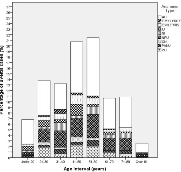
Figure I: Uveitis cases (%) organized according to age interval (years)
Legend: AU – Anterior Uveitis, IOSD – Inflammatory Ocular Superficial
Disease, IU – Intermediate Uveitis, NIPU – Non-Infectious Posterior Uveitis, PANU – Pan-Uveitis, PIU – Posterior Infectious Uveitis
Anexo 1
Normas de Publicação – European Journal of Ophtalmology
Manuscripts submitted to The European Journal of Ophthalmology (EJO) must be an original contribution not previously published in any language or country (except as an abstract or preliminary report) and must not be under
consideration for publication elsewhere. Likewise, updates of previously published studies that add little data to an existing publication will not be considered.
The Editor in Chief and the Editorial Board are primarily responsible for ensuring a fair review process. The final responsibility rests with the authors and the statements and opinions are solely those of the individual authors and contributors.
Original research articles
Previously unpublished manuscripts, describing clinical, pre-clinical,
epidemiological investigations, clinical trials, clinical observations, and other relevant investigations that are based on sound patient series, validated analytical methods, and appropriate statistical evaluation.
Original research articles should be structured as follows: Introduction (clearly stating an objective or hypothesis), Methods (describing the study design and methods applied, including the study setting and dates, patients or participants with inclusion and exclusion criteria, and/or participation or response rates, or data sources, and how these were selected for the study), Results (describing the results of the study in context with the published literature and addressing study limitations), and Discussion (addressing relevant implications for clinical practice or health policy). A structured abstract is required.
Words: max 3000 (excluding figures and tables) Figures/Tables: max 6
References: max 50
Manuscript preparation
Submissions need to be of sufficient editing quality in order that they are easily interpreted by the readership of the Journal. If submitted work does not meet this standard, it will be returned to the authors. The Journal follows the AMA Manual of Style for manuscripts submitted to biomedical journals.
Set your document as A4 (International Standard: ISO 216) paper, use double line spacing, Arial font size 12, number all pages, do not justify the right margin, and do not use line numbers. Save your manuscript as a Word document (.doc, .docx, or previous).
Structure your manuscript file as follows: Title page, Abstract and key words, Text, Acknowledgments, References, Tables, Figure legends.
Title page
The first page (title page) of your manuscript file must include the following information:
Full title (max 135 characters including letters and spaces), which must be concise and informative.
Short title (max 75 characters, including letters and spaces).
All authors listed as first name, initials, and last name (i.e., Paul M. Smith) with highest academic or medical degree first.
Institutional affiliation for each author, using superscripts and not symbols (e.g., Paul M. Smith1).
Corresponding author’s information (full mailing address, phone and fax numbers, email address); this is usually the submitting author.
Clinical Trial Protocol number when submitting a Clinical Trial Protocol. Online-only supplementary material, with a short description.
If you are submitting a manuscript that has been rejected previously, please inform the Journal of the previous review comments, and subsequent revision of the manuscript.
Manuscript word count (excluding figures and tables).
Abstract and key words
The abstract must not exceed 250 words and must be structured and divided in the sections indicated in each article type.
Below the abstract, identify 3 to 6 key words in alphabetical order under which you believe the article should be indexed. Use terms from the Medical Subject Headings list from lndex Medicus whenever possible. A library of terms is available at http://www.nlm.nih.gov/mesh/meshhome.html.
Manuscript text
Divide the text into the sections described above depending on content type. Use commas (,) to separate thousands and full stop (.) for decimals (e.g. 12,354.55). Include tables in the manuscript file, after the references. Number all figures (graphs, charts, photographs, and illustrations) in the order of their citation in the text. Figures must be submitted as separate files and not embedded in the Word document.
Units of measure
Laboratory values are expressed using conventional units of measure, with relevant Système International (SI) conversion factors expressed secondarily (in parentheses) only at first mention. Articles that contain numerous conversion factors may list them together in a paragraph at the end of the Methods section.
In tables and figures, a conversion factor to SI units should be provided in a footnote or legend. The metric system is preferred for the expression of length, area, mass, and volume. For more details, see the Units of Measure conversion table on the website of the AMA Manual of Style.
Names of drugs, devices, and other products
Use non-proprietary names of drugs, devices, and other products, unless the specific trade name of a drug is essential to the discussion. In such cases, use the trade name once and the generic or descriptive name thereafter. Do not include trademark symbols.
Cancer classification scheme
Authors are encouraged to use the American Joint Commission on Cancer TNM Classification scheme when describing tumor stage (see AJCC Cancer Staging Manual, 7th Edition, Springer, New York). The classification scheme can also be found at https://cancerstaging.org/references-tools/Pages/What-is-Cancer-Staging.aspx
Abbreviations
Use only standard abbreviations: the full term for which an abbreviation stands for should precede its first use in the text. Do not use abbreviations in the title. All abbreviations must be spelled out when they are used for the first time in the abstract and again when they are used for the first time in the text.
Abbreviations should appear first in parentheses immediately after the term or phrase to which they refer. Every abbreviation used in any table or figure should be defined in each corresponding legend. Please refer to the AMA Manual of Style for a listing of acceptable abbreviations and acronyms.
Acknowledgments
List in this section:
Any substantial contribution when provided by a person different from the author and list all other persons who do not fulfil authorship criteria.
The assistance of medical writing experts.
All participating group authors who do not meet the full authorship criteria. All sources of funding for the manuscript and the financial disclosures for all authors.
Written permission must be obtained to include the names of all individuals included in the Acknowledgments section.
If the manuscript has been presented at a meeting, please indicate in this section its name, location, and date.
References
Authors are responsible for the accuracy and completeness of their references and for correct text citation. Personal communications, unpublished data,
abstracts, and oral or poster presentations should be limited and incorporated in parentheses within the text without a reference number. A signed permission should be included from each individual identified in a personal communication or as a source for unpublished data, as well as the date of communication.
References should follow the text and begin on a separate page.
References must be double line spaced and numbered consecutively in order of appearance within the text, using the automated numbering tool of Word. Identify references in text, tables, and legends in Arabic numerals in parentheses, i.e. (7).
List all authors when six or fewer; when seven or more, list only the first three and add et al.
References used within tables or figure legends should be included in the reference list and numbered in consecutive order according to the table/figure citation in the text.
Journals’ names should be abbreviated according to Index Medicus/Medline. If there is any doubt about abbreviation of a journal name, it should be spelled out completely.
Any references to studies (including books or articles) that have been accepted for publication, but not yet published, should indicate where they will be published and have the term “in press” in the reference in place of volume and page numbers. These must be updated prior to publication, if possible. Do not add a discussion or comment to a reference.
Suffixes such as Jr, Sr, and III follow author’s initials.
Examples of reference style:
1. Standard journal article
Yealy DM, Kellum JA, Huang DT, et al. A randomized trial of protocol-based care for early septic shock. N Engl J Med. 2014;370(18):1683-1693.
References to web resources must always include the full link and the date the information was accessed and the link was live. (E.g. U. S. Food and Drug Administration. Postmarket drug safety information for patients and providers. http://www.fda.gov/Drugs/DrugSafety/PostmarketDrugSafetyInformationforPatie ntsandProviders/default.htm. Accessed June 10, 2014.)
Tables
Submit tables in your manuscript file after references. Do not submit them as separate files. As a general rule, tables should not unnecessarily offer duplicate information given within the text. Starting on a new page, type each table on a separate sheet, using double line spacing. Tables should be created in a Word
document using the table tool. Do not format tables as columns or tabs and do not submit tables as figures. Tables should be numbered consecutively in Roman numerals by order of citation in the text. Each table must include title, appropriate column headings, and explanatory legends, including definitions of any abbreviations used. References used within tables should be included in the reference list and numbered in consecutive order according to the table citation in the text. Identify statistical measures of variations such as SD and SEM. Follow the guidelines for creating tables.
Figure legends and legends for supplementary online-only material
At the end of the manuscript, include a short title and a legend for each figure. When symbols, arrows, numbers, or letters are used to identify parts of the figures, identify and explain each one clearly in the legend.
For photomicrographs, include the type of specimen, original magnification or a scale bar, and stain in the legend. For gross pathology specimens, label any rulers with unit of measure. Digitally enhanced images (CT/MRI, blots,
photographs, photomicrographs, ultrasound images, x-ray films, etc.) must be clearly identified in the figure legends as digitally processed images.
References used within figure legends should be included in the reference list and numbered in consecutive order according to the figure citation in the text.
Any figure that has been published elsewhere should have an acknowledgment to the original source; a copy of the permission to publish the figure, signed by the copyright holder, must accompany the submission.
Figures and illustrations
Number all figures (graphs, charts, photographs, and illustrations) in the order of their citation in the text. Include a title for each figure (a brief phrase,
preferably no longer than 10-15 words). Do not embed figures in the Word document. Figures must be submitted as individual .jpg or .tif files and have a high enough resolution for publishing. Do not submit figures as Word,
PowerPoint or PDF files.
Clinical photographs that identify an individual must be accompanied by a signed statement by the patient or legal guardian granting permission for publication of the pictures for educational purposes or must be masked to prevent identification of the patient.