UNIVERSIDADE DA BEIRA INTERIOR
Ciências
Construction of an immunosensor for human
cytomegalovirus infection diagnosis
Filipa Andreia Velez Pires
Tese para obtenção do Grau de Doutor em
Bioquímica
(3º ciclo de estudos)
Orientadora: Profª. Doutora Ana Cristina Mendes Dias Cabral
Coorientadora: Profª. Doutora María Julia Arcos Martínez
iii
To my grandma
To my parents
v
After this journey, I would like to thank those who were supportive and offered a word of motivation helping me to never give up and achieve my goals.
To professor Ana Cristina Cabral, my supervisor,
For the confidence she has put on me to integrate this project. This path was not always easy, but her availability, scientific guidance, enthusiasm, dedication, warmth and friendliness shown were crucial to me to fulfil this project. She was tireless in trying to keep me positive when everything seemed to go wrong.
To professor Julia Arcos Martínez, my co-supervisor,
For her availability, scientific guidance, professionalism, and all the knowledge and recommendations during these years. Her enthusiasm and positivism was truly motivator.
To CICS-UBI,
For being my “home” since I have started on this journey. Thank you to all the collaborators who are part of this institution, all of you made a small contribution for the development of this work.
To Juan Carlos Vidal and Juan Ramón Castillo,
For the warm way they welcomed me during my stay in Zaragoza. My stay there was a very enriching experience and was a great opportunity to learn.
To my lab mates (and friends),
For their invaluable collaboration, friendship and spirit of mutual aid. Always supporting me in the darkest hours, even when we were apart from each other. For this reason, they will always be my boys.
To my friends
I am grateful for your friendship, concerning and support. You have made this journey easier.
To my parents and grandpa
For your love, fondness, advices, recommendations and unconditional support. I owe everything to you. Thank you!
vii
O citomegalovírus humano (HCMV) é o maior vírus da família Herpesviridae e da subfamília β-herpesviridae. Como em todos os vírus herpes, a infeção pelo HCMV resulta no estabelecimento de uma infeção latente ao longo da vida do hospedeiro. Assim, sempre que uma pessoa é infetada, o vírus persiste num estado de latência celular, no qual as células infetadas não produzem nenhuma partícula infeciosa do vírus, mas retêm o seu genoma completo, tendo potencial para começar a produzir partículas virais mais tarde. Após infeção primária, o HCMV é excretado em fluidos corporais, como urina, sangue, saliva, lágrimas, secreções vaginais e cervicais, sêmen e leite materno. Este processo pode durar de meses a anos. Dessa forma, o HCMV pode ser transmitido por via oral, congénita, sexual, através da exposição à urina, por transfusão de sangue e transplante de órgãos ou medula óssea, sendo extremamente difícil identificar a sua via de transmissão.
O HCMV é considerado um vírus de paradoxos, pois este pode ser um potencial assassino ou um companheiro silencioso para toda a vida. Isto deve-se ao facto de a infeção pelo HCMV não induzir doença evidente em portadores saudáveis, devido a um controle imunológico efetivo, contudo a infeção pode ser grave e até fatal em indivíduos imunocomprometidos, como é o caso de transplantados, infetados pelo vírus da imunodeficiência humana (HIV) e aqueles com um sistema imunológico imaturo, como fetos e recém-nascidos. O HCMV também é considerado um dos mais bem-sucedidos parasitas, pois pode ser encontrado tanto em sociedades industrializadas e desenvolvidas como em grupos indígenas isolados, sendo a infeção por este vírus relativamente comum entre mulheres em idade reprodutiva, com seroprevalência variando de 45 a 100%.
O diagnóstico da infeção por HCMV permanece controverso, pois é difícil separar os pacientes assintomáticos (mas que excretam HCMV em fluidos corporais) e que poderão vir a necessitar de terapia, de pacientes com doença sintomática (pneumonia ou retinite). Atualmente, os métodos laboratoriais para o diagnóstico da infeção por HCMV podem ser divididos em técnicas sorológicas e virológicas. Os métodos sorológicos são usados principalmente para avaliar os anticorpos do doador ou do recetor em situações de transplante e prever o risco de os pacientes imunocomprometidos virem a desenvolver doença sintomática. Por outro lado, o diagnóstico virológico da doença por HCMV é geralmente baseado no isolamento do vírus por métodos de cultura. Estes métodos podem ser usados mediante a utilização de amostras de sangue, urina, saliva, fezes, lágrimas, leite materno, secreções cervicais e vaginais e sêmen. Os métodos mais comuns para o diagnóstico da infeção por HCMV são então: - testes sorológicos baseados na deteção de IgM e IgG; - a deteção direta de HCMV através de isolamento viral em cultura de fibroblastos e deteção de antigénios virais em amostras de tecido, urina ou saliva; e - PCR, que se baseia na amplificação de fragmentos específicos do genoma do HCMV e sua posterior hibridização. No entanto, estes métodos apresentam alguns inconvenientes na sua aplicação como métodos de triagem em laboratórios
de análises clínicas, pois requerem um longo período de tempo até à obtenção de um diagnóstico ou são caros. Assim, existe a necessidade de desenvolver um método que seja rápido, eficaz e barato para o diagnóstico deste vírus, capaz de ser usado em série.
Nos últimos anos, os biossensores eletroquímicos foram amplamente utilizados na determinação de variadas substâncias com diferentes propriedades e para a monitorização contínua de processos biológicos. A deteção eletroquímica é usada devido a sua sensibilidade aprimorada e custos de instrumentação reduzidos em comparação com outros métodos de transdução. Para além disto, para desenvolver dispositivos eletroquímicos confiáveis, miniaturizados e gerenciáveis, a tecnologia screen-printing é uma escolha inteligente. Os elétrodos serigrafados (SPE) contribuem para o desenvolvimento de novos biosensores em dispositivos miniaturizados, que apresentam as vantagens acima descritas, permitindo a obtenção de resultados em poucos minutos. Adicionalmente, os SPEs permitem uma produção massiva de sistemas eletródicos com tamanho e geometria uniformes, garantindo reprodutibilidade entre medições a baixo custo. Outra mais-valia destes sensores é o facto de serem descartáveis, o que evita alguns problemas frequentemente associados aos elétrodos tradicionais, como a necessidade de um processo de limpeza. Eles são igualmente bastante versáteis, uma vez que uma ampla gama de designs e materiais podem ser aplicados para na sua construção.
Na literatura podemos encontrar relatos do uso de dispositivos de deteção miniaturizados para o reconhecimento eletroquímico de sequências amplificadas de ADN provenientes de HCMV. Num desses trabalhos, baseado em elétrodos serigrafados, o ADN alvo foi adsorvido e hibridado com uma sonda de ADN biotinilada e os híbridos formados foram determinados com estreptavidina conjugada com peroxidase de rábano (HRP). Apesar da amplificação de sinal ter sido conseguida, a atividade do conjugado tem de ser controlada periodicamente devido à estabilidade da enzima. Para superar essa limitação, um outro grupo explorou outra estratégia recorrendo a marcação do ADN com nanopartículas de ouro. Apesar de terem tido melhores resultados, ambos os métodos descritos não descartam a utilização de PCR, o que os torna dispendiosos e inúteis como métodos de triagem. Um sensor piezoelétrico também foi descrito para detetar a glicoproteína do HCMV. Embora a técnica não dependa de ADN amplificado, requer o uso de instrumentação cara. Adicionalmente, um dispositivo de deteção baseado em imunofluorescência foi desenvolvido por outro grupo, aqui a amostra biológica é aplicada sobre uma superfície de ouro revestida com anticorpos específicos para HCMV (se presente em amostras biológicas, o HCMV é aprisionado na superfície deste). Ensaios positivos e negativos eram discriminados pelo uso de uma sonda fluorescente. A principal desvantagem deste dispositivo é a baixa sensibilidade que compromete a sua aplicabilidade em amostras com baixas cargas virais. Recentemente, foi ainda proposto um imunoensaio para a deteção do antígeno pp65 do HCMV utilizando HPR e nanopartículas de Pt-Pd funcionalizadas com single-walled nanohorns de carbono. A abordagem permitiu a deteção rápida de HCMV, no
ix
deteção e quantificação de HCMV gB. O objetivo é construir um imunossensor que determine a presença de gB em amostras de urina. O uso de anticorpos de captura contra as glicoproteínas do envelope do HCMV abre a possibilidade para o desenvolvimento de novos métodos de análise imunoquímica. A glicoproteína B do HCMV (gB) é uma glicoproteína viral que desempenha um papel crucial na entrada do vírus na célula e surge durante os estágios iniciais de uma infeção pelo mesmo vírus. A gB também é o antigénio dominante presente no envelope do HCMV, sendo possível a sua determinação em fluídos corporais como a urina e saliva, onde as cargas virais são maiores. Como consequência, o desenvolvimento de novos métodos baseados na deteção de gB em fluídos corporais é de grande interesse. Para a construção dos dispositivos, usamos sempre imunoensaios com configuração em sandwich, pois a gB é colocada entre um anticorpo primário, previamente imobilizado numa superfície sólida, e um anticorpo secundário marcado. Os imunoensaios em sandwich são atualmente os mais frequentemente usados, principalmente devido a sua alta sensibilidade e correspondente minimização de interferências. Para além disto, podem ser realizados em qualquer tipo de superfície, sendo o principal critério destes ensaios a disponibilidade de dois anticorpos com sítios de ligação diferentes para o mesmo antigénio-alvo.
Durante o decorrer deste trabalho foram desenvolvidos três imunoensaios diferentes. O primeiro foi um imunoensaio eletroquímico. Foram usados anticorpos de captura anti-gB absorvidos em elétrodos de carbono serigrafados e um anticorpo secundário anti-gB marcado com nanopartículas de ouro. A deteção de gB foi realizada por meio da análise eletroquímica de nanopartículas de prata depositadas quantitativamente no imunossensor através de catálise por nanopartículas de ouro, as quais foram utilizadas como marcadores do anticorpo secundário. A reprodutibilidade do método (RSDs de cerca de 12%) não foi muito boa devido à imobilização aleatória do anticorpo primário no elétrodo de trabalho, o que resultou numa pequena eficiência de deteção do antígeno (foram observados baixos sinais considerando a grande quantidade de anticorpo utilizado). Contribui-o também para a baixa RSD observada a deposição não específica de prata na superfície do sensor. Por estas razões, decidiu-se desenvolver outra abordagem para superar as limitações observadas.
Desenvolvemos um imunoensaio enzimático espectrofotométrico baseados em partículas magnéticas (mpEIA). O uso de esferas magnéticas (MBs) funcionalizadas com proteína G (MBs-prG) como superfície sólida para a imobilização do anticorpo primário (mAb1) permite a sua fixação orientada, resultando num reconhecimento mais efetivo do gB. Para além disto, estas partículas melhoram a interação de afinidade graças a uma cinética de análise mais rápida. O anticorpo secundário foi marcado com HRP para possibilitar a deteção espectrofotométrica. Os resultados obtidos com este mpEIA espectrofotométrico são favoravelmente comparáveis com outros relatos de deteção de gB em termos de desempenho analítico. No entanto, apesar das vantagens, os leitores ELISA não podem ser aplicados como dispositivos portáteis para fazer medições in situ.
Para superar essa limitação, o método mpEIA mencionado acima foi adaptado à transdução eletroquímica recorrendo ao uso de elétrodos serigrafados. Esta variação visou a obtenção de um dispositivo simples, sensível, descartável e portátil. É mantido o esquema de imunoensaio com base na proteína analítica gB intercalada entre um anticorpo monoclonal primário e o anticorpo secundário anti-gB marcado com HRP, que permite igualmente deteção eletroquímica. Da mesma forma, partículas magnéticas funcionalizadas com proteína G (MBs-prG) são usadas para permitir a imobilização orientada ao anticorpo (mAb1). O imunossensor desenvolvido mostrou ser um método portátil, rápido, preciso, rigoroso, de baixo custo e, portanto, eficaz na deteção de gB em amostras de urina humana para a valiosa triagem de infeções por HCMV.
Palavras-chave
Citomegalovírus humano, glicoproteína B, imunoensaios, mpEIA, imunossensores eletroquímicos, elétrodos serigrafados
xi
Human Cytomegalovirus (HCMV) is a herpes virus that establish a lifelong latent infection of the host, so once a person is infected, the virus persists in a state of cellular latency. Following primary infection, HCMV is excreted in body fluids and its transmission occurs through mucous contact and exposure to urine, blood transfusion and organ or bone marrow transplant procedures, being extremely difficult to identify the transmission route.
HCMV infection induces no overt disease in healthy carriers, owing to effective immune control, but this infection can be severe or even fatal in immunosuppressed individuals, fetuses and newborns. Furthermore, HCMV is also relatively common among women in reproductive age, with seroprevalence ranging from 45 to 100%.
The diagnosis of HCMV disease remains controversial because of the difficulty of separating patients who are asymptomatic but shedding HCMV in body fluids, from patients who have the symptomatic disease. Nowadays the most common methods for diagnosis of HCMV infection are: - serological tests based on IgM and IgG detection; - direct free HCMV detection by viral isolation and viral antigens detection in tissue, urine or saliva samples; and - PCR, which is based on amplification of selected segments of the HCMV genome and its hybridization. However, these methods are disadvantageous to be routinely used in clinical diagnosis as point of care because they require a long time to perform or are costly. Thus, there is a need to develop a method which is fast, effective and inexpensive for this virus diagnosis.
As an alternative, the use of capture antibodies against the envelope glycoproteins of HCMV open the possibility of faster immunochemical methods. Glycoprotein B of HCMV (gB) is the dominant antigen in the envelope of HCMV, being possible its determination in body fluids like urine and saliva, where viral loads are higher. In consequence, the development of new methods based on the accurate detection of gB in body fluids, is of great interest.
In recent years, electrochemical biosensors were widely used to determine various substances with different properties and for continuous monitoring of biological processes. Bioanalytical assays such as immunoassays (IAs), are also very important in many fields. IAs are based on antibodies ability to form complexes with the corresponding antigen, making them highly specific and selective. Thus, electrochemical immunoassays offer enhanced sensitivities and reduced instrumentation costs compared to their counterparts using other transducing elements. Also, screen-printed electrodes (SPE) contribute to develop miniaturized, easy to handle and reliable IAs devices. In addition, SPEs allow for a high-volume production of electrode systems with uniform size and geometry, ensuring measurement reproducibility at low cost. They are also very versatile, since a wide range of designs and materials can be applied in their construction.
The present work describes the development of an alternative method for HCMV gB detection and quantification. It is intended the development of an immunosensor to quantify the presence of gB in urine samples. For the construction of this device we made use of a
sandwich type immunoassay, wherein HCMV gB is sandwiched between a primary antibody, previously immobilized on a solid surface, and a labelled secondary antibody. Sandwich immunoassays are currently the most commonly and successfully used, mainly due to their high sensitivity and minimized background signal. Moreover, they can be performed on any kind of sensing surface, being the main criterion for these assays the availability of two antibodies with different binding sites on the target antigens.
Three different immunoassays were developed. The first one was an electrochemical immunoassay, gB detection was carried out over electrochemical stripping analysis of silver nanoparticles quantitatively deposited on the immunosensor through catalysis by nanogold labels. Capture anti-gB antibodies were absorbed on screen-printed carbon electrodes, and a secondary anti-gB antibody labelled with gold nanoparticles. Nevertheless, the reproducibility of the method (RSDs ≈ 12%) was not very good owing to the random immobilization of the primary antibody on the working electrode, which resulted in small efficiency of antigen detection. Contributing to the low observed RSD was also the nonspecific deposition of silver on the sensor surface. For these reasons, it was decided the development of another approach to overcome the observed limitations. A spectrophotometric magnetic particle-based enzyme immunoassays (mpEIA) was constructed. The use of magnetic beads (MBs) functionalized with protein G (MBs-prG) as solid surface for primary antibody (mAb1) immobilization allows its oriented attachment, resulting in a more effective recognition of gB. Additionally, they improve the affinity interaction thanks to a faster assay kinetics of the dispersed beads in urine samples. The results obtained with this spectrophotometric mpEIA compared favorably to those obtained in other reports of gB detection in terms of analytical performance. Despite the advantages, ELISA readers cannot be applied as portable devices to make in situ measurements. It was then proposed an adaptation to electrochemical transduction on screen-printed electrodes. This variation aimed the achievement of a simple, sensitive, disposable and portable device. It was maintained the immunoassay scheme based on the analyte protein gB sandwiched between the primary monoclonal antibody and the secondary anti-gB-HCMV HRP labelled antibody. Similarly, magnetic particles functionalized with protein G (MBs-prG), were used.
The developed immunosensor was shown to be a portable, fast, accurate, rigorous, low cost and an effective method of detecting gB in human urine samples for the valuable diagnosis/screening of HCMV infections.
Keywords
Human cytomegalovirus, glycoprotein B, immunoassays, mpEIA, electrochemical immunosensors, screen printed electrodes.
xiii
This doctoral thesis is structured in five chapters appendices, herein summarized:
CHAPTER I consists on a brief introduction aiming literature review and the definition of PhD thesis objectives. State of the art description emphases:
• Topics on human cytomegalovirus, with focus on virus structure as well as the problems associated to viral infection in humans;
• Basics underlining the construction of an immunoassay along with immunoassay advantages and disadvantages;
• Fundamentals on the construction of an electrochemical biosensor. With focus on the steps required for its assembly, from the choice of the biological recognition system to the choice of the transducer. Praticular importance was given to electrochemical immunosensors, once they represent the main objective of this work.
CHAPTER II discusses and integrates all the results obtained for the development of a sandwich type immunosensor for the detection of human cytomegalovirus glycoprotein B through electrochemical stripping analysis of silver nanoparticles quantitatively deposited on the immunosensor through catalysis by nanogold labels.
CHAPTER III discusses and integrates all the results obtained for the development of a magnetic particle-based enzyme immunoassay for human cytomegalovirus glycoprotein quantification CHAPTER IV discusses and integrates all the results obtained for the development of an amperometric immunosensor for Human cytomegalovirus glycoprotein B detection
CHAPTER V presents the conclusions of this thesis and future remarks
APPENDIX consists on the presentation of the electrochemical immunosensors national patent granted during the course of this work
xv
F. Pires, J. C. Vidal, J. R. Castillo, M. J. Arcos-Martínez, A. C. Dias-Cabral. Magnetic
particles-based amperometric immunosensor for Human Cytomegalovirus glycoprotein B detection. (paper submitted to Talanta)
F. Pires, A. C. Dias-Cabral, M. J. Arcos-Martínez, J. C. Vidal, J. R. Castillo. A rapid magnetic
particle-based enzyme immunoassay for human cytomegalovirus glycoprotein B quantification. Journal of Pharmaceutical and Biomedical Analysis 165 (2018): 372-378.
F. Pires, Hugo Silva, O. Domínguez-Renedo, M. A. Alonso-Lomillo, M. J. Arcos-Martínez, A. C.
Dias-Cabral. Disposable immunosensor for human cytomegalovirus glycoprotein B detection. Talanta 136 (2015): 42-46.
F. Pires, F. Marques, S. Almeida, F. C. Domingues, O. Domínguez-Renedo, M. A.
Alonso-Lomillo, M.J. Arcos Martínez, A. C. Dias-Cabral. “Dispositivo electródico para deteção de glicoproteína B, procedimento de fabrico e uso dos ditos dispositivos”. National invention patent nº 107029. Published in Boletim de Propriedade Industrial nº 248/2014 edited on 2014-12029. Patente granted on 2015 - 07- 08.
xvii
obtained during the PhD Course
J. Carvalho, S. Miguel, A. Moreira, J. Boga, F. Pires. Viruscan-sensors for viral detection. SCIENT Entrepreneurship Academy and Business Competition, Nicosia, Cyprus, September, 2017 (First prize).
F. Pires, M.J. Arcos-Martínez; A.C. Dias-Cabral, J.C. Vidal, J.R. Castillo, “mpEIA for Human
Cytomegalovirus glycoprotein B detection”. XII annual CICS-UBI symposium. Covilhã, Portugal, July 2017 (award of best PhD oral communication).
F. Pires, J.C. Vidal, J.R. Castillo, M.J. Arcos-Martínez; A.C. Dias-Cabral;
“Magneticparticles-based enzyme immunoassay for Human Cytomegalovirus detection”. II International Congress in Health Sciences Research. Covilhã, Portugal, May 2017.
F. Pires, J.C. Vidal, J.R. Castillo, M.J. Arcos-Martínez, A.C. Dias-Cabral. "New
spectrophotometric immunosensor for human cytomegalovirus glycoprotein B detection". XI Annual CICS Symposium. Covilhã, Portugal. July 2016.
F. Pires, H.Silva, O. Domínguez-Renedo, M. A. Alonso-Lomillo, M.J. Arcos Martínez, A. C.
Dias-Cabral. “Disposable immunosensor for human cytomegalovirus glycoprotein B detection”. XXXVI Reunión del grupo de electroquímica de la Real Sociedad Española de Química XVII Encontro Ibérico de Electroquímica. Vigo, Spain. July 2015
F. Pires, H.Silva, O. Domínguez-Renedo, M. A. Alonso-Lomillo, M.J. Arcos Martínez, A. C.
Dias-Cabral. “Imunossensor descartável para a detecção de glicoproteína B do citomegalovírus humano”. 4º Ciclo de Conferências da Faculdade de Ciências da Universidade da Beira Interior. Covilhã, Portugal. January 2015
F. Pires, H.Silva, O. Domínguez-Renedo, M. A. Alonso-Lomillo, M.J. Arcos Martínez, A. C.
Dias-Cabral. “Detection of Human Cytomegalovirus glycoprotein B using a disposable immunosensor”. IX Annual CICS Symposium. Covilhã, Portugal. July 2014
xix
Course
F. Pires, J. C. Vidal, J. R. Castillo, M. J. Arcos-Martínez, A.C Dias-Cabral. “New immunosensor
for human Cytomegalovirus detection”. Encontro Ciência, Lisbon, Portugal, July 2016.
F. Pires, J.C. Vidal, J.R. Castillo, M.J. Arcos-Martínez A.C. Dias-Cabral. "New Amperometric
immunosensors for Human Cytomegalovirus glycoprotein B detection". BioIberoAmerica 2016. Salamanca, Spain, June 2016.
F. Pires, J.C. Vidal, J.R. Castillo, M.J. Arcos-Martínez, A.C. Dias-Cabral. “Nuevos
inmunosensores amperométricos para la determinación de la glicoproteína B del citomegalovirus humano”. IV Jornadas del IUCA. Zaragoza, Spain, November 2015.
F.Pires, O. Domíngues-Renedo, M.A. Alonso-Lomillo, M.J.Arcos Martínez, A.C. Dias-Cabral.
“Development of an immunosensor for human cytomegalovirus glycoprotein b detection. X annual CICS-UBI symposium. Covilhã, Portugal, July 2015.
F. Pires, H. Silva, O. Dominguez-Renedo, M.A. Alonso-Lomillo, A.C. Dias-Cabral, M.J.
Arcos-Martínez. “Studies towards the development of a disposable immunosensor for diagnosis of Human Cytomegalovirus congenital infection- immobilization methods”. 10th European Symposium on Biochemical Engineering Sciences and 6th International Forum on Industrial Bioprocesses in collaboration with ACS. Lille, France, September 2014.
F. Pires, H.Silva, O. Domínguez-Renedo, M. A. Alonso-Lomillo, A. C. Dias-Cabral, M.J. Arcos
Martínez. “Disposable Immunosensor for diagnosis of Human Cytomegalovirus congenital infection”. XXXV Meeting of Electrochemistry of the Spanish Royal Society of Chemistry e 1st E3 Mediterranean Symposium: Electrochemistry for Environment and Energy. Burgos, Spain, July 2014.
F.Pires, O. Domíngues-Renedo, M.A. Alonso-Lomillo, M.J.Arcos Martínez, A.C. Dias-Cabral.
“Disposable Immunosensor for diagnosis of Human Cytomegalovirus congenital infection”. MicroBiotec. Aveiro, Portugal, December 2013.
xxi
Chapter I ... 1
I.1. Human Cytomegalovirus ... 3
I.1.1 Human Cytomegalovirus Overview
... 5
I.1.2 The Herpesviridae family
... 6
I.1.3. About the Human Cytomegalovirus
... 11
I.1.3.1. Structure ... 11
I.1.3.1.1. Glycoprotein B ... 15
I.1.3.2. Infectious cycle ... 16
I.1.3.3. Pathogenesis and infection Routes ... 18
I.1.3.4. Host defense ... 20
I.1.3.5. Seroprevalence ... 22
I.1.3.6. Prevention of HCMV infection ... 23
I.1.3.7. Clinical features and management of patients ... 25
I.1.3.8. Diagnosis ... 28
I.2. Immunoassays ... 35
I.2.1. Immunoassays overview
... 37
I.2.2. Antigens and antibodies
... 37
I.2.2.1. Antigen ... 37
I.2.2.2. Antibody ... 38
I.2.2.2.1. Antibody structure ... 38
I.2.2.2.2. Classes of antibodies ... 40
I.2.2.2.3. Antibody formats ... 42
I.2.3. Immunoassay Systems
... 45
I.2.3.1 Immunofluorescence assays ... 45
I.2.3.2. Radioimmunoassay ... 46
I.2.3.3. Enzyme immunoassays ... 46
I.2.3.4. Alternative immunoassays ... 49
I.3. Biosensors ... 51
I.3.1. Biosensor concept
... 53
I.3.2. Biological recognition system
... 54
I.3.2.1. Immobilization of the sensitive element ... 55
I.3.3. Transducer
... 57
I.3.3.1.Electrochemical transductors ... 59
I.3.4. Screen-printed electrodes
... 60
I.3.4.1. Screen printed electrodes fabrication ... 61
I.3.4.2. Materials used on screen printed electrodes and their modification ... 62
I.3.5. Electrochemical immunosensors
... 64
I.3.5.1. Antibody immobilization and its monitoring ... 64
I.3.5.2. Antibody labels ... 67
I.4. Aims ... 71 I.4.1. Aim of the thesis
... 73
Bibliography ... 75 Chapter II ... 85 II.1. Summary
... 89
II.2. Introduction
... 89
II.3. Material and Methods
... 91
II.3.1. Reagents and apparatus ... 91 II.3.2 Preparation of Au NP-labeled antibodies ... 92 II.3.3. Immunosensor preparation and measurement procedure ... 92 II.4. Results and discussion
... 93
II.4.1. Optimization of Detection Conditions... 93 II.4.1.1. Effect of Silver Deposition time on stripping current ... 93 II.4.1.2. Effect of Silver Enhancer Concentration ... 94 II.4.1.3. Effect of Antibody Concentration ... 95 II.4.1.4.Effect of BSA Concentration ... 96 II.4.1.5. Effect of gB Incubation Time ... 97 II.4.2. Immunosensor performance ... 98 II.4.3. Application in the analysis of spiked urine human samples ... 100 II.5. Conclusions
... 101
Acknowledgements... 102
Bibliography... 103
Chapter III ... 105 III.1. Summary... 109
III.2. Introduction... 109
III.3. Material and methods
... 111
III.3.1. Apparatus ... 111 III.3.2. Reagents ... 111 III.3.3. Primary antibody immobilization on functionalized magnetic beads ... 111 III.3.4. Immunoassay determination of gB ... 112 III.4. Results and Discussion
... 112
III.4.1.Optimization of the experimental conditions ... 113 III.4.2. Immunosensor analytical performance ... 116 III.5. Conclusions ... 123 Acknowledgments
... 123
Bibliography... 125
Chapter IV ... 129 IV.1. Summary... 133
IV.2. Introduction... 133
IV.3. Material and methods
... 135
IV.3.1. Apparatus ... 135 IV.3.2. Reagents ... 135 IV.3.3. Primary antibody immobilization on functionalized magnetic beads ... 136
xxiii
IV.4.2. Immunosensor Validation ... 143 IV.5. Conclusions
... 147
Acknowledgments
... 147
Bibliography
... 149
Chapter V ... 153 V.1.Concluding remarks and future perspectives
... 155
xxv
Figure I.1. Representation of Human Cytomegalovirus (HCMV). 5
Figure I.2. The 906 aa sequence of HCMV strain AD169 gB. a) Several structural features of gB. b) Antibody binding sites (AD-1, AD-2 and AD-3) and regions which are thought to contribute to assembled, conformational dependent antibody binding sites. Adapted from Ref. 23 and 30.
15
Figure I.3 HCMV replication summary. Adapted from Ref. 31. 16
Figure I.4. Owl’s eye inclusion. A single epithelial cell bearing an intranuclear inclusion. Withdrawal from Ref.29.
19
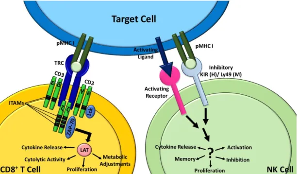
Figure I.5. a) T cell recognition and signaling. b) NK cell recognition and signaling. NK cell surface activating and inhibitory receptor–ligand interactions mediate the recognition and signaling of a NK cell. The combinatorial threshold that must be reached to activate or inactivate the NK cell is largely unknown. Adapted from Ref.43.
21
Figure I.6 Worldwide HCMV seroprevalence among women of reproductive age. Reproductive age was generally defined as between 12 and 49 years of age. Adapted from Ref.6.
23
Figure I.7. Antibody structure. Adapted from Ref.86. 39
Figure I.8. The four subclasses of human IgG. Adapted from Ref. 83. 40
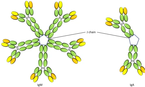
Figure I.9. The structure of the IgM pentamer and IgA. Adapted from Ref.83. 41
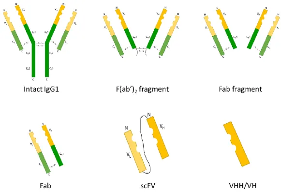
Figure I.10. Schematic representation of different antibody fragments types. Adapted from Ref.95.
44
Figure I.12. Schematic representation of direct ELISA. 47
Figure I.13. Schematic representation of indirect ELISA. 48
Figure I.14. Schematic representation of sandwich ELISA. 49
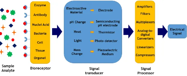
Figure I.15. Schematic representation of a biosensor. Adapted from Ref. 78, 110
and 115.
53
Figure I.16. Different immobilization methods a) Adsorption; b) entrapment; c) encapsulation; d) covalent binding and e) cross-liking. Adapted from Ref. 114.
56
Figure I.17. Traditional screen-printed electrodes constitution. Adapted from one of screen-printed electrode design made in University of Burgos.
60
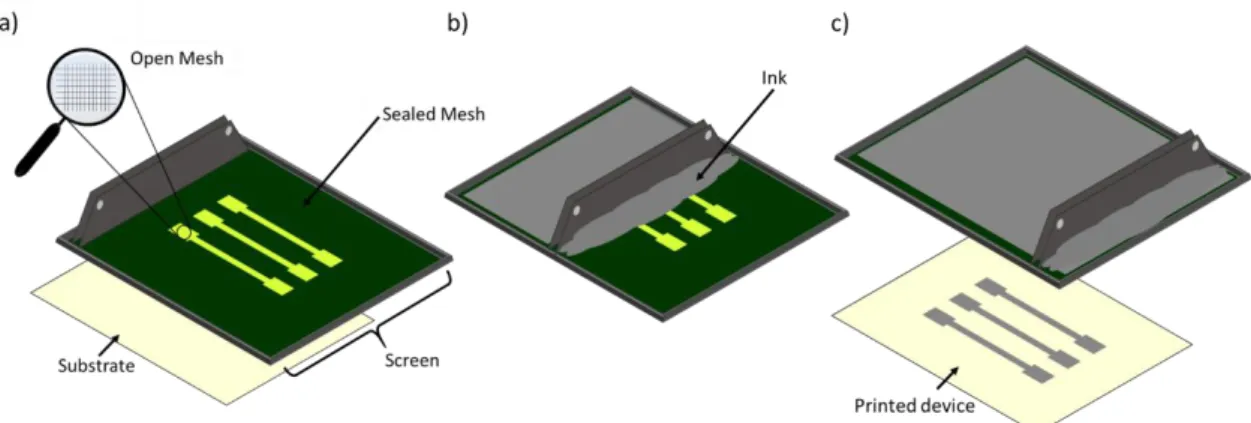
Figure I.18. Scheme of a screen-printed device fabrication process: a) screen poured; b) Squeegee traversing; c) Ink deposited on the substrate surface. Adapted from Ref.134.
61
Figure I.19. Schematic representation of the screens required for the construction of screen-printed electrodes. Adapted from screens used in University of Burgos.
62
Figure I.20. Schematic representation on antibody immobilization. a) random immobilization. b) oriented immobilization. Adapted from Ref.
148,151.
65
xxvii
for 15 ng mL-1 gB in 1.0 M KCl. Results are the mean±SD of n=3
measurements.
Figure II.3. Electrochemical signal intensity, for 15 ng mL-1 of gB in 1.0 M KCl, at
different concentrations of silver enhancer (study made in triplicate). (---) bare electrode; ( ---) electrode modified with Ab-AuNPS. a) 20-fold dilution; b) 140-fold dilution.
95
Figure II.4. Effect of antibody concentration on stripping-current response of AgNPs for 20 ng mL-1 of gB in 1.0 M KCl. Results are the mean±SD of
n=8 measurements. ---Blank; ---Assay.
96
Figure II.5. Effect of BSA concentration on stripping-current response of AgNPs for 20 ng mL-1 gB in 1.0 M KCl. Results are the mean±SD of n=8
measurements.
97
Figure II.6. Effect of incubation time on stripping-current response of AgNPs for 20 ng mL-1 gB in 1.0 M KCl. Results are the mean±SD of n=8 measurements.
95
Figure II.7. Linear-sweep stripping voltammetric curves of AgNPs a) and calibration curve b) for gB detection using the proposed strategy. gB was diluted in Tris-HNO3 buffer. a) Curves are the differential pulse
voltammetry responses for gB at concentrations from 0 to 15 ng mL-1.
98
Figure II.8. Linear-sweep stripping voltammetric curves of AgNPs a) and calibration curve b) for gB detection using the proposed strategy. gB was diluted in urine samples diluted 1:2 in tris-HNO3 buffer. a) Curves
are the differential pulse voltammetry responses for gB at concentrations from 0 to 15 ng mL-1.
99
Figure III.1. a) Schematic representation of the mpEIA sandwich assay method. --- ---Mbs-PrG-mAb1;- gB; --- Ab2-HRP b) Identification of the steps where blockage is needed.
Figure III.2. Absorbances measured after the well blockage with SuperBlock, Pierce ProteinFree and BSA blocking agent solutions, after incubations with Ab2-HRP for 60 min. The absorbance values are the mean±SD of n=5 replicates.
114
Figure III.3 Comparison between the use of PBS and PBS with 0.02% tween 20 during Ab2-HRP incubation and study of tween 20 effect in nonspecific antibody binding. The absorbance values are the mean±SD of n=5 replicates.
115
Figure III.4. Influence of the MBs-PrG-mAb1 blockage on background absorbances from NSA of Ab2-HRP and on absorbances from gB detection. An excess of concentration of 50 ng mL-1 gB was used. Results are the mean±SD
of n=5 replicates.
115
Figure III.5. Absorbance signals of the mpEIA assay method for gB concentrations from 0.00 to 6.000 ng mL-1 in 0.1 M. buffer matrix. a) and calibration
curve from 0.07 to 0.80 ng mL-1 b). Experimental conditions: 5 µg of
saturated with antibody MBs added to the wells (8 µg of mAb1 per mg of MBs-PrG). Results are the mean±SD of n=3 replicates.
116
Figure III.6. Calibration curve for mpEIA in urine diluted 1:2 (v/v) in PBS buffer from 0.09 to 0.70 ng mL-1. Experimental conditions: 5 µg of saturated
with antibody MBs added to the wells (8 µg of mAb1 per mg of MBs-PrG). Results are the mean±SD of n=3 replicates.
117
Figure III.7. Study of the cross-reactivity of EBV and VZV viruses on gB determination, and comparison with control solutions without gB. Results are the mean±SD of n=3 replicates.
121
Figure IV.1. Schematic representation of the developed mpEIA-based electrochemical immunosensor for the sensitive gB quantification.
xxix
modifications onto the electrode surface. An excess concentration of 1 µg mL-1 gB was used. Results are the mean ±SD of n=5 measurements.
Figure IV.3. Influence of the enzymatic reaction incubation time on TMBox reduction signal. An excess concentration of 1 µg mL-1 gB was used. Results are the mean ±SD of n=5 measurements.
139
Figure IV.4. Optimization of the amount of MBs-PrG-mAb1 to be used on the sensor surface. An excess concentration of 1 µg mL-1 gB was used. Results are the mean ±SD of n=5 measurements.
140
Figure IV.5. Temperature effect on the immunoassay response. An excess concentration of 1 µg mL-1 gB was used. Results are the mean ±SD of n=5 measurements.
140
Figure IV.6. Effect of antigen-antibody incubation time on current signal. An excess concentration of 1 µg mL-1 gB was used. Results are the mean ±SD of n=5 measurements.
141
Figure IV.7. Effect of Ab2-HRP concentration on current signal. An excess concentration of 1 µg mL-1 gB was used. Results are the mean ±SD of n=5 measurements.
142
Figure IV.8. a) Immunosensor current response for gB concentrations from 0 to 100 ng mL-1 in 0.1 M. buffer matrix. b) calibration curve from 0 to 60 ng mL-1. Experimental conditions: 6 µg of MBs-PrG-mAb1 added to the sensor surface (8 µg of mAb1 per mg of MBs-PrG). Results are the mean ±SD of n=3 measurements.
144
Figure IV.9. Immunosensor current response for gB concentrations from 0 to 40 ng mL-1 in urine matrix (dilute 1:2 in 0.1M buffer pH 7.4). Experimental conditions: 6 µg of MBs-PrG-mAb1 added to the sensor surface (8 µg of mAb1 per mg of MBs-PrG). Results are the mean ±SD of n=3 measurements.
Figure IV.10. Study of the cross-reactivity of EBV and VZV viruses on gB determination, and comparison with control solutions without gB. Results are the mean ±SD of n=3 measurements.
xxxi
Table I.1. Human herpesviruses basic properties. Adapted from Ref.17. 9
Table I.2 Selection of expression structure HCMV proteins. Adapted from Ref. 5 and 19.
12
Table I.3. Summary of diagnostic tests for identification of HCMV. 33
Table I.4. Physical properties of immunoglobulins. Adapted from Ref.
83. 42
Table II.1. Calibrations parameters for gB prepared in buffer and urine samples (1:2 dilution). RSD of analyte detection on different electrodes.
99
Table II.2. Results of HCMV gB determination in urine samples by application of the standard addition method. (N is the number of experimental data points).
100
Table III.1. Calibration linear parameters for gB determination in buffer and urine matrix samples. Urine samples were diluted 1:2 (v/v)) in PBS 0.1 M.
118
Table III.2. Comparison of the analytical performance of methods for the detection of HCMV antigens/antibodies.
119
Table III.3. Results of gB determination in spicked urine samples.
Calibration equation: .
Results are the mean±SD of n=5 replicates.
122
Table IV.1. Summary of the optimal parameters of mpEIA-based sandwich electrochemical immunosensor. 142
Table IV.2. Calibration linear parameters for gB determination in buffer and urine matrix samples. Urine samples were diluted 1:2 (v/v)) in PBS 0.1 M.
144
Table IV.3. Results of gB determination in spicked urine samples. Calibration equation:
Results are the mean±SD of n=3 measurements.
xxxiii
A
aa Aminoacid Ab Antibody
Ab2-HRP Antibody labelled with Horseradish peroxidase Ab-AuNPs Antibody labelled with gold nanoparticles ABS Antigen binding sites
AD Antigenic domain AFM Atomic force microscopy Ag Antigen
AgNPs Silver nanoparticles AuNPs Gold nanoparticles
B
BTM Bone marrow transplantation BSA Bovine serum albumin
C
C Antibody constant region CE Counter electrode CH Heavy constant region
CL Light constant region
CDR Complementarity-determining region CNS Central nervous system
CNTs Carbon nanotubes CTL Cytotoxic T lymphocytes CVC Capsid vertex-capping
D
DPI Dual polarization interferometry
E
EBV Epstein Barr vírus
ELISA Enzyme-linked immunosorbent assay EMEA European Medicines Agency
ERGIC Endoplasmic reticulum and Golgi complex intermediate compartment
F
Fab Antigen-binding fragments
FTIR Fourier transform infrared reflection
G
gB Glycoprotein B gH Glycoprotein H gL Glycoprotein L gM Glycoprotein M gM Glycoprotein M gO Glycoprotein OH
HCMV Human cytomegalovirus HDAC Histone deacetylase HHV Human herpes virusesHIV Human immunodeficiency virus HRP Horseradish peroxidase
HSV-1 Herpes simplex virus type 1 HSV-2 Herpes simplex virus type 2
I
IAs Immunoassays IE Immediate-early IF Immunofluorescence Ig Immunoglobulin IgA Immunoglobulin A IgG Immunoglobulin G IgM Immunoglobulin Mxxxv
KSHV Kaposi’s sarcoma associated herpes
L
L Late
LOD Limit of detection LTP Large tegument protein
LTPbp Large tegument binding protein
M
mAb Monoclonal antibody
mAb1 Primary monoclonal antibody MBs Magnetic micro-beads
MBs-PrG Magnetic particles functionalized with protein G
MBs-PrG-mAb1 Complex of magnetic particles functionalized with protein G with primary monoclonal antibody
mCBP Minor capsid binding protein MCP Major capsid protein
mCP Minor capsid protein MIE Major immediate early
mpEIA Magnetic particle-based enzyme immunoassay MWCNT Multi-wall carbon-nanotubes
N
NHS N-hydroxysuccinimide NK Natural killer
NMNPs Noble metal nanoparticles NPs Nanoparticles
NR Neutron reflectometry NSA Non-specific adsorptions
O
ORF Open Reading frame
P
pAb Polyclonal antibody
pAP Assembly protein precursor PBL Peripheral blood leukocytes PCR Polymerase chain reaction
pMHC Peptide-major histocompatibility complexes pNP1 Proteinase precursor
PoC Point of care PORT Portal protein
Q
QD Quantum dots
R
RE Reference electrode RIA Radioimmunoassay
RSD Relative standard deviation
RT-PCR Reverse transcriptase polymerase chain reaction
S
SAXs Small-angle X-ray scattering SCP Small capsid protein
SPE Screen printed electrode SPR Surface plasmon resonance SU Surface subunit
SWCNT Single-wall carbon-nanotubes
T
TCR T cell antigen receptors TER1 Terminase subunit 1 TER2 Terminase subunit 1 TM Transmembrane
TMB Peroxidase substrate 3,3’,5,5’-tetramethylbenzidine liquid substrate system for ELISA
xxxvii
V Antibody variable region VH Heavy variable region
VL Light variable region
VZV Varicella-zoster virus
W
WE Working electrode
β
C
HAPTER
I
3
5
I.1.1 Human Cytomegalovirus Overview
Human Cytomegalovirus (HCMV) is the most usual name for herpesvirus 5, a virus that belongs to Herpesviridae family and β-herpesviridae subfamily 1. It is the largest virus of the
family, with 200 nm in diameter, 240 kb in size and a molecular weight of 155 kDa, and is morphological indistinguishable from other herpes viruses, with a linear double-stranded DNA genome packaged in an icosahedral capsid (figure I.1). The capsid is surrounded by a protein layer known as tegument, and this is enclosed in a lipid bilayer that contains 6 encoded glycoproteins, gpUL55 (gB, glycoprotein B), gpUL73 (gN, glycoprotein N), gpUL74 (gO, glycoprotein O), gpUL75 (gH, glycoprotein H), UL100 (gM, glycoprotein M) and gpUL115(gL, glycoprotein L) 1–5. These glycoproteins perform an important role in the initial process of
interaction with the host cell 3,5. Specialy, glycoprotein B plays a crucial role in virus binding,
entry, cell-to-cell spread and cell fusion 5. In addition, gB is the major antigen, capable to
induce neutralizing antibodies against HCMV 3.
Figure I. 1. Representation of Human Cytomegalovirus (HCMV).
As for other herpesviruses, the assembly of the infectious particle is a complex and poorly understood process 2.
Human being is the only known receptor for HCMV and its transmission can occur both vertically and horizontally. HCMV is one of the most successful parasites, it can be found in both developed industrial societies and in isolated aboriginal groups, being relatively common among women in reproductive age, with seroprevalence ranging from 45 to 100% 5,6.
HCMV is considered a virus of paradoxes, because it can be a potential killer or a lifelong silent companion. The infection by HCMV results in the establishment of a lifelong latent infection of the host, so once a person is infected, the virus persists in a state of cellular latency, in which infected cells are not producing any infectious virus, but retain the complete genome and have the potential to start producing virus at a later time 1,3,4,7. HCMV infection
induces no overt disease, due to effective immune control, but the infection can be severe and even fatal in immunosuppressed individuals, as transplanted ones, persons infected by human immunodeficiency virus (HIV) and those with an immature immune system, like fetuses and newborns 7,8. Nevertheless, reactivation from latency to a state of active replication, is the
major cause of disease and can occur in a situation of immune system dysfunction 3,4.
In the case of immunosuppressed individuals infected by HCMV, pneumonia, retinitis, colitis and encephalopathies may be diagnosed, while newborns can manifest microcephaly, small body size, hepatomegaly, blindness, deafness, mental retardation, among other pathologies 9. Moreover, the infection by HCMV is the most frequent cause of embryonic and
fetal pathology induced by a virus in the whole world, although the majority of the infected children does not manifest any symptom at birth 5,9. In newborns, HCMV may be acquired in
utero via placenta or by exposure to maternal genital tract during labor. In this case, most perinatal infection by HCMV are asymptomatic5 and severe disease acquired perinatally occurs
in most of the cases in child underweight 9,10.
Primary infection does not usually result in a clinical illness, except in cases of congenital infection 5,7. Nevertheless, reactivation from latency to a state of active replication,
is the major cause of disease and can occur in a situation of immune system dysfunction, which can results from other illnesses 3,4.
In immunocompromised patients HCMV infection is mostly controlled by available antiviral drugs, yet it continues to maintain its role as one of the most dangerous infectious agent for the unborn infant 9.
I.1.2 The Herpesviridae family
Herpesviridae is a family of large and complex ubiquitous viruses that are constituted
by three major structural regions, capsid, tegument, and a lipid-containing envelope 2,11. Aside
from a similar morphology and some similarities in replication cycles, the major feature shared by all herpesviruses is the capacity to establish latent infection. Latent infection is defined as a type of persistent infection in which the viral genome is present but there is no production of infectious virus, except during intermittent episodes of reactivation 1,12–16. Following initial
infection, the viruses can be reactivated from the latent state producing, occasionally, episodes of significant, or serious disease. In this way, latency is the central feature of herpesviruses, however, the basic mechanisms involved in the processes of establishment and maintenance of latency and reactivation are not totally understood2,16,17.
Eight human herpesviruses (HHV) are known. These viruses are morphologically identical (figure I.1.), with a virion consisting of an icosahedral nucleocapsid of about 100 nm
7
HHV are divided into the α, β, and γ-herpesvirus subfamilies (Table I.1.) and this division was based on shared biological properties (i.e., host range, replication kinetics, and ability to spread in culture) and genetic relatedness 11.
The α herpesvirus subfamily consists ofherpes simplex viruses types 1 and 2 (HSV-1 and HSV-2, respectively) and varicella-zoster virus (VZV). These HHV have relatively short reproductive cycles (measured in days) and cause cytopathic effects on infected cells. They establish latent infections predominantly in the sensory ganglia17. Human cytomegalovirus
(HCMV), HHV-6A and B and HHV-7 make part of β-herpesviruses. These viruses have longer reproductive cycles (about weeks) and the infected cells often become enlarged. During latency period they can be maintained in leukocytes, kidneys, secretory glands and other tissues. Finally, γ- herpesviruses are Epstein-Barr virus (EBV) and HHV-8. This subfamily of herpesviruses is tropic for either T- or B-lymphocytes, and latency is often established in lymphoid tissue 17.
Herpes simplex viruses types 1 and 2 (HSV-1 and HSV-2) are among the most common human infectious viral pathogens 15. Primary HSV-1 and HSV-2 infection usually involve
ectodermally derived tissues, being the mucosal membranes of the mouth, throat, genitals and corneal epithelium the most affected tissiues 17. As the site of infection is often the site at
which the lesions appear, HSV clinical manifestation can be cold sores, genital ulcerations and corneal blindness 15–17. After initial infection, virus (or at least a subviral particle with an
associated viral genome) enter in the peripheral sensory nerves and migrate along nerve axons to associated in sensory ganglia 16.Upon reactivation from the latent state, the viruses migrate
back along sensory nerves to the body surface 17.
VZV is a ubiquitous virus, which causes varicella (chicken pox) and herpes zoster (shingles). While varicella results from primary VZV infection, and it is a common childhood illness associated with fever and a generalized pruritic vesicular, herpes zoster is caused by VZV reactivation. Herpes zoster is characterized by a localized, painful and vesicular rash involving one or adjacent dermatomes. The incidence of herpes zoster increases with age or immunosuppression 12. After initial replication in the respiratory epithelium, virus spreads to
regional lymph nodes and to the liver and spleen. Secondary viremia is mediated by mononuclear cells, which transport infection to cutaneous epithelial cells and respiratory mucosa. During this process, vesicles appear (chickenpox). VZV persists in the sensory ganglia of the central nervous system (CNS) 17.
HCMV is one of the most common cause of congenital infection in humans 18. During the
acute infection, epithelium-derived cells (e.g. ductal cells in the kidneys, alveolar cells in the lungs, and hepatocytes) are most commonly involved in viral replication. There is also evidence for leukocyte involvement during acute disease, with mononuclear cells being the most prominent ones 16.
HHV-6 has two distinct subtypes: HHV-6A and HHV-6B, being A variant more common14.
Primary infection occurs commonly in early childhood. Exanthema subitum (roseola infantum) is caused by HHV-6B, whereas disease associations for HHV-6A remain ambiguous.Both HHV-6A
and HHV-6B are lymphotropic 17. HHV-7 is similar to HHV-6A and -6B in many ways (for example,
its genome sequence and biology and it may also cause exanthema subitum) 17.This virus was
initially isolated from CD4+ T cells of a healthy individual when the activated cells in culture showed cytopathic effects. It can be detected in peripheral blood 14. Nevertheless, little is
known about HHV-7 17.
EBV virus infection results in clinical manifestation that include infectious mononucleosis, fever, sore throat, cervical and generalized lymphadenopathy, hepatosplenomegaly, and somatic complaints of fatigue and malaise 13. EBV is transmitted
through saliva, specially through the handling of toys, in the case of toddlers, or by kissing, being this the reason why infectious mononucleosis is commonly called “kissing disease” 13.
During latent stage, virus is maintained in B-lymphocytes 16.
HHV-8 or Kaposi’s sarcoma associated herpes virus (KSHV) is the most recent addition to the HHV family 17. HHV-8 is detectable in Kaposi’s sarcoma lesions and may also be associated
with other malignancies, since it seems that infect lymphocytes and is associated with cell immortalization and transformation 14,17.
Table I. 1.Human herpesviruses basic properties. Adapted from Ref. .
COMMON NAME GENOME SIZE PRIMARY TARGET SITE OF LATENCY Human α-herpesviruses HHV-1 Herpes simplex virus type 1 (HSV-1) ≈152 kb Mucoepothelial cells
(predominantly orofacial tract)
Neurons
HHV-2 Herpes simplex virus type 2 (HSV-2) ≈154 kb Mucoepothelial cells
(predominantly genital tract)
Neurons
HHV-3 Varicella zoster virus (VZV) ≈125 kb Respiratory epithelium Neurons
Human β-herpesviruses HHV-5 Human cytomegalovirus (HCMV) ≈240 kb Epithelial cells, monocytes, fibroblasts and more.
Leukocytes, epithelial cells
HHV-6 (A and B)
Roseleovirus ≈160 kb Epithelial cells, monocytes, fibroblasts and more.
Leukocytes, epithelial cells
HHV-7 Roseleovirus ≈145 kb Epithelial cells, monocytes, fibroblasts and more.
Leukocytes, epithelial cells
Human γ-herpesviruses HHV-4 Epstein Barr virus (EBV) ≈184 kb T- or B-lymphocytes Lymphoid tissue B cells
HHV-8 Kaposi’s sarcoma associated herpes virus (KSHV)
11
I.1.3. About the Human Cytomegalovirus
I.1.3.1. Structure
HCMV virion appears to be structurally similar to those of other herpesviruses, with a DNA core inside of a icosahedral capsid made up of 162 capsomeres surrounded by an envelope derived from host cell membrane containing viral glycoproteins tocontrol attachment and entry into cells19. As the larger virus of the family, HCMV has an icosahedral nucleocapsid with a
diameter of 100 nm that accommodates a 240 kb double stranded linear DNA genome. The nucleocapsid of HCMV is surrounded by protein layers, called tegument that, in its turn, is enclosed by an lipidic bilayer (envelope) that harbor a large number of viral glycoproteins 1–4.
Overall, the HCM virion is the most structurally complex of the herpesviruses and the mature virion presents a diameter of 200 nm, approximately 3,19.
HCMV capsids are composed of four core proteins: major capsid protein (MCP), encoded by UL86, minor capsid protein (mCP), encoded by UL85, minor capsid binding protein (mCBP) encoded by UL46 and the smallest capsid protein (SCP) encoded by UL48.5 20 (Table I.2).
Organization of these four proteins into a capsid appears to be coordinated by two genetically related, internally situated proteins, called the proteinase precursor (pNP1), encoded by UL80a, and the assembly protein precursor (pAP), encoded by UL80.5 20. One specialized penton
composed of the portal protein (PORT), encoded by UL104 acts as a channel for both encapsidation and release of viral DNA together with two principal subunits of the terminase, subunit 1 (TER1), encoded by UL89 gene and subunit 2 (TER2), encoded by the UL56. A capsid vertex-capping (CVC) complex composed of UL77 and UL93 proteins decorates all pentons and the proteins encoded by UL51 and UL52 provide stability20.Therefore, hexons from MCP are
responsible for the triangular faces of the capsid and their conjugation with pentons from MCP and PORT form the bulk of the 15-nm–thick capsid walls19.
The viral tegument is the region located between the capsid and the envelope and contains approximately 40% of the virion protein mass (Table I.2). Nevertheless, little is known about its structure or function 21,22. Nowadays, at least 32 virus-encoded proteins are known on
tegument, many of which are phosphorylated. These proteins, may play important roles in viral gene regulation, in modification of the host cell metabolism and in virion assembly 22. The most
abundant tegument protein in virions is pp65, encoded by UL83. This protein is highly immunogenic, however, despite its abundance and importance during natural infection, UL83 is dispensable for virus replication 19.
Virions, dense bodies and other noninfectious virus particles are enclosed in a lipid bilayer envelope. The envelope is derived from the endoplasmic reticulum, Golgi complex intermediate compartment (ERGIC) or endosomal membranes. The envelope has approximately 66 virus encoded proteins that play diverse roles during infection, ranging from mediating and
modulating entry and egress virus in the cell, influencing cell tropism, and interacting with the host response to infection. Nevertheless, just six envelope glycoproteins (gB, gH, gL, gM, gN and gO) provide essential replication functions and are targets of neutralizing antibody 19 (Table
I.2).
Glycoprotein B (gB) or gpUL55 is perhaps the most highly conserved envelope component and is also the most abundant envelope protein. gB function in the replicative cycle of HCMV is undefined, but studies suggest that it participates in both attachment and fusion of the virion to the cell 23. The characteristics and role of this protein will be discussed with more
detail in section I.1.3.1.1
Glycoprotein H (gH) or gpUL75 is lesser abundant than gB, however is perhaps the next most abundant protein component in the envelope. It has been proposed that gH mediates viral/host cell membrane fusion in the initial steps of virus infectivity. Also, monoclonal anti-gH antibodies exhibit potent virus neutralizing activity. This glycoprotein has been reported to associate covalently with glycoprotein L (gL) or gpUL100 and glycoprotein O (gO) or gpUL74 23.
gH/gL/gO complex mediate virus replication, more specifically its entry into epithelial and endothelial cells, but its basic structure is uncharacterized 24,25
Glycoprotein M (gM) or gpUL100 is also important during virus fusion and/entry in the cell and cell-to-cell virus spread. This protein forms a complex with glycoprotein N (gN) or gpUL55 and little is known about this complex immunogenicity during natural infection, nevertheless, as envelope glycoproteins, it is believed that they equally induce neutralizing antibodies 26. In conclusion, and as already was mentioned, these envelope glycoproteins linked
covalently to associate and form complexes that are highly conserved. Presently, the HCMV envelope consists of at least three distinct glycoprotein complexes, designed gCI, gCII, gCIII 27.
gCI is composed by homodimeric gB molecules, gCII is composed by gM and gN and gCIII is a heteroligomeric complex composed of gH, gL, and gO and all of these three complexes induce neutralizing antibodies 5,25,28.
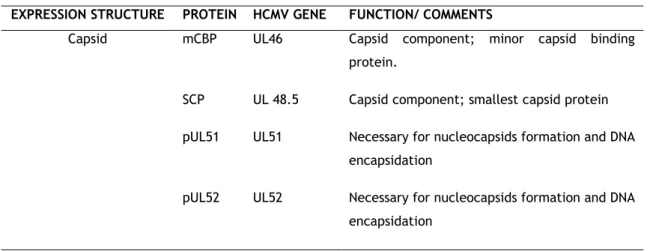
Table I. 2. Selection of expression structure HCMV proteins. Adapted from Ref. 5 and 19.
EXPRESSION STRUCTURE PROTEIN HCMV GENE FUNCTION/ COMMENTS
Capsid mCBP UL46 Capsid component; minor capsid binding protein.
SCP UL 48.5 Capsid component; smallest capsid protein pUL51 UL51 Necessary for nucleocapsids formation and DNA
encapsidation
pUL52 UL52 Necessary for nucleocapsids formation and DNA encapsidation
13 Table I. 1. Selection of expression structure HCMV proteins. Adapted from 5 and 19 (continued).
EXPRESSION STRUCTURE PROTEIN HCMV GENE FUNCTION/ COMMENTS
Capsid TER2 UL56 Binds to DNA packaging motif, exhibits nuclease activity (DNA encapsidation) pUL77 UL77 Supposed capsid vertex component; DNA
encapsidation pNP1 UL80A Capsid assembly pAP UL80.5 Capsid assembly
mCP UL85 Minor capsid protein; capsid component MCP UL86 Major capsid protein; capsid component TER1 UL89 Inhibition by antiviral compounds, DNA
encapsidation
pUL93 UL93 Supposed capsid vertex component; DNA encapsidation
PORT UL104 DNA encapsidation Tegument pUL36 UL36 (IE) IE; blocks apoptosis
LTPbp UL47 Associates with pUL48 and supports intracellular capsid transport
LTP UL48 Largest tegument protein, associates with pUL47 and is responsible for intracellular capsid transport
pp65 UL83 Gene regulation
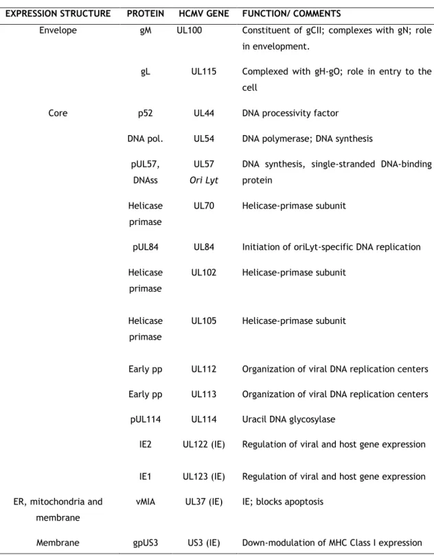
pIRS1 IRS1 (IE) Transactivator of viral gene expression pTRS1 TRS1 (IE) Transactivator of viral gene expression Envelope gB UL55 Major envelope glycoprotein, Constituent of
gCI, mediator of viral entry in the cell gN UL73 Constituent of gCII, complexes with gM to
support envelopment
gO UL74 Constituent of gCIII; enhances gH-gL delivery and release of virions
gH UL75 Constituent of gCIII; complexed with gL and gO; role in entry in the cell
Table I. 2. HCMV proteins discussed in this work. Adapted from Ref. 5 and 19 (continued).
EXPRESSION STRUCTURE PROTEIN HCMV GENE FUNCTION/ COMMENTS
Envelope gM UL100 Constituent of gCII; complexes with gN; role in envelopment.
gL UL115 Complexed with gH-gO; role in entry to the cell
Core p52 UL44 DNA processivity factor DNA pol. UL54 DNA polymerase; DNA synthesis
pUL57, DNAss
UL57
Ori Lyt
DNA synthesis, single-stranded DNA-binding protein
Helicase primase
UL70 Helicase-primase subunit
pUL84 UL84 Initiation of oriLyt-specific DNA replication Helicase
primase
UL102 Helicase-primase subunit
Helicase primase
UL105 Helicase-primase subunit
Early pp UL112 Organization of viral DNA replication centers Early pp UL113 Organization of viral DNA replication centers
pUL114 UL114 Uracil DNA glycosylase
IE2 UL122 (IE) Regulation of viral and host gene expression
IE1 UL123 (IE) Regulation of viral and host gene expression ER, mitochondria and
membrane
vMIA UL37 (IE) IE; blocks apoptosis
15
I.1.3.1.1. Glycoprotein B
Glycoprotein B (gB), as the most abundant protein in HCMV envelope, represents more than 50% of the envelope protein mass, and its encoding open reading frame (ORF), UL55, is probably the most highly conserved genome segment of the virus 23. Additionally, it is the most
easily detected envelope glycoprotein in infected cells, having, as mentioned before, an important role in replicative HCMV cycle, participating in both attachment and fusion of the virion to the cell 23,29.
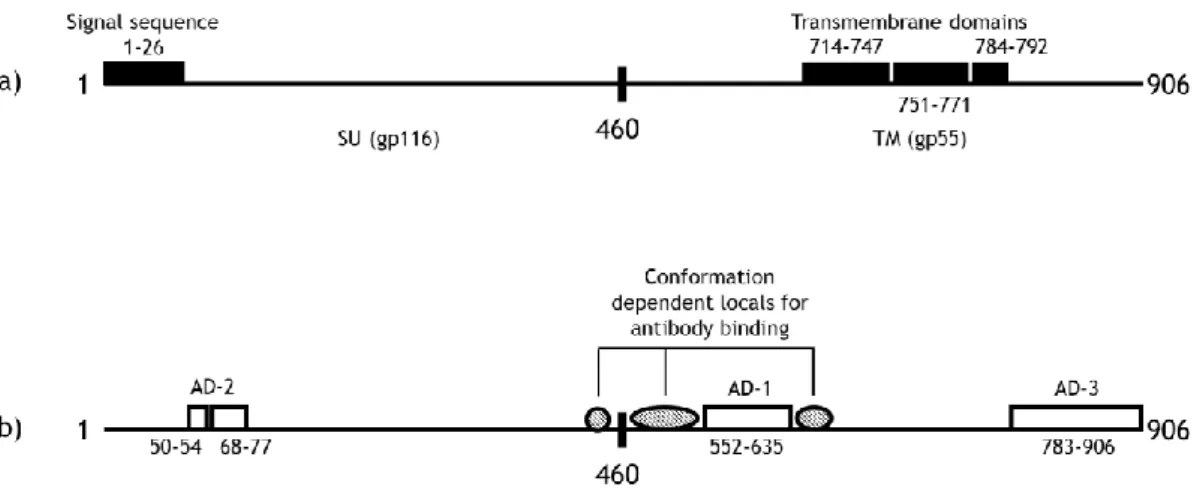
gB is a type I membrane glycoprotein, being composed by a transmembrane (TM), gp55 subunit and a surface subunit (SU), gp 116 (Fig. I.2a.). The analysis of aminoacids (aa) sequence from strain AD169 has suggested the presence of cleavable signal aa sequence between 1-26 aa, and three potential hydrophobic regions. These regions are aa between 714-747, 751-771 and 784-792 (figure I.2b.). The deletion of the first hydrophobic region leads to the failure in protein secretion, resulting in a protein that is incompletely processed. The second hydrophobic region, 751-771 aa, and the most hydrophobic domain is responsible for the gB insertion in the membrane. The importance of the third hydrophobic fourth region is still undefined 23.
Figure I. 2. The 906 aa sequence of HCMV strain AD169 gB. a) Several structural features of gB. b) Antibody binding sites (AD-1, AD-2 and AD-3) and regions which are thought to contribute to assembled, conformational dependent antibody binding sites. Adapted from Ref. 23 and 30.
The 906 aa polypeptide gB of strain AD169 is post-translationally modified into a 160 kDa glycosylated precursor molecule which is subsequently proteolytically cleaved in the position 460. Both subunits remain covalently linked by disulphide, representing the mature intracellular as well as the virion form of gB 10.
Several studies have demonstrated that a considerable fraction of the virus- neutralizing activity found in human serum following natural infection is directed against gB, addressing to gB the role of the dominant antigen on the envelope of HCMV 10,30
.
Three antibody-binding sites have been identified on gB: antigenic domain 1 (AD-1), located between aa 552-635; AD-2, aa 50–77; and AD-3, aa 783–906 (figure I.2b.) 30. AD-1
represents the dominant antibody-binding site on gB, once nearly all of the infected individuals who are seropositive for gB have antibodies against AD-1. Antibody binding requires the entire AD-1 sequence 30. AD-2comprises two sites, local I (residues 68-77 ) and local II (residues 50-54) 10. Of these three domains, just domain AD-1 and local II of AD-2 are able to induce
virus-neutralizing antibodies during the natural infection10.
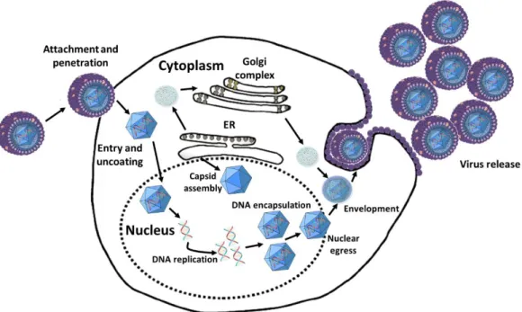
I.1.3.2. Infectious cycle
Virus entry occurs in distinct steps: (I) binding to specific cell surface receptors, (II) viral envelope fusion with cellular membranes to release nucleocapsids into the cytoplasm, either directly at the plasma membrane (as occurs in fibroblasts) or after endocytosis into cells (as occurs in endothelial and epithelial cells), (III) nucleocapsid translocation toward the nucleus on cytoskeletal filaments, (IV) nucleocapsid interaction with nuclear pores, and (V) release of the viral genome into the nucleus 19 (figure I.3.).
Figure I. 3. HCMV replication summary. Adapted from Ref. 31 .
The receptors for HCMV are widely distributed among host cell types, and contributes to the broad viral tropism observed during natural infections 5. The first step in this process