Article
Printed in Brazil - ©2018 Sociedade Brasileira de Química
*e-mail: [email protected]
Antimicrobial Activity of
Paepalanthus
planifolius
and its Major Components
against Selected Human Pathogens
MarceloR.deAmorim,aFelipeHilário,bPauloT.Sano,cTaisM.Bauabband
LourdesC.dosSantos*,a
aDepartamento de Química Orgânica, Instituto de Química and bDepartamento de Ciências Biológicas, Escola de Ciências Farmacêuticas,
Universidade Estadual Paulista (UNESP), 14800-900 Araraquara-SP, Brazil
cDepartamento de Botânica, Instituto de Biociências, Universidade de São Paulo (USP),
05508-090 São Paulo-SP, Brazil
The chemical investigation of ethyl acetate extract from Paepalanthus planifolius capitula resulted in the identification of 1H-naphtho[2,3-c]pyran-1-one,9-[(6-O-β-D -glucopyranosyl-β-D-glucopyranosyl)oxy]-3,4-dihydro-10-hydroxy-7-methoxy-3-methyl, semi-vioxanthin 9-O-β-D-glucopyranoside, toralactone-9-O-β-D-glucopyranoside, paepalantine-9-O-β-D-glucopyranoside, semi-vioxanthin, 1H-naphtho[2,3-c ]pyran-1-one,3,4-dihydro-9,10-dihydroxy-5,7-dimethoxy-3-methyl, vioxanthin and paepalantine dimer, and also the isolation and identification of a new naphthopyranone dimer named planifoliusin A. The chemical structures of two compounds were elucidated by performing spectroscopic 1D and 2D nuclear magnetic resonance (NMR) experiments and spectrometric HRMS (high-resolution mass spectrometry) analysis. Other six naphthopyranone dimers were proposed by MS fragmentation patterns. The minimum inhibitory concentration (MIC) values for vioxanthin (7.8 µg mL-1), planifoliusin A
(15.6 µg mL-1) and the ethyl acetate extract (31.2 µg mL-1) showed antimicrobial activity against Staphylococcus aureus (ATCC 25923).
Keywords:Paepalanthus planifolius, naphthopyranone, antimicrobial activity
Introduction
Staphylococcus aureus is a leading cause of bacterial infections in hospitals and communities worldwide, and is one of the most important agents responsible for healthcare-associated infections, it has been estimated that around 20-30% of the human population are carriers of
S. aureus. The increased occurrence of methicillin resistant
S. aureus (MRSA) strains has created genuine clinical and therapeutical problems.1
Considering that the antimicrobial resistances have been raised as a concern and the necessity to search for new antimicrobials agents as potential candidates for the treatment of several bacteria and fungi species, it is even more relevant to discover antimicrobial drugs.2
Eriocaulaceae are ornamental plants popularly known as “evergreens” and comprise about 1200 species
in 10 genera.3 Among these genus, Paepalanthus has approximately 357 species which 95% are endemic species restricted to Espinhaço Range in Brazil.4,5 Although Eriocaulaceae are used in the manufacture of ornamental products, literature reports studies with Eriocaulaceae species that have promising antimicrobial activity.6,7 Other studies have previously demonstrated the importance of extract and isolated substances in the Eriocaulaceae family, mainly because they present a great biological potential, such as the already identified antioxidant activities,8,9 cytotoxic,10,11 antiulcerogenic,12 antibacterial and vulvovaginal candidiasis treatment.13,14
Thus, in search of new biological compounds we described the antimicrobial activity of ethyl acetate (EtOAc) extract of capitula from Paepalanthus planifolius
Antimicrobial activities and identification of naphthopyranones in the extract
The minimum inhibitory concentration (MIC), minimum bactericidal concentration (MBC) and minimum fungicidal concentration (MFC) for the antimicrobial activity of the capitula EtOAc extract were evaluated against four human pathogenic microorganisms:
Escherichia coli (ATCC 25922), Salmonella setubal
(ATCC19196), Staphylococcus aureus (ATCC 25923) and
Candida albicans (ATCC 10231). Antimicrobial activity is considered good if the extract MIC values displayed lower than 100 µg mL-1; moderate from 100 to 500 µg mL-1; weak from 500 to 1000 µg mL-1; and over 1000 µg mL-1 the extract is considered inactive.15 Then the EtOAcextract showed a moderate activity against the Gram-negative bacteria tested, which displayed MIC values of 500 µg mL-1. For the Gram-positive bacteria, it was considered a good activity (MIC value of 31.2 µg mL-1), while for the yeast fluconazole-resistant C. albicans, it was shown a low activity (Table 1). Once that the extract showed promised activity against S. aureus, we performed its chemical study.
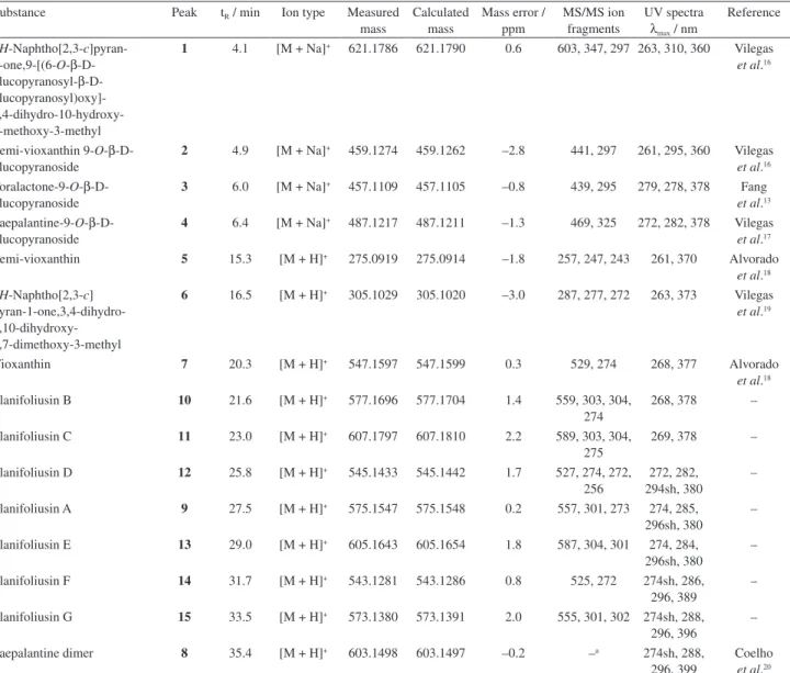
A preliminary analysis of the EtOAc extract of
P. planifolius capitula by LC-ESI-TOF-HRMS (liquid chromatography-electrospray ionization-time-of-flight-high resolution mass spectrometry) and LC-ESI-IT-MS/MS (liquid chromatography-electrospray ionization-ion trap-tandem mass spectrometry) revealed the presence of naphthopyranone derivatives, 1H-naphtho[2,3-c ]pyran-1-one,9-[(6-O-β-D-glucopyranosyl-β-D-glucopyranosyl) oxy]-3,4-dihydro-10-hydroxy-7-methoxy-3-methyl (1), semi-vioxanthin 9-O-β-D-glucopyranoside (2), toralactone-9-O-β-D-glucopyranoside (3), paepalantine-9-O-β-D-glucopyranoside (4), semi-vioxanthin (5), 1H- n a p h t h o [ 2 , 3 -c] p y r a n 1 o n e , 3 , 4 d i h y d r o
-showed absorption spectra in the UV of naphthopyranones, but were not found in the literature (Table 2, Figure 2). Then, the extract was carried out by the fractionation of gel permeation chromatography (Sephadex LH-20 column) and subjected to a purification using semipreparative HPLC-DAD (high-performance liquid chromatography-diode array detector), resulting in the isolation of a new compound named planifoliusin A (9) and the known vioxanthin (7) (Figure 1). The analogues planifoliusin B (10), planifoliusin C (11), planifoliusin D (12), planifoliusin E (13), planifoliusin F (14) and planifoliusin G (15) were proposed comparing the MS fragmentation patterns with planifoliusin A (9) and vioxanthin (7) and its UV spectra (see Supplementary Information).
Naphthopyranones exhibit a wide biological activity, including antimicrobial, anti-inflammatory, mutagenic, cytotoxic properties, among others.21 Thus, it is possible to relate that the good antimicrobial activity presented by the EtOAc extract of P. planifolius comes from this major naphthopyranones identified in the extract.
Elucidation of planifoliusin A (9)
Planifoliusin A (9) was isolated as an amorphous yellow powder and the LC-HRMS analysis exhibited an ion at
m/z 575.1547 [M + H]+ (calcd. 575.1548), suggesting the molecular formula C31H26O11.
Compound 9 was elucidated by nuclear magnetic resonance (NMR) spectra, including 1H, 13C, HSQC (heteronuclear single quantum correlation), HMBC (heteronuclear multiple bond correlation) and 1H-1H COSY (correlation spectroscopy), and showed some similarities when compared to paepalantine dimer and vioxanthin isolated from the Paepalanthus species.18,20 The 1H NMR spectrum displayed four signals of aromatic hydrogens at
Table 1. Antimicrobial activity of P. planifolius extract and compounds 7 and 9
Sample
MIC (MBC) / (µg mL-1) MIC (MFC) / (µg mL-1)
Gram-negative Gram-positive Yeast
E. coli S. setubal S. aureus C. albicans
Extract 500 (1000) 500 (1000) 31.2 (500) a
7 500 (1000) 1000 (1000) 7.8 (250) a
9 1000 (1000) 500 (1000) 15.6 (62.5) a
Ampicillin 6.25 12.5 0.15 a
Amphoterecin B NA NA NA 10.0
Fluconazole NA NA NA ND
dH 6.72 (s, 1H, H-6), 6.97 (s, 1H, H-5), 6.57 (s, 1H, H-4’) and 7.06 (s, 1H, H-6’). Other signals at dH 3.03 (m, 2H, H2-4) and 4.78 (m, 1H, H-3) were assigned to the methylene and methine groups, respectively (Table 3). It was observed the signals of three methoxyl groups at dH 3.93 (s, 3H, H-13’), 3.91 (s, 3H, H-12’) and 3.86 (s, 3H, H-12) and two methyl groups at dH 2.32 (s, 3H, H-11’) and 1.56 (d,
3H, J 6.0 Hz, H-11). The signals at dH 13.81 (s, 1H, H-10), 13.44 (s, 1H, H-10’), 9.74 (s, 1H, H-9) and 9.75 (s, 1H, H-9’) were attributed to hydroxyl groups.
Substance Peak tR / min Ion type Measured mass
Calculated mass
Mass error / ppm
MS/MS ion fragments
UV spectra
λmax / nm
Reference
1H-Naphtho[2,3-c]pyran- 1-one,9-[(6-O-β-D -glucopyranosyl-β-D - glucopyranosyl)oxy]- 3,4-dihydro-10-hydroxy-7-methoxy-3-methyl
1 4.1 [M + Na]+ 621.1786 621.1790 0.6 603, 347, 297 263, 310, 360 Vilegas et al.16
Semi-vioxanthin 9-O-β -D-glucopyranoside
2 4.9 [M + Na]+ 459.1274 459.1262 –2.8 441, 297 261, 295, 360 Vilegas et al.16 Toralactone-9-O-β-D
-glucopyranoside
3 6.0 [M + Na]+ 457.1109 457.1105 –0.8 439, 295 279, 278, 378 Fang et al.13 Paepalantine-9-O-β-D
-glucopyranoside
4 6.4 [M + Na]+ 487.1217 487.1211 –1.3 469, 325 272, 282, 378 Vilegas et al.17 Semi-vioxanthin 5 15.3 [M + H]+ 275.0919 275.0914 –1.8 257, 247, 243 261, 370 Alvorado
et al.18 1H-Naphtho[2,3-c]
pyran-1-one,3,4-dihydro- 9,10-dihydroxy-5,7-dimethoxy-3-methyl
6 16.5 [M + H]+ 305.1029 305.1020 –3.0 287, 277, 272 263, 373 Vilegas et al.19
Vioxanthin 7 20.3 [M + H]+ 547.1597 547.1599 0.3 529, 274 268, 377 Alvorado
et al.18 Planifoliusin B 10 21.6 [M + H]+ 577.1696 577.1704 1.4 559, 303, 304,
274
268, 378 –
Planifoliusin C 11 23.0 [M + H]+ 607.1797 607.1810 2.2 589, 303, 304, 275
269, 378 –
Planifoliusin D 12 25.8 [M + H]+ 545.1433 545.1442 1.7 527, 274, 272, 256
272, 282, 294sh, 380
–
Planifoliusin A 9 27.5 [M + H]+ 575.1547 575.1548 0.2 557, 301, 273 274, 285, 296sh, 380
–
Planifoliusin E 13 29.0 [M + H]+ 605.1643 605.1654 1.8 587, 304, 301 274, 284, 296sh, 380
–
Planifoliusin F 14 31.7 [M + H]+ 543.1281 543.1286 0.8 525, 272 274sh, 286, 296, 389
–
Planifoliusin G 15 33.5 [M + H]+ 573.1380 573.1391 2.0 555, 301, 302 274sh, 288, 296, 396
–
Paepalantine dimer 8 35.4 [M + H]+ 603.1498 603.1497 –0.2 –a 274sh, 288, 296, 399
Coelho et al.20 aauto-MS/MS mode acquisition did not acquire the fragmentations. t
R: retention time.
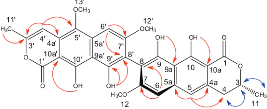
Comparing and contrasting the above information with the data derived from the analysis of the 2D NMR spectra, it was possible to conclude that 9 is a naphthopyranone dimeric with a monomeric lactone, similar to paepalantine and the other monomer similar to semi-vioxanthin.18,19 The 1H-1H COSY correlations indicated the sequence H-4 ↔ H-3 ↔ H-11 confirming the presence of semi-vioxanthin derivatives.19 In the HMBC spectrum, diagnostic long range correlations were observed for the unit derived from the semi-vioxanthin between H-5 with the carbons C-4, C-4a and C-5a. The unit of the paepalantine derivative was evident by the correlation of the H-4’ with C-4a’, C-3’ and C-5’. Additional evidence was given in the HMBC spectrum of which correlations to 2J and 3J of the OH-9 (dH 9.74) with C-9a (dC 108.5) and OH-10 (dH 13.81) with C-10a (dC 99.5) were observed, positioning these hydroxyls in the semi-vioxanthin monomer. The hydroxyls belonging to the paepalantine unit were also observed by the
correlations in the HMBC spectrum of the hydrogen signal of the hydroxyl OH-9’ (dH 9.75) with C-8’ (dC 108.6) and the OH-10’ (dH 13.44) with C-10a’ (dC 96.9). Therefore, what remains is to connect the monomer of the paepalantine and of the semi-vioxanthin. The non-observance of H-6/H-8
meta coupling and H-6’/H-8’ coupling combined with the chemical shift of C-8 (dC 108.1) and C-8’ (dC 108.6) made it possible to establish that the linkage between two monomeric units is between C-8/C-8’. Thus, the compound 9 was proposed to be made up by two different monomeric portions, semi-vioxanthin and paepalantine, linked through a C-8/C-8’ bond (Figure 3).
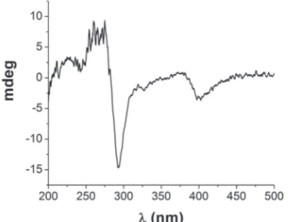
T h e C D ( c i r c u l a r d i c h r o i s m ) s p e c t r u m o f planifoliusin A (9) showed negative Cotton effect at 293 nm that corresponds to n →π* transition (CO lactone) (Figure 4). Comparison of this negative Cotton effect with (R)-semivioxanthin revealed the absolute configuration at C-3 of planifoliusin A to be R.22
Table 3. 1H (600 MHz) and 13C (150 MHz) spectroscopic data of planifoliusin A (9) in CDCl
3 [d in ppm, multiplicity (J in Hz)]
Position 1H 13C Position 1H 13C
1 – 171.7 1’ – 168.4
3 4.78 m 76.7 3’ – 152.4
4 3.03 m 34.9 4’ 6.57 s 99.5
4a – 133.1 4a’ – 122.3
5 6.97 s 116.3 5’ – 140.6
5a – 140.2 5a’ – 135.6
6 6.72 s 98.3 6’ 7.06 s 92.4
7 – 161.6 7’ – 162.1
8 – 108.1 8’ – 108.6
9 (OH) 9.74 s 155.5 9’ (OH) 9.75 s 155.9
9a – 108.5 9a’ – 108.9
10 (OH) 13.81 s 163.0 10’ (OH) 13.44 s 158.9
10a – 99.5 10a’ – 96.9
11 (CH3) 1.56 d (6.0) 20.9 11’ (CH3) 2.32 s 19.8
12 (OCH3) 3.86 s 56.1 12’ (OCH3) 3.91 s 56.2
13’ (OCH3) 3.93 s 62.0
d: chemical shift; J: coupling constant.
Vioxanthin (7) was identified by comparison of its 1H NMR, 13C NMR and MS data with literature values.18
Additionally, the CD spectrum of vioxanthin (7) allowed to confirm the (P,R,R)-vioxanthin.23
Antimicrobial activities of isolated compounds
Planifoliusin A (9) and vioxanthin (7) showed the respective MIC values of 1000 and 500 µg mL-1 for E. coli; 500 and 1000 µg mL-1 for S. setubal; both compounds exhibited significant activity against S. aureus with MIC values of 15.6 (9) and 7.8 µg mL-1 (7); and MIC values above 1000 µg mL-1 for C. albicans (Table 1). It has been commonly described that plant extracts are more active against Gram-positive bacteria than against Gram-negative bacteria.24 The existing difference between them can be explained because the cell walls of Gram-negative bacteria are less permeable to antimicrobial metabolites.25 In addition, the results for the MBC showed that the EtOAc extract and the compounds 9 and 7 displayed bactericidal activity from 62.5 to 1000 µg mL-1, while for S. aureus these compounds showed MBC of 62.5 (9) and 250µg mL-1 (7), indicating bacteriostatic behavior of these compounds for S. aureus.
The compounds 9 and 7 displayed a good antibacterial activity for the strains tested and, together with the results of the EtOAc extract, have shown pronounced efficacy against the Gram-positive bacteria S. aureus.
Conclusions
The LC-ESI-MSn analysis established a method for identifying naphthopyranones in the EtOAc extract of capitula from Paepalanthus planifolius in which 15 compounds were characterized. This is the first report on naphthopyranones dimers in Paepalanthus by LC-MS and this methodology could be used to identify these naphthopyranones in other Paepalanthus species.
due to the presence of naphthopyranones derivatives, including the new planifoliusin A (9) compound and vioxanthin (7). Future studies may be made to isolate the new naphthopyranones dimers proposed (10-15) to obtain spectroscopic data and their antimicrobial activities.
Experimental
General experimental procedures
1H NMR (600 MHz), 13C NMR (150 MHz), gHMBC,
gHSQC and gCOSY experiments were conducted on a Bruker Avance III 600 spectrometer using the non-deuterated residual solvent signal as a reference. Optical rotation was measured on a PerkinElmer 341-LC polarimeter. Circular dichroism was measured on a Jasco J-815 CD spectrometer. HPLC separations were carried out on a Jasco (PU-2089 Solvent Delivery Module and AS-2055 AutoSampler) coupled with a Jasco MD-2018 photodiode array detector (DAD) system using an RP-18 column (Knauer, Eurospher II 250.0 × 8.0 mm, 5 µm, flow rate 3.2 mL min-1) along with the protective guard column Phenomenex (4 × 3 mm). All solvents were purchased from Sigma-Aldrich (St. Louis, MO, USA), and Tedia (Fairfield, OH, USA) for HPLC analysis. The chromatography column was carried out on Sephadex LH-20 (75 × 2.0 cm, i.d.) obtained from Merck (Darmstadt, Germany).
LC-MS analysis
For the LC-MS analysis, 2.0 mg of the extract was dissolved in 2 mL of methanol:water (7:3, v/v). For both the analyses, the solution was filtered through a 0.22 µm polytetrafluoroethylene (PTFE) membrane and aliquots of 10 µL were directly injected into the LC-MS system.
The LC-ESI-TOF-HRMS experiment was performed using a UFLC (Shimadzu, Japan) containing two LC20AD solvent pumps, a SIL20AHT auto sampler, a CTO20A column oven and a CBM20A system controller and a diode array detector (SPD-M20AV), coupled with a microTOF (Bruker Daltonics, USA) mass spectrometer. The analyses were performed under the following conditions: capillary voltage, 3.5 kV; capillary temperature, 220 °C; end plate offset voltage, 490 V; nebulizer gas pressure, 5.5 bar; dry gas (N2), 10 L min-1. Mass spectra were measured from
m/z 50-1300 in positive ion mode.
The LC-ESI-IT-MS/MS analyses were performed using a UFLC (Shimadzu), coupled with an AmaZon SL ion trap mass spectrometer (Bruker). The ion trap acquisition Figure 4. CD spectrum (solvent: acetonitrile) of planifoliusin A (9)
parameters were as follows: capillary voltage, 3.5 kV; end plate offset, 500 V; nebulizer gas pressure, 60 psi; dry gas (N2), 10 L min-1; dry gas temperature, 320 °C. CID fragmentation was achieved in auto-MS/MS mode.
The analysis was performed using a gradient mode, eluted with a water:methanol gradient (70-92% methanol, 45 min) containing 0.1% formic acid, the flow rate was 1.0 mL min-1, RP-18 column (Knauer, Eurospher II 250.0 × 4.6 mm, 5 µm) along with the protective guard column (Phenomenex, 4 × 3 mm) and the column oven was set at 30 °C. The software Bruker Compass DataAnalysis 4.2 was used to control the system, for data collection, and processing. The mass analyzer was calibrated using a solution of sodium trifluoroacetic acid as the internal standard.
Plant material
Authenticated Paepalanthus planifolius (Bong.) Körn (Eriocaulaceae) was collected in the Serra do Cipó, Minas Gerais, Brazil, 19°13’21.64’’S, 43°30’04.06’’W, in October of 2013 and identified by Prof Dr Paulo Takeo Sano. A voucher specimen (Sano 4979) was deposited at the Institute of Biosciences, USP, São Paulo, Brazil.
Extraction and isolation
The dried powder of the P. planifolius capitula (50.1 g) was extracted with ethyl acetate (EtOAc, 1 L) for 48 h at room temperature by maceration and the same process was repeated for five times until exhaustion of the plant material. The solution was evaporated to dryness in vacuo to give the EtOAc extract (1.7 g, 3.4%), which was stored at 4 °C.
A portion of the extract (200 mg) was solubilized in EtOAc (5.0 mL) using ultrasound for 10 min and then centrifuged for 15 min. The precipitate (14 mg) was separated from the supernatant. The supernatant was filtered and fractionated by gel permeation chromatography using a Sephadex LH-20 column and eluted with EtOAc to give 5 fractions which were analyzed by silica gel thin layer chromatography (TLC, chloroform:EtOAc, 1:1, v/v, organic phase). Fractions 3-5 were combined and further purified by semipreparative HPLC-DAD which led to the isolation of planifoliusin A (9, 2.5 mg) and vioxanthin (7, 11.2 mg).
Antimicrobial activities
Antibacterial activity and minimum bactericidal concentration (MBC)
The evaluation of the antibacterial activity and the minimal inhibitory concentration (MIC) were determined
by the broth microdilution method, as described in the M7-A6 reference guideline of the Clinical and Laboratory Standards Institute.26 The biological activity was evaluated against the bacteria E. coli (ATCC 25922),
S. setubal (ATCC19196) and S. aureus (ATCC 25923), all the microorganisms were obtained from the American Type Culture Collection (Rockville, MD, USA). They are incubated in the Muller-Hinton broth for 24 h at 37 °C. These inoculums were standardized at 1.0 × 10⁸ CFU mL-1 (corresponding to 0.5 McFarland standards) by adjusting the optical density to 0.10-0.15 at 620 nm. The assay was carried out in 96-well microplates containing 80 µL of Muller-Hinton broth and 20 µL of standardized inoculum.
The extract and the substances 9 and 7 were dissolved in DMSO:H2O (2:8, v/v) to initial concentrations of 1000 µg mL-1. A two-fold serial dilution was carried out in order to obtain concentration ranging from 7.81 to 1000 µg mL-1 and 100 µL of each concentration were added to 96-well microplates. Ampicillin was used as positive control while DMSO:H2O (2:8, v/v) was used as the negative control. The plates were incubated at 37 °C for 24 h. The assay was displayed in triplicate.
The MIC of the samples was detected using spectrophotometric reading at 595 nm and with the addition of 30 µL of resazurin solution (100 µg mL-1), incubated at 37 °C for 2 h. The growth of bacteria changes the blue dye resazurin into a pink. The color pink indicates positive growth, whereas the color blue indicates growth inhibition. MIC was defined as the lowest sample concentration which prevented this change and exhibited inhibition of microorganism growth. For the determination of minimal bactericidal concentration (MBC), a portion from each well that showed antibacterial activity was plated on Muller-Hinton agar and incubated at 37 °C for 24 h. The lowest concentration that showed no bacteria growth in the subcultures was used as the MBC.6
Antifungal activity and minimum fungicidal concentration (MFC)
The evaluation of the antifungal activity and the minimal inhibitory concentration (MIC) were determined by the broth microdilution method, as described in the M27-A3 reference guideline of the Clinical and Laboratory Standards Institute, with modifications.27,28 The biological activity was evaluated against the fluconazole-resistant
Candida albicans (ATCC 10231).
of RPMI 1640 followed by a two-fold serial dilution to obtain concentration ranges of 7.81-1000 µg mL-1 to the extract and the isolated compounds. Fluconazole and amphotericin B were used as a positive control while DMSO:H2O (2:8, v/v) was used as the negative control. The plates were incubated at 37 °C for 48 h. The assay was displayed in triplicate.
The MIC of the samples was detected using spectrophotometric reading at 595 nm after the addition of 20 µL triphenyl-tetrazolium chloride (TTC) solution (0.02 g mL-1) incubated at 37 °C for 2 h. Yeast growth changes the colorless TTC to red. MIC was defined as the lowest sample concentration that prevented this change and exhibited inhibition of microorganism growth. For the determination of minimal fungicidal concentration (MFC), a portion from each well that showed antibacterial activity was plated on Sabouroud agar and incubated at 37 °C for 48 h. The lowest concentration that demonstrated no yeast growth in the subcultures was used as the MFC.6
Supplementary Information
Supplementary information (1H and 13C spectra for compounds 7 and 9, CD spectrum of vioxanthin (7), UV spectra and LC-MS analyses for compounds 1-15 and proposed fragmentation pathways of compounds 7-15) is available free of charge at http://jbcs.sbq.org.br as a PDF file.
Acknowledgments
We would like to thank the Coordenação de Aperfeiçoamento de Pessoal de nível Superior (CAPES) for the scholarships awarded to M. R. A. We also thank the Fundação de Amparo à Pesquisa do Estado de São Paulo (FAPESP) which provided a fellowship for F. H. (grant No. 2013/12564-6) and a project for L. C. S. (grant No. 2015/04899-3) and T. M. B. (grant No. 2013/25432-0). We would also like to thank Conselho Nacional de Pesquisa (CNPq) for the scholarships awarded to the L. C. S. The authors would like to thank the Prof Dr Marcelo Trovó Lopes de Oliveira for providing the photo of the
Paepalanthus planifolius.
References
1. Plata, K.; Rosato, A. E.; Wegrzyn, G.; Acta Biochim. 2009, 56, 597.
2. Cragg, G. M.; Newman, D. J.; Biochim. Biophys. Acta 2013, 1830, 3670.
4. Forzza, R. C.; Baumgratz, J. F. A.; Bicudo, C. E.; Carvalho Jr., A. A.; Costa, A.; Costa, D. P.; Hopkins, M.; Leitman, P. M.; Lohmann, L. G.; Maia, L. C.; Martinelli, G.; Menezes, M.; Morim, M. P.; Coelho, M. A. N.; Peixoto, A. L.; Pirani, J. R.; Prado, J.; Queiroz, L. P.; Souza, V. C.; Stehmann, J. R.; Sylvestre, L. S.; Walter, B. M. T.; Zappi, D.; Catálogo de Plantas e Fungos do Brasil, vol. 1.; Andrea Jakobsson Estúdio: Rio de Janeiro, 2010, p. 86.
5. Giulietti, A. M.; Pirani, J. R.; Harley, R. M. In Centres of Plant Diversity: A Guide and Strategies for the Conservation, vol. 3.; Davis, S. D.; Heywood, V. H.; Herrera-MacBryde, O.; Villa-Lobos, J.; Hamilton, A. C., eds.; WWF-IUCN: Cambridge, 1997, p. 397.
6. Araújo, M. G. F.; Hilário, F.; Nogueira, L. G.; Vilegas, W.; Santos, L. C.; Bauab, T. M.; Molecules 2011, 16, 10479. 7. Araújo, M. G. F.; Pacífico, M.; Vilegas, W.; Santos, L. C.; Icely,
P. A.; Miró, M. S.; Scarpa, M. V. C.; Bauab, T. M.; Sotomayor, C. E.; Med. Mycol. 2013, 51, 673.
8. Amaral, F. P.; Napolitano, A.; Masullo, M.; Santos, L. C.; Festa, M.; Vilegas, W.; Pizza, C.; Piacente, S.; J. Nat. Prod. 2012, 75, 547.
9. Amorim, M. R.; Rinaldo, D.; Amaral, F. P.; Vilegas, W.; Magenta, M. A. G.; Vieira Jr., G. M.; Santos, L. C.; Quim. Nova 2014, 37, 1122.
10. Santos, L. C.; Piacente, S.; Pizza, C.; Albert, K.; Dachtler, M.; Vilegas, W.; J. Nat. Prod. 2001, 64, 122.
11. Kitagawa, R. R.; Raddi, M. S. G.; Santos, L. C.; Vilegas, W.; Chem. Pharm. Bull. 2004, 52, 1487.
12. Coelho, R. G.; Batista, L. M.; Santos, L. C.; Rev. Bras. Cienc. Farm. 2006, 42, 413.
13. Fang, J.-J.; Ye, G.; Chen, W.-L.; Zhao, W.-M.; Phytochemistry 2008, 69, 1279.
14. Ramos, M. A. S.; Toledo, L. G.; Calixto, G. M. F.; Bonifácio, B. V.; Araújo, M. G. F.; Santos, L. C.; Almeida, M. T. G.; Chorilli, M.; Bauab, T. M.; Int. J. Mol. Sci. 2016, 17, 1368.
15. Holetz, F. B.; Pessini, G. L.; Sanches, N. R.; Cortez, D. A.; Mem. Inst. Oswaldo Cruz 2002, 97, 1027.
16. Vilegas, W.; Dokkedal, A. L.; Rastrelli, L.; Piacente, S.; Pizza, C.; J. Nat. Prod. 1999, 62, 746.
17. Vilegas, W.; Santos, L. C.; Alécio, A. C.; Pizza, C.; Piacente, S.; Pauw, E. D.; Sano, P. T.; Phytochemistry 1998, 38, 207. 18. Alvarado, J. G.; Abad-Reyes, J. A.; Montealegre, R.;
Amaro-Luis, J. M.; Av. Quim. 2013, 8, 131.
19. Vilegas, W.; Roque, N. F.; Salatino, A.; Giesbrecht, A. M.; Davino, S.; Phytochemistry 1990, 29, 2299.
20. Coelho, R. G.; Vilegas, W.; Devianne, K. F.; Raddi, M. S. G.; Fitoterapia 2000, 71, 497.
21. Saeed, A.; Eur. J. Med. Chem. 2016, 116, 290.
23. Bode, S. E.; Drochner, D.; Muller, M.; Angew. Chem., Int. Ed. 2007, 46, 5916.
24. Vlietinck, A. J.; Van Hoof, L.; Totté, J.; Lasure, A.; Vanden Berghe, D.; Rwangabo, P. C.; Mvukiyumwami, J.; J. Ethnopharmacol. 1995, 46, 31.
25. Adamu, M.; Naidoo, V.; Eloff, J. N.; BMC Vet. Res. 2014, 10, 1. 26. Clinical and Laboratory Standards Institute (CLSI); Methods
for Dilution Antimicrobial Susceptibility Tests for Bacteria
that Grow Aerobically, 6th ed.; Document M7-A6; Clinical and
Laboratory Standards Institute (CLSI): Wayne, Pennsylvania, USA, 2006.
27. Clinical and Laboratory Standards Institute (CLSI); Reference Methods for Broth Dilution Antifungal Susceptibility Tests for
Yeasts; Document M27-A3; Clinical and Laboratory Standards Institute (CLSI): Wayne, Pennsylvania, USA, 2008.
28. Duarte, M. C. T.; Figueira, G. M.; Sartoratto, A.; Rehder, V. L. G.; Delarmelina, C.; J. Ethnopharmacol. 2005, 97, 305.
Submitted: May 8, 2017
Published online: November 10, 2017