Artigo
*e-mail: [email protected]
THE PHENOLIC COMPOSITION OF THE HEPATOPROTECTIVE AND ANTIOXIDANT FRACTIONS OF Albizia lebbeck L.
Nadia M. Sokkara,b,*, Seham M. El-Hawaryb, Amany M. Slemc and Zeinab Talaatd
a Natural Products and Alternative Medicine Dept. Faculty of Pharmacy, King Abdulaziz University, Jeddah, 80200, Kingdom of
Saudi Arabia
b Pharmacognosy Dept., Faculty of Pharmacy, Cairo University, Cairo, 11562, Egypt c National Research Institute Dokki, Giza, Egypt
dNational Organization for Drug Control and Research, Giza, Egypt
Recebido em 01/02/2016; aceito em 07/04/2016; publicado na web em 17/06/2016
An investigation of the hepatoprotective and antioxidant activities of the chloroform, ethyl acetate and n-butanol fractions of the leaves of Albizia lebbeck L. was performed. The irst two fractions expressed the best results regarding the suppression of the increased levels of plasma amino-transferases and alkaline phosphatase in liver-damaged mice (after intoxication with CCl4) compared with silymarin and the signiicantly increased GSH content of alloxan- induced diabetic rats compared with vitamin E (tests and reference drugs were orally administered). The bioactive fractions of Albizia lebbeck L. were subjected to chromatographic analysis to investigate their phenolic contents using a HPLC-PDA-ESI-MS/MS technique in the negative ion mode. The results constitute the irst report of the presence of seven compounds in the genus Albizia, three of which were identiied as 3-O-caffeoylquinic acid, cafeic acid, myricetin; four other lavonoids (in mg 100 g-1 dry powder ± SD), myricetin 3-O-rhamnoside (0.129 ± 0.0052), quercetin 3-O-dideoxypentoside (0.011 ± 0.001), kaempferol 3-O-glucoside (0.015 ± 0.002), quercetin 3-O-dihexoside (0.138 ± 0.002); quercetin 3-O-rutinoside at a level of 0.135 ± 0.004; and the aglycones quercetin, luteolin and kaempferol. Method validation was performed, providing an analytical technique that can be used to detect trace amounts of the identiied compounds in Albizia extracts with rapid sample preparation.
Keywords: Albizia lebbeck; hepatoprotection; antioxidant; phenolics; LC-PDA-ESI-MS/MS
INTRODUCTION
The Albizia lebbeck L. tree is native to tropical Africa, Asia, and northern Australia and is widely planted and naturalized throughout the tropics. It occurs as a popular ornamental tree in Egypt and is described as a potent herbal drug.1 The tree is traditionally used in
the treatment of ophthalmia, bronchial asthma and other allergic disorders, including chronic cough and bronchitis.2 Biologically,
Albizia lebbeck L. has anticonvulsant,3,4 anthelmintic,5,6 antioxidant,7
antifertility,8,9 analgesic,10 spermicidal,2 nootropic and anxiolytic,11
antimicrobial12 and in vitro antiprotozoal13 effects. Previous
phyto-chemical studies have reported the presence of lavonoids,14-18
sapo-nins,19-21 β-lactam derivatives, triterpenes, sterols and hydrocarbons,16
macrocyclic alkaloids22,23 and tannins24 in different tissues of the tree.
The lower of A. lebbeck L. has been used in chronic cough, helped in removing black spots, used as an anti-allergenic, for urine retention and also as a sex tonic. Analysis of the loral odor25-27 has revealed
its chemical composition.
In continuation to the work that focused on the medicinal value of the different organs of the A. lebbeck L. tree cultivated in Egypt,28 an investigation of its fractions was performed, showing suppressive effects against carbon tetrachloride (CCl4)-induced liver injury in mice
and an antioxidant effect on alloxan-induced diabetic rats. In this study, we investigated the hepatoprotection and antioxidant activity of the chloroform, ethyl acetate and n-butanol fractions prepared from an ethanol extract of the leaves of A. lebbeck L. and carried out liquid chromatography tandem mass spectrometry (LC-MS/MS) analysis to explore their phenolic contents. The occurrence of pheno-lic acid derivatives, myricetin, quercetin and kaempferol glycosides
other than those previously published in the bioactive fractions are reported for the irst time, and no research on the hepatoprotection and antioxidant properties of active fractionsof A. lebbeck L. has previously been carried out.
EXPERIMENTAL
Plant material
Leaves of A. lebbeck L. were obtained from trees grown in the Agricultural Research Center and EL-Orman garden during 2013. The taxonomical identity was kindly veriied by Dr. M. Abd El Hafez, Agricultural Research Center, Giza. A voucher specimen (A-123) has been deposited in the Herbarium, Pharmacognosy Department, Faculty of Pharmacy, Cairo University.
Standards and reagents
3-O-Caffeoylquinic acid, caffeic acid, quercetin, luteolin, kaempferol, myricetin, quercetin 3-O-rutinoside, myricetin 3-O-rhamnoside, and kaempferol 3-O-glucoside were kindly supplied by the Laboratory of Phytochemistry, Natural Products Department, NODCAR, Giza-Egypt; silymarin, from Grand Pharma Co.; vitamin E (dl-α-tocopherol acetate), from Pharco Phytopharmaceutical Co., Egypt; aloxan, from Sigma Co., USA; glutathione kit, from Wok Co., Germany; and biodiagnostic kits for estimation of serum liver enzymes (AST, ALT, and ALP), from Biomerieux, France.
Solvents
(Merck), and deionized H2O was treated with pure aqua RC655. The
chemical reagents and solvents were all of analytical grade (BDH).
Extraction of A. lebbeck leaves
Air-dried and defatted (using petroleum ether) powdered leaves (1 kg) of Albizia lebbeck L. were exhaustively extracted with 70% ethanol. The combined extract was evaporated under reduced pres-sure (93 g, dry residues). The residue was suspended in water and portioned successively with chloroform, ethyl acetate and n-butanol saturated with water to afford 12.62 g (CFL - chloroform fraction of the leaves), 7.6 g (EAFL - ethyl acetate fraction of the leaves) and 5.5 g (BFL - n-butanol fraction of the leaves) dry residues, respectively. LC-MS/MS-ESI analysis
Mass detection was performed on Thermo LCQ Advantage Max ion trap mass spectrometer (Thermo Finnigan, San Jose, CA, USA). The analysis was carried out applying the following settings: the heated capillary and voltage were maintained at 400 °C and 4 kV, respectively; the nebulizer gas was air; the curtain gas was N2; the collision gas was He; ionization was performed in the
ne-gative mode; and the collision energy was 35%. The full-scan mass infusion was performed using a syringe pump (Hamilton syringe, 500 μL) connected to the ESI unit at a low rate of 10 μL mL−1. The
total ionmapping experiment was used as a LC-MS/MS technique, in which the production scans for each parent ion were used to determine which parent ions lost a fragment to yield a particular product ion.
The HPLC analysis was performed using a PDA detector with an Intersil ODS-2 C18 column (2.1 mm × 50 mm, particle size 3 μm, Alltech). Mobile phase: (A) 0.1% formic acid-water; and (B) acetonitrile-methanol (60:40, v/v). The gradient program was 30% B (0-2 min), 50% B (2-6 min), 70% B (6-9 min), and 70% A (9-12 min) at a low rate of 0.2 mL min−1 and injection volume of
20 μL. The Xcalibur software (version 1.4) linked to the instrument was used for the calculation of the corresponding concentrations.
Sample preparation for qualitative analysis of phenolics by HPLC
CFL (250 mg) was chromatographed by vacuum liquid chro-matography (VLC) using silica gel and eluted with gradients of chloroform to methanol increasing the polarity with a 5% stepwise addition of methanol to give ive main fractions (200 mL each): a (40 mg), b (25 mg), c (32 mg), d (34 mg) and e (28 mg) by TLC proile, performed with cyclohexane:dichloromethane:formic acid:ethyl formate (35:30:5:30, v/v) (S1) and ethyl acetate:formic acid:acetic acid:water (100:11:11:26, v/v) (S2) as mobile phases. An aliquot of each fraction (5 mg) was separately dissolved in 5 mL of methanol and iltered through a 0.45-μm membrane ilter before injection.
Sample preparation for the quantitative analysis of flavonoids by LC-ESI-MS
Air-dried powdered leaves (10 g) were defatted with petroleum ether in a Soxhlet extractor for 12 h; the plant residue was extract with 70% ethanol by sonication at room temperature (1 h). The extract was iltered, concentrated under reduced pressure, suspended in 25 mL of water and fractionated between chloroform and ethyl acetate (4 x 100 mL, each). The concentrated EAFL residue was quantitatively dissolved in 5 mL of methanol and iltered through a membrane ilter (0.45 pore size) before injection.
Quantitative determination of flavonoids
Compounds used as external standards were prepared at different dilutions. Rutin and kaempferol-3-O-glucoside were diluted to 5 mg mL-1 using 1/10 dilutions each. Myricetrin was diluted to 0.087, 0.87,
4.35, 8.7, 43.5, and 87 μg each. The stock solutions were stored at -20 ºC. The standard solutions were iltered through 0.45-μm ilters before injection into the HPLC and diluted as necessary with metha-nol. Each concentration of the standards was analyzed in triplicate. Quantiication of the lavonoid glycosides was achieved by the external standard method. The analytical curves were prepared with six concentrations of each standard, and each sample was analyzed in triplicate.
Q u a n t i t a t iv e d e t e r m i n a t i o n s o f r u t i n , q u e r c e t i n 3-O-dideoxypentoside and quercetin 3-O-dihexoside were expressed as rutin; myricetrin as myricetrin; and kaempferol 3-O-glucoside as kaempferol 3-O-glucoside. The limits of detection were calculated as the concentrations corresponding to three times the signal-noise ratio. The precision test was carried out by injecting each sample so-lution six times. The measurements of intra- and inter-day variability were utilized to determine the repeatability of the developed method. The intra-day variability was determined by analyzing each sample three times within the same day, and the inter-day reproducibility was performed on three different days. The RSD (relative standard deviation) was considered as the measure of precision.
Sample preparation for the hepatoprotective effect
Aliquots of the residues of CFL, EAFL, and BFL of leaves (4 g, each) of A. lebbeck L. were separately dissolved in distilled water (20%, w/v) containing a few drops of TWEEN® 80 for antihepato-toxicity testing.
Experimental animals
Albino mice (25-30 g) and adult male albino rats of the Sprague Dawley® Strain (130-150 g) were used. The animals were kept on a standard laboratory diet and under the same hygienic conditions. Water was supplied ad lib.
Toxicity
The median lethal dose (LD50) values of the fractions CFL, EAFL,
and BFL prepared from leaves of A. lebbeck L. were determined according to OECD.29
Hepatoprotective effect
The experimental procedures were performed according to the recommendations of the Ethical Committee of the National Research Centre and followed the guidelines of the proper care and handling of animals.
The tested fractions (CFL, EAFL, BFL) of A. lebbeck were evaluated and compared with a standard sample of silymarin (a po-werful hepatoprotective drug that was used as a positive control),30,31
according to a previously published method.32 Fifty adult male
albino rats were randomly divided into 5 groups (10 animals each) as follows: the irst group received 1 mL of saline and was kept as the reference control group; the second group received silymarin (25 mg kg-1 b. wt.); and groups 3-5 received the three test solutions
5 mL kg-1 of 25% CCl
4 in liquid parafin; then, the treatments were
continued for another two weeks. After an overnight fast, blood was obtained from the rat orbital venous plexus through the eye canthus of the anaesthetized animal. The blood samples were collected at time zero and after two and four weeks. Serum was isolated by centrifu-gation and divided for the determination of the biochemical markers aspartate amino-transferase (AST), alanine amino-transferase (ALT) and alkaline phosphatase (ALP).
Antioxidant activity
Rats were divided into 6 groups (6 animals, each). The irst group was kept as a negative control and received 1 mL of saline orally. For the other groups, diabetes mellitus was induced by an intraperitoneal injection of a single dose of alloxan (150 mg kg-1 b.
wt.) in each animal followed by an overnight fast.33 The second group
of diabetic rats remained untreated, the third group received vitamin E as a reference drug (7.5 mg kg-1 b. wt.) and the other three groups
received CFL, EAFL, and BFL (100 mg kg-1 b. wt., orally) of leaves
of A. lebbeck L. Blood glutathione was determined after one week.34 RESULTS AND DISCUSSION
Biochemical analysis
The toxicity study revealed that oral administration of CFL, EAFL, or BFL of A. lebbeck in doses up to 1.3 g kg-1 b. wt. did not
cause any signs of toxicity. Previous work has discussed the hepato-protective activity of a 70% ethanolic extract of leaves of A. lebbeck in experimental liver damage induced by thioacetamide (100 mg kg-1) in
albino rats35,36 and it was demonstrated that the extract had a positive
effect in lowering serum enzymes. In the present study, an experi-ment was performed to deine the bioactive fractions prepared from a 70% extract of leaves after induction of liver damage using CCl4, a
highly hepatotoxic industrial chlorinated solvent. The effects of CCl4
intoxication (Table 1) were high recorded levels of liver enzymes, (AST, ALT and ALP) 15 days after intoxication. The hepatotoxicity of CCl4 has been reported to be due to its biotransformation by the
cytochrome P450 system (CYP2E1) in the endoplasmic reticulum of the liver to generate a highly reactive trichloromethyl (CCl3•)
radical that reacts rapidly with oxygen to form a trichloromethyl peroxy (CCl3O2•) radical.37 These reactive radicals possibly attack
the polyunsaturated fatty acids of the endoplasmic reticulum, thereby stimulating lipid peroxidation and disrupting Ca2+ homeostasis,38,39
resulting in cellular leakage, loss of functional integrity of the cell membrane and release of the hepatic enzymes.40 Meanwhile, the oral
administration of silymarin (25 mg kg-1 b. wt.) decreased the level
of the liver enzymes AST, ALT and ALP by 48, 49.1 and 57.9%, respectively, compared to the control group. However, simultaneous treatment of liverdamaged rats with A. lebbeck fractions or silymarin were signiicantly able to preserve biochemical changes during CCl4
intoxication and conirmed their potential hepatoprotection activity to accelerate the regeneration of parenchymal cells, which decreased in the order of silymarin > EAFL > CFL > BFL. The reduction in the serum enzymes activities revealed the stabilization of the plasma membrane and the severity of the hepatopathy.
The results (Table 2) demonstrated that intoxication with alloxan caused a disturbance in the antioxidant defence systems and oxida-tive stress, as evident from a marked decrease in the GSH content of 39.84%. In addition, the depleted level of GSH may also be due to decreased synthesis or increased utilization to counteract the excess free radicals produced.41 In our investigation, the oral administration
(100 mg kg-1 b. wt.) of CFL, EAFL, and BFL of the leaves of A.
lebbeck or vitamin E signiicantly increased the GSH content (% change from control 14.28, 2.20, 18.41, respectively), consequently preventing oxidative stress and establishing a potential therapeutic role of fractions prepared from the leaves of A. lebbeck in free-radical--mediated diseases in the order of vitamin E > EAFL > CFL > BFL. The antioxidant properties of the phenolics in the EAFL and CFL are related to their redox properties and their chemical structures, which allow them to act as hydrogen donors and singlet oxygen quenchers. Some of them also display a metal chelation effect, which hinders the oxidationpromoting effect of transition metals.
For this reason, our chromatographic investigation was directed towards the highly bioactive EAFL and CFL.
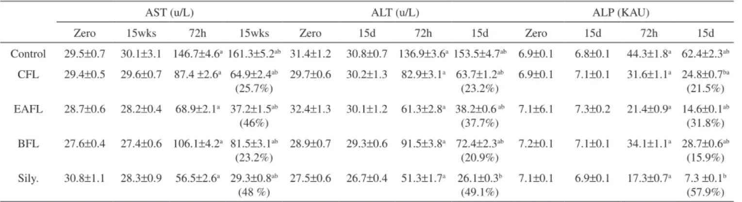
Table 1. Effect of CFL, EAFL, BFL prepared from A. lebbeck L. and silymarin on the serum
AST (u/L) ALT (u/L) ALP (KAU)
Zero 15wks 72h 15wks Zero 15d 72h 15d Zero 15d 72h 15d
Control 29.5±0.7 30.1±3.1 146.7±4.6a161.3±5.2ab 31.4±1.2 30.8±0.7 136.9±3.6a153.5±4.7ab 6.9±0.1 6.8±0.1 44.3±1.8a 62.4±2.3ab CFL 29.4±0.5 29.6±0.7 87.4 ±2.6a 64.9±2.4ab
(25.7%)
29.7±0.6 30.2±1.3 82.9±3.1a 63.7±1.2ab (23.2%)
6.9±0.1 7.1±0.1 31.6±1.1a 24.8±0.7ba (21.5%) EAFL 28.7±0.6 28.2±0.4 68.9±2.1a 37.2±1.5ab
(46%)
32.4±1.3 30.1±1.2 61.3±2.8a 38.2±0.6 ab (37.7%)
7.1±6.1 7.3±0.2 21.4±0.9a 14.6±0.1ab (31.8%) BFL 27.6±0.4 27.4±0.6 106.1±4.2a 81.5±3.1ab
(23.2%)
28.9±0.7 29.3±0.6 91.5±3.8a 72.4±2.3ab (20.9%)
7.2±0.1 7.1±0.1 34.1±1.1a 28.7±0.6ab (15.9%) Sily. 30.8±1.1 28.3±0.9 56.5±2.6a 29.3±0.8ab
(48 %)
27.5±0.6 26.7±0.4 51.3±1.7a 26.1±0.3b (49.1%)
7.1±0.1 6.9±0.1 17.3±0.7a 7.3 ±0.1b (57.9%) aStatistically signiicant from time zero at p < 0.01; bstatistically signiicant from 72 h after CCl
4 at p < 0.01. A daily dose of 100 mg kg-1 b. wt. of different extracts and 50 mg kg-1 b. wt. of silymarin.; d, day; h, hour; Sily., silymarin as a positive control; wks, weeks.
Table 2. Effect of CFL, EAFL, and BFL of A. lebbeck L. leaves on the blood glutathione level of alloxan-induced diabetic rats (n=6).
Group Blood glutathione
(mg %) % Potency
Control (1 mL saline) 36.4±1.1 _ Diabetic control 21.9±0.4a 39.84 Diabetic + vitamin E 35.9±1.2 1.37
Diabetic + CFL 31.2±1.2 14.28
Phenolics analysis
Generally, the mobile phases that were mainly used in the HPLC analysis of phenolics (lavonoids and phenolic acids) are aqueous acetonitrile, aqueous methanol, or their mixtures in combination with different concentrations of an acid as a proton source as needed for ionization, viz. formic acid (0.1 or 0.5%), acetic acid (0.25, 0.5, 1%), triluoroacetic acid (0.05%), phosphoric acid (2, 0.1%), ammonium acetate (10 mmol L-1) or formate (10 mmol L-1). In the present study,
the method development was intended to provide a reliable rapid technique for the separation of 9 lavonoids using an Intersil ODS-2 C18 column and a mobile phase consisting of mixtures of water acidulated with formic acid and acetonitrile-methanol (60:40, v/v). The analysis of the phenolics in CFL and EAFL was performed on an HPLC-PDA (Figures 1 and 2), and the peak identities were further conirmed by a LC-ESI-MSn (n = 2) system in the negative mode. This analysis was used for the separation, detection and characterization of the structure of the phenolic acids and lavonoids in the bioactive fractions in which no extensive sample preparation is required, and this analysis was shown to have a higher sensitivity for the subject compounds than MS analysis in the positive mode.
In Table 3, the MS behavior of fractions (a-e) prepared by VLC of the CFL revealed the presence of phenolic acids 1 and 2, which were positively identiied by comparison with available standards. The spectrum of compound 1 showed a deprotonated molecular ion at m/z 353 of a monocaffeoylquinic acid, which was irst detected through a loss scan of 162 μ (a caffeic acid unit) and gave a base peak at m/z 191 (for quinic acid). This fragmentation is typical of 3-O-caffeoylquinic acid (chlorogenic acid).42 The mass spectrum of compound 2 showed a typical loss of CO2 for caffeic acid, giving
[M–H–44] – as a characteristic ion.43
MSn fragmentation of the ion [M–H]– of the standards produced
a major fragment speciic for lavone and lavonols at m/z 151 with different intensities, originating from an RDA reaction.44 There was
a major fragment for the aglycones detected in fractions (a-e) of the CFL. Compound 3 displayed an [M–H]– ion at m/z 301, while
compounds 4 and 5 had a deprotonated ion [M−H]– at m/z 285. Their
fragmentation patterns match with those of standards for quercetin, luteolin and kaempferol.44 The detection of free aglycones is
com-monly an indication of the presence of their glycosylated forms, but no glycosylated luteolin was detected here. Compound 6 has an [M–H]– ion at m/z 317, and its fragmentation behavior agreed with
that of a myricitin standard.45
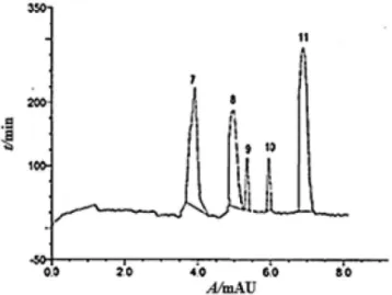
The LC-MS/MS chromatogram of the EAFL (Table 4) of A. leb-beck L. identiied ive lavonoid glycosides that are O-glycosylated, and their fragmentation was characterized by a cleavage of the glycosidic bonds and elimination of the sugar moieties with charge retention on the aglycone (Y0-). The data for the retention times
(Rt), UV, deprotonated molecules [M–H]– and mass fragmentation
patterns (MSn) of the lavonoids detected in the EAFL are listed in
Table 3. Retention times, UV spectra and product ions of phenolic acids and lavonoid aglycones in fractions (a-e) prepared from CFL of A. lebbeck L.
Peak No. Compounds Rt
(min) UV max (nm) MW
MS/MS [M – H]– Fraction
m/z E d c b a
1 3-O-Caffeoylquinic acid 0.85 240, 298 (sh), 326 354 191, 179, 135 353 - - - + +
2 Caffeic acid 1.25 325 180 135 (100%) 179 - - + + +
3 Quercetin 8.62 256, 372 302 151 (34%) 301 - + + +
-4 Luteolin 9.64 348 286 151 (65%) 285 + + + + +
5 Kaempferol 10.45 264, 370 286 151 (55%) 285 - - + + +
6 Myricetin 11.24 254, 372 314 151 (58%) 317 - - - +
-Figure 1. HPLC/PDA chromatogram of VLC fractions (a-e) prepared from
Table 4. The ions corresponding to the deprotonated aglycone (Y0-)
products were compared with deprotonated molecules [M–H]– of
the aglycones detected in the CFL fractions; similar data are present in the literature and have been compiled for known standards that conirmed the identiication of glycosides of the aglycones 3, 5, 6 as quercetin 3-O-rutinoside (rutin) (7),16 myricetin 3-O-rhamnoside
(8)46 and kaempferol 3-O-β-glucoside (10).47
Because reference samples of compounds 9 and 11 (Table 4) were not available, LC- MS proved to be extremely helpful for their assignment and further characterization of individual substances with the aid of literature data. Compound 9 had a deprotonated ion [M-H]- at m/z 593 and (MS2, [M-H]-) and other ions at m/z 447 (23%,
[M-146]-) and m/z 301 [100%, Y
0-] due to the successive loss of two
deoxypentoses from the aglycone quercetin (compared with com-pound 3). The two sugars are attached to the same aglycone carbon. This was mainly demonstrated by comparing their peak intensities to that of rutin, indicating their link at the 3-position.47 These ions arising
from the cleavage of the glycosidic bonds are weak, and the presence of a quasi-molecular ion with low abundance (< 25) indicated the attachment of the two sugars at the same position as noticed from rutin. The lavonoid is identiied as quercetin 3-O-dideoxypentoside. Compound 11 showed MS at m/z 625 [M-H]- and a peak at m/z 301
(100%, Y0-) due to the loss of two hexose moieties (324 mu) linked
to the aglycone and a fragment at m/z 463 [M-H-162]-. By comparing
these fragments with those of compound 7 and other similar compoun-ds in the literature,46 in which the two hexoses are mostly attached to
C-3, we propose the structure to be quercetin 3-O-dihexoside. The attachment of the sugars at this position is a recurrent characteristic of quercetin glycosides in Albizia species.14,47
Natural products derived from natural resources worldwide have a potential for protection and are successfully used to treat liver dise-ases.48-50 Approximately half of the pharmaceuticals in use today are
derived from natural products.51 Their positive effects are mainly due
to the presence of different phenolics viz., lavonoids, coumarins, and phenolic acids.52 The hepatoprotective effect of silymarin is mainly
due to its bioactive lavonoid content.31,53 Similarly, the promising
hepatoprotective effect of Albizia lebbeck is mainly attributed to the lavonoids content in the fraction prepared from a 70% ethanol extract. Evidence for this reasoning is that the ethyl acetate fraction, which contains mainly lavonoid glycosides that are polyphenols with hepatoprotective and antioxidant properties, has highest activity.
Previous publications discussed the identiication of quercetin and kaempferol 3-O-α rhamnopyranosyl (1→6)-β-glucopyranosyl (1→6)-β-galactopyranosides in leaves,14 in addition to rutin and
kaempferol-3-O-rutinoside in lowers16of A. lebbeck.Top of Form To our knowledge the two phenolic acids 1 and 2, the aglycone myricetin (6) and the glycosides myricetin 3-O-rhamnoside (8), quercetin 3-O-dideoxypentoside (9), kaempferol 3-O-glucoside (10), and quercetin 3-O-dihexoside (11) have not previously been reported in the genus Albizia. As EAFL exhibited the highest potency in the hepatotoxicity assays, it is reasonable to standardize the extracts from Albizia based on the contents of these major glycosides, which are probably related to the biological activity in question.
Validation
The standard compounds quercetin 3-O-rutinoside (rutin), myri-cetin 3-O-rhamnoside (myricetrin) and kampferol 3-O-glucoside showed good linearity, with r2 = 0.9659, 0.9989, 0.9634, respectively,
over a relatively wide concentration range. The inter-day precision showed that the RSD (relative standard deviation) values were 2.21, 1.98, and 2.12% and the intra-day variations were 2.22, 1.688, and 1.36%, respectively. Their average regression equations were y = 415754.2 + 56122.2x (rutin); y = 314224.5 + 7112.5x (myricitrin); and y = 322235.3 + 29813.6x (kampferol 3-O-glucoside). The limits of detection were 2.14 (rutin), 1.22 (myricetrin), and 6.44 (kampferol 3-O-glycoside) ng mL-1. The percentage accuracy ranged between 97.40 and 99.98%. The glycosides were quantitatively estimated (in units of mg 100 g-1 dry powder ± SD) as being from the EAFL
Table 4. Retention times, UV spectra and product ions of lavonoid glycosides in EAFL of A. lebbeck L.
Peak No. Compounds Rt
(min)
UV max (nm)
MW [M – H]- LC-MS/MS
m/z
7 Quercetin 3-O-rutinoside 3.88 257, 267 (sh),359 610 609 463, 447, 301([A-H] -80%)
8 Myricetin 3-O-rhamnoside 4.65 480 479 317(60%)
9 Quercetin 3-O-dideoxypentoside 5.12 256, 267 (sh), 359 594 593 447(42%), 301([A-H]-100%)
10 Kaempferol 3-O-glucoside 5.91 266, 345 448 447 285(43%)
11 Quercetin 3-O-dihexoside 6.42 257, 267(sh), 352 626 625 463, 301(100%)
Figure 2.HPLC/PDA chromatogram of the EAFL fraction of A. lebbeck
Table 5. Quantitative determination of the identiied lavonoids in the EAFL of A. lebbeck
of A. lebbeck by LC/MS, with the results in Table 5, quercetin 3-O-dihexoside was present in the highest concentration (ca. 0.138 mg 100 g-1) followed by quercetin 3-O-rutinoside (ca. 0.135 mg 100 g-1).
CONCLUSION
Air-dried powdered leaves were extracted with 70% ethanol. The concentrated residue was fractionated with different organic solvents (chloroform, ethyl acetate and n-butanol) to yield the CFL, EAFL and BFL residual fractions, respectively. The fractions were examined for their hepatoprotection on induced liverdamaged mice and compared to silymarin as well as for their antioxidant activity on the diabetic rats compared with vitamin E. The results showed the following decreasing order of activity: silymarin > EAFL > CFL > BFL and vitamin E > EAFL > CFL > BFL, respectively. The bioactive fractions were sub-jected to LC-ESI-MS/MS analysis in the negative ion mode to explore their phenolics content. The results revealed the presence of 11 com-pounds: two phenolic acids, four lavonoid aglycones and ive lavonoid glycosides. Seven of these compounds were identiied here for the irst time in the genus Albizia. Quantiication of the identiied lavonoid glycosides revealed that quercetin 3-O-rutinoside was predominant. ACKNOWLEDGMENTS
This work was funded by the Deanship of Scientiic Research (DSR), King Abdulaziz University, Jeddah, under grant no. (166-011-d1434). The authors acknowledge the technical and inancial support from DSR.
REFERENCES
1. Faisal, M.; Singh, P. P.; Irchhaiya, R.; Int. Res. J. Pharm.2012, 3, 63. 2. Orwa, C.; Mutua, A.; Kindt, R.; Jamnadass, R.; Simons, A.;
Agrofor-estree Database: a tree reference and selection guide, World Agrofor-estry Centre, Kenya, 2009.
3. Kasture, V. S.; Chopde, C. T.; Deshmukh, V. K.; J. Ethnopharmacol. 2000, 71, 65.
4. Kasture, V. S.; Kasture, S. B.; Pal, S. C.; Indian J. Exp. Biol. 1996, 34, 78.
5. Galal, M.; Bashir, A. K.; Salih, A. M.; Adam, S. E.; J. Ethnopharmacol. 1991,31, 333.
6. El Garhy, M. F.; Mahmoud, L. H.; J. Egypt. Soc. Parasitol.2002, 32, 893.
7. Resmi, C. R.; Venukumar, M. R.; Latha, M. S.; Indian J. Physiol. Phar-macol. 2006, 50, 297.
8. Gupta, R. S.; Kachhawa, J. B.; Chaudhary, R.; Asian J. Androl. 2004,6, 155.
9. Gupta, R. S.; Chaudhary, R.; Yadav, R. K.; Verma, S. K.; Dobhal,M. P.; J. Ethnopharmacol. 2005, 96, 31.
10. Saha, A.; Ahmed, M.; Pak. J. Pharm. Sci. 2009,22, 74.
11. Une, H. D.; Sarveiya, V. P.; Pal, S. C.; Kasture, V. S.; Kasture, S. B.; Pharmacol. Biochem. Behav. 2001, 69,439.
12. Rahul, C.; Pankaj, P.; Sarwan, S. K.; Mahesh, J. K.; J. Chem. Pharm. Res. 2010, 2, 476.
13. Al-Musayeib, N. M.; Mothana R. A.; Al-Massarani, S.; Matheeussen, A.; Cos, P.; Maes, L.; Molecules 2012, 17, 11379.
14. El-Mousallamy, A. M. D.; Phytochemistry, 1998, 48,759.
15. Rashid, R. Chawdhury R. B.; Jabbar, A.; Hasan, C. M.; Rashid, M. A.; Saudi pharm. J. 2003, 11, 52.
16. El Halim, A.; Mohamed, F. A.; El Gamal, A. A.; Master Thesis, College of Pharmacy, King Saudi University, Saudi Arabia, 2011.
17. Nazneen, B.; Wesely, E. G.; Johnson, M.; Int. J. Adv. Pharm. Res. 2012, 3, 830.
18. Sulaiman, C. T.; Balachandran, I.; Indian J. Pharm. Sci. 2012, 74, 258. 19. Pal, B. C.; Achari, B.; Yoshikawa, K.; Arihara, S.; Phytochem. 1995, 38,
1287.
20. Ueda, M. L.; Tokunaga, T. O.; Okazaki, M. Sata, P; N. U.; Ueda, K. O.; Yamamura, S. P.; Nat. Prod. Res. 2003, 17,329.
21. Scott, J. P.; Tinto, W. F.; Reynolds, W. F.; Nat. Prod. Commun. 2008, 3, 1763.
22. Misra, L. N.; Dixit, A. K.; Wagner, H.; Phytochemistry 1995, 39, 247. 23. Dixit, A. K.; Misra, L. N.; J. Nat. Prod. 1997, 60, 1036.
24. Ma, Y. T.; Hsiaob, S. C.; Chen, H. F.; Hsu, F. L.; Phytochem. 1997, 46, 451.
25. Jain, A. S.; Mishra, K. L.; Tetrahedron Lett. 1963, 1, 19.
26. Holman, R. T.; In North American Terrestrial Orchids Symposium II, Proceedings and Lecture, Michigan Orchid Society, Livonia, 1981, pp. 32-40.
27. Aiyelaagbe, O. O.; Oyewole, A. E.; Oladosu, I. A.; J. Essent. Oil-Bear. Plants 2010, 13, 644.
28. El- Hawary, S.; El- Fouly, K.; Sokkar, N. M.; Talaat, Z; Asian J. Bio-chem. 2011, 6, 122.
29. OECD; Guidelines for the testing of chemicals, Organisation for eco-nomic co-operation and development: Paris, 2001.
30. Pandey, G.; Sahni Y. P.; Int. J. Res. Ayurveda Pharm. 2011, 2, 75. 31. Abenavoli, L.; Capasso, R.; Milic, N.; Capasso, F.; Phytother. Res. 2010,
24, 1423.
32. Klassen, C. D.; Plaa, G. L.; Biochem. Pharmacol. 1969, 18, 2019. 33. Eliasson, S. G.; Samet, J. M.; Life Sci. 1969, 8, 493.
34. Beutler, E.; Duron, O.; Kelly, B.M.; J. Lab. Clin. Med.1963, 61, 882. 35. Shirode, D. S.; Hirode Jain, B. B.; Mahendra Kumar, C. B.; Setty, S. R.;
J. Chem. Pharm. Sci. 2012, 5, 199.
36. Kokila, K.; Priyadharshini, S. D.; Sujathai, V.; Int. J. Pharm. Sci.2013, 5, 70.
37. Samudram, P.; Vasuki, R.; Rajeshwari, H.; Geetha, A.; Moorthi, P.; J. Medicinal Plants Res. 2009, 3, 1078.
38. Heibatollah, S.; Reza, N. M.; Izadpanah, G.; Sohailla, S.; Afr. J. Biomed. Res. 2008, 2, 141.
39. Gutiérrez, R.; Solís, R.; Rec. Nat. Prod. 2009, 3, 46.
40. Tiwari, B. K.; Khosa, R. L.; The Internet Journal of Tropical Medicine 2010, 6, 1540.
41. Dang, S. S.; Zhang, X.; Jia, X. L.; Cheng, Y. A.; Song, P.; Liu, E. Q.; He, Q.; Li, Z. F.; Chin. Med. J. 2008, 121, 1010.
42. Rabaneda, F. S.; Jáuregui, O.; Casals, I.; Lacueva, C. A.; Pulido, M. I.; Lamuela-Raventós, R. M.; Mass Spectrom. 2003, 38, 35.
43. Queralt, A. V.; Uregui, O. J.; Remo, A. M.; Lacueva, C. A.; Ravento, R. M. L.; Rapid Commun.Mass Spectrom. 2010, 24, 2986.
44. Justesen, U.; J. Chromatogr. A 2000, 902, 369.
45. Hifnawy, M.; Sokkar, N.; Ezzat, S.; Raslan, M.; Sleim, A.; Asian J. Plant Sci. 2012, 11, 124.
46. Ferreres, F.; Llorach, R.; Gil-Izquierdo, A.; J. Mass Spectrom. 2004, 39, 312.
47. Melek, F. R.; Ghaly, N. S; El-Kady, M.; Nabil, M.; Egypt. J. Pure Appl. Sci. 2011, 79.
48. Seeff, L. B.; Lindsay, K. L.; Bacon, B. R.; Kresina, T. F.; Hoofnagle, J. H.; Hepatology 2001, 34, 595.
49. Santillán, E. M.; Bujaidar, E. M.; González, I. Á.; Martínez, M. T. S.; Salinas, J. G.; Bautista, M.; González, Á. M.; Rubio, M. G. L. G.; Faisal, L. A.; González, J. A. M.; World J. Gastroenterol. 2014, 20, 14787. 50. Zachariah, S. M.; Aleykutty, N.; Vishwanad, V.; Halima, O. A.; Res. J.
Pharm. Technol. 2012, 5, 317.