João Carlos Canossa Ferreira
Characterization of vacuole permeabilization
and HMGB1 nuclear release in the yeast cell
death induced by a benzo[a]phenoxazine
derivative
Universidade do Minho
Escola de Ciências
Março de 2017 Jo ão C ar lo s C an os sa F er re ir a C h a ra c te ri za ti o n o f v a c u o le p e rm e a b il iz a ti o n a n d H M G B 1 n u c le a r r e le a se in t h e y e a st c e ll d e a th i n d u c e d b y a b e n zo [ a ]p h e n o xa zi n e d e ri va ti ve Minho | 20 1 7 UJoão Carlos Canossa Ferreira
Characterization of vacuole permeabilization and
HMGB1 nuclear release in the yeast cell death
induced by a benzo[
a
]phenoxazine derivative
Tese de Mestrado
Mestrado em Genética Molecular
Trabalho realizado sob orientação de
Professora Doutora Maria João Marques Ferreira Sousa Moreira
Professora Doutora Maria Sameiro Torres Gonçalves
iv
Agradecimentos
Apesar deste trabalho deste trabalho ter sido de caracter individual, não poderia deixar de exprimir o meu mais sincero agradecimento a todos os que me ajudaram e apoiaram na sua realização.
Em primeiro lugar, agradeço as minhas orientadoras, Doutora Maria João Sousa e Doutora Maria do Sameiro Gonçalves, pela possibilidade de realização deste trabalho. À Doutora Maria João, por todo o apoio, toda a disponibilidade e orientação prestada e pelos ensinamentos, aconselhamentos e criticas ao longo deste trabalho que foram muito importantes para o meu crescimento cientifico. À Doutora Sameiro, por se ter mostrado sempre disponível em ajudar e por ter sido incansável em disponibilizar o composto com que trabalhei, sem ela seria impossível a realização deste trabalho.
Agradeço a todos os colegas da Micro I, Lisandra Castro, Cátia Fernandes, Joana Guedes, Anabela Ferreira, Paulo Veloso, Nuno Machado, António Rego, Filipa Dias, Diana Silva, Catarina Afonso e Vasco Lobato muito obrigado pela ajuda e pelo bom ambiente que sempre criaram no laboratório.
Ao departamento de biologia, a todos os técnicos e funcionários, em especial ao Sr. Luís por ser incansável em garantir todas as condições de trabalho.
Obrigado aos meus amigos, em especial ao João Pacheco, Luís Ferraz, Bruno Oliveira, Carlos Eloy, Henrique Ribeiro, João Ferreira, Rui Sousa e Beatriz Domingues por estarem sempre presentes e por me terem apoiado em todos os momentos. Agradeço também aos meus amigos da terra, Miguel Moreira, Hugo Moreira, Ana Luísa, Sandrina Rodrigues, Fábio Daniel, Rosa Silva e Kelly Dias por me terem ajudado a ultrapassar mais esta etapa.
Á minha família, o meu muito obrigado, por terem acreditado em mim e por terem permitido esta oportunidade, sem vocês nada disto seria possível.
v
Abstract
The number of antifungal agents approved for use in humans is still very limited and resistance to the existing ones is often found, thus revealing the need for the development of new antifungal drugs.
Phenoxazine derivatives have assumed an increasing importance in life sciences since they present antiproliferative proprieties which potentiate their utilization as antitumor and antimicrobial agents. We have recently found that one new phenoxazine derivative (MSG-111-cd3), induces cell death in Saccharomyces cerevisiae and accumulates at the vacuolar membrane and endoplasmic reticulum. Initial studies revealed that MSG-111-cd3 toxic effect may be mediated through vacuolar membrane damage and vacuolar permeabilization. Furthermore, it was observed that the compound lead to Nhp6Ap (the yeast HMGB1 orthologue) release from the nucleus to the cytosol, where it displayed a punctuated pattern, without loss of plasma membrane integrity.
Aiming to further elucidate the cell death mechanism we analyzed the consequences of vacuolar permeabilization, the role of the HMGB1 protein in the cell death process and the mechanisms underlying its nuclear release. We find out that MSG-111-cd3 yeast cell death is dependent on the vacuolar protease Pep4p and that the vacuole permeabilization resulted in its translocation from the vacuole to the cytosol. We observed that autophagy is not involved in the cell death process, and although MSG-111-cd3 leads to mitochondrial network fragmentation, apparently, the cell death process is independent of the mitochondrial pathway, since no other alterations were significantly induced in this organelle. Our results suggest that Nhp6Ap is apparently not crucial for the cell death process. Furthermore, we found out that Nhp6Ap is not the only DNA binding protein to be released into cytosol, since the histone Hta2p also suffer translocation exhibiting the same phenotype as Nhp6Ap. Furthermore, the treatment lead to nuclear envelope disorganization, which show the high susceptibility of the nucleus to the MSG-111-cd3 effects.
vi
Resumo
O número de agentes antifúngicos aprovados para utilização em seres humanos é ainda muito limitado e é frequentemente encontrada resistência aos existentes, revelando assim a necessidade de desenvolvimento de novos fármacos antifúngicos.
Os derivados de fenoxazina assumiram uma importância crescente nas ciências da vida, uma vez que apresentam propriedades antiproliferativas que potenciam a sua utilização como agentes antitumorais e antimicrobianos. Recentemente descobrimos que um novo derivado de fenoxazina (MSG-111-cd3) induz morte celular em Saccharomyces cerevisiae e acumula-se na membrana vacuolar e no retículo endoplasmático. Estudos iniciais revelaram que o efeito tóxico de MSG-111-cd3 parece ser mediado através de danos na membrana vacuolar e permeabilização vacuolar. Além disso, observou-se que o composto conduziu à libertação de Nhp6Ap (o ortólogo de HMGB1 em levedura) do núcleo para o citosol, onde apresenta um padrão pontuado, sem perda da integridade da membrana plasmática.
Com o objetivo de melhor clarificar o mecanismo de morte celular induzido pelo composto MSG-111-cd3, analisamos as consequências da permeabilização vacuolar, o papel da proteína HMGB1 e os mecanismos subjacentes à sua liberação nuclear em resposta ao tratamento com este composto. Descobrimos que a morte das células de levedura causada pelo composto MSG-111-cd3 é dependente da protéase vacuolar Pep4p e que a permeabilização do vacúolo resultou na sua translocação para o citosol. Observamos que a autofagia não está envolvida no processo de morte celular e, embora o MSG-111-cd3 leve à fragmentação da rede mitocondrial, aparentemente, o processo de morte celular é independente da via mitocondrial. Os resultados sugerem que Nhp6Ap aparentemente não é crucial para o processo de morte celular. Além disso, mostrámos que Nhp6Ap não é a única proteína de ligação ao DNA a ser libertada do núcleo para o citosol, uma vez que a histona Hta2p também sofre translocação exibindo o mesmo fenótipo que Nhp6Ap. Além disso, o tratamento conduz à desorganização do envelope nuclear, o que mostra a elevada suscetibilidade do núcleo aos efeitos do MSG-111-cd3.
vii
Table of contents
Agradecimentos ... iv
Abstract ... v
Resumo ... vi
Table of contents ... vii
List of abbreviations and acronyms ... ix
List of figures ... xi
List of tables ... xii
1. Introduction ... 13
1.1 Cell death ... 14
1.1.1 Cell death Pathways ... 14
1.1.2 Cell death in yeast ... 20
1.2 Cell death by lysosomal permeabilization ... 23
1.3 HMGB1 protein... 28
1.4 Phenoxazine derivatives ... 32
2. Aim ... 36
3. Material and Methods ... 38
3.1 Yeast strains ... 39
3.2 Media and Growth conditions ... 39
3.3 MSG-111-cd3 treatment ... 41
3.4 Viability assays ... 41
3.5 Yeast pUG35-nhp6a-GFP, pESC (Ø), pESC-Pep4p(FL) and pESC-DPM-Pep4p transformation ... 42
3.6 Assessment of Pep4p, Nhp6Ap, Hta2p and Nup49p localization and vacuole membrane permeabilization ... 43
3.7 Assessment of calcium fluctuation levels ... 43
3.8 Terminal deoxynucleotidyl transferase dUTP nick end labeling (TUNEL) ... 43
3.9 Assessment of mitochondrial fragmentation ... 44
3.10 Assessment of mitochondrial potential and plasma membrane integrity ... 44
3.11 Assessment of GFP-ATG8 protein localization ... 45
3.12 Evaluation of GFP-ATG8 cleavage by SDS gel electrophoresis/Western Blot ... 45
3.12.1 Cell extracts preparation ... 45
3.12.2 SDS gel electrophoresis/Western Blot ... 45
3.13 Epifluorescence microscopy and flow cytometry ... 46
4. Results ... 47
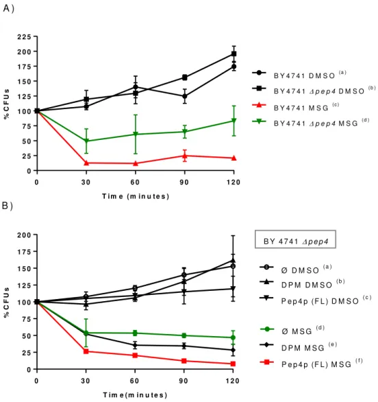
4.1 MSG-111-cd3 induces cell death of S. cerevisiae by a process that is dependent on Pep4p ... 48
viii
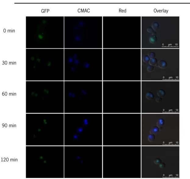
4.2 Pep4p localization and vacuole permeabilization ... 50
4.3 Assessment of calcium fluctuations ... 53
4.4 Assessment of DNA fragmentation by TUNEL assay ... 54
4.5 Assessment of mitochondrial fragmentation ... 56
4.6 Analysis of mitochondrial membrane potential and plasma membrane integrity ... 58
4.7 Analysis of autophagy involvement in the cell death process ... 61
4.8 Assessment of Nhp6Ap translocation from the nucleus ... 65
4.9 Assessment of Hta2p localization and nuclear envelope disorganization ... 70
5. Discussion and Future perspectives ... 73
ix
List of abbreviations and acronyms
AcLi Lithium acetate
AIF Apoptosis inducing factor APC Antigen-presenting cell ATP Adenosine triphosphate CFU Colony formation units DAPI 4’,6-diamidino-2-phenylindole DIC Differential Interference Contrast DMSO Dimethyl sulfoxide
DPM Double point mutation ENDOG Endonuclease G H2O2 Hydrogen Peroxide
HMGB1 High Mobility group box 1
LAMP Lysosome-associated membrane proteins LC3 Light chain 3
LIMP Lysosomal integral membrane proteins LMP Lysosomal membrane permeablization LPS Lipopolysaccharides
MIC Minimum inhibitory concentration
MMP Mitochondrial membrane permeabilization MSG MSG-111-cd3
x NLS Nuclear localizations signals
O.D Optical density PCD Programed cell death PEG Polyethylene glycol PI Propidium iodide
PMSF Phenylmethylsulfonyl fluoride PVDF Polyvinylidene fluoride membrane
RAGE Receptor for advanced glycation end products ROS Reactive oxygen species
S. cerevisiae Saccharomyces cerevisiae
SDS-PAGE Sodium dodecyl sulphate polyacrilamide gel electophoresis ssDNA Salmon sperm DNA
TUNEL Terminal deoxynucleotidyl transferase dUTP nick end labeling WT Wild type
YEPD Yeast Extract Peptone Dextrose
xi
List of figures
Figure 1 – Representation of extrinsic a) and intrinsic apoptotic pathways b)... 16
Figure 2 – Macroautophagy process ... 18
Figure 3 – Assays routinely used in the field of yeast cell death ... 22
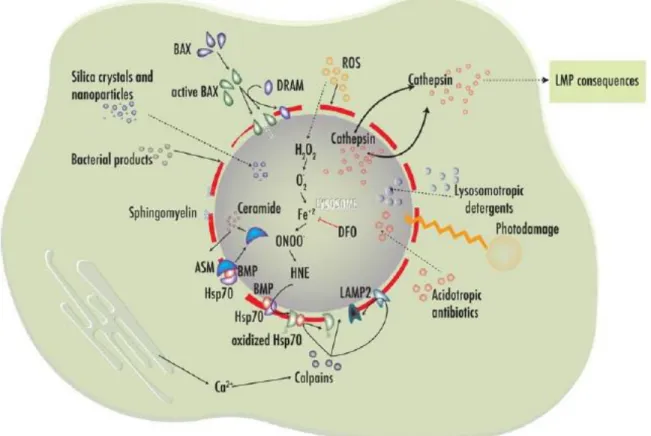
Figure 4 – Inducers of lysosomal membrane permeabilization.. ... 24
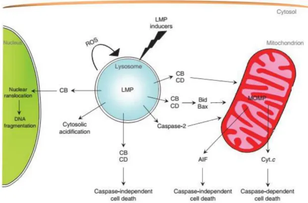
Figure 5 – LMP downstream lethal pathways ... 26
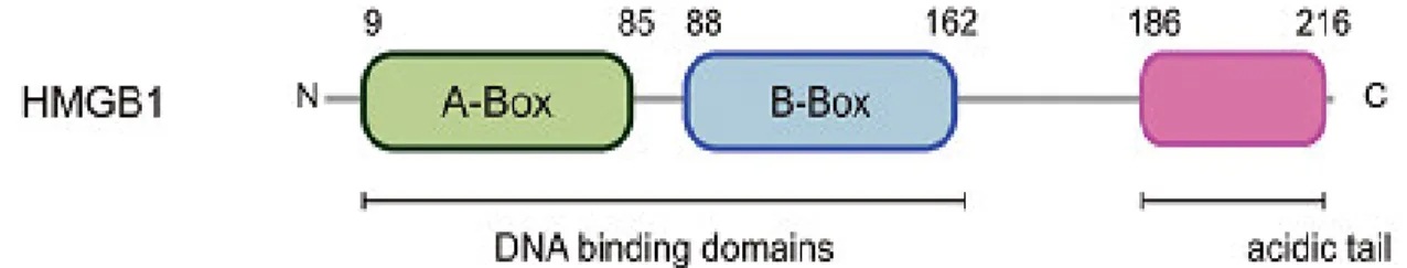
Figure 6 – HMGB1 structure.. ... 28
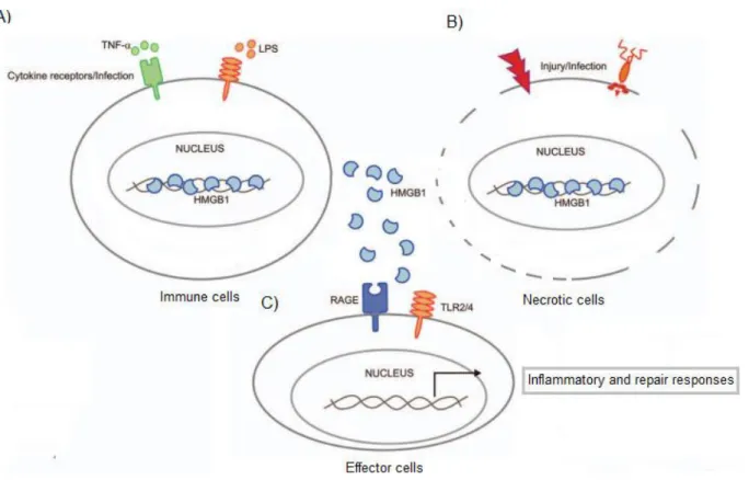
Figure 7 – Release of HMGB1 protein. ... 29
Figure 8 – Molecular structure of a) Oxazine, b) Phenoxazine and c) Benzo[a]phenoxazine. ... 32
Figure 9 – N‐(5‐((4‐ethoxy‐4‐oxobutyl)amino)‐10‐methyl‐9H‐benzo[a]phenoxazin‐9‐lidene) (MSG-111-cd3) ... 35
Figure 10 - Influence of Pep4p on the effect of MSG-111-cd3 on yeast cellular viability ... 49
Figure 11 – Effect of DMSO (negative control) on Pep4p localization. ... 51
Figure 12 – Effect of MSG-111-cd3 on Pep4p localization.. ... 52
Figure 13 – Effect of MSG-111-cd3 on Intracellular calcium accumulation ... 53
Figure 14 – Effect of MSG-111-cd3 on DNA fragmentation ... 55
Figure 15 – Effect of MSG-111-cd3 on mitochondrial network ... 57
Figure 16 – Effect of MSG-111-cd3 on mitochondrial membrane potential. ... 59
Figure 17 – Effect of MSG-111-cd3 on plasma membrane integrity ... 60
Figure 18 – Analysis of Atg8p localization in response to MSG-111-cd3 ... 62
Figure 19 – Western-blot analysis of GFP- Atg8p processing ... 63
Figure 20 – Analysis of Atg8p localization in response to MSG-111-cd3 and Rapamycin. ... 64
Figure 21 – Effect of MSG-111-cd3 on BY 4741 Δnhp6a viability ... 66
Figure 22 – Effect of MSG-111-cd3 on Nhp6Ap nuclear release. ... 67
Figure 23 – Effect of MSG-111-cd3 on Nhp6Ap nuclear release of BY 4741 Δstp22 strain ... 68
Figure 24 – Effect of MSG-111-cd3 on Nhp6Ap nuclear release of BY 4741 Δsnf7 strain. ... 69
Figure 25 – Effect of MSG-111-cd3 on Hta2p localization ... 71
Figure 26 – Effect of MSG-111-cd3 on nuclear envelope organization ... 72
xii
List of tables
Table 1 – S. cerevisiae strains used in this study. ... 40 Table 2 – List of plasmids used in this study. ... 42 Table 3 – Transformation mix. ... 42
14
1.1 Cell death
1.1.1 Cell death Pathways
All living organisms from the simplest to the most complex share the same structural and functional base unit - the cell. Starting from this basic unit of life and through more or less complex biological processes, the cellular organization and its rearrangement will give the enormous biodiversity of living beings that exist today. The final stage of a living organism after reaching its maturity is death. However, at the cellular level the dying process is not always associated with something bad and neither should be unwanted (Baehrecke, 2002). In reality, cells also need to die to allow the development and to contribute to the homeostasis of the individual. Thus, cell death is an important biological process in the development of an organism, being involved in eliminating abnormal cells, in the control of cell numbers, and also in the formation and deletion of structures (Jacobson et al., 1997). In fact, genetics studies have shown that organisms so different as worms and humans have conserved genes that encode the core cell death machinery (Aravind et al., 2001).
On the other hand, aberrant cell death is associated with the occurrence of various complex diseases such as cancer, autoimmune diseases, neurodegenerative syndromes and myelodysplastic syndromes (Thompson, 1995; Krammer, 2000; Sastry and Rao, 2000). Cell death can be classified taking into account the morphological appearance of the process without a reference to precise biochemical mechanisms, but also taking into account the enzymological criteria (with or without the involvement of nucleases, or proteases, such as calpains, caspases, cathepsins, and transglutaminases), functional aspects (if the lethal process is accidental or programed, pathological or physiological), and considering the immunological characteristics (immunogenic or non-immunogenic) (Melino et al., 2001; Kroemer et al., 2009).
The first descriptions of programmed cell death mechanisms date back to around the mid 1960’s (Lockshin and Williams, 1964, 1965). Since then, there has been much research in order to classify the cell death modalities based on morphological characteristics. Schweichel and Merker were able to propose a classification of the several cell death processes, that includes the type I cell death that is an heterophagic processes also known as apoptosis, the type II cell death that is associated
15 with autophagy, and also the type III cell death known as necrosis, that does not involve any type of digestion (Schweichel and Merker, 1973).
Apoptosis or type I cell death is an active form of PCD (programed cell death), that was first presented by Kerr (Kerr et al., 1972). It is a process that has a direct involvement in several fundamental biological events, and is characterized by specific alterations in the dying cells, both morphological and biochemical. In terms of morphological alterations apoptotic cells present reduction of cellular volume, nuclear condensation (pyknosis) and fragmentation (karyorhexis) (Kroemer et al., 2009). Apoptotic cells also present a few or no ultrastructural modifications of cytoplasmic organelles, loss of adhesion to neighbors cells and plasma membrane blebbling that results in the formation of apoptotic bodies but with the maintenance of the cell integrity until the final stages of the process (Nishida et al., 2008). In terms of biochemical alterations, apoptotic cells display expression of cell surface markers such as phosphatidylserine externalization, internucleosomal cleavage of chromosomal DNA, protein cross-linking and also cleavage of a number of intracellular substrates by specific proteolysis (Cohen et al., 1994; Martin and Green, 1995). However, is important to consider that apoptosis and programed cell death are not synonyms, since when cell death occurs in a context of physiological development, can manifest non-apoptotic features (Roach and Clarke, 2000; Baehrecke, 2002). Apoptosis is a highly sophisticated and complex process, that involves an energy-dependent cascade of molecular events. There are two main apoptotic pathways, the intrinsic (mitochondrial pathway) that is dependent or independent of caspases, and the extrinsic also known as the death receptor pathway (figure1) (Galluzzi et al., 2012).
Mitochondrial membrane permeabilization (MMP) and caspase activation are the two prominent processes responsible for the occurrence of these pathways. MMP determines the point of no return of the intrinsic pathway, and is regulated by Bcl-2 proteins that can act as inducers or blockers of the process, the result of MMP is the release of pro-apoptotic molecules like cytochrome c, that acts as an activator of caspases which are responsible for the degradation of many nuclear and cytoplasmic proteins in the apoptotic cell (Youle and Strasser, 2008). However, the activation of caspases is not strictly dependent on MMP, and, for example, in the death receptor pathway caspase 8 can undergo an autocatalytic activation when it is recruited by the activated dead receptors, leading then to the directly cleavage and activation of the effector caspases 3 and 7 (Taylor et al., 2008). During MMP,
16 besides cytochrome c, there are other mitochondrial proteins that can be released, such as apoptosis inducing factor (AIF) and endonuclease G (ENDOG). These proteins translocate to the nucleus and originate a large-scale DNA fragmentation leading to occurrence of apoptosis in a caspase-independent intrinsic pathway (Modjtahedi et al., 2006).
Furthermore, there is evidence of the crosstalk between the intrinsic and extrinsic pathways, one example is the role of the BCL-2-family member BID, that when cleaved by caspase 8 is capable of activating the mitochondrial pathway acting as way to amplify the apoptotic signal (figure 1) (Galluzzi et al., 2009b).
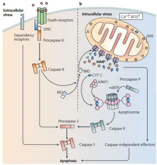
Figure 1 – Representation of extrinsic a) and intrinsic apoptotic pathways b) The activation of the extracellular pathway occurs at the level of the plasma membrane by some specific transmembrane receptors, followed by the activation of the initiator caspase 8 that can catalyze the proteolytic maturation of executioner caspases, such as caspase 3. The mitochondrial pathway is activated by intercellular alterations such as ROS accumulation or Ca2+ overload, that results in MMP. MMP is the point of no return in this pathway, since this permeabilization
leads to the occurrence of cell death by the activation of both caspase-dependent and caspase-independent mechanisms. Adapted from (Galluzzi et al., 2009b).
17 Autophagy is associated with another form of programmed cell death, also known as type II cell death, first introduced by Deter and Duve (Duve, 1967). Autophagy which means “self-eating” is a lysosomal degradative pathway that allows cells to recycle and degrade macromolecules and organelles, and has as primary functions the maintenance of cell homeostasis and the protection of cells under stress conditions, such as the presence of toxic substances and starvation (Klionsky, 2007). Furthermore, this process is also involved in normal organism development (Levine and Klionsky, 2004), lifespan extension (Vellai et al., 2009), senescence (Young et al., 2009), immunity and defense against microbial incursion (Deretic and Levine, 2009). On the other hand, autophagy is also involved in several human diseases, such as gastrointestinal disorders, myopathies, neurodegeneration, liver and heart diseases and cancer (Klionsky, 2005; Mizushima et al., 2008).
Researchers have found that this process is highly conserved since genes that regulate autophagy in yeast have been conserved in organisms that are as different as humans (Levine and Klionsky, 2004). There are three main types of autophagy, microautophagy, chaperone-mediated autophagy and macroautophagy. Among those, macroautophagy is the process that is mostly associated with type II autophagic cell death (Baehrecke, 2005).
During macroautophagy (Figure 2), proteins, organelles and other cytoplasmic components are sequestered within a double-or multi-membrane structure (the autophagosome) that then fuses with hydrolase and protease containing lysosomes forming the autolysosomes for bulk degradation (Maiuri et al., 2007). The resulting products are then recycled and used for synthesis of other molecules and ATP production. During the last decade, extensive research has shown the existence of at least 38 autophagy-related proteins that are involved in the autophagy initiation, elongation, cargo recruitment and fusion with lysosomes (Rubinsztein et al., 2012). However, besides the high level of regulation, in some cases, autophagy is excessively induced, in an attempt to eliminate toxic molecules or damaged organelles, and this excessive induction can result in an autophagic cell death (Maiuri et al., 2007).
Morphologically, autophagic cell death is defined by the high amount of autophagic vacuolization of the cytoplasm, which often, but not always, is associated with an increase in the autophagic flux (Stunkard, 2009). However, is incorrect to assume the occurrence of autophagic cell death based on this morphological alteration since in most cases autophagy is induced in a
18 cytoprotective way, in an attempt to cope with stress, and the inhibition will result with an increase in the cell death (Boya et al., 2005). So, the occurrence of ‘autophagic cell death’ should be defined considering functional and biochemical aspects. It’s only possible to assume that autophagic is responsible for the death process if it can be suppressed by the inhibition of the autophagic pathway (Galluzzi et al., 2012). This could be achieved using autophagy inhibitor agents (such as agents that target PI3K), and/or using gene knockout/mutations or RNAi targeting in essential autophagic modulators like ATG1, ATG5 and ATG12 (Galluzzi et al., 2012).
So, we should not classify as an autophagic cell death process the cases where the autophagy inhibition does not result in a decreases of cell death even if is possible to observe autophagy occurrence both through morphological alterations, or by an increased degradation of autophagic substrates.
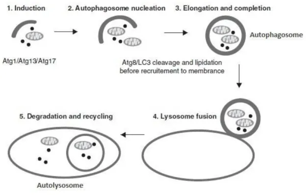
Figure 2 - Macroautophagy process. Macroautophagy is inducted by the formation of the ATG1 complex followed by the recruitment of protein and lipids mediated by the class-III phosphatidylinositol-3-kinase (Vps34) and Beclin-1/Atg6 necessary for autophagosome formation. Elongation and autophagosome completion is regulated by the conjugation of microtubule-associated protein 1 light chain 3 (LC3). In yeast this function is performed by Atg8p. Autolysosome formation occurs when the completed autophagosome fuses with the lysosome. Adapted from (Denton et al., 2012)
19 Necrosis or type III cell death has been defined over the years as an accidental, uncontrolled type of cell death, that occurs when cells are subjected to an excessive external stress, such as a pathogen infection, ischemia and heat (Galluzzi et al., 2012). This type of cell death exhibits several morphological alterations such as an increase in the cellular volume, swelling of organelles, rupture of plasma membrane and release of cytosolic contents (such as high mobility group Box 1 - HMGB1, that is a pro-inflammatory factor) (Ziegler, 2004). The leak of the cellular contents to the extracellular environment usually acts as a ‘danger signal’ that alerts the innate immune system and leads to an inflammatory process (Zitvogel et al., 2004).
Despite its importance in human pathology, necrosis has been referred as a passive and unregulated process and therefore very little efforts had been made to investigate its mechanism. However, evidences have emerged that necrosis occurrence and course might be regulated. Today, there are several data that support both programmed course and programmed occurrence of necrosis: necrosis can occur during development (example of that is the death of chrondrocytes that control the longitudinal growth of bones) (Roach and Clarke, 2000) and in tissue homeostasis (in intestinal epithelial cells) (Barkla and Gibson, 1999). Furthermore, the facts that necrosis can be triggered by plasma membrane surface receptors when they are bound with their ligands, the inhibition of some enzymes and processes can prevent necrosis and also that genetic and epigenetic factors can regulate the susceptibility of necrotic cell dead are some of evidences that necrosis is not strictly an accidental type of cell death (Golstein and Kroemer, 2007).
More recently there has been a substantial advance in the comprehension of the molecular pathways that regulate and execute the cell dead processes, which have allowed the classification of cell dead not only based on morphological characteristics but also using biochemical assays for monitoring cell death-related phenomena (Galluzzi et al., 2009a). Besides, those advances have allowed to observe that similar cell death morphologies present high biochemical, immunological and functional heterogeneity (Green et al., 2009; Kroemer et al., 2009). So, now it becomes clear that we cannot apply the equation “programmed cell death – apoptosis – caspase activation – non-immunogenic cell death” in all the cases, because it will constitute an incorrect generalization. Supporting this conclusion is fact that necrosis may occur and be regulated by a programed process (Galluzzi et al., 2007). Also, apoptosis can lead to a lethal occurrence without caspase activation, and
20 caspase activation does not necessarily mean that will occur a death process (Galluzzi et al., 2007). These examples clearly show the complexity of the cell death subroutines, and that a lot of work is still necessary in order to improve the knowledge of these processes with the main goal to improve treatments of diseases associated with deregulated cell death.
1.1.2 Cell death in yeast
Yeast has been used as an excellent model in the understanding of basic cellular processes such as intracellular trafficking (Nakano, 2004), protein folding (Coughlan and Brodsky, 2003), autophagy (Abeliovich and Klionsky, 2001) and cell cycle regulation (Hartwell, 2004). After the first description of apoptosis in a Saccharomyces cerevisiae strain, that dates from more than eighteen years ago (Madeo et al., 1997), yeast, mainly S. cerevisiae, has also been used as an experimental model for cell death study. In fact, S. cerevisiae has several characteristics that enhance its use as an excellent model for cell death study, such as being inexpensive and non-pathogenic, displaying rapid growth and easy genetic manipulation accompanied by simple mutant isolation (Carmona-Gutierrez et al., 2010b).
Furthermore, researchers found out that the apoptotic core machinery is conserved in yeast, which makes this organism a great model to approach human apoptosis and its deregulation in several human diseases (Carmona-Gutierrez et al., 2010b). Today, crucial molecular apoptotic regulators have been identified in yeast such as the apoptosis inducing factor (AIF) (Wissing et al., 2004), effector caspases like caspase 7 (Madeo et al., 2002), endonuclease G (yeast Nuc1p) (Buttner et al., 2007), as well as complex apoptotic scenarios such as chromatin modifications, mitochondrial fragmentation and depolarization, cytochrome c release and cytoskeleton perturbations (Carmona-Gutierrez et al., 2010b). Furthermore, yeast is a ‘clean room system’ to heterologously express some mammalian apoptotic proteins that retain their function but do not have obvious orthologues in yeast, in a way to analyze and to elucidate their role (Greenwood and Ludovico, 2010) . Example of that is the expression of pro-apoptotic protein Bax in yeast, that results in cell death that can be prevented by the co-expression of Bcl-2 protein, suggesting the protective role of Bcl-2 in the process (Ligr et al., 1998; Renault et al., 2016). Moreover, yeast undergoes nonapoptotic types of cell death such as programmed
21 necrosis and autophagic cell death. In fact, programmed necrosis has recently been described as an important regulatory mechanism during chronological aging in yeast (Eisenberg et al., 2010). Furthermore, yeast undergo autophagic cell death following activation of protein phosphatase 2A or when cells arrested at any stage of the cell cycle (Ludovico et al., 2005).
Today there is a large amount of cell death markers that can be used to evaluate the yeast cell death process (Figure 3). The cell death rate is usually obtained by the quantification of Colony Formation Units (CFU) or using fluorescent vital and dead dyes (Carmona-Gutierrez et al., 2010a). Apoptotic cells can be identified by the ‘terminal deoxynucleotidyl transferase dUTP nick end-labeling’ (TUNEL), that allows to determine DNA fragmentation and the existence of DNA stand breaks a hallmark of apoptotic cell death (Madeo et al., 1997). Furthermore, DAPI staining is usually used to observe DNA condensation. Alternatively, externalization of phosphatidylserine by Annexin V staining is also used to visualize apoptotic cells. Mitochondrial fragmentation, changes in the mitochondrial membrane potential, ROS accumulation and cytochrome c are also some common apoptotic markers (Carmona-Gutierrez et al., 2010a).
Necrotic yeast cells are characterized by alterations in the plasma membrane integrity by propidium iodide staining (PI). Furthermore, PI staining is usually combined with Annexin V staining, in a way to distinguish early apoptotic (AnnV+/PI-) from late apoptotic and secondary necrotic (AnnV+/PI+) as well as primary necrotic cells (AnnV-/PI+). The nuclear-cytosolic translocation of HMGB1 yeast ortholog Nhp6Ap has been also used as a necrotic marker in yeast cell death (Eisenberg et al., 2010).
22
Figure 3 - Assays routinely used in the field of yeast cell death. Viability assays are used to determine the cell death rate. Co-staining of Annexin V and propidium iodide (PI) allows the distinction between early apoptotic (AnnV+/PI-) from late apoptotic and secondary necrotic (AnnV+/PI+) as well as primary necrotic cells (AnnV-/PI+). TUNEL is used to analyze DNA fragmentation and chromatin condensation and fragmentation is observed by DAPI staining. ROS accumulation is commonly detected using dihydroethidium (DHE). Mitochondrial morphology can be evaluated using a GFP labeled protein as well asthe Nhp6Ap nuclear-cytosolic translocation can be evaluated using a GFP bonded to Nhp6Ap, whereas changes in the mitochondrial membrane potential are usually assessed using a probe that accumulates on mitochondria upon membrane potential such as DiOC6(3). Cytochrome c release can be assessed by
immunodetection or by spectroscopy. In all the cases stained cells are usually analyzed by flow cytometry and/or florescence microscopy observations. Adapted from (Carmona-Gutierrez et al., 2010a)
23
1.2 Cell death by lysosomal permeabilization
Lysosomes are single membrane-enclosed cytoplasmic organelles that are present in almost all kinds of eukaryotic cells and were discovered in 1955 by Christian de Duve (De Duve and Wattiaux, 1966) . They are the main digestive organelle in eukaryotic cells and are characterized by the presence of a high amount of hydrolytic enzymes, as they contain more than 50 different hydrolases that include proteases, glycosidases, lipases, nucleases, phosphatases, phospholipases and sulfatases, and which usually have their maximal enzymatic activity at low pH (Turk et al., 2002). The lysosomes are also characterized by the presence of an acidic milieu (pH - 3.8 - 5) maintained by a vacuolar ATPase that pumps protons from the cytosol into the lysosomal lumen using metabolic energy derived from ATP (Eskelinen et al., 2003).
They are the major degradative compartment of the endosomal/lysosomal system and have as prime function the degradation of organelles, proteins, nucleic acids and glycoconjugates. However, they also present some other functions, being involved in the repair of plasma membrane, in the release of endocytosed material, in the removal of pathogens as well as central hubs for signal transduction in the control of cellular responses to nutrients and energy metabolism (Luzio et al., 2007; Puertollano, 2014). Besides, lysosomes are essential organelles in autophagy. In macroautophagy they fuse with autophagosomes to form autolysosomes, where the degradation and recycling of the autophagosome material occurs (Mizushima et al., 2008). As referred above, in microautophagy lysosomes form surface invaginations in order to trap cytosolic material destined for degradation whereas in chaperone-mediated autophagy, certain proteins are recognized by chaperones such as the heat shock cognate 70 (Hsc70) which deliver the substrate to the membrane of the lysosome (Boya et al., 2013).
As stated above, there is a large arsenal of hydrolytic enzymes in the lysosome lumen, which makes it a potentially harmful organelle for the cell. In fact, 50 years ago, Christian de Duve described lysosomes as “suicide bags” that can lead to an indiscriminate degradation of cellular components if they release their contents into the cytosol due to a lysosomal membrane damage (Turk and Turk, 2009). However, the inner leaflet of the lysosome is coated by a thick glycocalyx, composed by heavily glycosylated lysosomal integral membrane proteins (LIMPs) and lysosome-associated membrane
24 proteins (LAMPs) such as LAMP1 and LAMP2 that protect the lysosomal membrane from the acidic hydrolases avoiding an intrinsic membrane damage and accidental release of the lysosomal content. Furthermore, compared with other cellular organelles, lysosome membranes present a different lipid composition characterized by a low percentage of cholesterol and sphingolipids which contribute for its stability (Eskelinen et al., 2003; Eskelinen, 2006).
Today, there are evidences that show that lysosomes play a major role in cell death, both necrosis and apoptosis, due to the occurrence of lysosomal membrane permeabilization (LMP). LMP can be induced by several agents and molecules (figure 3) such as ROS that are one of the principal inducers (Terman et al., 2006), lysosomotropic agents (molecules that accumulate inside the lysosomes and act as detergents) such as sphingosine (Kagedal et al., 2001), photodamage, acidotropic antibiotics (Ichinose et al., 2006), lipids, such as fat acids, sphingosine and bile salts as
Figure 4 – Inducers of lysosomal membrane permeabilization. ROS accumulation in vacuole lumen is associated with the production of toxic intermediates that damage lysosomal protective proteins such as Hsp70, calpain calcium activation also leads to cleavage and damage in lysosomal protective proteins. Bax is capable to induce lysosomal membrane permeabilization by the rupture of the vacuolar membrane. Lysosomotropic agents and acidotropic antibiotics are able to enter in the vacuole lumen and induce LMP, this effect is enhanced by photodamage. Silica crystals, nanoparticles and bacterial products are other agents capable to induce LMP. Adapted from (Serrano-Puebla and Boya, 2016).
25 well as by some pro-apoptotic Bcl-2-like proteins such as Bax. In fact, it was shown that Bax translocates from the cytosol to lysosomes and induces permeabilization of the lysosomal membranes via a mechanism analogous to Bax mediated pore formation in mitochondria during apoptosis (Kagedal et al., 2005). Calpain calcium activation has also been reported to participate in cell death by LMP, example of this is the cleavage of several lysosomal associated membrane proteins such as LAMP2 and Hsp70 that normally confer lysosomal stability (Villalpando Rodriguez and Torriglia, 2013). Furthermore, researchers found out that LMP can be induced by some apoptotic regulators such as p53 that is activated by DNA damage (Paquet et al., 2005), as well as by the activation of dead receptors of tumor necrosis factor (TNF) receptor family (Werneburg et al., 2004).
The factor that determines the type of cell death is the extent of LMP caused by the distinct agents. Complete disruption of lysosomes and release of all its content is usually associated with uncontrolled cell death by necrosis due to the cytosolic acidification and indiscriminate degradation of cellular components, whereas partial and selective LMP induces a controlled cell death process by apoptosis (Bursch, 2001). Furthermore, LMP has also been associated with autophagy blockade. In fact, is already stablished that lysosomal dysfunction by LMP leads to autophagosome accumulation and the blockade of autophagic flux (Serrano-Puebla and Boya, 2016).
As referred above the lysosomal lumen is fully packed with lysosomal proteases. Among them, cathepsins that are cysteine proteases represent the largest group of proteolytic enzymes in the lysosomes and assume a large importance in the cell death process when LMP occurs. In humans there are 11 cysteine cathepsins (cathepsin B, L, H, K, S, F, C, W, X, V and O), and also the only lysosomal aspartic protease cathepsin D (Turk et al., 2002). They all share the same core structure and are all monomers of 30 kDa, with the exception of cathepsin C, which is a homotetramer. Furthermore, cathepsins are recycle enzymes exhibiting considerable redundancy, presenting a broad specificity for substrates (Turk et al., 2012).
The cathepsins that have been mostly implicated in cell death are cathepsins B (CB), cathepsin D (CD) and cathepsin L (CL) mostly due to the fact that they remain active at neutral cytosolic pH (Turk et al., 2012). When these proteins are present in the cytosol they are responsible for the activation of apoptotic effectors such as caspases and mitochondria. There are several studies that show that cathepsin B, D and L are capable to activate the BH3-only protein (Bid) by a proteolytic cleavage, which
26 results in conformational changes in the pro-apoptotic Bcl-2 family proteins members Bax and Bak leading to MMP with cytochrome c release and caspase-dependent apoptosis (Reiners et al., 2002; Blomgran et al., 2007). Moreover, several reports have shown that cathepsins degrade the anti-apoptotic Bcl-2 family proteins Bcl2, Mcl-1, Bcl-xL and XIAP, which means that lysosomal cathepsins have a large role in apoptosis mediated by LMP (Droga-Mazovec et al., 2008).
However, the type of cell death does not only depend on the type of cathepsin that is present in the cytosol after LMP induction, but also on the amount of cathepsins, on the intensity of LMP as well as on the presence of cathepsin inhibitors such as cystatins. There is a range of distinct modalities of cellular dismantling processes that depend on these properties. Thus, LMP associated with cathepsin release can lead to the occurrence of different lethal pathways that include: cell death through direct activation of calpains and caspases, MMP followed by caspase-dependent cell death, MMP followed by a caspase-independent cell death as well as caspase-independent cell death (figure 5) (Boya and Kroemer, 2008). Is also important to note that the role of cathepsins in cell dead is not
Figure 5 – LMP downstream lethal pathways. The LMP inducer, the expression level of lysosomal hydrolases, the cytosolic concentration of cathepsins as well as cathepsins inhibitors, the MMP, AIF and cytochrome c concentration, determinates the effector pathway that leads to cell death. Adapted from (Boya and Kroemer, 2008)
27 completely understood especially in the case of cathepsin D, where its role is cell type and context dependent, in some situations it enhances apoptosis whereas in others situations it inhibits apoptosis (Sagulenko et al., 2008). Additionally, it was demonstrated that in cancer cells when CD is present outside the cells, it induces proliferation, angiogenesis, invasion and metastasis (Benes et al., 2008). So it is clear that the molecular mechanisms underlying LMP and cathepsin release associated with cell death are very complex and are not completely understood and an intense research in this field is still necessary.
Yeasts do not possess lysosomes, but they also present membrane-bound acidic organelles the vacuoles, which share many similarities with lysosomes, and these organelles were also shown to be directly involved in cell death. There are several studies that relate cell death with vacuolar permeabilization. One of them was the study made by Cunningham group, were they show that tunicamycin (a drug that induce endoplasmic reticulum stress) leads to vacuolar membrane permeabilization of yeast cells, that resulted in a nonapoptotic cell death by a process that involves the vacuolar V-ATPase activity (Kim et al., 2012).
Furthermore, Mason et al. observed the translocation of vacuolar Pep4p (the yeast ortholog of cathepsin D) to cytosol, leading to the degradation of nucleoporins during H2O2-induced apoptosis
(Mason et al., 2005). Another study, found that Pep4p is released from intact vacuoles by a partial permeabilization of the vacuolar membrane when yeast cells undergo acetic acid induced apoptosis (Pereira et al., 2010). However, in this case the authors observed that Pep4p has a protective role in the cell death process, since the deletion of Pep4p leads to a higher vulnerability to acetic acid treatment (Pereira et al., 2010). Importantly, similar results were then also observed in colorectal carcinoma cells (Marques et al., 2013), which supports the use of yeast as a model to understand the function of lysosomes in cell death.
28
1.3 HMGB1 protein
HMGB1 (High-mobility group box 1) also known as amphoterin, was originally identified as a highly conserved nuclear DNA-binding protein and was the first member of the HMGB family identified (Štros, 2010). This family is composed by HMGB1, -2 and -3. HMGB4 was also identified as a member of this family, however, it was found that this protein is identical to HMGB3, so its name remained HMGB3 (Štros, 2010; Yang et al., 2013). These proteins have a highly conserved structure showing over 80% of sequence identity.
However, HMGB1 has assumed a greater importance due to its ubiquitous presence in all nucleated eukaryotic cells in comparison with HMGB2 which expression is limited to lymphoid tissues and testis of adult animals (Ronfani et al., 2001; Muller et al., 2004), and with HMGB3 which expression is restricted to embryos and hematopoietic stem cells (Nemeth et al., 2003).
Structurally, the human HMGB1 is a 30 kDa protein with 216 amino acids that contains two folded helical DNA-binding motifs, called A-Box and B-Box, followed by a negatively charged acidic tail that contains a string of glutamic and aspartic acid (figure 6) (Weir et al., 1993; Hardman et al., 1995). HMGB1 contains two nuclear localizations signals (NLS), one in A-Box and another in B-Box that are responsible for its nuclear localization (Yang et al., 2013). However, this protein is not always present in the nucleus, since the NLS’s are susceptible to suffer acetylation modifications in there conserved lysine residues, which results in the protein exclusion from the nucleus (Bonaldi et al., 2003; Lu et al., 2012). In fact, HMGB1 is a protein with two functions. Inside the cell it is bound to DNA, while outside the cell it acts as a cytokine.
Figure 6 – HMGB1 structure. The human HMGB1 protein is composed by 216 amino acids and is characterized by the existence of two DNA binding domains the A-Box and the B-Box, followed by a C terminal negatively charged acidic tail. Adapted from (Vande Walle et al., 2011).
29 Although its nuclear role it is still incompletely understood, it is known that in the nucleus HMGB1 acts as a non-histone DNA-binding factor, where it supports the structure of chromatin by binding to the DNA minor grove in a nonspecific manner, being involved in: chromatin stabilization (Gerlitz et al., 2009), transcription regulation (Bianchi and Agresti, 2005), chromosome assembly (Celona et al., 2011), cell replication (Bianchi and Agresti, 2005), DNA repair (Lange et al., 2008) and also in the assembly of site-specific DNA binding proteins like p53 at their binding sites within the chromatin (Thomas and Travers, 2001).
On the other hand, HMGB1 is an example of an alarmin molecule (molecules that can alarm the innate immune system) (Bianchi, 2007), since outside the cells this protein act as a cytokine and binds to RAGE (receptor for advanced glycation end products) and to Toll-like receptors (TLR-2 and -4) of effector cells, mediating inflammatory and repair responses caused by both infectious and
Figure 7 – Release of HMGB1 protein. A) HMGB1 is secreted by LPS and TNF- activated immune cells (monocyte and macrophage cell lines). The translocation of HMGB1 protein from the nucleus to the cytoplasm is accompanied by post-translational modifications, such as acetylation, phosphorylation and methylation. Once in the cytoplasm HMGB1 is encapsulated in secretory lysosomes for secretion (Gardella et al., 2002). B) HMGB1 is released in necrotic cells usually by a passive mechanism. C) Outside the cells, HMGB1 acts as an alarmin molecule and bind to its receptors RAGE and TLR2/4 on effector cells in order to induce inflammation and repair responses. Adapted from (Vande Walle et al., 2011)
30 autoimmune disorders (figure 7) (Vande Walle et al., 2011). Actually, several studies have shown that HMGB1 is secreted by LPS (lipopolysaccharides) and TNF-α activated monocyte and macrophage cell lines, which is consistent with its role as a prototypical alarmin (Figure 7 A)) (Kaminska et al., 1999; Andersson et al., 2000). However, to exert its alarmin role, HMGB1 must translocate from the nucleus to the extracellular medium.
Although this transition was first observed in immune cells activated by LPS, several studies subsequently showed that necrotic cells also release this protein (figure 7 B). One of these studies, was made by Paola Scaffidi group, were they used a chimeric protein (HMGB1-GFP) to observe the location of the HMGB1 protein (Scaffidi et al., 2002). In fact, in necrotic HeLa cells the researchers observed a rapid dissociation of the chimeric protein from the chromatin followed by its leak into the extracellular medium (Scaffidi et al., 2002). As such, HMGB1 has been implicated as a cause of inflammation that occurs secondary to necrotic cell death. This is supported by Patrizia Rovere-Querini work, where the authors showed that necrotic HMGB1-/- cells have reduced ability to activate antigen-presenting cells (APCs). This was concluded by comparing the activation of APCs by supernatants from necrotic HMGB1-/- or will-type cells (Rovere-Querini et al., 2004).
However, it should be noted that more recent studies showed that the extent of HMGB1 release during necrosis might depend on the agent used for the induction. In these studies it is shown that certain agents cause a drastic extracellular protein translocation whereas other agents may lead to a small or event inexistent release (Beyer et al., 2012). Regarding the apoptotic release of these proteins, both studies referred above (Scaffidi et al., 2002; Rovere-Querini et al., 2004), observed that HMGB1 release did not occur during apoptotic cell death, in contrast to necrosis. Interestingly, Scaffidi group showed that in HeLa cells treated with etoposide to induce apoptosis, post-translational modifications on HMGB1 occur, which increased the protein adherence to chromatin, resulting in the fixation of the protein in the nucleus. Moreover, in cells that transit to a late apoptotic state (where it occurs cell permeabilization) it was observed retention of HMGB1 in the nucleus. Indeed, the failure to release HMGB1 in apoptotic cells can explain why apoptosis is an immunological silence non-inflammatory process.
Today, is established that cells undergoing apoptosis can release several molecules from the nucleus as well as DNA fragments (Li et al., 2003; Choi et al., 2005). As stated above, in an apoptotic
31 process there is an increase in the adherence of HMGB1 to chromatin, so if apoptotic cells can release DNA fragments from the nucleus the failure of apoptotic cells to show HMGB1 externalization is surprising. More recent studies have been based on this problematic. In fact, Bell et al. showed that in Jurkat cells induced to undergo apoptosis by different apoptotic stimuli, HMGB1 is released in a time-dependent manner, during late apoptosis (a state where the cell membrane permeability increase and nuclear molecules such as DNA and histones shift to an extracellular location). Besides, they observed that this release is blocked when cells are treated whit apoptotic inhibitors (Bell et al., 2006). These results demonstrate that we cannot generalize that HMGB1 release only occurs in necrotic cells.
Although HMGB1 nuclear release can occur during cell death and cell activation an intriguing question that has attracted researchers’ attention is how this protein travel from the nucleus to the extracellular medium. Actually, it does not present the classical peptide secretion signals that allow the exit of proteins to the extracellular medium in Golgi-derived secretory vesicles by the classic Endoplasmic reticulum – Golgi secretory pathway (Lee et al., 2004). There are already several models that have been proposed to explain the extracellular release of this kind of proteins like HMGB1 in lysosomes, microvesicles and exosomes (Nickel and Rabouille, 2009).
Studies have shown that in cell activation, HMGB1 suffers post-translation modifications like phosphorylation, acetylation and methylation which lead to the loss of chromatin adherence and subsequent cytoplasmic release followed by the protein entry into secretory lysosomes that lead to their extracellular release (Gardella et al., 2002; Bonaldi et al., 2003). In contrast, in necrosis, the process of protein release is passive, since there is a huge increase of nuclear and cellular membrane permeability that may allow the protein exit (Figure 7 A and B).
Furthermore, recent studies made by Pisetsky (Pisetsky, 2014) showed that HMGB1 appears to be present on microparticles (small membrane bound vesicles, that contain nuclear and cytoplasmic components), which appear to be extracellularly released by blebbing processes during cell activation and cell death. In this study RAW 264.7 macrophages were stimulated with LPS and treated with apoptotic inducers (staurosporine and etoposide); after the treatments the microparticles were isolated by centrifugation and the presence of HMGB1 was determined by Western blotting. The results showed that in both treatments HMGB1 was present in the microvesicles (Pisetsky, 2014). So it is clear that a
32 lot of research is still necessary to fully understand the molecular mechanisms underlying the extracellular release of HMGB1 both in cell activation and cell death.
Although yeasts do not express HMGB1, they express a homologue protein, the Nhp6Ap (Kolodrubetz et al., 1988). The nuclear release of this protein has also been observed in necrotic yeasts and has therefore being used as a marker of necrosis in this organism (Eisenberg et al., 2009; Santos et al., 2012).
1.4 Phenoxazine derivatives
Over the last years there has been a huge research in the design and synthesis of novel organic compounds for life science applications. Among these, are the cationic polycyclic phenoxazine derivatives, that are florescent markers emitting at long wavelength light and that have assumed a large importance as probes but have also attracted interest as antiproliferative compounds. They absorb and emit florescence in the 600-900 nm region of the spectrum, where there is minimum the interference caused by the natural auto-fluorescence of the biological molecules (Jose and Burgess, 2006). Their applications include the covalent labeling of amino acids (Frade et al., 2007) and proteins (Salomi et al., 2005), however they have assumed a more common utilization in non-covalent labeling of nucleic acids in various contexts such as blotting experiments, gel electrophoresis and also living cell assays (Soto et al., 2002).
Phenoxazine dyes, namely benzo[a]phenoxazines are derived from the oxazin heterocycle (figure 8a). Structurally, they are characterized by presenting a benzene ring fused with the a face of the phenoxazine structure (figure 8b and 8c) (Jose and Burgess, 2006). The main similarity between
33 them, apart from the central structure of benzophenoxazine, is the presence of an amine function at the 9-position of the system (which may be primary, secondary or tertiary and positively charged or neutral) (Jose and Burgess, 2006). Furthermore, some benzo[a]phenoxazines also present a functional group (amine, carboxyl or hydroxyl group) at the position 5 (figure 8c) that is essential for covalent labelling molecules, but also confer them the possibility of other chemical modifications, in addition to its intrinsic noncovalent character (Frade et al., 2007).
As referred above, besides their use as fluorophores, oxazine heterocycles, such as phenoxazine and benzo[a]phenoxazine derivatives have assumed also significance in life sciences due to their antiproliferative proprieties that have prompted their study as antimicrobial (Patil et al., 2015) or antitumor agents (Shimamoto et al., 2001; Bolognese et al., 2006). In fact, there are several reports in literature that reveals that this compounds possess a wide spectrum of biological activities as antibacterial, antifungal (Kumar et al., 2006), cytotoxic (Motohashi et al., 1991), antitumor (Hendrzak-Henion et al., 1999), anti-inflammatory (Silva et al., 2004), etc.
Usually, these compounds exert their antiproliferative activity by the formation of stable complexes with DNA established by the intercalation of their aromatic planar structure between the DNA base pairs. This leads to the formation of hydrogen bounds and π–π stacking interactions that disrupt the normal function of DNA, leading to cell death (Bolognese et al., 2002). In addition, Alberti group showed that some phenoxazine derivatives can suffer a metabolic conversion into free radical intermediates leading to oxidative stress and DNA damage (Alberti et al., 2003).
Studies made by Lewis group showed that benzo[a]phenoxazines derivatives administered orally to neoplastic rats lead to tumor labeling and shrinkage, showing that this class of compounds accumulate in neoplastic cells in comparison with normal cells and induce cell death (LEWIS et al., 1949). Besides, they were able to observe the huge versatility of this class of compounds, since they find out that different substitutions in the positions 5 and 9 originate significant differences in the tumoral labeling, toxicity and selectivity.
Suzuki group compared the cytotoxicity of several phenoxazine derivatives on tumor cells and normal human cells and they observed that two benzo[a]phenoxazine salts the WM7 and WM8, presented one of the highest specificity indices for the tumor cells among the tested compounds
34 (Suzuki et al., 2007). Furthermore, they observed that the type of cell death caused by the two benzo[a]phenoxazine salts did not lead to the appearance of apoptotic features such as caspase activation and DNA fragmentation, and rather inhibited autophagosome formation.
Furthermore, there are some benzo[a]phenoxazines derivatives that have been shown to lead to an apoptotic cell death in different neoplastic cell lines, both in a caspase-dependent and independent manner (Abe et al., 2001; Shirato et al., 2007). Akihisa Abe observed that the addiction of Z-VAD-fmk, a caspase family inhibitor, reverted the apoptotic cell death induced by a novel phenoxazinone in human lung carcinoma cells. Whereas, Shirato group showed that the addiction of Z-VAD-fmk didn’t affect the apoptotic cell death process induced by two benzo[a]phenoxazines derivatives on human glioblastoma cell lines, suggesting that the cell death process is mediated by the caspase-independent apoptotic cell death pathway.
So it is clear that there is an enormous potential in the use of these phenoxazine derivatives as antimicrobial and antitumor agent for therapeutic purposes. However, information available about their use is very limited so a deeper research in the synthesis and application of these compounds is still necessary in order to maximize their use.
In these context, in the pass years our group have synthesized several Benzo[a]phenoxazine compounds, and have found their interesting for used as antifungal agents (Frade et al., 2007, 2008). Among those, one in particular has attracted our interest, due to its high antifungal activity, the N‐(5‐ ((4‐ethoxy‐4‐oxobutyl)amino)‐10‐methyl‐9H‐benzo[a]phenoxazin‐9‐lidene), also designated as MSG-111-cd3 (Figure 9) (Frade et al., 2007). So far, the studies using Saccharomyces cerevisiae as a model, suggest that MSG-111-cd3 accumulates at the vacuolar membrane and leads to vacuolar membrane damage and vacuolar permeabilization (Carvalho, 2011; Lopes, 2015). Furthermore, it was also observed that the treatment led to the release of Nhp6Ap (the yeast ortholog of the mammalian HMGB1) from the nucleus to the cytoplasm, however, no loss of plasma membrane integrity was observed (Ferreira, 2014; Lopes, 2015). Furthermore, the release pattern of the Nhp6Ap appears to be different in comparison to the cells undergoing necrosis, the results evidencing a punctuated disposition of the protein in the cytoplasm instead of being uniformly dispersed as is observed in necrotic cells (Eisenberg et al., 2009; Santos et al., 2012). These observations may be due to a possible vesicular localization of Nhp6Ap (perhaps similar to the one recently described for the HMGB1
35 in microparticles by David S. Pisetsky) that is dependent of the cell death process induced by the compound. So this protein can have a crucial role in the cell death process induced by MSG-111-cd3. So further studies will be necessary to clarify the role of these proteins in the S. cerevisiae cell death induced by MSG-111-cd3.
37 As previously referred, in the pass years our group have synthetized several benzo[a]phenoxazines compounds, and the N‐(5‐((4‐ethoxy‐4‐oxobutyl)amino)‐10‐methyl‐9H‐ benzo[a]phenoxazin‐9‐lidene), also designated as MSG-111-cd3, have attracted our interest, due to its high antifungal activity. So based on its great potential we have tested its effect on the yeast S. cerevisiae. Used as a model of eukaryotic cell. In previous work, we observed that at concentrations close to the minimum inhibitory concentration (MIC), MSG-111-cd3 accumulates in the vacuolar membranes and in the endoplasmic reticulum (Carvalho, 2011). At higher concentrations, it was observed that MSG-111-cd3 leads to loss of viability of several yeast strains and mutants by a process that is partial dependent on protein synthesis.
Several cell death markers, have already been tested, such as ROS accumulation, chromatin condensation, nuclear release of Nhp6Ap, loss of plasma membrane integrity, vacuole membrane permeabilization and fragmentation. It was observed that the compound lead to Nhp6Ap release from the nucleus to the cytosol in yeast cells without loss of plasma membrane integrity (Ferreira, 2014; Lopes, 2015). However, the previous results suggest that MSG-111-cd3 toxic effects may be mediated through the vacuolar membrane damage and vacuolar permeabilization (Lopes, 2015).
So, based on these previous observations, the goal of this work was to further elucidate the cell death process mechanism. To achieve this goal, we analyzed the consequences of vacuolar permeabilization, mainly the role of Pep4p in the cell death process. We assessed several cell death markers such as intracellular calcium fluctuation, DNA fragmentation, mitochondrial fragmentation, alterations on mitochondrial potential, and also the involvement of autophagy by evaluation of the processing of a GFP-ATG8 fusion. We also study the role of the HMGB1 protein in the cell death process and the mechanisms underlying its nuclear release, evaluating the compound effect on the nuclear envelope and assessing if other DNA binding protein, in this case the histone Hta2p, suffer any localization alteration.
39
3.1 Yeast strains
The Saccharomyces cerevisiae strains used in this work are listed below in the table 1. As wild type strains were used the BY 4741 and the W303-1A. BY 4741 WT, BY 4741 Δnhp6a, BY 4741
Δpep4, BY 4741 Δpep4 pESC(Ø), BY 4741 Δpep4 pESC-Pep4p(FL) and BY 4741 Δpep4
pESC-DPM-Pep4p were used in viability assays.
For florescence microscopy assays the following strains were used, BY 4741 pUG35-nhp6a-GFP, BY 4741 Δstp22 pUG35-nhp6a-GFP, BY 4741 Δsnf7 pUG35-nhp6a-GFP, W303-1A pYX-mt-GFP, W303-1A p416 ADH-pep4-EGFP, W303-1A GFP-ATG8, W303-1A Δatg5 GFP-ATG8, W303-1A Δatg32 GFP-ATG8, MEY 337, SCY 363.
3.2 Media and Growth conditions
Cells of the S. cerevisiae strains, BY 4741 WT, BY 4741 Δnhp6a, BY 4741 Δpep4, MEY 337 and SCY 363 were grown on YEPD (Yeast Extract Peptone Dextrose) medium plates (1% yeast extract, 2% bactopeptone, 2% glucose and 2% agar) at 30 °C during 2 days. Then, they were transferred to liquid YEPD (without agar) at 30 °C with agitation at 200 rpm, and allowed to reach the exponential phase (OD640 nm ≈ 0.5).
The strains that were transformed with plasmids, were selected and grown in synthetic complete medium (SC: 2% glucose; 0.5% (W/V) ammonium sulphate; 0.7% yeast nitrogen base w/o amino acids; 0.2% dropout mix; 0.01% histidine, uracil and tryptophan; 0.02% leucine). In the case of BY 4741 Δpep4 pESC(Ø), BY 4741 Δpep4 pESC-Pep4p(FL), BY 4741 Δpep4 pESC-DPM-Pep4p and W303-1A pYX-mt-GFP strains, the same medium with galactose 2% instead of glucose was used in order to induce the expression of the proteins. All the strains were growth at 30 °C with agitation at 200 rpm, until they reach the exponential phase (OD640 nm ≈ 0.5). For solid media, 2% of agar was
40 Table 1 – S. cerevisiae strains used in this study.
Yeast strains Genotype Source
BY 4741 MATa, his3Δ1, leu2Δ0, met15Δ0, ura3Δ0 EUROSCARF W303-1A MATa, ura3-52, trp1Δ2, leu2-3, 112 his3-11,
ade2-1
EUROSCARF BY 4741 Δnhp6a MATa, his3Δ1, leu2Δ0, met15Δ0, ura3Δ0;
YPR052c::kanMX4
EUROSCARF BY 4741 Δpep4 MATa, his3Δ1, leu2Δ0, met15Δ0, ura3Δ0;
YPL154c::kanMX4
EUROSCARF BY 4741 Δpep4 pESC (Ø) MATa, his3Δ1, leu2Δ0, met15Δ0, ura3Δ0;
YPL154c::kanMX4, pESC (Ø) (HIS3)
This study BY 4741 Δpep4 pESC-Pep4p(FL) MATa, his3Δ1, leu2Δ0, met15Δ0, ura3Δ0;
YPL154c::kanMX4, pESC-Pep4p(FL) (HIS3)
This study BY 4741 Δpep4 pESC-DPM-Pep4p MATa, his3Δ1, leu2Δ0, met15Δ0, ura3Δ0;
YPL154c::kanMX4, pESC-DPM-Pep4p (HIS3)
This study BY 4741 pUG35-nhp6a-GFP MATa, his3Δ1, leu2Δ0, met15Δ0, ura3Δ0,
pUG35-nhp6a-GFP (URA3)
This study BY 4741 Δstp22 pUG35-nhp6a-GFP MATa, his3Δ1, leu2Δ0, met15Δ0, ura3Δ0,
YCL008c::kanMX4, pUG35-nhp6a-GFP (URA3)
This study BY 4741 Δsnf7 pUG35-nhp6a-GFP MATa, his3Δ1, leu2Δ0, met15Δ0, ura3Δ0,
YLR025w::kanMX4, pUG35-nhp6a-GFP (URA3)
This study W303-1A pYX-mt-GFP MATa, ura3-52, trp1Δ2, leu2-3, 112 his3-11,
ade2-1, pYX-mt-GFP (URA3)
Nadine Camougrand W303-1A p416 ADH-pep4-EGFP MATa, ura3-52, trp1Δ2, leu2-3, 112 his3-11,
ade2-1, p416 ADH-pep4-EGFP (URA3)
David Galdfarb W303-1A GFP-ATG8 MATa, ura3-52, trp1Δ2, leu2-3, 112 his3-11,
ade2-1, GFP-ATG8 (URA3)
Nadine Camougrand W303-1A Δatg5 GFP-ATG8 MATa, ura3-52, trp1Δ2, leu2-3, 112 his3-11,
ade2-1, YPL149w::kanMX4, GFP-ATG8 (URA3)
Nadine Camougrand W303-1A Δatg32 GFP-ATG8 MATa, ura3-52, trp1Δ2, leu2-3, 112 his3-11,
ade2-1, YIL146c::kanMX4, GFP-ATG8 (URA3)
Nadine Camougrand MEY 337 MATa, ho::LYS2/ho::LYS2, lys2/lys2,
his3::hisG/his3::hisG, leu2::hisG/leu2::hisG,
ura3/ura3, trp1::hisG/trp1::hisG,
Hta2-GFP::kanMX6/HTA2, Pep4-mCherry::URA3/PEP4
Marc Meneghini SCY 363 MATa, ho::LYS2/ho::LYS2, lys2/lys2,
his3::hisG/his3::hisG, leu2::hisG/leu2::hisG,
ura3/ura3, trp1::hisG/trp1::hisG,
Nup49-GFP::HIS3/NUP49, Hta2-mCherry::hygMX6/ HTA2
Marc Meneghini
41
3.3 MSG-111-cd3 treatment
A 80.0 mM, stock solution of MSG-111-cd3 was prepared by diluting the compound in dimethyl sulfoxide (DMSO).
As referred above the yeast strains were growth in its respective liquid mediums at 30 °C with agitation at 200 rpm, until they reach the exponential phase (OD640 nm ≈ 0.5). After that, the cells were
collected, washed and resuspended in filtered YEPD. BY 4741 Δpep4 pESC(Ø), BY 4741 Δpep4 pESC-Pep4p(FL), BY 4741 Δpep4 pESC-DPM-Pep4p and W303-1A pYX-mt-GFP strains were resuspended in filtered YEPG (galactose instead of glucose) to continue the plasmid expression.
The compound treatment was done by adding MSG-111-cd3 to the resuspended cells at final concentration of 300 μM. The same volume of DMSO (≈ 0.35 %) was added to another tube that functioned as negative control. The cells were then incubated at 30 °C with agitation at 200 rpm, until the end of the assay. Cells were collected every 30 mim along 120 minutes, the 0 min sample was collected before adding the compound and the DMSO.
3.4 Viability assays
Cells were treated as described in 3.3. After adding the MSG-111-cd3 and DMSO (negative control) samples of 50 μl of culture were collected every 30 min along 120 minutes and diluted 10-4
in sterile deionized water. The 0 min sample was collected before adding the compound or DMSO. Seven drops of 30 μl from the 10-4 dilution were placed on YEPD plates. The plates were
incubated during 2 days at 30 ºC and cell viability was assessed by measuring the Colony-Forming Units (%CFU).
42
3.5 Yeast pUG35-nhp6a-GFP,
pESC (Ø),
pESC-Pep4p(FL) and
pESC-DPM-Pep4p transformation
All the plasmids (table 2) were first extracted from Escherichia coli strains using GenElute Plasmid Miniprep Kit (Sigma Aldritch).
Table 2 – List of plasmids used in this study.
Plasmid Description Source
pUG35-nhp6a Nhp6a-GFP inserted on pUG35, URA3, ampR, MET25-Promoter Frank Madeo pESC (Ø) Empty pESC, HIS3, ampR, GAL1-Promoter Frank Madeo pESC-Pep4p(FL) Pep4p Full length inserted on pESC, HIS3, ampR, GAL1-Promoter Frank Madeo pESC-DPM-Pep4p Double point mutation Pep4p inserted on pESC, HIS3, ampR, GAL1-Promoter Frank Madeo
All the yeast strains (BY 4741, BY 4741 Δpep4, BY 4741 Δstp22 and BY 4741 Δsnf7) were grown over-night in YEPD medium (1% yeast extract, 2% bactopeptone and 2% glucose), in the next day they were diluted to an OD640 nm = 0.2, and incubated until reaching an OD640 nm = 0.8. After reaching the
OD640 nm = 0.8, for each transformation 100 μl of cells were collected (centrifuged at 5000 rpm for 2
minutes), the pellet was washed with deionized sterile water and centrifuged again.
The transformation of the yeast cells with the plasmids was performed by the lithium acetate method. So, the competent cells pellet was resuspended in 360 µl of the mix presented in the table 3. After resuspension, the mix was incubated at 42 ºC for 40 minutes. Cells were then pelleted by centrifugation at 14800 rpm for 1 minute and, resuspended in 200 μl of deionized sterile water. Finally, the cells were plated on appropriate selective medium.
Table 3 – Transformation mix.
Reagents Negative Control Plasmid
Lithium acetate (LiAc) (1M) 36 μL 36 μl PEG (50%) 240 μL 24 μl H20 72 μL 72 μl
ssDNA (carrier 10mg/ml) 10 μL 10 μl Plasmid --- 2 μl

![Figure 8 – Molecular structure of a) Oxazine, b) Phenoxazine and c) Benzo[ a ]phenoxazine](https://thumb-eu.123doks.com/thumbv2/123dok_br/17575061.818318/31.918.141.743.880.1013/figure-molecular-structure-oxazine-b-phenoxazine-benzo-phenoxazine.webp)