Sofia Alexandra Camacho Pereira
Licenciada em Ciências de Engenharia do Ambiente
Analysis of the interaction of polycyclic
aromatic compounds in a model organism:
integration of genotoxic and histopathological
effects
Dissertação para obtenção do Grau de Mestre em Engenharia do Ambiente
Perfil de Engenharia de Sistemas Ambientais
Orientador: Prof. Doutora Maria Helena Ferrão Ribeiro da
Costa, Professora Associada com Agregação, Faculdade
de Ciências e Tecnologia da Universidade Nova de Lisboa
Co-orientador: Doutor Pedro Manuel Broa Costa,
Investigador Sénior do IMAR
–
Instituto do Mar
Júri:
Presidente: Prof.ª Doutora Maria Luísa Faria de Castro Castro e Lemos Arguente: Doutor Mário Emanuel Campos de Sousa Diniz
Vogal: Doutor Pedro Manuel Broa Costa
Analysis of the interaction of polycyclic aromatic
compounds in a model organism: integration of genotoxic
and histopathological effects
Copyright © Sofia Alexandra Camacho Pereira, Faculdade de Ciências e Tecnologia, Universidade Nova de Lisboa.
vii Agradecimentos
Não poderia deixar de agradecer a todas as pessoas que foram um importante apoio e ajuda constante ao longo do meu Mestrado, sem os quais não seria possível a realização desta dissertação e aos quais estarei eternamente grata.
À Professora Maria Helena Costa pela oportunidade de trabalhar neste projeto, pela orientação, e apoio, e por todo o conhecimento que me transmitiu ao longo do meu percurso académico.
Ao Pedro Costa pelo apoio constante no laboratório e orientação em toda a elaboração da tese, pela disponibilidade, por todos os conhecimentos que me transmitiu, por todos os conselhos, críticas e incentivo que me estimularam a trabalhar mais e não desistir, e por ter acreditado em mim.
À Marta Martins pelo apoio e disponibilidade em ajudar, pelos conselhos e incentivo ao longo do trabalho.
Aos meus colegas de laboratório Ana Patrícia, Carla, Cátia, João, Jorge e à Filipa por estarem sempre disponíveis para ajudar, pela partilha de conhecimentos e dicas importantes, pelas conversas e gargalhadas que me proporcionaram momentos de descontração.
Aos migos Cátia, Rita, Sílvia, Vanessa, Joana, José e Francisco pela amizade, pelo apoio, pela força nos momentos mais difíceis e também por todos os momentos de descontração e alegria.
Aos amigos “da Madeira” pela amizade e mensagens de apoio, e por me terem acompanhado nesta grande “viagem”, mesmo estando longe.
Aos meus tios, tias, primos e primas e à família de coração, que mesmo longe estiveram sempre por perto. Especial obrigado à Joana e à Mariana pelos vossos conselhos, apoio e pelas viagens e aventuras que partilharam comigo.
À minha irmã por toda a força e amizade, pelos momentos de alegria e passeios ao fim da tarde que me fizeram esquecer os problemas e por me ter aturado nos momentos mais difíceis.
Por último, aos meus pais, um obrigado do tamanho do universo, por me terem permitido
embarcar nesta “viagem”, por terem acreditado em mim, por serem os principais responsáveis pela pessoa que sou hoje e pelo vosso apoio e amor incondicional que me permitiram ultrapassar todos os obstáculos e dificuldades.
ix Abstract
Due to their toxicity, especially their carcinogenic potential, polycyclic aromatic hydrocarbons (PAHs) became priority pollutants in biomonitoring programmes and environmental policy, such as the European Water Framework Directive. The model substances tested in this study, namely benzo[b]fluoranthene (B[b]F), considered potentially carcinogenic to humans and an effector carcinogenic PAH to wildlife, and phenanthrene (Phe), deemed a non-carcinogenic PAH, are common PAHs in coastal waters, owning distinct properties reflected in different, albeit overlapping, mechanisms of toxicity. Still, as for similar PAHs, their interaction effects remain largely unknown. In order to study the genotoxic effects of caused by the interaction of carcinogenic and non-carcinogenic PAHs, and their relation to histopathological alterations, juvenile sea basses, Dicentrarchus labrax, a highly ecologically- and economically-relevant marine fish, were injected with different doses (5 and 10 µg.g-1 fish ww) of the two PAHs, isolated
or in mixture, and incubated for 48 h. Individuals injected with B[b]F and the PAH mixture exhibited higher clastogenic/aneugenic effects and DNA strand breakage in blood cells, determined through the erythrocytic nuclear abnormalities (ENA) and Comet assays, respectively. Also, hepatic histopathological alterations were found in all animals, especially those injected with B[b]F and the PAH mixture, relating especially to inflammation. Still, Phe also exhibited genotoxic effects in sea bass, especially in higher doses, revealing a very significant acute effect that was accordant with the Microtox test performed undergone in parallel. Overall, sea bass was sensitive to B[b]F (a higher molecular weight PAH), likely due to efficient bioactivation of the pollutant (yielding genotoxic metabolites and reactive oxygen species), when compared to Phe, the latter revealing a more significant acute effect. The results indicate no significant additive effect between the substances, under the current experimental conditions. The present study highlights the importance of understanding PAH interactions in aquatic organisms, since they are usually present in the aquatic environment in complex mixtures.
xi Sumário
Os hidrocarbonetos aromáticos policíclicos (PAHs), devido à sua elevada hidrofobicidade e à sua toxicidade, especialmente o seu potencial carcinogénico, são considerados substâncias prioritárias em programas de biomonitorização e política ambiental, tais como a Diretiva-Quadro da Água. As substâncias modelo testadas neste estudo, benzo[b]fluoranteno (B[b]F), considerado potencialmente carcinogénico para os humanos, e fenantreno (Phe) considerado um PAH não carcinogénico, são compostos comuns em água costeiras, que possuem propriedades distintas refletidas em diferentes mecanismos de toxicidade. Ainda assim, como para PAHs semelhantes, os efeitos da sua interação são ainda desconhecidos. Com o objetivo de estudar os efeitos genotóxicos causados pela interação de PAHs carcinogénicos e não-carcinogénicos, e a sua relação com as alterações histopatológicas, robalos juvenis, Dicentrarchus labrax, foram injetados com diferentes doses (5 e 10 µg.g-1 peso fresco) de dois PAHs, isolados e em mistura, e
incubados por 48 h. Os indivíduos injetados com B[b]F e a mistura de PAHs apresentaram maiores efeitos clastogénicos/aneugénicos e quebras da cadeia de ADN nas células do sangue, determinados através do teste das anomalias nucleares eritrocíticas (ANE) e do ensaio Comet, respetivamente. Alterações histopatológicas hepáticas foram também encontradas em todos os animais, especialmente aqueles injetados com o B[b]F e a mistura de PAHs, relacionadas principalmente com resposta inflamatória. Ainda, Phe também causou efeitos genotóxicos no robalo, especialmente em doses elevadas, revelando um efeito agudo muito significativo, de acordo com o teste de Microtox realizado em paralelo. No geral, o robalo demonstrou sensibilidade ao B[b]F (PAH de maior peso molecular), provavelmente devido a bioactivação eficiente do poluente (produzindo metabolitos genotóxicos e espécies reativas de oxigénio), quando comparado com o Phe. Os resultados indicam que não existiu efeito aditivo significativo entre as duas substâncias, de acordo com as condições experimentais atuais. O presente estudo destaca a importância de compreender as interações dos PAHs em organismos aquáticos, uma vez que estão normalmente presentes em misturas complexas.
xiii Table of Contents
Agradecimentos ...vii
Abstract ...ix
Sumário ...xi
Abbreviations ... xv
List of Figures... xvii
List of Tables ... xix
1. Introduction ... 1
2. Objectives ... 5
3. Material and Methods ... 7
3.1. Experimental procedure... 7
3.2. Determination of genotoxicity ... 7
3.3. Histopathological procedure... 9
3.4. Microtox test procedure ... 9
3.5. Statistical Analysis ... 10
4. Results ... 11
4.1. Mortality ... 11
4.2. Genotoxicity Assessment ... 11
4.2.1. Erythrocytic nuclear abnormalities analysis ... 11
4.2.2. The alkaline single-cell gel electrophoresis (Comet) assay ... 13
4.3. Histopathological alterations ... 17
4.3.1. Liver histopathology ... 17
4.3.2. Spleen histopathology ... 20
4.4. The Microtox test ... 21
5. Discussion ... 23
6. Conclusions ... 31
xv Abbreviations
Ahr – Aryl Hydrocarbon Receptor AO – Acridine Orange staining
ARNT – Aryl Hydrocarbon Nuclear Translocator B[b]F – Benzo[b]fluoranthene
CYP – Cytochrome P450 DMSO – Dimethyl Sulfoxide
EDTA – Ethylenediamine Tetraacetic Acid ENA – Erythrocytic Nuclear Abnormalities EU – European Union
H&E – Hematoxylin and Eosin stain
IARC – International Agency for Research on Cancer LMPA – Low Melting-Point Agarose
MFO – Mixed-Function Oxygenases MMC – Melanomacrophage Centers
MSFD – Marine Strategy Framework Directive NMPA – Normal Melting-Point Agarose PAH – polycyclic aromatic hydrocarbons PBS – Phosphate-Buffered Saline
Phe – Phenanthrene
ROS – Reactive Oxygen Species
SCGE – Single-Cell Gel Electrophoresis SDI – Strategic Diagnostics Inc.
TAE – Tris-Acetate-EDTA
USEPA – United States Environmental Protection Agency WFD – Water Framework Directive
xvii
List of Figures
Figure 2.1. Thesis layout.. ... 6 Figure 4.1. Normal nuclei and common ENA observed in erythrocytes of Dicentrarchus labrax.. ... 12
Figure 4.2. Mean percentage of cells showing ENA (1000 mature erythrocytes counted per individual).. ... 13
Figure 4.3. Comet examples from injected Dicentrarchus labrax.. ... 15 Figure 4.4. Mean percentages of the three Comet assay parameters used (100 comets measured per individual)... 16
xix
List of Tables
1 1. Introduction
Aquatic ecosystems are constantly subjected to a cocktail of toxic chemicals that result from the combination of different anthropogenic pressures, such as industry, agriculture, transport and urbanism. As a consequence, water pollution became a serious threat to human populations and the biota that may be exposed to contaminants, e. g. through water and food, besides severe socio--economical impacts. In the developed world, legislation has been issue to try to meet the growing threat of water pollution. On October 2000, the European Union (EU) adopted the EU Water Framework Directive (WFD, Directive 2000/60/EC), updated in 2008, to which is added the Marine Strategy Framework Directive (MSFD, Directive 2008/56/EC), aiming at establishing the legislative grounds for the safeguard of inland and coastal aquatic ecosystems and water quality. These directives set guidelines and standards to minimize and monitor the adverse impacts of human activities on aquatic ecosystems. The WFD is complemented by the List of Priority Substances (Annex X of the WFD) within which eight Polycyclic Aromatic Hydrocarbons (PAHs) are included: naphatalene, anthracene, fluoranthene, benzo[b]fluoranthene, benzo[k]fluoranthene, benzo[a]pyrene, benzo[ghi]perylene and indenol[1,2,3-cd]pyrene.
Polycyclic aromatic hydrocarbons are a particular class of organic pollutants, widely dispersed in the aquatic environment and constituted by hundreds of individual substances (see Douben, 2003). Environmental toxicology of PAHs and monitoring of aquatic environments can be changeling because they are usually present in the environment as complex mixtures and not as single chemicals. The compounds are typically formed by the combustion of organic matter, such as fossil fuels and forest fires (pyrogenic sources), or by geological process (petrogenic sources), such as petroleum (see Douben, 2003). These compounds consist essentially of carbon and hydrogen and have two or more fused aromatic rings. Variations in molecular weight will result in different physical and chemical characteristics (such as solubility in water), which determine its movement and environmental fate (see Douben, 2003, for review). The PAHs with lower-molecular-weight are usually associated with the two or three aromatic rings, whereas higher-molecular-PAHs are composed with four or more rings. Two or three PAH-ringed tend to diffuse more readily in water, whereas larger PAHs, due to their high hydrophobicity, tend to be trapped and stored in aquatic sediments (Douben, 2003).
2
potentially carcinogenic and carcinogenic to humans. However, the differences between the toxicological mechanisms of either type (carcinogenic and non-carcinogenic) are not yet fully understood and, moreover, their interaction effects. These toxicants owe their toxicity to their ability of being metabolized by CYP (cytochrome P450) mixed-function oxygenases (MFOs) during phase I of detoxification (a process termed bioactivation), which results in highly reactive and toxic PAH metabolites and oxygen radicals as a secondary metabolites. In fish as for other vertebrates, the liver is the main organ involved in accumulation and detoxification of xenobiotics like PAHs and the majority of pollutants (see Tuvikene, 1995, for a review on PAH metabolism). Depending on parent compound and CYP isozyme to which it preferentially binds to, some PAH metabolites are highly genotoxic, eliciting strand breakage and/or adducts. The lesions, depending on whether they may be repaired or not, may result in mutations, potentially leading to carcinogenic effects (e.g. Aas et al., 2000; Shimada and Fujii-Kuriyama, 2004; Vijayalakshmi and Suresh, 2008 ). In addition, ROS produced during bioactivation affects almost every aspect of intracellular metabolism, causing, inclusively direct DNA base oxidation (Livingstone, 2001). Although, the role of CYP P450 monooxygenase system in PAHs metabolic activation is long known to be linked with genotoxicity and carcinogenicity of these compounds (e.g. Conney,
1982), the division between “carcinogenic” and “non-carcinogenic” PAHs is mostly based on
information retrieved from studies with mammals and applies especially to human risk, even though the mechanisms of PAH bioactivation and mutagenesis are ubiquitous at least between all vertebrates. Furthermore, studies focusing on the interaction effects between these two types of toxicants are essentially absent even though PAH bioactivation and detoxification processes are complex, interlinked and known to be affected by many factors, including the co-effects of other toxicants (see e.g. Whyte et al., 2000).
3 the chromosomal level, respectively. The ENA assay are recorded the frequency of cells exhibiting nuclear alterations, such as micronuclei, fragmentations and budding (Costa and Costa, 2007). The alkaline version of the Comet assay determined DNA chain fragmentation resulting from the combination of single- and double-strand breaks, alkali labile sites (base or phosphate alkylation decomposed to form strand breaks, dependending on pH and time of alkaline treatment) and PAH-DNA adducts, that eventually break during electrophoresis (Singh et al., 1988). The co--employment of these two techniques has already proposed before for a more efficient assessment of genotoxicity in fish (e.g. Costa et al., 2008, 2011a; Neuparth et al., 2009).
Histopathological analyses in fish have become an important tool in aquatic biomonitoring programs and are acknowledged to be efficient and sensitive tools, since histopathological alterations tend to reflect the true state of health of the organism (e.g. Stentiford et al., 2003; Costa
et al., 2009, 2011b, 2013). However, there is a lack of knowledge on the specificity of lesions and alterations on anatomy and pathology of fish and other aquatic organisms, being far better described for mammals (see Au, 2004; Costa et al., 2009 and references therein). Histopathological alterations can be determined qualitatively (description of histological lesions and alterations), semi-quantitatively or quantitatively (quantification of histological lesions and alterations by direct measurements to provide numerical data). However, it is the first case that produces the bulk of histological references on fish toxicological studies, especially those integrating multiple endpoints. In fact, although semi-quantitative and quantitative histopathological analyses may allow establishing cause-effect relationships statistically, it is not always possible or logistically feasible to undertake direct measurements on histopathological traits.
4
5 2. Objectives
The main objective of this thesis is to evaluate the interaction effects, and at least part of their underlying mechanisms, between carcinogenic and non-carcinogenic PAHs in an ecologically relevant model teleost, the sea bass Dicentrarchus labrax, with particular respect to genotoxicity, for being the basis of potential mutagenic and carcinogenic effects. Two PAHs were surveyed, as model toxicants: phenanthrene (Phe), classified as non-carcinogenic to humans, and benzo[b]fluoranthene (B[b]F), considered potentially carcinogenic to humans and effectively carcinogenic to fish (IARC, 1983). Phenanthrene is a low molecular weight PAH is composed of three fused benzene rings. Although is not considered as mutagenic or carcinogenic (IARC, 1983), its toxic effects to aquatic organisms, such as ROS production, has been reported by various authors (Martins et al., 2013; Yin et al., 2007; Sun et al., 2006; Mu et al., 2012; Hannam
et al., 2010; Xu et al., 2009; Correia et al., 2007) and it is included in the 16 USEPA priority PAHs (USEPA, 2008). Benzo[b]fluoranthene, which toxicological data is still scarcer, is a high molecular weight PAH, composed of five aromatic rings. B[b]F is considered as a high-risk environmental pollutant and its genotoxic effects to marine aquatic organisms has already been reported (Martins et al., 2013; Kerambrun et al., 2012). The choice of these two substances as models also related to their ubiquitous presence in marine environments, especially sediments, which act as the major storage of PAHs in coastal environments (see Martins et al., 2013).
The layout of the thesis is present at Figure 2.1. Specifically, it is aimed at:
Comparing the genotoxic and histopathological effects of carcinogenic (B[b]F) and non-carcinogenic (Phe) PAH in a marine fish known to be able to bioactivate PAHs.
Determining the type of DNA lesions (i.e. strand breakage or clastogenesis/aneugenesis) triggered preferentially by either compound.
Identifying histological lesions and alterations in the liver (due to its role in toxicant metabolism) and the spleen (a hematopoietic organ) of Dicentrarchus labrax.
Determining the potential interaction effects between the two substances in vivo through bioassays with isolated and mixed PAHs and several doses.
6
Figure 2.1. Thesis layout. ENA - erythrocytic nuclear abnormilites. B[b]F - benzo[b]fluoranthene. Phe -
7 3. Material and Methods
3.1. Experimental procedure
Juvenile sea basses were injected intraperitoneally with two different doses of PAHs, 5 µg/g fish ww and 10 µg/g fish ww, prepared as solution in dimethylsulfoxide (DMSO), termed experiments A and B, respectively. For each experiment, A (“low dose”) and B (“high dose”), animals (standard length = 76.62 ± 5.18 mm; total wet weight = 9.54 ± 1.93 g) were injected with either compound, isolated or in mixture, and divided by four treatments groups. These treatments were control (DMSO only), benzo[b]fluoranthene, phenanthrene and mixture, identified by C, B[b]F, Phe and M, respectively. Animals were injected with 100 µL of DMSO. The parental solutions contained 0.5 or 1 µg PAH/ µL DMSO for experiment A and B, respectively. After administration of the treatments, fish were maintained in 12 L-capacity white polyvinyl tanks with blunt edges to which 10 L of clean water was added. The bioassay was performed under temperature = 18 ± 1 °C, salinity = 32 ± 1g L-1, pH = 7.9 ± 0.2, dissolved O
2 ranged between 92 and 95% and total
ammonia was less than 0.05 mg L-1. Photoperiod was set at 16:8 h light:dark. The tanks were
continuously aerated and water parameters were monitored weekly, where 25% of total water volume was changed in order to maintain the bioassay conditions and ensuring minimal stress to the tested fishes. Sea basses were fed once a day with M2 grade commercial fish pellets (AQUASOJA, Ovar, Portugal). At the end of the incubation period (48 h) at least twelve individuals (six from each experiment A and B) per treatment, were sacrificed for genotoxic assessment and histological analyses.
3.2. Determination of genotoxicity
Genotoxic effects were assessed by the erythrocytic nuclear abnormalities (ENA) assay and the alkaline version of single-cell gel electrophoresis (Comet) assay in fish peripheral blood. Blood was collected just above the lateral line system with a syringe previously washed with 0.1 M EDTA anti-coagulant. To ensure maximum cell viability and minimum accessory damage analysis was run immediately after blood collection. Blood aliquots were smeared on glass microscopy slides for ENA analysis and diluted (1:200) in cold 50 mM PBS (phosphate-buffered saline) with 0.7% NaCl, pH 7.4 for the Comet assay.
8
maximum absorbance at 488 nm). Slides were afterwards mounted with DPX (from BDH). Approximately 1000 intact mature erythrocytes were scored per slide in order to determine the percentage of cells with nuclear abnormalities. The ENA considered were: micronuclei, nuclear buds, polynucleated cells and fragmenting nuclei. Results are expressed as the percentage of mature erythrocytes showing nuclear abnormalities.
The Comet assay was prepared according to the protocol described by Costa et al. (2008), adapted from Singh et al. (1988). Blood cell suspensions (10 µL) were diluted in 180 µL of liquid (35-40ºC) 1% (m/v) low melting-point agarose (LMPA; Sigma) in PBS and placed (2 × 75 µL) on slides pre-coated with dried (for at least) 1% (w/v) normal melting-point agarose (NMPA) in TAE (Tris-Acetate-EDTA) buffer. After LMPA solidification (15 min, 4 ºC, in dark) slides were immersed for 1 h, in the dark, at 4 ºC, in lysis solution (2.64% NaCl w/v, 3.72% EDTA w/v and 5mM Tris), to which was added 10% (v/v) DMSO and 1% (v/v) Triton-X 100 just before use. To allow DNA unwinding and to enhance alkali-labile sites expression, slides were immersed in cold (4 ºC) alkaline electrophoresis solution (0.1 µM EDTA, 0.3 M NaOH; pH 13) for 40 min. Electrophoresis was performed at 25 V using a Sub-Cell model 96 apparatus (Bio-Rad), at 4 ºC for 30 min. Afterwards, slides were neutralized in 0.1 M Tris-HCl buffer (pH 7.5) for 15 min. All steps were executed under dim light and in cold (4 ºC) to prevent accessory DNA damage. The
room temperature was controlled (≈ 20 ºC) to avoid agarose lifting from slides as well as to
minimize procedural damage to DNA. One-hundred cells were scored per slide, after staining with 0.02 mg.mL-1 ethidium bromide.
The percentage DNA in the tail, tail moment and Olive tail moment were considered as Comet metrics (Lee and Steinert, 2003). Tail parameters were calculated automatically using the software CometScore 1.5 (TriTek Corp., Summerduck, USA). The Olive tail moment was calculated by multiplication of percentage of DNA in tail and the length between the center of the head and the tail of the comet (Olive et al., 1990). However, the tail moment, also expressed in some studies as the extent tail moment (Lee and Steinert, 2003; Lee et al., 2004), was defined by percentage of DNA in tail multiplied by the tail length (length of DNA migration). Results are expressed in average percentage of DNA strand breakage per individual.
9 3.3. Histopathological procedure
All surviving animals, from each experiment A and B, were sacrificed by cervical sectioning and dissected immediately. Portions of liver and spleen were prepared for histological analyses essentially according to Martoja and Martoja-Pierson (1967). Liver was chosen as target organ due to its role in toxicant metabolism and spleen as a hematopoietic organ to evaluate the toxic effects of the PAHs in the immune system of the fish. Liver and spleen samples were immersed in Bouin-Hollande’s solution (10% v/v formaldehyde and 7% v/v acetic acid to which picric acid was added till saturation) and fixed for 36 h, at 4 ºC. After fixation, the Bouin-fixed samples were washed in distilled water to remove excess picric and dehydrated in a progressive series of ethanol dilutions. Afterwards, samples were washed in xylene for intermediate impregnation and embedded in paraffin.
Sections of liver and spleen (5 µm thickness) were cut with a Jung RM2035 model rotary microtome (Leica Microsystems). A minimum of eight serial sections, for at least two slides per individual and per organ, were obtained and allowed to dry for 24 h at room temperature. Afterwards, liver and spleen samples were deparaffinated in of xylene, rehydrated to water following a regressive series of ethanol and stained with haematoxylin, for 2 minutes, being blued in tap water. Counterstaining was achieved with alcoholic eosin Y, for 1.5 minutes (H&E staining). Afterwards, samples were dehydrated in a progressive series of ethanol (70, 95 and 100%), cleared with xylene, allowed to dry completely at room temperature and mounted with DPX resin (from BDH).
The aforementioned microscopy apparatus was used for histological analyses.
3.4. Microtox test procedure
10
Test reagent, diluent, osmotic adjusting solution, reconstitution solution and all the other materials necessary for analysis were supplied by Strategic Diagnostics Inc. (SDI, USA). The light emission was measured using the Microtox 500 analyzer (SDI, USA). Acute toxicity determination was performed accordingly to the basic test protocol, using the MicrotoxOmni software (SDI, USA). Results are expressed in percentage of effect (inhibition of luminescence) to Vibrio fischeri.
3.5. Statistical Analysis
After invalidation of the normality and the homogeneity of variances assumption for parametric analyses (determined through Kolmogoroff-Smirmoff and Levene’s tests, respectively), ENA assay data were analyzed by the non-parametric Mann-Whitney U test for pairwise comparisons between treatments. Comet assay data were validated for the assumption for parametric analyses, and thus analyzed using one-way ANOVA followed by Fisher’s LSD test as post-hoc procedure.
11 4. Results
4.1. Mortality
Overall mortality was different between A and B experiments and, additionally, a distinct pattern was observed between the four treatments (C, B[b]F, Phe and M), as presented in Table 4.1.
Table 4.1. Mortality (given as number of casualties per six biological replicates) observed for the treatments
C (control treatment), B[b]F (the potentially carcinogenic PAH), Phe (the non-carcinogenic PAH) and M (the combination of the two compounds). Results are shown for each experiment: A (lower dose, 5 µg.g-1
fish ww) and B (higher dose, 10 µg.g-1 fish ww).
C B[b]F Phe M
A 1 (17%) 1 (17%) 2 (33%) 0 (0%)
B 2 (33%) 1 (17%) 3 (50%) 4 (67%)
In experiment A (lower dose, 5 µg.g-1 fish ww), exposure to Phe yielded the highest level of
mortality (2 out of 6 individuals), followed by treatment C (control) and exposure to B[b]F, whereas the mixture treatment caused no mortality. Conversely, experiment B (higher dose, 10 µg.g-1 fish ww) caused the highest level of mortality: 67% for treatment M followed by Phe and
C, attaining 50% and 33%, respectively.
4.2. Genotoxicity Assessment
4.2.1. Erythrocytic nuclear abnormalities analysis
12
Costa (2007). Overall, the observed nuclear alterations are in accordance with previously studies for analysis of ENA in fish erythrocytes based on acridine orange staining (AO) (Costa and Costa, 2007).
Figure 4.1. Normal nuclei and common ENA observed in erythrocytes of Dicentrarchus labrax. (a) normal
immature erythrocyte; (b) normal mature erythrocyte; (c-i) mature erythrocytes exhibiting nuclear abnormalities: (c) lobed nucleus, (d-e) stages of fragmenting nuclei to form a binucleated cell, (f) lobed nucleus with nuclear bud (arrow), (g-i) different stages of nuclear bud until the formation of a fully individualized micronucleus (arrow); (j) lymphocyte.
Analysis of ENA frequencies showed a distinct pattern between experiments A and B, as presented in Figure 4.2. For experiment A, the overall differences were moderate, with the percentage of cells exhibiting ENA was similar between treatments C and Phe (%ENA > 11%) and between B[b]F and M (%ENA > 16%). Animals injected with PAH mixture and benzo[b]fluoranthene isolated exhibited moderate frequencies of mature erythrocytes with evidence of clastogenic and aneugenic alterations (Figure 4.2 A). However, in this experiment, the lowest induction of ENA was observed in fish treated with phenanthrene (Figure 4.2 A). Fish erythrocytes from the M treatment showed a significant increase of ENA frequency compared to the Phe treatment (Mann-Whitney U test, p < 0.05).
13 0.05). The lowest induction of ENA was observed in fish injected with DMSO only (the control treatment) and the PAH mixture (Figure 4.2 B). However, animals injected with phenanthrene showed moderate frequencies of mature erythrocytes exhibiting nuclear abnormalities (Figure 4.2 B).
Overall, experiment B was the responsible for the highest induction of ENA, led by exposure to benzo[b]fluoranthene isolated (Figure 4.2 B). Still, both experiments yielded similar values for the control treatment (injection with DMSO only), revealing unaltered solvent effect between experiments (Figure 4.2).
Figure 4.2. Mean percentage of cells showing ENA (1000 mature erythrocytes counted per individual). (A) Experiment with the lower dose (5 µg.g-1 fish ww). (B) Experiment with the higher dose (10 µg.g-1 fish
ww). Error bars represent 95% confidence intervals. Different letters indicate significant differences (Mann-Whitney U test, p < 0.05). C - control treatment. B[b]F - treatment with benzo[b]fluoranthene (higher molecular weight PAH). Phe - treatment with phenanthrene (lower molecular weight PAH). M - treatment with the combination of the two PAH compounds.
4.2.2. The alkaline single-cell gel electrophoresis (Comet) assay
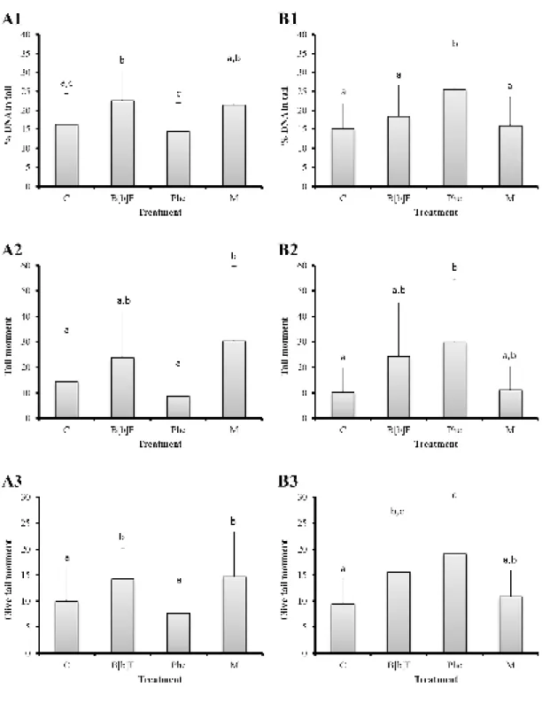
The Comet assay yielded distinct patterns of total DNA strand breakage (inferred from the % of DNA in tail) between experiments A and B (Figure 4.3). Overall, these patterns were consistent between metrics, i.e., %DNA in tail, tail moment and Olive moment (Figure 4.4).
14
between B[b]F and M (%DNA in tail > 21%) (Figure 4.4 A1). Fish injected with benzo[b]fluoranthene isolated, from experiment A, exhibited a significant increase of percentage of DNA in tail (Fisher’s LSD test, p < 0.05) compared to the control and Phe treatments (Figure 4.4 A1). Animals exposed to the PAH mixture also showed a significant increment of DNA strand breakage (Fisher’s LSD test, p < 0.05) when compared to the Phe treatment (Figure 4.4 A1). However, although no significant differences of percentage of tail moment (Fisher’s LSD test, p < 0.05) were found for fish treated with B[b]F, fish exposed to the PAH mixture showed a significant increase (Fisher’s LSD test, p < 0.05) compared to the control and Phe treatments (Figure 4.4 A2). Nevertheless, animals injected with benzo[b]fluoranthene and the combination of the two PAHs exhibited a significant increase of percentage of Olive moment (Fisher’s LSD
test, p < 0.05) when compared with the control and Phe treatments (Figure 4.4 A3).
With respect to experiment B, fish injected with phenanthrene revealed an significant increment of percentage of DNA in tail (Fisher’s LSD test, p < 0.05) compared to the other treatments, however no significant differences were found between control, B[b]F and M treatments (Figure 4.4 B1). Although, animals treated with phenanthrene exhibited a significant increase of percentage of tail moment (Fisher’s LSD test, p < 0.05) compared to control treatment (Figure 4.4 B2), and a significant increase of percentage of Olive moment (Fisher’s LSD test, p < 0.05)
compared to control and M treatments (Figure 4.4 B3), these tail moments showed no significant differences (Fisher’s LSD test, p < 0.05) between the B[b]F and Phe treatments, unlike the % DNA in tail (Figure 4.4 B1, B2 and B3). On the other hand, % DNA in tail and Olive tail moment parameters showed significant differences (Fisher’s LSD test, p < 0.05) between Phe and M treatments, contrary to what was observed for the tail moment parameter (Figure 4.4 B1, B2 and B3).
15
Figure 4.3. Comet examples from injected Dicentrarchus labraxshowing nucleoids with ≈ 0% (a), ≈ 10%
16
Figure 4.4. Mean percentages of the three Comet assay parameters used (100 comets measured per individual). (A) Experiment with the lower dose (5 µg.g-1 fish ww). (B) Experiment with the higher dose
(10 µg.g-1 fish ww). (1) Mean percentage of DNA in tail. (2) Mean percentage of tail moment. (3) Mean
percentage of olive tail moment. Error bars represent 95% confidence intervals. Different letters indicate
17 4.3. Histopathological alterations
4.3.1. Liver histopathology
Dicentrarchus labrax from the control treatment of either experiment, A and B, presented a normal parenchyma consistent with juveniles, composed of polyhedral hepatocytes with a clear cytoplasm and regular-sized nuclei (Genten et al., 2009). The sea bass liver, similar to that of other fish species, is characterized by the presence of other tissue elements such as, bile ducts and exocrine pancreatic tissue (hepatopancreas) (see Figure 4.5 F). Small capillaries (sinusoids) branching from large blood vessels were observed, containing few blood cells, mostly erythrocytes (Figure 4.5 A). Also, the liver of these animals showed no significant signs of histopathological lesions and alterations. Fish injected with phenanthrene displayed high resemblances to control fish, exhibiting little or no signs of inflammatory response (Figure 4.5 B), indicated by infiltration of leukocytes and, typically, by congested adjacent blood vessels (hyperemia). Likewise, livers of fish treated with phenanthrene from experiment A (low dose) displayed high similarities with fish from experiment B (high dose) of the same treatment. However, fish injected with benzo[b]fluoranthene and the PAH mixture presented greater severity and dissemination of lesions and alterations in the hepatic parenchyma than the control animals. Overall, livers of fish exposed to the PAH mixture from experiment B (high dose) showed more damage-related lesions than fish injected with the PAH mixture from experiment A (low dose).
Between all of alterations most usually observed in livers of fish injected with PAHs, circulatory disturbances and fat vacuolation were the most conspicuous. Although, inflammatory response-related alterations, such as hyperemia and infiltration of defense cells, was observed in all treatments, B[b]F and the PAH mixture treatments presented signs of greater inflammatory response (Figure 4.5 C, D, E and F). For these treatments was frequently observed a more pronounced hyperemia, likely caused by inflammation evidenced by proliferation and swelling of sinusoids. Inflammation occurred in all treatments in both experiments A and B, however limited to small foci in control livers (Figure 4.5 A). Hepatocyte fat vacuolation was observed in livers of all treatments of both experiments A and B, even in some control animals and fish injected with phenanthrene (Figure 4.5 B). However, fish injected with benzo[b]fluoranthene and the PAH mixture presented greater degree and dissemination of this progressive change, exhibiting highly vacuolated hepatocytes, enlarged due to increase lipid storage (Figure 4.5 D and E).
18
areas was more significant in fish exposed to benzo[b]fluoranthene and the PAH mixture, from both experiments A and B (Figure 4.5 C and D). Occasionally, focal hemorrhage was observed in necrotic areas (Figure 4.5 D and F). Nuclear alterations such as pyknosis were recurrent in areas where focal cell death was occurring (necrosis or apoptosis, in some cases), indicating alterations to chromatin (Figure 4.5 C). Livers of fish injected with benzo[b]fluoranthene and the PAH mixture from experiment B (high dose) also presented a higher prevalence of macrophages intruding the damaged tissue, suggesting inflammation (Figure 4.5 D and F). In the most damage livers of fish injected with benzo[b]fluoranthene from experiment B (high dose) was observed different forms of hepatocellular degeneration (Figure 4.5 D). In addition, this particular liver presented lipidic droplets inside blood vessels, which could possibly be an artefact, but nevertheless confirms that was in fact a fat liver (Figure 4.5 D).
19
20
border (arrowheads). Proliferation and swelling of blood vessels (bv) are also evident. Lipid droplets (ld) inside hepatic portal vein (hpv), as a consequence of a fatty liver. Inset: hemorrhage characterized by blood cells and defense cells invading the liver parenchyma at a necrotic focus (arrow). (E) Liver from a fish injected with benzo[b]fluoranthene and phenanthrene (mixture treatment) from experiment B (high dose), exhibiting a small area of hepatocellular alteration similar to adenomatous tissue (ad). The anaplastic tissue seems to compress the edge of the normal parenchyma (arrowheads). Proliferation of hepatocytes containing fat vacuoles (v) was also observed. (F) Necrotic focus (n) of pancreatic tissue in the liver of a fish injected with benzo[b]fluoranthene and phenanthrene (mixture treatment) from experiment B (high dose). Hemorrhage (arrow) is indicated by the infiltration of erythrocytes (e) into necrotic tissue.
4.3.2. Spleen histopathology
Overall, the majority of the fish presented the normal spleen microanatomy common among vertebrates, regardless of treatment. The spleen was composed essencially by two components, white and red pulp without clear boundaries, enclosed by a capsule (Figure 4.6 A). The spleen of control animals from both experiments A and B, exihibited increased volume of red pulp relatively to white pulp, comparatively to other treatments, as expected from a normal organ (Figure 4.6 A and B). In the sea bass, white pulp is poorly developed, composed essentially of lymphoid tissue, interconnected by a system of sinusoids and splenic cords forming the red pulp (Quesada et al., 1990).
The most frequent histopathological alteration was hyperemia. This alteration was observed in all treatments from both experiments A and B, however fish injected with benzo[b]fluoranthene and phenanthrene (mixture treatment) presented signs of greater inflammatory response, indicated by a more pronounced hyperemia and infiltration of defense cells, mostly lymphocytes and macrophages, into blood vessels (Figure 4.6 C).
21
Figure 4.6. Common histopathological lesions and alterations observed in the spleen of tested Dicentrarchus labrax (H&E). (A) Overall aspect the normal splenic parenchyma, which is composed by white (wp) and red (rp) pulp, enclosed by a capsule (c). s sinusoids. (B) Hyperemia, here in the spleen of a control fish, which was commonly revealed by congestion of sinusoids (s). Inset: Melanomacrophage centres (arrowhead) were commonly observed scattered through the splenic parenchyma. They are indicated by bodies containing of dark brown (melanin) or brown yellow (lipofuscin) materials. (C) Swollen spleen of a fish injected with benzo[b]fluoranthene and phenanthrene resulting in a high inflammatory response. Agglomerates of inflammatory cells (arrows) were frequently observed in blood vessels of inflammated spleens.
4.4. The Microtox test
22
Figure 4.7. Acute toxicity (% inhibition of luminescence of Vibrio fischeri) obtained for each treatments C (control treatment), B[b]F (the potentially carcinogenic PAH), Phe (the non-carcinogenic PAH) and M (combination of the two PAH compounds). Results are shown for each experiment: A (low dose, 5 µg.g-1
23 5. Discussion
The present study revealed that the two PAHs, benzo[b]fluoranthene (considered carcinogenic) and phenanthrene (non-carcinogenic), isolated or combined, caused different patterns of toxic effects to a marine fish. However, both compounds demonstrated ability to induce DNA damage at the level of nucleotide chains and at the chromosomal level, as well as histopathological alterations. Still, Dicentrarchus labrax injected intraperitoneally with benzo[b]fluoranthene only and the combination of benzo[b]fluoranthene and phenanthrene showed, in general, more severe genotoxic and histopathological lesions than fish treated with phenanthrene only. Overall, benzo[b]fluoranthene caused moderate clastogenic and aneugenic alterations and significant hepatic histopathological alterations, especially concerning inflammation-related responses. It must be noted that fish were subjected to a short-time test (48 h) after injection and, therefore, the overall histopathological hepatic alterations are more consistent with acute effects than chronic alterations, which would require longer bioassays to become developed. In spite of the lack of knowledge on the differences between the toxicological pathways of carcinogenic and non-carcinogenic PAHs, the results are accordant with previous findings, under different experimental conditions, on fish (Neuparth et al., 2009; Machado et al., 2014) and even bivalves (Martins et
al., 2013), which confirms that both phenanthrene and benzo[b]fluoranthene possess different genotoxic potentials.
24
It should also be stressed that PAH activation is regulated by a positive feedback loop that regulates CYP transcription. It is known that PAHs may induce CYP1A gene expression through the binding to an intracellular receptor complex, the Aryl Hydrocarbon Receptor (Ahr), which, on its turn, will form a complex with the aryl hydrocarbon nuclear translocator (ARNT). This complex will bind to a specific part of the DNA, the xenobiotic response element (XRE), promoting transcription (see for reviews Goksøyr and Förlin, 1992; Bucheli and Fent, 1995).
Billiard et al. (2002) observed that the common model PAH benzo[a]pyrene, also a five-ring PAH like B[b]F, although far more extensively studied, and phenanthrene had differential affinities towards Ahr and, subsequently, different abilities for CYP1A induction in juvenile rainbow trout (Oncorhynchus mykiss). Actually, five-ring PAH, like B[a]P and B[b]F, are regarded as stronger Ahr agonists than lower molecular weight PAHs, therefore more able to induce CYP1A transcription and, consequently, the bioactivation of PAHs into genotoxic metabolites (see Mu et
al., 2012). This may explain the higher genotoxic damage observed in fish injected with B[b]F when compared to the fish injected with Phe following injection with the lower dose, albeit not the higher dose treatment (Figure 4.4). Nonetheless, it is highly likely that the high-dose treatment
induced significant cytotoxic effects in the animals’ peripheral blood cells, which is accordant
with the results from the Microtox test (Figure 4.7), in spite of the differences between blood cells and the prokaryote V. fischeri.
25 In fact, Costa et al. (2011b) reported that fat vacuolation could be caused by other unspecific factors since fatty livers are a common alteration observed in farmed fish and may depend on diet. In addition, some authors suggest that fatty livers may account for reduced energy production and weakened anti-oxidative responses, potentially leading to hepatocellular dysfunctions (Vendemiale et al., 2001; Sánchez-Pérez et al., 2005). So, it is possible that control animals already had their fat metabolism altered and as consequence, had presented in their livers some degree of fat vacuolation.
Polycyclic aromatic hydrocarbons are known to induce both DNA strand breakage as well chromosomal breakage, leading to the formation of erythrocytic nuclear abnormalities (ENA) (see, for example, Costa et al., 2008, 2011a; Neuparth et al., 2009). Although, DNA fragmentation, formation of DNA-PAH adducts and chemically altered nucleotide (alkali-labile sites) may result from direct action of mutagens to the DNA chains (e.g. Neuparth et al., 2009; Costa et al., 2011a), chromosomal clastogenesis, assessed through the ENA assay, may be a consequence of the errors occurred during cellular division, where DNA damage was passed on to daughter cells, biomagnifying the initial baseline DNA damage. For this reason, considering that DNA strand breakage damages may repaired depending on type and extent, chromosomal clastogenesis may be considered a more severe type of mutagenesis, since it is unlikely to be repaired (Costa et al., 2011a). Therefore, it is likely that benzo[b]fluoranthene can, overall, induce more severe genotoxic damage (Figure 4.2).
Since the frequency of nuclear abnormalities are linked to the frequencies of apoptotic and necrotic cells (e.g. Ghiraldini and Mello, 2010), the greater frequency of ENA observed in blood cells of fish treated with benzo[b]fluoranthene could be linked with acute lesions and alterations of the hepatic parenchyma of fish injected with the same treatment. In fact, focal hepatic necrosis, nuclear pleomorphism and inflammatory response were usually observed together (Figure 4.5 C, D). The presence of nuclear pyknosis and inflammation on livers clearly confirms the evidence of hepatic necrosis and is usually associated with toxicant-induced necrosis (Newman, 2010). It must be noted that necrotic foci allied with nuclear pyknosis and hemorrhage (indicating inflammation) has already been reported in livers of juvenile soles exposed to sediments mostly contaminated by organic compounds (Costa et al., 2009, 2011b), indicating that these compounds are strong inducers of severe, likely acute, hepatic lesions albeit unspecific.
26
and Santos, 1997; Gravato and Santos 2002, 2003) and invertebrates (Venier et al., 1997) and PAHs with four or more condensed benzene rings (like B[b]F) are potential mutagenic and/or carcinogenic as benzo[a]pyrene (Gravato and Santos, 2002), present results showed that phenanthrene (PAH with three condensed rings) is also genotoxic to sea bass (by eliciting DNA strand breaks), when fish were injected with higher doses for a short-term period (Figure 4.4), apparently in accordance with the results of the Microtox test (Figure 4.7).
Since phenanthrene is a lower molecular weight PAH, its metabolites are generally acknowledged to be less genotoxic than those of larger PAHs, which results from a poorer interaction with DNA (Thakker et al., 1978; Wood et al. 1979; Martins et al., 2013). It is possible that exposure to benzo[b]fluoranthene yielded both genotoxic metabolites and ROS, while phenanthrene caused genetic damage, mainly, as a result of the formation of ROS alone. Although exposure to PAH might produce reactive oxygen species, under normal physiological conditions they can be removed by antioxidant defense systems (Livingstone, 2001; Meyer et al., 2002). Yin et al. (2007) observed, in fish (Carassius auratus) exposed to different concentrations of phenanthrene, an
increase of ROS production, mostly hydroxyl radical (OH). This type of radical is the most reactive towards the DNA molecule (Cadet et al., 2010). Yin et al. (2007) also demonstrated that fish can experience severe oxidative stress when exposed to high concentrations of phenanthrene. Therefore, it is possible that fish for experiments B, injected with higher doses of phenanthrene, suffered severe oxidative stress, suppressing the antioxidant defense system, and led to a higher induction of total DNA strand breakage (Figure 4.4). Once more, these findings are accordant with the results from the Microtox test (Figure 4.7), indicating higher acute effects induced by this PAH. It should be noted that the high doses administered in sea basses are not ecologically relevant, which, allied with the stress caused by the injection treatments may explain the highest level of mortality in fish treated with phenanthrene and the PAH mixture, from experiment B (Table 4.1).
The exposure of the luminescent marine bacterium Vibrio fischeri to phenanthrene appears to pose a higher short-term acute toxicity than benzo[b]fluoranthene. Actually, the biotransformation of phenanthrene, leading to the formation of phenanthrene quinones, leads to inhibition of V. fischeri luminescence, even at low concentrations (Wang et al., 2009). Wang et
27 five or more rings is still limited. Nevertheless, Seo et al. (2009) and Kanaly and Harayama (2000) reported that benzo[a]pyrene, more liposoluble than phenanthrene, poses a higher resistance to microbial degradation, and therefore, the relation between biodegradation rates and PAH molecular weight must be considered in toxicity analyses. For this reason, it is possible that phenanthrene was metabolized by bacterium V. fischeri at a higher rate than benzo[b]fluoranthene, yielding more metabolites and present a higher toxic potential to bacteria, as shown by the Microtox test (Figure 4.7).
In contrast, despite the Microtox test revealed a similar acute toxic effect of phenanthrene and the PAH mixture (Figure 4.7), no similarities were found between this pattern of toxicity and those obtained for ENA and the Comet assays in animals from experiment B (high dose) (Figures 4.2 and 4.4). In fact, the present results suggest that interaction effect between the two PAHs depends on the dose of exposure. It should be noticed that, as far other toxicants, PAH interactions effects can be additive (when lead to an addition of the effect of both PAHs), supra-additive, synergistic (when the effect of the two PAHs yields an effect that cannot be attained by either isolates compound) and antagonistic (when a PAH could suppress the effect of the other compound) (Freedman, 1995; Staal et al., 2007; Gonçalves et al., 2008). Even though many studies report additive or synergistic effect in complex PAH mixtures in fish (see e.g. Basu et al., 2001; Billiard
et al., 2008; Fleming and Di Giulio, 2011), and also in humans (Staal et al., 2007), some researchers demonstrated antagonistic interactions of PAHs in multiple organisms, from mice (Springer et al., 1985) to Salmonella (Hermann, 1981; White, 2002). From these contradictory reports, it is clear that further research is needed to better understand the behavior of genotoxic compounds in complex mixtures and the relation of dose- and time- effect of PAHs and their interactions.
For animals subject to experiment B, the combined effects of the two PAHs seems to be less genotoxic than the effects of the two isolated compounds, which suggest that interactions between benzo[b]fluoranthene and phenanthrene, in higher doses, can produce antagonistic effects. Staal
28
PAH mixtures than cells treated with benzo[a]pyrene alone (Mahadevan et al.; 2004).
Actually, some authors suggest that the occurrence of more than one CYP1A inducers can interfere with the integrity of the enzyme causing its inactivation (Stegeman and Hahn, 1994). The inhibition of CYP1A enzymes in fish liver was showed in several studies at high PAH exposure (Haasch et al., 1993; Schlezinger and Stegeman, 2001; Correia et al., 2007). Therefore, it is highly likely that the high-dose treatment of PAH mixture may have led to inhibition of MFOs, generating less active metabolites, hence less DNA strand breakage and chromosomal damage (Figures 4.2 B and 4.4 B). Interestingly, the same was not observed in livers and spleens of fish treated with the mixture of PAH. Overall, fish injected with the PAH mixture, from experiment B, presented a more damage-related lesions than fish treated with lower doses of the two PAHs. The same relation was observed in livers treated with B[b]F only. Overall, livers of fish subjected to experiment B (high dose) presented a greater dissemination of lesions and alterations in the hepatic parenchyma than livers from experiment A (low dose) (Figures 4.5 C, D), suggesting that the damage observed in livers treated with the PAH mixture occurred, mainly, by the exposure to benzo[b]fluoranthene. Since benzo[b]fluoranthene is more lipossoluble (log Kow = 5.78) than phenanthrene (log Kow = 4.52) (IARC, 2010), this PAH tends to accumulate in
lipid-rich tissues (as livers) and therefore it is possible that its toxic effect was more evident in liver (yielding acute lesions) than in blood cells (Figures 4.4 and 4.5). Inflammatory response-related alterations, such as hyperemia and infiltration of defense cells, was observed in all treatments, however B[b]F and the PAH mixture presented signs of greater inflammatory response. Although inflammation-related responses and effects are highly non-specific, some researchers already related high prevalence of hyperemia in fish liver as a consequence of exposure to organic compounds (Noreña-Barroso et al., 2004; Agamy, 2012; Zorita and Cuevas, 2014). Even though inflammatory response is usually associated with other pathophysiological processes, in this case, the absence of defense cells other than macrophages indicates that this alteration was probably related to non-infectious agents. It must be noted that even though a probable hepatocellular adenoma (Figure 4.5 E), was found in the liver of a fish treated with the PAH mixture, it is highly unlikely that this hepatic alteration was caused by action of the PAHs, since the animals were allowed to incubate for just 48 h. Still, adenomas have been observed in fish after long-term exposure to PAHs (Reynolds et al., 2003; Zorita and Cuevas, 2010). However this type of hepatocellular change seems to be highly depended of fish age (Vethaak et al., 1996).
31 6. Conclusions
This study aimed at understanding the comparative and interaction effects between two PAHs, isolated or in mixture, in a model organism (Dicentrarchus labrax). The present results revealed that isolated benzo[b]fluoranthene (considered potentially carcinogenic to humans) and the PAH mixture caused the most severe genotoxic effects and histopathological alterations in sea bass. However phenanthrene, considered non-carcinogenic to humans by IARC, also poses genotoxic effects, especially at a higher dose (experiment B). The different effects between benzo[b]fluoranthene (five-ring PAH) and phenanthrene (three-ring PAH), with respect to DNA strand breakage and induction of ENAs, confirmed a positive relation between the number of PAH benzene rings and their genotoxicity to fish. Also importantly, the results indicated no significant additive effects between benzo[b]fluoranthene and phenanthrene, under the current experimental conditions, in the contrary, higher doses of PAHs seems to inhibit the activity of CYP1A-related isoenzymes, restraining the formation of genotoxic activated PAH metabolites and consequently, reducing their genotoxicity. The current findings also suggested that the interaction between the two PAHs, in the sea bass, depends on dose of exposure, since the pattern of genotoxic effects varied significantly between the two experimental conditions.
Nevertheless, the results also suggest a distinction between the type of toxicity of the PAHs, in the sense that benzo[b]fluoranthene appears to induce more chronic effects more significantly, whereas phenanthrene is acutely toxic to fish and Vibrio fischeri, as shown by higher mortality (especially at higher concentrations) and more significant effects obtained from the Microtox test. However, this issue still requires from further research. Furthermore, V. fischeri appears to be highly sensitive to DMSO, in spite of the low concentrations of the carriers used in testing, which may have hindered clear dose-effect relationships.
The Comet assay (alkaline version) seems to be a more sensitive method than ENA analysis, under the present conditions of assessment, since it reflected better the genotoxic effects of benzo[b]fluoranthene and phenanthrene, isolated or in mixture, between the two experiments A and B. The Comet metrics here employed (percentage of DNA in tail, tail moment and Olive tail moment) revealed to be equally good parameters, since they showed a similar pattern of measuring DNA damage at the molecular level, under the current experimental conditions.
32
showed sensitivity toward PAHs, however liver, likely for being the main organ of accumulation and detoxification of xenobiotics, revealed to be a better target than spleen, since it yielded more severe and diffuse alterations. Still, it must be noted that the lesions and alterations observed are rather unspecific and reflect an acute form of exposure (intraperitoneal injection of the toxicants). Also, peripheral blood cells of fish also proved to be a relevant target in evaluating PAH toxicity, since cell bloods are rapidly affected by the direct action of contaminants and yielded consistent results.
33 7. References
Aas, E., Baussant, T., Balk, L., Liewenborg, B., Anderson, O. K., 2000. PAH metabolites in bile, cytochrome P4501A and DNA adducts as environmental risk parameters for chronic oil exposure: a laboratory experiment with Atlantic cod. Aquatic Toxicology 51, 241-258.
Agamy, E., 2012. Histopathological liver alterations in juvenile rabbit fish (Siganus canaliculatus) exposed to light Arabian crude oil, dispersed oil and dispersant. Ecotoxicology and Environmental Safety 75, 171-179.
Agius C. 1979a. Aspects of the melano-macrophage centres in fish. PhD Thesis, University of Stirling.
Agius C. 1979b. The role of melano-macrophage centres in iron storage in normal and diseased fish. Journal of Fish Diseases 2, 337-343.
Au, D. W. T., 2004. The application of histo-cytopathological biomarkers in marine pollution monitoring: a review. Marine Pollution Bulletin 48, 817-834.
Basu, N., Billiard, S., Fragoso, N., Omoike, A., Tabash, S., Brown, S., Hodson, P., 2001. Ethoxyresorufin-O-deethylase induction in trout exposed to mixtures of polycyclic aromatic hydrocarbons. Environmental Toxicology and Chemistry 20, 1244-1251.
Biagianti-Risbourg, S., Pairault, C., Vernet, G., Boulekbache, H., 1996. Effect of lindane on the ultrastructure of the liver of the rainbow trout, Oncorhynchus mykiss, sac-fry. Chemosphere 33, 2065-2079.
Billiard, S. M., Meyer, J. N., Wassenberg, D. M., Hodson, P. V, Di Giulio, R. T., 2008.
Nonadditive effects of PAHs on early vertebrate development : mechanisms and implications for
risk assessment. Toxicological Sciences 105, 5-23.
Billiard, S. M., Hahn, M. E., Franks, D. G., Peterson, R. E., Bols, N. C., Hodson, P. V, 2002. Binding of polycyclic aromatic hydrocarbons (PAHs) to teleost aryl hydrocarbon receptors (AHRs). Comparative Biochemistry and Physiology Part B 133, 55-68.
Boldrin, B., Tiehm, A., Fritzsche, C., 1993. Degradation of phenanthrene, fluorene, fluoranthene, and pyrene by a Mycobacterium sp. Applied and Environmental Microbiology 59, 1927-1930.
Bucheli, T. D., Fent, K., 1995. Induction of cytochrome P450 as a biomarker for environmental contamination in aquatic ecosystems. Critical Reviews in Environmental Science and Technology 25, 201-268.
Cadet, J., Douki, T., Ravanat, J.- L., 2010. Oxidatively generated base damage to cellular DNA. Free Radical Biology and Medicine 49, 9-21.
Carlson, E. A., Li, Y., Zelikoff, J. T., 2002a. The Japanese medaka (Oryzias latipes) model: applicability for investigating the immunosuppressive effects of the aquatic pollutant benzo[a]pyrene (BaP). Marine Environmental Research 54, 565-568.
34
Carlson, E. A., Li, Y., Zelikoff, J. T., 2004. Benzo[a]pyrene-induced immunotoxicity in Japanese medaka (Oryzias latipes): relationship between lymphoid CYP1A activity and humoral immune suppression. Toxicology and Applied Pharmacology 201, 40-52.
Conney, A. H., 1982. Induction of microsomal enzymes by foreign chemicals and carcinogenesis by polycyclic aromatic hydrocarbons. Cancer Research 42, 4875-4917.
Correia, A. D., Gonçalves, R., Scholze, M., Ferreira, M., Henriques, M. A.-R., 2007. Biochemical and behavioral responses in gilthead seabream (Sparus aurata) to phenanthrene. Journal of Experimental Marine Biology and Ecology 347, 109–122.
Costa, P. M., Carreira, S., Costa, M. H., Caeiro, S., 2013. Development of histopathological indices in a commercial marine bivalve (Ruditapes decussatus) to determine environmental quality. Aquatic Toxicology 126, 442-454.
Costa, P. M., Costa, M. H., 2007. Genotoxicity assessment in fish peripheral blood: a method for a more efficient analysis of micronuclei. Journal of Fish Biology 71, 148-151.
Costa, P. M., Diniz, M. S., Caeiro, S., Lobo, J., Martins, M., Ferreira, A. M., Caetano, M., Vale, C., DelValls, T. A., Costa, M. H., 2009. Histological biomarkers in liver and gills of juvenile Solea senegalensis exposed to contaminated estuarine sediments: a weighted indices approach. Aquatic toxicology 92, 202-212.
Costa, P. M., Neuparth, T. S., Caeiro, S., Lobo, J., Martins, M., Ferreira, A. M., Caetano, M., Vale, C., DelValls, T. A., Costa, M. H., 2011a. Assessment of the genotoxic potential of contaminated estuarine sediments in fish peripheral blood: laboratory versus in situ studies. Environmental Research 111, 25-36.
Costa, P. M., Caeiro, S., Lobo, J., Martins, M., Ferreira, A. M., Caetano, M., Vale, C., DelValls, T. Á., Costa, M. H., 2011b. Estuarine ecological risk based on hepatic histopathological indices from laboratory and in situ tested fish. Marine Pollution Bulletin 62, 55-65.
Costa, P. M., Lobo, J., Caeiro, S., Martins, M., Ferreira, A. M., Caetano, M., Vale, C., Delvalls, T. Á., Costa, M. H., 2008. Genotoxic damage in Solea senegalensis exposed to sediments from the Sado Estuary (Portugal): effects of metallic and organic contaminants. Mutation Research 654, 29-37.
Douben, P. E. T., (Ed.), 2003. PAHs: An ecotoxicological perspective. Unilever Colworth R&D, Safety and Environmental Assurance Centre. Bedford, 391pp.
Fleming, C. R., Di Giulio, R. T., 2011. The role of CYP1A inhibition in the embryotoxic interactions between hypoxia and polycyclic aromatic hydrocarbons (PAHs) and PAH mixtures in zebrafish (Danio rerio). Ecotoxicology 20, 1300-1314.
Freedman, M. D., 1995. Drug interactions: classification and systematic approach. American Journal of Therapeutics 2, 433-443.
Genten, F., Terwinghe, E., Danguy, A. 2009. Atlas of fish histology. Science Publishers, Enfield, NH, USA, 215 pp.
35 Reports 17, 27-31.
Goksøyr, A., Förlin, L., 1992. The cytochrome P-450 system in fish, aquatic toxicology and environmental monitoring. Aquatic Toxicology 22, 287-312.
Gonçalves, R., Scholze, M., Ferreira, A. M., Martins, M., Correia, A. D., 2008. The joint effect of polycyclic aromatic hydrocarbons on fish behavior. Environmental Research 108, 205-213.
Gravato, C., Santos, M. A, 2002. Juvenile sea bass liver P450, EROD induction, and erythrocytic genotoxic responses to PAH and PAH-like compounds. Ecotoxicology and Environmental Safety 51, 115-127.
Gravato, C., Santos, M. A., 2003. Genotoxicity biomarkers’ association with B(a)P biotransformation in Dicentrarchus labrax L. Ecotoxicology and Environmental Safety 55, 352-358.
Gravato, C., Santos, M. A., Magalhães, I., 2000. Juvenile Dicentrarchus labrax L. biochemical and genotoxic responses after short-term exposure toβ-naphthoflavone and contaminated harbor waters. Fresenius Environmental Bulletin 9, 269-274.
Haasch, M. L., Quardokus, E. M., Sutherland, L. A., Goodrich, M. S., Lech, J. J., 1993. Hepatic Cyp1A1 induction in rainbow trout by continuous flow through exposure to β-naphthoflavone. Fundamental and Applied Toxicology 20, 72-82.
Hannam, M. L., Bamber, S. D., Galloway, T. S., Moody, A. J., Jones, M. B., 2010. Effects of the model PAH phenanthrene on immune function and oxidative stress in the haemolymph of the temperate scallop Pecten maximus. Chemosphere 78,779-784.
Hermann, M., 1981. Synergistic effects of individual polycyclic aromatic hydrocarbons on the mutagenicity of their mixtures. Mutation Research 90, 399-409.
IARC, 1983. IARC monographs on the evaluation of the carcinogenic risk of chemicals to humans. Polynuclear aromatic hydrocarbons, Part 1, Chemical, Environmental and Experimental Data, Vol. 32. International Agency for Research on Cancer, Lyon, France.
IARC, 2010. IARC monographs on the evaluation of carcinogenic risks to humans. Some non-heterocyclic polycyclic aromatic hydrocarbons and some related exposures, Vol. 92. International Agency for Research on Cancer, Lyon, France.
Kanaly, R., Harayama, S., 2000. Biodegradation of high-molecular-weight polycyclic aromatic hydrocarbons by bacteria. Journal of Bacteriology 182, 2059-2067.
Kang, H., Hwang, S. Y., Kim, Y. M., Kim, E., Kim, Y. S., Kim, S. K., 2003. Degradation of phenanthrene and naphthalene by a Burkholderia species strain. Canadian Journal Microbiology 49, 139-144.
Kerambrun, E., Le Floch, S., Sanchez, W., Thomas Guyon, H., Meziane, T., Henry, F., Amara, R., 2012. Responses of juvenile sea bass, Dicentrarchus labrax, exposed to acute concentrations of crude oil, as assessed by molecular and physiological biomarkers. Chemosphere 87, 692-702.
![Figure 2.1. Thesis layout. ENA - erythrocytic nuclear abnormilites. B[b]F - benzo[b]fluoranthene](https://thumb-eu.123doks.com/thumbv2/123dok_br/16655803.741984/28.892.138.760.117.654/figure-thesis-layout-erythrocytic-nuclear-abnormilites-benzo-fluoranthene.webp)
![Table 4.1. Mortality (given as number of casualties per six biological replicates) observed for the treatments C (control treatment), B[b]F (the potentially carcinogenic PAH), Phe (the non-carcinogenic PAH) and M (the combination of the two compounds)](https://thumb-eu.123doks.com/thumbv2/123dok_br/16655803.741984/33.892.134.754.412.520/mortality-casualties-biological-replicates-potentially-carcinogenic-carcinogenic-combination.webp)


![Figure 4.7. Acute toxicity (% inhibition of luminescence of Vibrio fischeri) obtained for each treatments C (control treatment), B[b]F (the potentially carcinogenic PAH), Phe (the non-carcinogenic PAH) and M (combination of the two PAH compounds)](https://thumb-eu.123doks.com/thumbv2/123dok_br/16655803.741984/44.892.159.747.151.472/inhibition-luminescence-treatments-treatment-potentially-carcinogenic-carcinogenic-combination.webp)
