w w w . r b h h . o r g
Hematology, Transfusion and Cell Therapy
Review article
The role of ClpX in erythropoietic protoporphyria
Jared C. Whitman, Barry H. Paw
a, Jacky Chung
∗Brigham and Women’s Hospital, Harvard Medical School, Boston, MA, United States
a r t i c l e
i n f o
Article history:
Received 28 February 2018 Accepted 2 March 2018 Available online 28 March 2018
Keywords:
Heme biosynthesis enzymes Porphyria
Erythropoietic protoporphyria ClpXP
ALASgene
a b s t r a c t
Hemoglobin is an essential biological component of human physiology and its production in red blood cells relies upon proper biosynthesis of heme and globin protein. Disruption in the synthesis of these precursors accounts for a number of human blood disorders found in patients. Mutations in genes encoding heme biosynthesis enzymes are associated with a broad class of metabolic disorders called porphyrias. In particular, one subtype – erythro-poietic protoporphyria – is caused by the accumulation of protoporphyrin IX. Erythroerythro-poietic protoporphyria patients suffer from photosensitivity and a higher risk of liver failure, which is the principle cause of morbidity and mortality. Approximately 90% of these patients carry loss-of-function mutations in the enzyme ferrochelatase (FECH), while 5% of cases are asso-ciated with activating mutations in the C-terminus ofALAS2. Recent work has begun to uncover novel mechanisms of heme regulation that may account for the remaining 5% of cases with previously unknown genetic basis. One erythropoietic protoporphyria family has been identified with inherited mutations in the AAA+ protease ClpXP that regulatesALAS
activity. In this review article, recent findings on the role of ClpXP as both an activating unfoldase and degrading protease and its impact on heme synthesis will be discussed. This review will also highlight the role of ClpX dysfunction in erythropoietic protoporphyria.
© 2018 Associac¸ ˜ao Brasileira de Hematologia, Hemoterapia e Terapia Celular. Published by Elsevier Editora Ltda. This is an open access article under the CC BY-NC-ND license (http://creativecommons.org/licenses/by-nc-nd/4.0/).
Introduction
Heme is a chemical moiety that is essential for all life.1–6In
humans, heme is most commonly known for its role in red blood cells (RBCs) as the oxygen-binding prosthetic group in hemoglobin. Given its importance in RBC physiology, defec-tive heme metabolism is strongly associated with hematologic diseases in humans.
∗ Corresponding author at: BWH Hematology Division, Harvard Institutes of Medicine Research Postdoctoral Fellow, 77 Avenue Louis
Pasteur, Room 612, Boston, MA 02115, United States.
E-mail address:jchung12@rics.bwh.harvard.edu(J. Chung).
a
Posthumously (seeAppendix A).
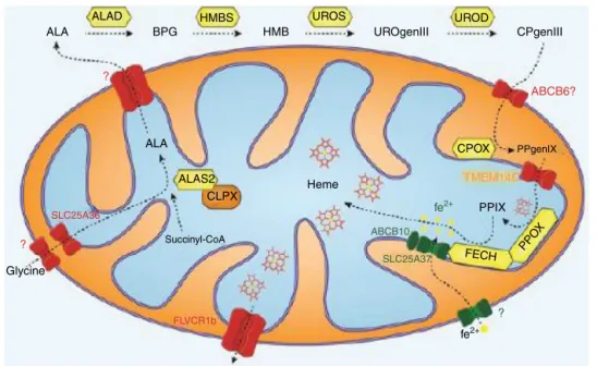
With few exceptions, heme is synthesized via a highly conserved set of eight enzymatic reactions. The first com-mitted step of heme biosynthesis involves the condensation of glycine and succinyl-CoA in the mitochondria to gen-erate ı-aminolevulinic acid (ALA) utilizing the enzyme ı-aminolevulinic acid synthesis (ALAS1). ALA is then
trans-ferred into the cytosol where it is converted to tetrapyrrole coporphyrinogen III (CPgenIII) by four enzymes – ALA
https://doi.org/10.1016/j.htct.2018.03.001
ALAD HMBS UROS UROD
ALA BPG HMB UROgenIII CPgenIII
ALA
Heme
CPOX
Glycine
Succinyl-CoA
PPgenIX ABCB6?
ALAS2 CLPX
SLC25A36
?
?
FLVCR1b
TMEM14C
PPO X
FECH
ABCB10
SLC25A37
?
fe2+
fe2+ PPIX
Figure 1 – Highly conserved heme/protoporphyrin IX (PPIX) synthesis in eukaryotic cells. Precursors, glycine and succinyl-CoA, undergo 8 enzymatic reactions withı-aminolevulinic acid synthesis (ALAS), ALA dehydratase (ALAD), hydroxymethylbilane (HMBS), uroporphyrinogen III synthase (UROS), uroporphyrinogen decarboxylase (UROD), coproporphyrinogen oxidase (CPOX), protoporphyrinogen oxidase (PPIOX), and ferrochelatase (FECH) in both the mitochondria and cytoplasm to form heme.
Figure illustration courtesy of Johannes G. Wittig (Technische-Universität-Dresden, Germany). Reprinted with permission from Yien et al.37and Oncotarget (Impact Journals, LLC).
dehydratase (ALAD), hydroxymethylbilane (HMBS), uropor-phyrinogen III synthase (UROS), and uroporuropor-phyrinogen decarboxylase (UROD).7 After entering the mitochondrial
inner membrane space, the enzyme coproporphyrinogen oxidase (CPOX) catalyzes the conversion of CPgenIII to proto-porphyrinogen IX (PPgenIX).8PPgenIX is then transported into
the mitochondrial matrix where it is the reactant for proto-porphyrinogen oxidase (PPOX), to produce protoporphyrin IX (PPIX). The penultimate enzyme in the cascade, ferrochelatase (FECH), is responsible for the final insertion of iron into PPIX to form heme (Figure 1).9
Erythroid heme production must meet the vast production of hemoglobin and accounts for roughly 85% of the daily heme synthesis.2 All heme biosynthesis genes are
upregu-lated during RBC differentiation.2 However, RBCs also have
unique mechanisms in addition to the conical heme biosyn-thetic pathway. For example, the first step of heme synthesis, involves an erythroid-specific isoform ofALAS(ALAS2) that becomes transcriptionally induced during RBC maturation.
ALAS2 differs from ALAS1 through an iron responsive ele-ment (IRE) inALAS2that can interact with iron responsive proteins (IRPs) allowing for the association ofALAS2to iron availability.10
The final enzyme, FECH, is also regulated in a red cell spe-cific manner. FECH is an iron-sulfur cluster protein located on the inner mitochondrial membrane. In RBCs, FECH is post-translationally stabilized through the formation of a complex with mitoferrin1 and ABCB10.11–13Recent work has
also found that erythropoietin (EPO) signaling together with the GATA-1 transcriptional program regulates the phospho-rylation and activation of FECH in RBCs.10–12 Based on the
number of regulatory pathways converging on FECH in red cells, it is not surprising that FECH is a rate-limiting enzyme in erythroid heme biosynthesis.10
Porphyrias
The intermediates produced by each enzyme in heme synthe-sis are cytotoxic and their accumulation can have deleterious effects. Clinically, the accumulation of heme intermediates cause porphyrias – a diverse group of metabolic disorders. Patients with porphyria suffer from a variety of symptoms including cutaneous photosensitivity, behavioral changes, restlessness, insomnia, seizures, abnormal liver function, and several other life threatening concerns.2,14 The
com-plex genotype-phenotype correlations in combination with environmental factors have given rise to a broad range of disease presentation. Mutations in nine genes are currently known to be associated with porphyrias. Of the nine, defects in seven genes are associated with hepatic porphyrias, such as porphyria cutanea tarda (PCT), and characterized by the accumulation of heme precursors in the liver.2 In contrast,
defects in the remaining two genes –ALAS2andFECH– encode enzymes particularly important in RBC biology and give rise to erythropoietic porphyrias with the distinctive feature of accu-mulating heme precursors in the bone marrow.2
FECH is the gene most commonly associated with ery-thropoietic protoporphyria (EPP), accounting for 90% of diagnosed cases.2These patients have reduced FECH activity.2
mutations lead to an accumulation of photoreactive PPIX. In EPP patients, accumulated PPIX reacts with sunlight result-ing in the production of cytotoxic reactive oxygen species (ROS).4,15 For some patients, even a small amount of light
exposure can trigger skin lesions, while severe sun expo-sure can result in edema, erythema, and wax-like scarring. Increased amounts of free PPIX can also cause patients to suf-fer from more life threating concerns such as gallstones, liver disease, and liver failure.14
Genetic basis of EPP
EPP mutations can be inherited in an autosomal dominant, recessive, or X-linked fashion.2The genetics of EPP have been
extensively studied. EPP cases with FECH mutations most commonly exhibit an autosomal dominant pattern of inheri-tance in which the hypomorphic IVS3-48C FECH allele is found in all affected family members.16–18 When the other FECH
allele is mutated, FECH enzymatic activity is substantially reduced to 15–25% of its normal activity level.2,17,19 Over 60
differentFECHmutations including partial deletions, frame shifts, and substitutions, have been identified to be associ-ated with the IVS3-48C allele and found to cause decreased FECH activity.16–18 The most common mutations found in
this region associated with EPP involve partial deletions. Par-tial deletions within theFECHallele, varying among patients, have been found to alter the structure, activation regions, or stability of FECH resulting in reduced enzyme acitivity.17
Patients suffering from frame shift mutations result in either a defective protein or a premature stop codon.18These changes
disrupt the secondary structure of FECH, leading to a decrease in the effectiveness of the enzyme.18 Research preformed
by Schneider-Yin et al. also found that FECH substitution mutations also cause EPP through changes of the structural components of the [2Fe-2S] cluster of FECH.16The changes in
the [2Fe-2S] cluster, found to play a structural role in FECH, destabilize the FECH enzyme structure resulting in a reduced level of enzymatic activity.16 Even though the mechanisms
through which FECH activity is disrupted are diverse, they all hinder heme production and result in high PPIX.
Although the majority of EPP cases arising fromFECH muta-tions are inherited in an autosomal dominant fashion, there are also a fewFECH-associated EPP cases that present as auto-somal recessive.20 In one family, two healthy parents, each
containing a different mutation to theFECHallele, developed a minor increase in PPIX levels, but no other symptoms.20
When their children inherited both mutant alleles, both chil-dren developed severe EPP and presented with liver failure and extreme photosensitivity.20In this particular case, the two
mutations were a substitution and an insertion that reduced FECH activity to 57% and 64%, respectively.20Although each
mutation alone only partially hindered FECH activity, in com-bination, the mutations led to an extreme depletion of FECH activity, 13–25%.20 Even though these double mutations are
rare, they can result in severe EPP.
Another 5% of EPP cases result from gain-of-function muta-tions inALAS2, which presents as an X-linked disorder.2These
mutations cluster in the inhibitory C-terminal region ofALAS2
and cause either the elongation or deletion of the C-terminus
on ALAS2.2,21 Alterations to the C-terminus of ALAS2
trigg-ers increased ALAS activity resulting in abnormally high PPIX levels.21,22 However, the remaining 5% of EPP patients do
not harborFECHorALAS2mutations, suggesting additional genetic modifiers still exist.
ClpX – a novel regulator of heme production
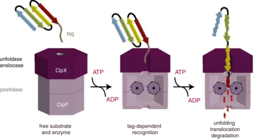
ClpX is a mitochondrial AAA+ ATPase found in tandem with the mitochondrial tetradecameric peptidase hexamer, ClpP, to form the AAA+ protease ClpXP.23Throughin vitrostudies
using the model substrate casein, a model for ClpXP func-tion has been developed.24–26 ClpX is involved in substrate
binding, unfolding, and translocation to ClpP, which mediates protein degradation (Figure 2).23 Independent of ClpP, ClpX
is still able to unfold proteins without degradation.23 While
substrate recognition by ClpX requires only ATP binding, ATP hydrolysis is required for the unfoldase and translocase activi-ties of ClpX.23Although the biochemical function of ClpXP has
been known for some time, physiologic substrates of ClpXP in mammalian cells are largely unknown.
This has recently changed with a series of elegant studies using both yeast (S. cerevisiae) as well as vertebrate models.27
Yeasts lack ClpP and, thus, they are an excellent system to study ClpX unfoldase function without substrate degradation. Researchers, first, found that the yeast equivalent of ClpX, MCX1, promoted the activity of HEM1, which is the yeast homolog of ALAS1.23,27 Similarly, Kardon et al. discovered
that mammalian ClpX, independent of ClpP, activated ALAS2
in vitroby facilitating PLP insertion. Based on these results, the authors proposed that ClpX activates ALAS2 in mammalian cells. In support, expression of ClpX but not ClpP was induced in maturing murine erythroid cells.27 Introduction of
mam-malian ClpP led to a decrease in ALAS activity, suggesting that active ALAS2 is a substrate for ClpXP degradation.27Together
these findings led the authors to propose that ClpXP plays a dual role in heme biosynthesis with ClpX as an activator of ALAS function and ClpP as a repressor. To further confirm ClpX as an activator, Kardon et al. showed that ClpX was required for efficient erythroid heme synthesis in zebrafish (Danio rerio) embryos. Morpholino knockdown of the zebrafish homolog,
clpxa, in zebrafish embryos (termed morphants) resulted in a substantial decrease in hemoglobinization and the number of erythroid cells in vivo.25The anemia found in ClpX
mor-phants was rescued by ALA supplementation that by-passes the block in ALAS activity. These results suggest that the acti-vation of ALAS by ClpX is a conserved pathway and represents an important step in heme synthesis.25
Further work by Kubota et al. has shed light into how ClpXP functions in both capacities and when it is important to do so.25Using the human HepG2 liver cell line, the authors
found that siRNA-mediated knockdown of ClpX led to a robust accumulation of ALAS1. This is consistent with the idea that ClpX is required not only for substrate binding and unfolding, but also shuttling to ClpP for degradation.25 Interestingly,
unfoldase translocase
pestidase
free substrate and enzyme
tag-dependent recognition
unfolding translocation
degradation
ClpX
ClpP
ATP ATP
ADP ADP
tag
Figure 2 – ClpXP protease model of substrate recognition, unfolding, and degradation function. The peptide tag binds to the axial pore of ClpX. Subsequently, ClpX unfolds the substrate, with energy provided by ATP, and translocates the unfolded polypeptide to ClpP, which mediates proteolysis.
Reprinted with permission from Baker et al.23andBiochim. Biophys. Acta(Elsevier, Inc.).
for ClpX-dependent turnover of ALAS1. Alternatively, the mechanisms through which ClpXP regulates ALAS1 may differ from ALAS2.23,25,27It is also important to note that the authors
did not measure ALAS1 activity in ClpP-deficient cells.25There
is evidence to indicate that ClpP inhibits the ATPase activity of ClpX, which is crucial for ClpX unfoldase function.25,27
Presumably, in this fashion, ClpP would inhibit ALAS1 activity by blocking PLP insertion without affecting steady state ALAS1 expression. Nevertheless, based on these findings, Kubota et al. propose that ClpXP constitutes part of the negative feedback loop in which excess heme triggers ALAS1 degradation.
ClpX
is a novel EPP gene
Recently, a family was identified with a history of EPP but no mutations inFECHorALAS2that was inherited in an autoso-mal dominant fashion. Additional analysis found a common heterozygoteClpXmissense mutation in all affected family members. This substitution of glycine with aspartic acid at position 298 of ClpX is located in the conserved Walker A sequence motif involved in ATP-binding.23This ATP binding
region is thought to be absolutely essential for the formation of the ClpXP complex.23,27The alteration of an amino acid at
the ATP binding region, according to models found by Baker et al. and Kardon et al., can cause an alteration in ClpX-ClpP interaction resulting in a variation in the translocation and degradation of both ALAS1 and ALAS2 producing altered heme levels.23,27
When the G298D ClpX mutant protein was exogenously overexpressed in murine erythroid cells, ALAS activity was increased with concomitantly higher PPIX levels.28
Interest-ingly,in vitrostudies showed that the G298D mutation severely compromised ATPase activity of mutant ClpX. When mixed
togetherin vitrowith wild-type ClpX, mutant ClpX was also able to inhibit wild-type ATPase activity.28Since ATP
hydroly-sis is required to form the ClpXP complex, the authors propose that mutant ClpX is unable to shuttle ALAS2 to ClpP for degra-dation; but retains the ability to unfold ALAS2 to allow PLP insertion (Figure 3). These results are significant for several reasons. First, the ability of mutant ClpX to inhibit wild-type function suggests that it is a dominant negative and pro-vides a mechanistic explanation to the observed pattern of inheritance. Second, high ALAS activity even with dimin-ished ATPase function suggests that ATP hydrolysis is not required for the unfoldase properties of ClpX or PLP inser-tion. It would be interesting to test whether this aspect of mutant ClpX also holds true with model substrates in vitro
or if this is an effect specific for ALAS2. Furthermore, if ClpP is, indeed, a negative regulator, it is tempting to speculate that it too would be a candidate gene mutated in EPP. To date, the phenotype of ClpP knockdown or deletion remains unexplored.23,25,27,28 The potential role of ClpP in EPP and
heme biosynthesis remains an exciting avenue of future research.
Another remaining issue is whether ALAS2 gain-of-function mutations in EPP affect its interaction with ClpX. This is even more important since it has been proposed that ClpXP is a critical mediator of heme-dependent negative feed-back regulation on ALAS1 activity.25 Additional studies are
CLPX (wt) CLPX + CLPXGD
glycine + succinyl-CoA
glycine + succinyl-CoA glycine +
succinyl-CoA
activation activation
ALA
ALAS ALAS
ALAS
PPIX
degradation degradation
reduced heme
CLPX loss
(activation lost)
ALA
PPIX
(degradation lost) ALA
PPIX
heme
High ALAS activity (porphyria)
Normal ALAS activity Low ALAS activity (anemia)
heme CLPP
CLPX
Figure 3 – A model for how ClpXP regulates ALAS function and heme production. Under normal conditions (left), ClpX unfoldase activity is needed for PLP insertion and activation of ALAS proteins. ClpP mediated degradation constitutes negative feedback regulation in response to high heme levels.27In EPP patients with the heterozygous G298D ClpX
dominant negative mutation, active ALAS2 is unable to be shuttled to ClpP for degradation, resulting in excess production of intermediates (middle).27In contrast, the complete absence of ClpX not only reduces degradation but also fails to activate
ALAS. This causes a profound loss of heme production and anemia (right).26
Reprinted with permission from and Yien et al. and Proceedings of the National Academy of Sciences (PNAS).
Current EPP treatments
Current treatment options for patients are very limited. These options include avoiding sunlight exposure, reducing effect of sunlight exposure with oral -carotene, adminis-tering porphyrin absorbents, such as cholestyramine and activated charcoal, transfusions, and in extreme cases liver or bone marrow transplant.2,29Unfortunately, almost all current
treatment options only alleviate the symptoms rather than addressing the pathogenesis of the disease. Only a bone mar-row transplant has been found to cure a patient with EPP, but finding an efficient treatment plan and the stage to intervene has still not been fully determined.29
A proposed treatment for EPP involves the administration of ALADs to decrease intermediates upstream of PPIX. While this treatment has shown some promise in mice trials, ALADs has been shown to cause anemia if prescribed in excess.30
Therefore, this regimen must be closely monitored for adverse side effects.
Developing new treatments for EPP patients
For the majority of EPP patients with defective FECH, research needs to focus on boosting residual FECH activity. Emerg-ing evidence indicates that FECH in RBCs is activated by EPO signaling. EPO receptor signaling triggers activation of pro-tein kinase A (PKA) leading to phosphorylation and activation of FECH.31 Since EPP patients with FECH mutations retain
minor FECH activity, increasing the activity of FECH through EPO administration could help lower PPIX levels.2 However,
researchers also found that if a mutation is altering the posi-tion of theFECHgene that is required for phosphorylation by PKA there is no increased activity.31Therefore, this strategy
may only be effective if the phosphorylation site is intact. In addition to the treatment plans discussed earlier, EPP patients, without a FECHmutation, have found benefi-cial results when treated with iron supplements.27,28,32Iron
supplements help clear excess PPIX by driving the metala-tion reacmetala-tion catalyzed by FECH.27,28,32 However, long-term
effects of continual iron supplementation are still being investigated.32In addition, treating with iron supplements has
only been met with limited success. This is likely due to feed-back inhibition of iron uptakevia the bone morphogenetic protein (BMP) signaling pathway. Excess circulating iron leads to BMP-mediated stimulation of hepcidin production that, in turn, blocks cellular iron uptake into cells.33Thus, although
exogenous iron supplements place more iron in circulation, homeostatic mechanisms limit the extent of iron entering the cell. As a result, BMP inhibitors, such as inhibin and BMP-3, have been a major focus for research.34Current efforts have
failed to find a BMP inhibitor that is both potent and has a high enough selectivity to be effective in patients.35
Another approach is to use small molecules to increase iron uptake. In a recent small molecule screen, hinokitiol, found naturally in the leaves of a Japanese cypress tree, was found to bind and transport iron in and out of the cell.36Hinokitiol
has been shown to rescue zebrafish mutants with defects in cellular iron uptake as well as intracellular iron transport.36
Conflicts of interest
The authors declare no conflicts of interest.
Acknowledgments
We would like to thank members of our lab for insightful dis-cussions. We would like to dedicate this review to Dr. Barry H. Paw, who was a tremendous mentor and role model. This work was supported by the Diamond-Blackfan Anemia Foun-dation (BHP) and the National Institutes of Health Research (P01 HL032262, BHP and JC).
Appendix A. Posthumously Barry biographical
note
Barry Paw, MD, PhD, was a pediatric oncologist at the Dana-Farber/Boston Children’s Cancer and Blood Disorders Center and principal investigator at Brigham and Women’s Hospital, Harvard Medical School. Dr. Paw received his undergraduate degree in Biochemistry from University of California, Berkeley and his doctorate from University of California, Los Ange-les School of Medicine. He had a highly distinguished career and was a recipient of several awards, including the William Randolph Hearst Young Investigator Award in 2002, the Basil O’Connor Scholar Award from the March of Dimes Birth Defects Foundation in 2004, the Young Investigator President’s Award from the International BioIron Society in 2005, and was elected into the American Society for Clinical Investigation in 2008. Dr. Paw was a true pioneer in the field of heme and iron metabolism, combining his expertise of zebrafish genetics and biochemistry to study red cell development. His research has resulted in the identification of several novel genes involved in red cell development and disease. Dr. Paw will be remembered for his contributions to the scientific community. However, even more importantly, he will be forever cherished as a caring mentor, a good friend, and an impeccable person unmatched in his strength of character and love of science.
r e f e r e n c e s
1. Paoli M, Marles-Wright J, Smith A. Structure–function relationships in heme-proteins. DNA Cell Biol. 2002;21(4):271–80.
2. Balwani M, Desnick RJ. The porphyrias: advances in diagnosis and treatment. Blood. 2012;120(23):4496–504.
3. Li T, Bonkovsky HL, Guo J. Structural analysis of heme proteins: implications for design and prediction. BMC Struct Biol. 2011;11:13.
4. Sachar M, Anderson KE, Ma X. Protoporphyrin IX: the good, the bad, and the ugly. J Pharmacol Exp Ther.
2016;356(2):267–75.
5. Hamza I, Dailey HA. One ring to rule them all: trafficking of heme and heme synthesis intermediates in the metazoans. Biochim Biophys Acta. 2012;1823(9):1617–32.
6. Korolnek T, Hamza I. Like iron in the blood of the people: the requirement for heme trafficking in iron metabolism. Front Pharmacol. 2014;5:126.
7. Krishnamurthy PC, Du G, Fukuda Y, Sun D, Sampath J, Mercer KE, et al. Identification of a mammalian mitochondrial porphyrin transporter. Nature. 2006;443(7111):586–9.
8. Helias V, Saison C, Ballif BA, Peyrard T, Takahashi J, Takahashi H, et al. ABCB6 is dispensable for erythropoiesis and specifies the new blood group system Langereis. Nat Genet.
2012;44(2):170–3.
9. Oborník M, Green BR. Mosaic origin of the heme biosynthesis pathway in photosynthetic eukaryotes. Mol Biol Evol. 2005;22(12):2343–53.
10. Chiabrando D, Mercurio S, Tolosano E. Heme and
erythropoieis: more than a structural role. Haematologica. 2014;99(6):973–83.
11. Chen W, Dailey HA, Paw BH. Ferrochelatase forms an oligomeric complex with mitoferrin-1 and Abcb10 for erythroid heme biosynthesis. Blood. 2010;116(4):628–30. 12. Chen W, Paradkar PN, Li L, Pierce EL, Langer NB,
Takahashi-Makise N, et al. Abcb10 Physically interacts with mitoferrin-1 (slc25a37) to enhance its stability and function in the erythroid mitochondria. Proc Natl Acad Sci U S A. 2009;106(38):16263–8.
13. Seguin A, Takahashi-Makise N, Yien YY, Huston NC, Whitman JC, Musso G, et al. Reductions in the mitochondrial ABC transporter Abcb10 affect the transcriptional profile of heme biosynthesis genes. J Biol Chem. 2017;292(39):16284–99. 14. Puy H, Gouya L, Deybach JC. Porphyrias. Lancet.
2010;375(9718):924–37.
15. Lim HW. Mechanisms of phototoxicity in porphyria cutanea tarda and erythropoietic protoporphyria. Immunol Ser. 1989;46:671–85.
16. Schneider-Yin X, Gouya L, Dorsey M, Rüfenacht U, Deybach JC, Ferreira GC. Mutations in the iron-sulfur cluster ligands of the human ferrochelatase lead to erythropoietic protoporphyria. Blood. 2000;96(4):1545–9.
17. Whatley SD, Mason NG, Holme SA, Anstey AV, Elder GH, Badminton MN. Gene dosage analysis identifies large deletions of the FECH gene in 10% of families with erythropoietic protoporphyria. J Invest Dermatol. 2007;127(12):2790–4.
18. Gouya L, Schneider-Yin X, Rüfenacht U, Herrero C, Lecha M, Mascaro JM, et al. Mutations in the ferrochelatase gene of four Spanish patients with erythropoietic protoporphyria. J Invest Dermatol. 1998;111(3):406–9.
19. Gouya L, Martin-Schmitt C, Robreau AM, Austerlitz F, Da Silva V, Brun P, et al. Contribution of a common single-nucleotide polymorphism to the genetic predisposition for erythropoietic protoporphyria. Am J Hum Genet. 2006;78(1):2–14.
20. Sarkany RP, Alexander GJ, Cox TM. Recessive inheritance of erythropoietic protoporphyria with liver failure. Lancet. 1994;343(8910):1394–6.
21. Whatley SD, Ducamp S, Gouya L, Grandchamp B, Beaumont C, Badminton MN, et al. C-terminal deletions in the ALAS2 gene lead to gain of function and cause X-linked dominant protoporphyria without anemia or iron overload. Am J Hum Genet. 2008;83(3):408–14.
22. Ducamp S, Schneider-Yin X, de Rooij F, Clayton J, Fratz EJ, Rudd A, et al. Molecular and functional analysis of the C-terminal region of human erythroid-specific 5-aminolevulinic synthase associated with X-linked dominant protoporphyria (XLDPP). Hum Mol Genet. 2013;22(7):1280–8.
23. Baker TA, Sauer RT. ClpXP, an ATP-powered unfolding and protein-degradation machine. Biochim Biophys Acta. 2012;1823(1):15–28.
24. Kang SG, Ortega J, Singh SK, Wang N, Huang NN, Steven AC. Functional proteolytic complexes of the human
25. Kubota Y, Nomura K, Katoh Y, Yamashita R, Kaneko K, Furuyama K. Novel mechanisms for heme-dependent degradation of ALAS1 protein as a component of negative feedback regulation of heme biosynthesis. J Biol Chem. 2016;291(39):20516–29.
26. Farmer WH, Yuan ZY. A continuous fluorescent assay for measuring protease activity using natural protein substrate. Anal Biochem. 1991;197(2):347–52.
27. Kardon JR, Yien YY, Huston NC, Branco DS, Hildick-Smith GJ, Rhee KY, et al. Mitochondrial ClpX activates a key enzyme for heme biosynthesis and erythropoiesis. Cell.
2015;161(4):858–67.
28. Yien YY, Ducamp S, van der Vorm LN, Kardon JR, Manceau H, Kannengiesser C, et al. Mutation in human CLPX elevates levels of␦-aminolevulinate synthase and protoporphyrin IX to promote erythropoietic protoporphyria. Proc Natl Acad Sci U S A. 2017;114(38):E8045–52.
29. Thapar M, Bonkovsky HL. The diagnosis and management of erythropoietic protoporphyria. Gastroenterol Hepatol. 2008;4(8):561–6.
30. Ademuyiwa O, Ugbaja RN, Ojo DA, Owoigbe AO, Adeokun SE. Reversal of aminolevulinic acid dehydratase (ALAD)
inhibition and reduction of erythrocyte protoporphyrin levels
by Vitamin C in occupational lead exposure in Abeokuta Nigeria. Environ Toxicol Pharmacol. 2005;20(3):404–11. 31. Chung J, Wittig JG, Ghamari A, Maeda M, Dailey TA, Bergonia
H, et al. Erythropoietin signaling regulates heme biosynthesis. Elife. 2017:6.
32. Barman-Aksoezen J, Schneider-Yin X, Minder EI. Iron in erythropoietic protoporphyrias: Dr Jekyll or Mr. Hyde? J Rare Dis Res Treat. 2017;2(4):1–5.
33. Babitt JL, Huang FW, Wrighting DM, Xia Y, Sidis Y, Samad TA, et al. Bone morphogenetic protein signaling by hemojuvelin regulates hepcidin expression. Nat Genet. 2006;38(5):531–9. 34. Rosen V. BMP and BMP inhibitors in bone. Ann N Y Acad Sci.
2006;1068:19–25.
35. Hopkins CR. Inhibitors of the Bone Morphogenetic Protein (BMP) signaling pathway: a patent review (2008–2015). Expert Opin Ther Pat. 2016:1–14.
36. Grillo AS, SantaMaria AM, Kafina MD, Cioffi AG, Huston NC, Han M, et al. Restored iron transport by a small molecule promotes absorption and hemoglobinization in animals. Science. 2017;356(6338):608–16.