Patrícia Marçal Alves Correia
Dissertation presented to obtain the Ph.D degree in Biology | Neuroscience
Instituto de Tecnologia Química e Biológica António Xavier | Universidade Nova de Lisboa
The role of serotonin in behavior
Shining light on the maestro of the brain
Acknowledgments
Esta longa expedição começou com a entrada no programa de
doutoramento (PGCN), ainda em fase de criação. Obrigada aos
nove que me acompanharam e com quem descobri as maravilhas
do cérebro, em particular à Maria, à Margarida e ao Zé, pelas
aventuras partilhadas. Um agradecimento especial ao Zach, Rui e
Marta, por acreditarem em nós e por terem dedicado imensa
energia (juntamente com outros) para criar o que é hoje o INDP e
o Champalimaud Neuroscience Programme (CNP).
The next stop was San Francisco. Thank you Loren. It is known
how important baby steps are for the future development. I am so
thankful that my memorable first steps in neuroscience were
given in your lab. Thank you Maggie, Caleb and the rest of the
lab for your guidance and constructive support. Obrigada Rita
Tavares por teres tornado esta etapa ainda melhor.
Back in Lisbon, it was an immense privilege to be part of the
CNP construction. Thank you Zach for your eternal creative spirit,
the inspirational out-of-the-box thinking, the wings and freedom that forced me to grow. Susana, Marta e Rui pelo apoio, força e
motivação que sempre me transmitiram. To the lab for the
conceptual discussions and the unifying moments. To Guillaume,
Eran, Masa and Eric for the scientific guidance. To the 5-HT
team for the shared suffering in the unknown moments. Sara por
teres sido a minha 5-HT buddy. To Dhruba and Madalena for
precious help. André, aquelas conversas. Gil, pela (improvável)
nossas aventuras fora da caixa (PS-isto não pára aqui). Maria, por
seres a minha irmã científica nesta expedição e amiga no restante.
To all the CNP, for the magical moments. To Alfonso, for
coordinating the INDP in the last years. To the CNP Admin unit,
especially Teresa, for making all the bureaucracy easy. To Inês
Soeiro and Ana Nunes for the valuable organization skills. To all
the CNP Platforms for making sure everything runs smoothly in
the lab.
I am very thankful also to the main institutions that supported my
PhD: Instituto de Tecnologia Química e Biológica / Universidade
Nova de Lisboa, Instituto Gulbenkian de Ciência, Fundação para
a Ciência e Tecnologia and Fundação Champalimaud. It was an
extraordinarily stimulant opportunity for the beginning of my
career.
Por fim, constantes ao longo desta viagem... Obrigada aos meus
pais, pelos genes que me deram e pelo amor, apoio e força que
recebo cada dia. À minha irmã, pelo sempre. Aos meus amigos,
pela vida além da ciência. À Catarina, por me obrigar a fazer
zoom out nos momentos mais cinzentos. À Constança, por me relembrar a importância da investigação fundamental.
A Mario, per la rivoluzione in questa spedizione e por seres a
Resumo
A serotonina (5-hydroxytryptamine, 5-HT) é um neuromodulador
fundamental, envolvido em diversas funções cerebrais e doenças,
incluindo a depressão e a ansiedade. No entanto, a função
específica que a 5-HT desempenha no comportamento,
permanece um mistério. Atualmente, existem três teorias
principais que procuram explicar a ação deste complexo
neuromodulador no cérebro: 1) inibição comportamental; 2)
hipótese sensorio-motora; 3) ação de recompensa. A vasta
literatura sobre o sistema serotonérgico apresenta resultados
ambíguos e, por vezes, contraditórios, devido, em parte, às
limitações metodológicas das abordagens existentes de
farmacologia e de fisiologia.
A fim de ultrapassar esta limitação técnica, desenvolvemos uma
estratégia nova de optogenética para controlar especificamente
neurónios de 5-HT no núcleo dorsal da rafe (DRN), a principal
fonte de projeção de 5-HT para o prosencéfalo. Numa primeira
fase, validaram-se e otimizaram-se os parâmetros de estimulação,
a fim de ativar especificamente os neurónios de 5-HT no DRN, in
vitro e in vivo (Dugué et al. 2014). Posteriormente, aplicou-se esta metodologia para testar se a ativação rápida e específica dos
neurónios de 5-HT no DRN interferia com respostas sensoriais
num teste mecano-sensorial. Foi descoberto que a ativação dos
neurónios de 5-HT no DRN promovia uma diminuição rápida e
reversível da resposta do animal ao estímulo sensorial. Os
ambas as teorias, de inibição comportamental e da hipótese
sensorio-motora.
Para apurar a função da serotonina no comportamento, usou-se a
mesma metodologia optogenética para avaliar o papel da 5-HT no
movimento. Foi realizado um conjunto de experiências, com a
finalidade de testar o efeito da ativação dos neurónios de 5-HT no
DRN em diversas tarefas comportamentais, incluindo a arena
aberta (open field) e testes de coordenação motora. A estimulação
dos neurónios de 5-HT no DRN induziu a uma diminuição
drástica da velocidade dos animais que se moviam na arena, sem
quaisquer efeitos nas medidas clássicas de ansiedade. Por outro
lado, usando a mesma metodologia, não foram observadas
modificações significativas na coordenação motora dos animais,
usando os testes do rotarod e LocoMouse.
Em seguida, para explorar o efeito da 5-HT em ações de
recompensa, foi testado um protocolo de estimulação, confinado a
uma sub-região da arena. Não foram observados quaisquer efeitos
de reforço positivo ou negativo. Estes resultados revelam que a
ativação de neurónios de 5-HT no DRN é suficiente para inibir o
movimento dos animais, sem afectar a sua coordenação motora, e
induzir estados de ansiedade ou respostas de reforço
positivo/negativo.
Estudos prévios propuseram que a 5-HT desempenha um papel
inibitório no comportamento sexual masculino. Para aprofundar
esta questão, foram ativados os neurónios de 5-HT no DRN de
ratinhos macho, enquanto os animais interagiam sexualmente
mudanças significativas no número e na duração de montas, no
número de intromissões, nos intervalos entre montas e no tempo
de ejaculação. Postulou-se que a modulação serotonérgica do
comportamento sexual poderá envolver outras áreas específicas
do cérebro, formas alternativas de ativação, ou afectar aspetos
mais subtis do comportamento.
A técnica de optogenética permite controlar o efeito da 5-HT no
comportamento, de forma especifica e rápida. Com base nos
resultados obtidos e, aqui apresentados, propõe-se que a 5-HT
desempenha uma função inibitória no comportamento, excepto
quando o animal se encontra num estado de motivação elevada,
como, por exemplo, durante a interação sexual ou durante a
corrida para obter recompensa.
Os estudos publicados nesta dissertação prestam um contributo
fundamental às investigações já realizadas até ao momento com
resultados pertinentes que permitem distinguir o papel deste
neuromodulador na inibição do comportamento, para além de
fomentarem a realização de outros trabalhos de investigação que
promovam o conhecimento das funções da 5-HT no campo das
Abstract
Serotonin (5-hydroxytryptamine, 5-HT) is a major
neuromodulator linked to a disparate set of brain functions and
disorders including depression and anxiety, but its fundamental
roles in behavior remain a mystery. Three main theories for 5-HT
function attempt to unravel how this complex neuromodulator
acts in the brain: 1) behavioral inhibition, 2) sensorimotor
hypothesis and 3) reward action. The vast literature in the 5-HT
field is hard to reconcile and presents ambiguous results, due in
part to the limitations of traditional pharmacological and
physiological methods.
We developed an optogenetic strategy to specifically target and
manipulate 5-HT neurons in the dorsal raphe nuclei (DRN), the
main source of 5-HT to the forebrain. We started by optimizing
and validating the DRN 5-HT photostimulation parameters,
both in vitro and in vivo (Dugué et al. 2014). We then employed
this approach to test if transient and specific activation of DRN
5-HT neurons in behaving mice interfered with sensory responses
in a mechanosensitivity assay. We found that activation of DRN
5-HT neurons displayed a rapid and reversible decrease in the
animal’s responses to plantar stimulation. Our findings provided a
new level of evidence for both the sensorimotor hypothesis and
the behavioral inhibition theory.
To further explore the role of 5-HT in behavior, we used the same
optogenetic strategy to study the function of 5-HT in movement.
optogenetic activation of DRN 5-HT neurons in both an
unconstrained environment and in a series of tasks to assess
motor coordination. We found that photostimulation of DRN
5-HT neurons induced a dramatic decrease in speed of mice running
in an open field arena, with no effects in the classical measures of
anxiety. In contrast, using the same optogenetic approach, we
found no evidence of motor coordination impairment in the
accelerating rotarod and linear track locomotion tests. Next, we
confirmed the absence of an appetitive or aversive effect of DRN
5-HT activation, in a task with photostimulation constrained to a
sub-region of the open field, under rewarding or
non-punishment conditions. These results provide direct evidence of
sufficiency of DRN 5-HT activation in inhibiting movement,
without affecting motor coordination and anxiety-like parameters
or promoting reinforcing responses.
Finally, we investigated the role of 5-HT in male sexual behavior,
where it has been proposed to have an inhibitory effect. We
optogenetically activated DRN 5-HT neurons, in sexually aroused
males and found no significant changes in number and duration of
mounts, intromissions, inter-mount intervals and latency to
ejaculate. We hypothesized that 5-HT modulation of male sexual
behavior might involve other specific pathways and/or alternative
forms of activation, or it may affect more subtle aspects of the
behavior.
Optogenetic techniques allowed us to access the effect of 5-HT
transients in behavioral actions at a very fast timescale. Based on
inhibitory function, unless the animal is already engaged in a
highly motivated state (eg. mating, or running to get reward). The
present series of studies contribute to 5-HT research in
neuroscience, advancing the current understanding of 5-HT
function and more specifically clarifying its role in behavioral
Abbreviations list
5-HT 5-hydroxytryptamine, serotonin
AAVs Adeno-associated viruses
AOM Acousto-optical modulator
ChR2 Channelrhodopsin-2
DRN Dorsal raphe nuclei
GABA γ-Aminobutyric acid GPCR G protein coupled receptors
MRN Median raphe nuclei
ROI Region of interest
SERT Selective 5-HT transporter
SSRIs Serotonin reuptake inhibitors
TPH Tryptophan hydroxylase
VTA Ventral tegmental area
WT Wild type
Figure Index
Figure 2.1. Specificity of ChR2-YFP expression in DRN 5-HT
neurons and efficiency of photostimulation in vitro. _________ 23
Figure 2.2. Photostimulation of DRN 5-HT neurons in vivo ___ 25
Figure 2.3. Decreased responsivity to mechanical stimuli during
DRN 5-HT neurons photostimulation ____________________ 28
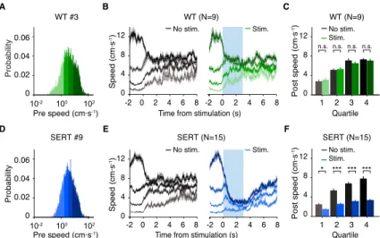
Figure 3.1. DRN 5-HT activation slows down animals in the open
field ______________________________________________ 51
Figure 3.2. Effect of DRN 5-HT photostimulation is persistent
regardless of previous speed of the animals ________________ 54
Figure 3.3. Effect of DRN 5-HT photostimulation is not exclusive
to spatial areas of the open field ________________________ 56
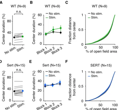
Figure 3.4. DRN 5-HT photostimulation does not affect
anxiety-like parameters in the open field ________________________ 58
Figure 3.5. DRN 5-HT photostimulation does not affect motor
coordination in the rotarod and LocoMouse assays __________ 60
Figure 3.6. Effect of DRN 5-HT photostimulation is not
explained by novelty and it is persistent across sessions and
blocks _____________________________________________ 65
Figure 3.7. Effect of DRN 5-HT photostimulation is sensitive to
open field floor, but not geometry _______________________ 67
Figure 3.8. DRN 5-HT photostimulation in a specific region of
interest of the open field ______________________________ 69
Figure 3.9. DRN 5-HT photostimulation in a specific region of
Figure 4.1. Schematic diagram of sexual behavior
neuromodulatory mechanism ___________________________ 95
Figure 4.2. C57BL/6 male sexual behavior ________________ 98
Figure 4.3. Optogenetic strategy to activate DRN 5-HT neurons
during sexual behavior ________________________________ 99
Figure 4.4. DRN 5-HT activation during sexual behavior for
ChR2 and YFP mice ________________________________ 101
Figure 4.5. DRN 5-HT activation during mounts does not affect
male sexual behavior in SERT-Cre mice _________________ 102
Figure 5.1. Schematic diagram for a proposed model of 5-HT
action in behavior ___________________________________ 120
Supplemental Figure Index
Figure S 2.1. Photostimulation of DRN 5-HT neurons in vitro _ 37
Figure S 2.2. Light propagation in the DRN _______________ 39
Figure S 3.1. DRN 5-HT activation in the LocoMouse test does
not produce changes in the nose and tail movement _________ 62
Figure S 3.2. DRN 5-HT photostimulation in the open field for a
subset of SERT-Cre mice (n = 3), performed after LocoMouse
experiment _________________________________________ 63
Figure S 3.3. DRN 5-HT photostimulation at 20mW slows down
Table Index
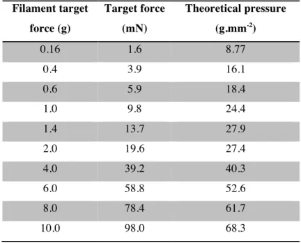
Table 2.1. Von Frey filaments force scale _________________ 45
Table 3.1. Comparison of significant variables across sessions for
ROI stimulation experiment ____________________________ 73
Contents
Acknowledgments _____________________________________________________ i
Resumo ________________________________________________________________ iii
Abstract ________________________________________________________________ vi
Abbreviations list _____________________________________________________ ix
Figure Index ____________________________________________________________ x
Supplemental Figure Index _________________________________________ xi
Table Index ___________________________________________________________ xii
1 General Introduction ____________________________________________ 1
1.1 Serotonin: the maestro of the brain ___________________________ 2
1.2 Serotonergic system ____________________________________________ 3
1.3 Theories and hypothesis of 5-HT _______________________________ 5
1.4 Technical barriers ____________________________________________ 14
1.5 Optogenetics: the new neuroscience era ____________________ 16
1.6 Thesis expedition guide ______________________________________ 18
2 Optogenetic recruitment of dorsal raphe serotonergic
neurons acutely decreases mechanosensory responsivity in
behaving mice ________________________________________________________ 19
2.1 Abstract _______________________________________________________ 20
2.2 Introduction __________________________________________________ 20
2.3 Results _________________________________________________________ 22
2.4 Discussion _____________________________________________________ 30
3 Optogenetic activation of dorsal raphe serotonergic
neurons reduces movement, without affecting motor
coordination or inducing reinforcing responses in behaving
mice ____________________________________________________________________ 46
3.1 Abstract _______________________________________________________ 47
3.2 Introduction __________________________________________________ 48
3.3 Results _________________________________________________________ 50
3.4 Discussion _____________________________________________________ 74
3.5 Experimental procedures ____________________________________ 81
4 Optogenetic activation of dorsal raphe serotonergic
neurons does not impair sexual behavior in male mice _______ 93
4.1 Abstract _______________________________________________________ 94
4.2 Introduction __________________________________________________ 94
4.3 Results _________________________________________________________ 97
4.4 Discussion ____________________________________________________ 103
4.5 Experimental procedures ___________________________________ 107
5 General Discussion ____________________________________________ 113
5.1 The role of 5-HT in behavior ________________________________ 114
5.2 Future directions to be explored ____________________________ 125
1
General Introduction
1.1
Serotonin: the
maestro
of the brain
Serotonin (or 5‐hydroxytryptamine, 5-HT) is one of the main
conductors of the brain. Discovered by the Italian scientist Vittorio Erspamer (Vialli & Erspamer 1937), 5-HT was noticed
for causing smooth muscle contraction in the gastrointestinal tract.
It was years after its characterization (Rapport et al. 1948) that
5-HT was proposed to be a neurotransmitter (Brodie & Shore 1957),
the third one to be found.
5-HT, together with other neuromodulators like dopamine,
acetylcholine and norepinephrine have the fundamental role of
modulating different neuronal populations, ensuring a balanced
functioning of the whole brain orchestra. In detail, 5-HT has a crucial role in regulating a huge variety of brain processes,
including sleep, movement and social behaviors. However, the
exact mechanism of 5-HT action in behavior is still enigmatic.
The present work has investigated the role of 5-HT in diverse
behavioral contexts, by employing a combination of optogenetic
and behavioral approaches.
In the following sections, we will: explore the anatomical
to investigate the role of 5-HT in behavior (1.5 Optogenetics: the new neuroscience era).
1.2
Serotonergic system
5-HT is a biogenic monoamine produced through the
hydroxylation of tryptophan and its subsequent decarboxylation
(Clark et al. 1954). The vast majority of body’s 5-HT is located in
the periphery, mainly stored in platelets and enterochromaffin
cells. Within the central nervous system, 5-HT is synthesized and
stored in the presynaptic neurons. Upon neuronal depolarization,
5-HT is released into the synaptic cleft. It can bind to
postsynaptic 5-HT receptors or 5-HT autoreceptors on the
presynaptic membrane (Cerrito & Raiteri 1979). Binding of 5-HT
to the autoreceptor acts as a negative feedback against further
release of 5-HT into the synaptic cleft (Cerrito & Raiteri 1979).
The highly selective 5-HT transporter (SERT) located on the
presynaptic membrane is responsible for removing 5-HT from the
synaptic cleft. Once transported into the presynaptic neuron,
5-HT is recycled back into presynaptic vesicles where it is protected
from metabolism.
The serotonergic system is incredibly complex, featuring seven
families of 5-HT receptors (5-HT1–7; Hoyer et al. 1994), further
divided into a total of at least seventeen 5-HT receptors (Bockaert
et al. 2010). The 5-HT receptor is a phylogenetically ancient
the lowest of invertebrates as well as the higher mammals
(Peroutka & Howell 1994). Most 5-HT receptors are G-protein
coupled receptors (except for 5-HT3 which is a ligand-gated ion
channel; Derkach et al. 1989) and exhibit heterogeneity with both
excitatory and inhibitory functions.
Within the central nervous system, 5-HT is located in nine groups
of neurons isolated in the pons and midbrain (Dahlstroem & Fuxe
1964). The raphe nuclei constitute the major 5-HT area in the
brain, with both ascending 5-HT fibers projecting to the forebrain
and descending fibers that extend to the medulla and spinal cord
(Dahlstroem & Fuxe 1964). Raphe nuclei are evolutionarily
conserved across fish, amphibians, up to mammals. In rodents,
the more posterior raphe nuclei provide serotonergic innervation
to the spinal cord, the brainstem itself, and some cerebellar areas.
The more anterior nuclei, dorsal (DRN) and median raphe (MRN)
provide extensive serotonergic innervation to the forebrain and
midbrain. DRN and MRN together constitute 80% of the
serotonergic innervation to the forebrain (Azmitia & Segal 1978).
In detail, DRN fibers are widely distributed throughout the
forebrain, including ventral midbrain, lateral hypothalamus,
midline thalamus, amygdala, striatum and most of the cortex. In
contrast, MRN projects preferentially to the hippocampus, septum
and other forebrain structures lying on or close to the midline
(Vertes & Linley 2008).
DRN constitutes approximately 50% of the total 5-HT neuronal
population in the brain (Steinbusch 1981; Descarries et al. 1982),
from the vast set of projections, DRN also receives afferents from
many brain structures, including cortical, limbic and brainstem
areas. Recent studies using rabies tracing strategies, combined
with cell-type-specific cre-driver mouse lines, confirm 5-HT
DRN inputs from prefrontal cortex, lateral habenula, ventral
striatum and lateral hypothalamus, highlighting the diversity of
the serotonergic brain map (Weissbourd et al. 2014; Pollak
Dorocic et al. 2014; Ogawa et al. 2014).
The complexity of 5-HT is not limited to receptor’s subtypes and
anatomical organization. DRN comprises also a variety of other
cell types, including dopamine, GABA, glutamate and other
neuropeptides (Araneda et al. 1980). In fact, 5-HT neurons
represent only about one third of the DRN neurons (Jacobs &
Azmitia 1992). Given such diversity, it is not surprising that
electrophysiological recordings from DRN putative 5-HT neurons
show a wide variety of responses (Jacobs & Azmitia 1992;
Veasey et al. 1997; Ranade & Mainen 2009).
In summary, DRN is the major container of 5-HT, with
impressive morphological, anatomical and neurochemical
heterogeneity.
1.3
Theories and hypothesis of 5-HT
5-HT is a critical neuromodulator, investigated in diverse animal
models, with a variety of different functions. Amongst these,
food deprivation (Groome et al. 1993) and sensitization of
defensive reflexes (Sahley 1995) in the leech and locomotion,
eating and foraging (Horvitz et al. 1982; Sawin et al. 2000;
Flavell et al. 2013) in the nematode C. elegans. Furthermore, 5-HT induces behavioral gregarization in the locusts
(transformation from solitary to group formation, Anstey et al.
2009) and affects social dominance in the lobster (Kravitz 1988).
In mammals, 5-HT is “leaving fingerprints in the scene of many
crimes” leading to a disparate set of theories and hypothesis. Some of the most relevant 5-HT theories are going to be reviewed
in the following section, with a primary focus on the rodent
literature (since the original studies here described use the mouse
as animal model).
1.3.1 5-HT in behavioral inhibition
Brodie and Shore originally proposed a general role for 5-HT in
behavioral inhibition, in contrast with the presumed excitatory
role of norepinephrine (Brodie & Shore 1957). Subsequent
studies focused on the hypothesis that 5-HT constrains the
response of organisms to external stimuli (usually aversive) and
that 5-HT absence potentiates that response (Soubrié 1986; Depue
& Spoont 1986). This argument is largely supported by a set of
5-HT depletion studies showing increase pain sensitivity (Tenen
1967), increase exploration or locomotor activity (Gately et al.
1985; Eagle et al. 2009), increase startle behavior (Davis &
Sheard 1974; Davis et al. 1980), increase the output of operant
2009), increase aggression (Vergnes et al. 1986) and facilitation
of male sexual behavior (Dewsbury & Davis 1970; Dewsbury et
al. 1972).
Consistent with the behavioral inhibition theory is the set of
studies relating 5-HT with impulse control, one of which from
our lab (Fonseca et al. 2015). DRN neurons are active while rats
wait for delayed rewards and delayed reward predictive cues
(Miyazaki et al. 2011) and blocking DRN 5-HT activity increases
premature responding (Miyazaki et al. 2012). More recently, it
was confirmed with optogenetic techniques that activation of
DRN 5-HT neurons suppresses impatient responses (Miyazaki et
al. 2014; Fonseca et al. 2015) .
Another line of research found 5-HT encoding aversive outcomes
very attractive, giving rise to the 5-HT and dopamine opponency
theory, with observations at the anatomical, physiological and
biochemical levels (Deakin & Graeff 1991; see also Cools et al.
2011 for review). The opponency hypothesis has also been
formally extended to a computational version (Daw & Daw 2002;
Dayan & Huys 2008). Dopamine has long been associated with
behavioral rewards, having two main behavioral functions: 1)
motor facilitation or activation and 2) reward-driven learning,
through reward prediction error signaling (Schultz et al. 1997).
Under the opponency hypothesis, 5-HT is suggested to play a
negative functional counterpart of DA with two proposed roles:
1) mediation of behavioral inhibition and 2) mediation of aversive
2009). However, diverse behavioral and clinical studies using
5-HT and dopamine manipulations demonstrate a rather cooperative
function between these two neuromodulators (reviewed in
Boureau & Dayan 2011). More recently, the opponency
framework has been experimentally tested in classical
conditioning learning paradigms, by monitoring DRN 5-HT
activity with electrophysiological recordings (Cohen et al. 2015)
and bulk fluorescence imaging (Matias et al. submitted). While
the first study reveals that 5-HT neurons respond to reward
predicting cues and punishments, the latter shows that, similar to
dopamine, 5-HT neurons are activated by prediction violations.
However, unlike dopamine neurons which are activated by
better-than-expected outcomes, for 5-HT neurons the valence of these
violations is not important. This finding suggests that the
opponency theory might need to be re-interpreted in a different
manner. It will be important to further explore this theory,
extending it to different behavioral contexts, also without the
conditional learning component. Social behaviors, in detail
male-female sexual interactions, are good candidates for testing the
opponency hypothesis, as dopamine and 5-HT seem to play a
crucial role in maintain a balanced sexual cycle (reviewed in
Pfaus 2009). However, the nature of the modulation and the exact
pathways involved in this interaction are still lacking
experimental support.
A second major theory of 5-HT function is based in the
sensorimotor hypothesis. Jacobs first proposed that the main role
of 5-HT is to facilitate motor output and suppress ongoing
processing of sensory input during motor behavior (reviewed in
Jacobs & Fornal 1997). Initial evidence to support this theory
comes from the finding that 5-HT activity is silent during REM
sleep, a state of movement paralysis (Lai & Siegel 1988).
Moreover, 5-HT infusion into the forebrain reduces the acoustic
startle reflex in rats, but enhances this response when infused
onto the spinal cord (Davis et al. 1980). The positive link between
5-HT and motor output became more clear with subsequent
electrophysiological studies, correlating raphe neurons with
central pattern generators (Martín-Cora et al. 2000; see also Clark
et al. 2004 for review). Although most of the putative 5-HT motor
control is associated with the more caudal raphe nuclei neurons,
Jacobs suggests an involvement of the cat DRN neurons in
oral-buccal behavior patterns, such as chewing, licking, biting,
grooming (Fornal et al. 1996). In the same experiment, DRN
activity seems to be resistant to somatosensory stimuli, but
surprisingly silent during orientation to a strong external stimulus
(sudden opening of a door). These results motivated Jacobs to
propose that 5-HT neurons tonically inhibit sensory processing
and facilitate motor output (reviewed in Jacobs & Fornal 1997).
The sensory interaction part of this hypothesis receives further
confirmation (Müller et al. 2007; Pum et al. 2008) and it is
supported by the findings that boosting 5-HT activity attenuates
Studies examining the effect of 5-HT release on sensory
processing reveal that raphe electrical stimulation inhibits
somatosensory responses in the neocortex (Sheibani & Farazifard
2006; Mantz et al. 1990), thalamus (Dong et al. 1991), and
brainstem (Chiang et al. 1989). Furthermore, local application of
broad-spectrum 5-HT receptor agonists attenuates odor-evoked
olfactory receptor neurons activity, supporting the idea that 5-HT
suppresses olfactory inputs (Petzold et al. 2009). More recently, a
study from our lab also described that DRN 5-HT activation
attenuates firing of anterior piriform cortex neurons (Lottem et al.
submitted).
Overall, the sensorimotor hypothesis also considers a general
inhibitory role for 5-HT. Although it sketches a relatively
coherent scenario of the physiological effects of 5- HT, it still
lacks a coherent connection to behavior itself. Unlike the
behavioral inhibition theory (more specifically the dopamine
opponency hypothesis), the sensorimotor theory also gives no
insight into why 5-HT would be an important modulator of mood,
emotion or any other of its psychoactive properties. These two
theories, while not completely inconsistent, remain to be further
connected.
1.3.3 5-HT in reward
An alterative theory suggests a role for 5-HT in reward and
reinforcing behaviors. This hypothesis is based on the evidence
that putative 5-HT neurons in the DRN respond to reward
Bromberg-Martin et al. 2010; Inaba et al. 2013; Cohen et al. 2015; Matias
et al. submitted) and that 5-HT depletion impairs reward
processing (Rogers et al. 2003; Seymour et al. 2012). A more
recent study shows that optogenetic activation of DRN 5-HT
neurons positively reinforces several behaviors (Liu et al. 2014).
Again, as with any other 5-HT theory, when a function is
proposed, other findings prove it wrong. Two studies used a
similar optogenetic approach, but failed to observe 5-HT
reinforcing effects (Miyazaki et al. 2014; Fonseca et al. 2015). It
was even proposed that the rewarding effect in the DRN is
preferentially non-serotonergic (McDevitt et al. 2014), thus
suggesting that different subsets of DRN neurons might have
different effects in behavior. In fact, in Liu et al.’s study, the
authors claim to affect also glutamatergic, besides 5-HT neurons
in DRN (Liu et al. 2014; reviewed in Luo et al. 2015). It seems
critical to further investigate the role of specific DRN 5-HT
neurons in appetitive behaviors, using unconstrained behavioral
paradigms.
In agreement with the 5-HT reward theory and its complexity is
the collection of psychopharmacological data implicating 5-HT in
animal models of depression and anxiety (Leonard 1987; Graeff
et al. 1996; Maier & Watkins 2005). In humans, selective 5-HT
reuptake inhibitors (SSRI) have been demonstrated to be effective
in treating major depression and are the most popularly
prescribed class of antidepressant drugs. SSRIs act by inhibiting
5-HT uptake and thus increasing synaptic levels of 5-HT. Dietary
patients who were successfully treated with SSRIs (Delgado et al.
1991; Heninger et al. 1996), thus suggesting that enhanced 5-HT
transmission is necessary to maintain the therapeutic
effectiveness of antidepressants.
Efforts to model human affective disorders in animals comprise
both animal models of depression and animal behavioral tests
sensitive and selective for detecting antidepressant drugs (Willner
1990). The forced swimming test (Porsolt et al. 1978) is one of
the most frequent behavioral assays used to measure potential
antidepressant activity. Effects of SSRIs were reported either to
be active, inactive, or active only at high doses in this behavioral
test (reviewed in Borsini 1995). A recent study using optogenetic
methods shows that activating the pathway that connects medial
prefrontal cortex with DRN has a robust impact in forced
swimming behavior (Warden et al. 2012).
Another commonly used behavioral test is the open field assay
(Seibenhener & Wooten 2015). Originally designed to score
defecation as a measure of animals’ ‘emotionality’ (Hall 1934),
the open field provides an initial screen for anxiety-related
behavior in rodents (Bailey & Crawley 2009). Once again,
pharmacological manipulations reveal all possible combinations:
5-HT promotes, reduces, or has no effect in anxiety-like
behaviors (see Prut & Belzung 2003 for review). A recent
optogenetic study shows no involvement of DRN in anxiety,
whereas photostimulation of MRN induces anxiolytic behavior
with other behavioral tests and significant effort has been made to
overcome the complexity of the serotonergic system and pinpoint
its mechanism in anxiety circuitry. As an example, the receptor
5-HT1a is expressed both in 5-HT (auto-receptor) and non-5-HT
neurons (hetero-receptor). The importance of both neuronal
populations has been highly debated, and it seems that 5-HT1a
auto-receptors are necessary, but not sufficient, to modulate
anxiety (Piszczek et al. 2013). Further studies manipulating the
activity of 5-HT neurons should help shine light on the exact role
of 5-HT in anxiety-like behaviors.
In summary, you have been guided through three main theories
for the role of 5-HT in behavior: 1) behavioral inhibition, 2)
sensorimotor and 3) reward functions. However, these three dots
are still very challenging to connect. Behavioral inhibition can
help explain the 5-HT connection to depression and anxiety, but
prediction of aversive outcomes seems opposite to reward coding.
The sensorimotor hypothesis focus on a different data set, giving
insight on 5-HT physiological function, but lacks behavioral
connection.
In the present set of studies, we aimed to experimentally test these
theories in spontaneous behaviors, as general locomotion and
sexual behavior. We hypothesize that behavioral inhibition is the
best candidate theory to describe the role of 5-HT in behavior, but
1.4
Technical barriers
Despite the valuable efforts of numerous pharmacological,
physiological and genetic experiments, the role of 5-HT in
behavior is not well understood. The reason for such mystery is in
part the heterogeneity of the 5-HT system, but also the technical
limitations of most of the methods employed so far.
While pharmacological experiments provide reasonable
specificity for 5-HT, they lack temporal resolution, acting like
modulators rather than controllers of 5-HT function. This is
problematic because transient bursts of activity in 5-HT neurons
are likely to be important to 5-HT function (Nakamura et al.
2008; Ranade & Mainen 2009). The sensitivity of receptors to the
time course of transmitter concentration depends on their affinity,
with higher affinity receptors, which unbind transmitter more
slowly, being more sensitive to slow, tonic changes. Therefore,
low-affinity 5-HT receptors (including 5-HT1a) and high-affinity
receptors (including 5-HT2a) will be more sensitive to phasic and
tonic HT levels respectively (Glennon et al. 1995). Because
5-HT1a and 5-HT2a receptors have opposite effects on signal
transduction pathways, tonic manipulations of 5-HT may in fact
have the opposite effect of phasic signals (reviewed in Dayan &
Huys 2009). Consequently, different affinity receptors may
influence similar 5-HT-mediated responses, but in very different
Another common approach is to use knockout mouse lines. While
an animal model lacking 5-HT neurons is non-viable, it is
possible to create viable knockouts lacking a particular 5-HT
receptor (Stark et al. 2007). Although behavioral changes can be
observed in these lines, the impact cannot be easily attributed to
the specific loss of that receptor subtype, given the many
compensatory changes in 5-HT neurons and other
neurotransmitter systems involved. Additionally, this genetic
manipulation affects the expression of the 5-HT receptors
throughout the brain.
Electrical stimulation is another used method that allows a
targeted manipulation to the raphe nuclei neurons, providing also
temporal specificity. The results of some electrical stimulation
studies are in agreement with the inhibition theory, producing
cessation of ongoing appetitive behavior, “freezing” responses
and/or sedation (Kostowski et al. 1969; Graeff et al. 1978).
However, others have produced equivocal or even contradictory
effects, such as increased arousal (Jacobs et al. 1973). Electrical
stimulation of the raphe nuclei also inhibits neural activity in
sensory systems (Sheibani & Farazifard 2006; Dong et al. 1991;
Mantz et al. 1990), but the behavioral effects on sensory
processing have not been assayed. The ambiguity and diversity of
results with electrical stimulation of the raphe nuclei are likely to
be due to its lack of cellular or neurochemical specificity. The
importance of this issue is especially critical because 5-HT
abundant non-5-HT neurons, including dopaminergic and
GABAergic neurons. Electrical stimulation may also activate the
dense nerve fascicles containing descending and ascending spinal
tracts (Fardin et al. 1984).
In order to overcome these technical limitations, a new method
has emerged, hopefully to shine light on the mysteries of 5-HT
functions.
1.5
Optogenetics: the new neuroscience era
I was fortunate to develop my thesis during an evolutionary
memorable moment for neuroscience: the optogenetics boom.
The introduction of genetically-based methods for stimulating,
silencing and monitoring neuronal activity has been an exciting
breakthrough discovery for the field. Most notable among the
(now) vast list of optogenetic tools is the photosensitive protein
channelrhodospin-2 (ChR2, Nagel et al. 2003). ChR2 is a
light-gated monovalent cation channel isolated from a green algae,
which opens upon exposure to blue light (activation maximum at
470 nm). Light can be delivered to ChR2- expressing neurons
either directly from the surface or through fiber optics inserted in
desired brain regions (Han & Boyden 2007). The efficacy of
ChR2 to drive neurons has been confirmed in acute slices (for
example, Aravanis et al. 2007; Arenkiel et al. 2007; Zhang et al.
A growing number of recent studies have shown that behavioral
responses can be evoked by light in mice engineered to express
ChR2 in specific brain regions and/or neuronal types (for an
overview see Boyden 2015; Deisseroth 2015). The use of ChR2
provides considerable advantages compared to the previous
described manipulations, since it allows for selective, rapid and
reversible activation of genetically defined populations of
neurons embedded in complex circuits.
The mysterious 5-HT system (as well as other neuromodulatory
systems) owns the most important flavors for the application of
optogenetic methods: clear genetic specification and compact
localization of the 5-HT neurons. There are now available two
mouse lines (Scott et al. 2005; Zhuang et al. 2005) and two
viruses lines (Benzekhroufa et al. 2009a; Benzekhroufa et al.
2009b) to allow specifically targeting of 5-HT neurons. Indeed,
such tools have recently placed the 5-HT system in the spotlight
and several groups are studying the effect of stimulating various
types of DRN neurons on several behavioral tasks (reviewed in
Luo et al. 2015).
Throughout this thesis, ChR2 was used to specifically activate
DRN 5-HT neurons. Because inactivation of ChR2 and
autoinhibition of 5-HT neurons may limit the efficiency of
photostimulation, we optimized the stimulation parameters
tested some 5-HT theories, by applying optogenetic perturbations
in diverse behavioral assays (Chapter 2-4).
1.6
Thesis expedition
guide
My primary motivation was to explore the “whats” and the “hows”
of 5-HT in the brain and behavior. This expedition started with the implementation of an optogenetic strategy to study the
serotonergic system, a novel method at that moment. We
validated and optimized the photostimulation conditions to
activate DRN 5-HT neurons in behaving animals (Chapter 2, Dugué et al. 2014) and kept this tool for the rest of the trip. Next,
we tested the effect of 5-HT in spontaneous movement, using the
open field test, where animals freely behave, in no reward and no
punishment conditions. We found that DRN 5-HT activation
induced a dramatic decrease of speed in the animals (Chapter 3). The effect was robust throughout many days and did not seem to
affect motor coordination, anxiety-like or aversive and appetitive
responses. Finally, we went on a preliminary journey to
investigate the role of 5-HT in sexual behavior, an ethological
relevant behavior. We confirmed the existence of variability in
biology and challenged the role of DRN 5-HT neurons in male
copulatory behavior (Chapter 4). We were able to narrow down some “whats”, leaving space for future explorations to resolve the
2
Optogenetic recruitment of
dorsal raphe serotonergic
neurons acutely decreases
mechanosensory
responsivity
in
behaving
mice
Results published: Dugué GP, Lorincz ML, Lottem E, Audero E, Matias S, Correia PA, Léna C, and Mainen ZF. (2014) Optogenetic recruitment of dorsal raphe serotonergic neurons acutely decreases mechanosensory responsivity in behaving mice. PLoS ONE 9(8): e105941. doi:10.1371/journal.pone.0105941
2.1
Abstract
The inhibition of sensory responsivity is considered a core
serotonin function, yet this hypothesis lacks direct support due to
methodological obstacles. We adapted an optogenetic approach to
induce acute, robust and specific firing of dorsal raphe
serotonergic neurons. In vitro, the responsiveness of individual dorsal raphe serotonergic neurons to trains of light pulses varied
with frequency and intensity as well as between cells, and the
photostimulation protocol was therefore adjusted to maximize
their overall output rate. In vivo, the photoactivation of dorsal raphe serotonergic neurons gave rise to a prominent light-evoked
field response that displayed some sensitivity to a 5-HT1A
agonist, consistent with autoreceptor inhibition of raphe neurons.
In behaving mice, the photostimulation of dorsal raphe
serotonergic neurons produced a rapid and reversible decrease in
the animals’ responses to plantar stimulation, providing a new
level of evidence that serotonin gates sensory-driven responses.
2.2
Introduction
The influences of central serotonin (hydroxytryptamine or
5-HT) impact a wide range of brain functions, from the control of
autonomic responses (Audero et al. 2008; Richerson 2004) to the
regulation of complex emotional behaviors (Lucki 1998; Cools et
considering possible core neurophysiological functions. Among
these, serotonergic neuromodulation has long been implicated in
the inhibition of sensory responsivity (Hurley et al. 2004; Davis
et al. 1980), an idea chiefly supported by gain-of-function
experiments. Pharmacological enhancement of 5-HT function
inhibits primary afferent neurotransmission in vitro (Choi et al. 2012; Chen & Regehr 2003), dampens sensory and
nociceptively-evoked firing in vivo (Petzold et al. 2009; Waterhouse et al. 1990;
Yoshida et al. 1984; Reyes-Vazquez et al. 1989) and decreases
acoustic startle responses and their pre-pulse inhibition (Davis et
al. 1980; Geyer et al. 2001). Similarly, electrical microstimulation
of the dorsal raphe nucleus (DRN), one of the largest sources of
ascending 5-HT projections (Abrams et al. 2004), reduces
forebrain sensory and nociceptively-evoked activity (Petzold et al.
2009; Yoshida et al. 1984; Reyes-Vazquez et al. 1989; Iwayama
et al. 1989; Kayama et al. 1989; Andersen & Dafny 1982) and
elevates vocalization thresholds to noxious stimuli (Fardin et al.
1984).
Despite these observations, technical limitations have impeded a
deeper understanding of the underlying mechanisms.
Pharmacological upregulation of 5-HT pathways neither mimics
phasic 5-HT release nor takes into account the effect of
co-released substances (Trudeau 2004; Gras et al. 2002), and may
exhibit paradoxical effects due to autoreceptor-mediated negative
feedback (Fornal et al. 1994) and drug-induced plastic changes
(Riad et al. 2001). Electrical stimulation, while spatially and
non-5-HT neurons and fibers-of-passage (Fardin et al. 1984). To
overcome these technical limitations, we optimized and validated
a direct and specific optogenetic stimulation of DRN serotonergic
neurons in mice. We then employed this strategy to test whether
transient and specific activation of DRN 5-HT neurons in
behaving mice can indeed interfere with sensory responsivity in a
simple test of mechanosensitivity.
2.3
Results
2.3.1 In vitro photostimulation of virally transduced dorsal raphe 5-HT neurons
To overcome the limitations of previous studies and explore the
specific functions of DRN 5-HT projections, we targeted
channelrhodopsin-2 fused to YFP (ChR2-YFP) to DRN 5-HT
neurons using a viral expression strategy. Adeno-associated
viruses (AAVs) carrying a Cre-activated expression cassette for
ChR2-YFP were injected into the DRN of SERT-Cre mice (Gong
et al. 2007) (Figure 2.1A-B). The specificity of this approach was assessed using anti-tryptophan hydroxylase (TPH)
immunocytochemistry (Figure 2.1C-E). TPH immunoreactivity was observed in 93.9 ± 2.0% of ChR2-YFP cells (n = 3 brains,
respectively 287/308, 293/305 and 507/549 counted
ChR2-YFP-positive cells also ChR2-YFP-positive for TPH), confirming that this method
yields 5-HT neuron-specific expression. The photosensitivity of
2.1F-G). Non-fluorescent cells displayed no photoevoked spike or current in response to the highest irradiance tested (1.5–10.4
mW·mm-2
, n = 7). In contrast, all ChR2-YFP cells fired in
response to brief (6 ms) light pulses (n = 34), with an average
photostimulation threshold of 0.26 ± 0.47 mW·mm-2
(calculated
for a subset of n = 21 cells; Figure 2.1G-I).
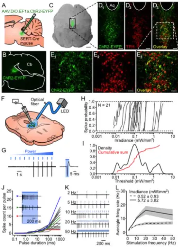
Figure 2.1. Specificity of ChR2-YFP expression in DRN 5-HT neurons and
efficiency of photostimulation in vitro.
Bar: 500 mm. (D1–3) Confocal pictures of the area delimited by a black rectangle in C. Aq: aquaduct. Bar: 100 mm. (E1–3) Magnified view of the area delimited by a white rectangle in D3. Bar: 50 mm. (F) Schematics of patch-clamp recordings in DRN slices. (G) Left: example of a loose cell- attached recording illustrating the protocol used to assess photo- stimulation thresholds (PTs). Right: blow-up of the trace showing a single photoevoked spike. (H) Spike probability versus incident irradiance for 21 cells. (I) Kernel density estimate of the distribution of logarithmically-scaled PTs (black) and superimposed normalized cumulative sum (red). (J) Spike count per pulse versus pulse duration for 17 cells at twice their PT. Inset: representative firing profiles of 3 different cells identified in the graph by colored arrowheads. (K) Response of a ChR2-YFP cell to trains of repeated light pulses (6 ms) at various frequencies (irradiance set at twice its PT). (L) Average firing rate (± SEM, shaded area) during a 5 s train versus photostimulation frequency at twice the PT (dashed line; n = 29, 28, 25 and 18 cells for 1–2, 5–10, 20 and 50 Hz respectively) and at a higher irradiance (~5 mW!mm-2; solid line, n = 14
and 13 cells for 1–20 and 50 Hz respectively).
Longer pulses (1 s) at twice the photostimulation threshold
evoked firing profiles ranging from non- to fully-inactivating
(Figure 2.1J). Firing evoked by repeated photostimulation (6 ms pulses at 1–50 Hz) generally adapted over time (Figure 2.1K). During 5 s photostimulation trains, ChR2-YFP cells could
reliably (spike probability > 0.85) follow up to 2 Hz at twice their
photostimulation threshold and up to 10 Hz at a higher irradiance
(~5 mW·mm-2). However the maximal average firing rates for
these two irradiances (6.8 ± 4.6 Hz, n = 18 and 16.0 ± 11.4 Hz, n
= 14) were attained with 50 and 20 Hz photostimulations
respectively (Figure 2.1L). Therefore irradiances one order of magnitude above photostimulation threshold are required to
induce firing rates comparable to those observed in behaving
animals during phasic episodes of increased DRN firing (Fornal
2.3.2 In vivo photostimulation of dorsal raphe 5-HT neurons
In order to estimate how far from the fiber tip ChR2-YFP neurons
could be recruited in vivo, we measured the spread of blue light in
the DRN of hemisected brains (Figure 2.2A-B); see Methods). Light intensity decayed in an exponential fashion away from the
fiber tip, with a space constant of 199 ± 35 µm (n = 3; Figure 2.2C-D).
Figure 2.2. Photostimulation of DRN 5-HT neurons in vivo
ChR2-YFP-expressing SERT-Cre (ChR2, solid lines) and a wild-type (WT, dotted lines) mouse. Top: normalized fluorescence (green, left axis) and OLFP amplitude (black, right axis) profiles plotted against the optrode position. Bottom: boxplots showing the optrode position at the point of maximal fluorescence (green, n = 8 mice) and largest OLFP amplitude (grey, n = 6 mice). Inset: average OLFP for the ChR2 mouse at the locations marked by the arrowheads. (G) Example showing the time course of the effect of the 5-HT1A receptor agonist 8-OH-DPAT on the OLFP amplitude (black), latency (purple) and width (blue). Bin: 158 s. Inset: average OLFP before (black) and after (red) 8-OH-DPAT injection (taken from the periods indicated by arrowheads). (H) Top: OLFP amplitude before and after 8-OH-DPAT injection (mean ± SD: 1506121 mV and 1216105 mV respectively, n = 10). Bottom: OLFP peak latency before and after 8-OH-DPAT injection (mean ± SD: 2.460.5 ms and 2.860.7 ms, n = 10). Changes are significant in both cases (P = 0.037 and P = 0.030 respectively, paired Wilcoxon rank sum test). Error bars represent the SEM. (I) Example illustrating the dependence of the OLFP amplitude (grey points) and latency (red points) on the irradiance at the fiber tip. The sum of two exponential functions was fitted to each curve (black lines). Inset: superimposed OLFPs for 0.2, 0.7, 1.6 and 6.0 mW!mm-2. (J) Normalized
average (6 SD, shaded area) OLFP amplitude (black, left axis) and latency (red, right axis) versus photostimulation frequency (n = 7 mice). (K) Average OLFP amplitude recovery curve (green; ± SD, shaded area) assessed by delivering test pulses at variable intervals (dt) after 10 Hz, 5 s trains of 6 ms light pulses (n = 4 mice). Blue: average amplitude during the trains.
The volume of tissue receiving more than 2% of the irradiance at
the brightest point extended 1.65 ± 0.21 mm, > 1.00 and 1.23 ±
0.10 mm in the antero-posterior, medio-lateral and fiber axes
respectively (n = 3). We then probed the response of DRN 5-HT
neurons in vivo by advancing a custom optrode toward the DRN of anaesthetized animals, while intermittently shining light pulses.
The tissue fluorescence (Figure 2.2E) displayed a peak 3.48 ± 0.43 mm below the cerebellar surface (n = 8), which correlated
spatially with a prominent multiphasic photoevoked potential
(OLFP, optically-evoked local field potential; Figure 2.2F). Systemic administration of the 5-HT1A receptor agonist
8-OH-DPAT, a drug commonly employed to inhibit 5-HT cell firing by
OLFP amplitude and prolonged its latency (Figure 2.2G-H). The OLFP peak amplitude and latency displayed a graded dependence
on the irradiance at the fiber tip (Figure 2.2I), approaching saturation (slope < 10 µV/mW·mm-2
) at 117 ± 34 mW.mm-2
(n =
9). This dependence could be described as the sum of two
exponential functions (!
fast and !slow of 7.4 ± 6.0 and 53.5 ± 18.7
mW.mm-2 accounting for 46% and 54% of the amplitude
respectively; !fast and !slow of 3.1 ± 5.2 and 29.6 ± 26.2 mW.mm-2,
accounting for 70% and 30% of the latency respectively; n = 9).
The OLFP waveform also rapidly adapted to trains of repeated
photostimulation (reduced amplitude and extended latency with
increased stimulation frequency, Figure 2.2J), an effect potentially mediated by a combination of factors such as partial
inactivation of voltage-dependent sodium channels, ChR2
desensitization, a build-up of afterhyperpolarization currents or
the activation of 5-HT1A autoreceptors. The OLFP recovery
kinetics, assessed for 10 Hz trains, had a time constant of 2.9 ±
0.5 s (n = 4; Figure 2.2K), and full recovery was observed after 8 seconds.
2.3.3 Decreased mechanosensory responsivity of behaving mice during acute dorsal raphe 5-HT neurons photostimulation
Having characterized the specificity and efficacy of the
photostimulation of DRN 5-HT neurons, we then tested this
mechanical sensitivity, the von Frey assay (Barrot 2012) (Figure 2.3A).
Figure 2.3. Decreased responsivity to mechanical stimuli during DRN 5-HT neurons photostimulation
In this test, a series of calibrated Nylon filaments (von Frey hairs)
of ascending stiffness are applied to the plantar surface of the
hind paws while monitoring the animal’s withdrawal response.
Groups of SERT-Cre (ChR2) and littermate wildtype (WT)
control mice (n = 17 for both) were infected with the same viral
vector. Both groups had the same sex ratio (9 males, 8 females)
and were implanted with optical fibers positioned over the DRN.
After a habituation period of 5 days (see Experimental
procedures), animals were tested during 3 to 4 sessions, with a
maximum of 1 session per day. Each session was divided in 3
blocks, designed to test the animal’s sensitivity prior to
(“baseline”), in conjunction with (“stim”) and after (“recovery”)
photostimulation (Figure 2.3B). For each block, a psychometric curve (response probability versus filament) was calculated and
the response threshold was taken as the interpolated filament
value corresponding to a response probability of 0.5. Baseline
responsivity to ascending forces applied with von Frey hairs (0.4–
8 g) to the hind paws did not differ between SERT-Cre and WT
mice (Figure 2.3D-E), with average response thresholds of 1.93 ± 0.63 g and 1.97 ± 0.61 g, respectively (P = 0.97, unpaired
Wilcoxon sum rank test). However in the presence of
concomitant photostimulation (a 12 s train of 10 ms light pulses
at 20 Hz, ~300 mW·mm-2 at the fiber tip, Figure 2.3B-C), response thresholds were significantly higher for ChR2 animals
than WT controls (2.49 ± 1.00 g vs 1.71 ± 0.69 g, P = 0.0076, unpaired Wilcoxon sum rank test). This difference was no longer
photostimulation train (Figure 2.3G-H). The effect observed in ChR2 mice during photostimulation corresponded to a significant
threshold elevation of 32.5 ± 52.0% (Figure 2.3I) which counteracted the slight sensitization observed in WT animals
(Figure 2.3G). This result shows that acute DRN 5-HT photostimulation induces a transient and fully reversible decrease
in responsivity to plantar stimulations in behaving mice.
2.4
Discussion
The initial optogenetic approaches used to study DRN functions
employed non-specific promoters (Warden et al. 2012; Varga et
al. 2009) or targeted local or distal neurons presynaptic to 5-HT
neurons (Warden et al. 2012; Challis et al. 2013). Recently,
specific optogenetic stimulation of DRN 5-HT neurons has been
achieved using transgenic mouse lines (Ito et al. 2013; Ohmura et
al. 2014) or viral injections (Liu et al. 2014) but these studies
have not provided a detailed account of optimal photostimulation
parameters. Here we devoted substantial efforts to optimizing the
yield of direct and specific photostimulation of DRN 5-HT
neurons. ChR2-EYFP-expressing cells were sensitive to low
irradiance in vitro (< 1 mW·mm-2, Figure 2.1H-I). However high irradiances (> 100 mW·mm-2
at fiber tip) were necessary to
saturate the photoevoked local field potential in vivo, a measure that might prove useful for optimally positioning optical fibers
Using irradiance of > 250 mW·mm-2
at the fiber tip, we estimated
that the entire DRN received irradiances over 5–6 mW·mm-2
, a
value at which the output of ChR2-EYFP neurons could be
maximized (up to 16 Hz) in vitro by using 20 Hz stimulations, despite their strong frequency-dependent adaptation. These
parameters are appropriate to attempt to mimic episodes of
increased DRN activity, which occur in association with a variety
of behavioral conditions such as oro-buccal movements (Fornal et
al. 1996) and defensive encounters (Walletschek & Raab 1982),
and in relation to reward outcome (Bromberg-Martin et al. 2010)
and waiting for delayed rewards (Miyazaki et al. 2011). Such
episodes typically last several seconds, during which the activity
of DRN neurons can peak up to 10‒20 Hz.
Our protocol for DRN 5-HT neuron photostimulation in behaving
mice (20 Hz for 12 seconds) evoked robust decreases in
behavioral responses to hind paw stimulation. Previous
observations have shown that chronic elevation of 5-HT levels
can increase response thresholds in rodent models of mechanical
allodynia (Ikeda et al. 2009; Xu et al. 2013; Katsuyama et al.
2013). Our result extends this observation by showing that a
similar effect can be reversibly induced on a faster timescale in
non-pathological conditions by recruiting DRN 5-HT neurons.
Whether the threshold calculated using von Frey filaments in
naïve animals is a measure of sensory or nociceptive sensitivity is
still a debated question (Barrot 2012). Therefore the question
whether the stimulation of DRN 5-HT neurons in our conditions
open. Nevertheless, our result helps to resolve the ambiguity of
previous gain-of-function experiments testing the influence of
DRN output by directly showing that DRN 5-HT neurons can
indeed tone down the influence of sensory and/or nociceptive
inputs, as opposed to what has been recently observed in
zebrafish (Yokogawa et al. 2012).
Given the projection pattern of DRN 5-HT cells (Abrams et al.
2004), this effect is likely to be mediated by the modulation of
anterior structures, as suggested by evidence highlighting a role
for 5-HT in the modulation of thalamic (Yoshida et al. 1984;
Reyes-Vazquez et al. 1989; Kayama et al. 1989; Qiao & Dafny
1988) and cortical (Hurley et al. 2004; Waterhouse et al. 1990)
sensory and nociceptive responses. It is not unlikely that other
co-released substances may play a role in the observed effect. In
particular, a recent study has shown that the glutamatergic
phenotype of certain 5-HT neurons seems to be partly responsible
for the effects produced by the photostimulation of DRN 5-HT
neurons on reward-related behaviors (Liu et al. 2014).
More refined targeting strategies, e.g. retrograde infection
(Rothermel et al. 2013) or intersectional genetics (Jensen et al.
2008), will allow the assessment of contributions of specific
sub-populations of DRN 5-HT neurons (Abrams et al. 2004). Overall
these results provide a new level of evidence for the involvement
of DRN 5-HT neurons in gating the access of sensory inputs to
behavioral output, a key physiological role which will help
2.5
Experimental procedures
2.5.1 Viral transduction of dorsal raphe neurons and optical fiber implantation
Adult (8–16 weeks) transgenic SERT-Cre (Audero et al. 2008) or
wild-type mice (C57BL/6 background, housed in a standard
12:12 hours light-dark cycle) were anaesthetized with isoflurane
mixed with O2 (3% for induction and 0.5–1% for maintenance) or
with ketamine xylazine (100 and 5 mg/kg) and placed in a
stereotaxic apparatus (David Kopf Instruments). Lidocaine (2%)
was injected subcutaneously before incising the scalp and
exposing the skull. A craniotomy was drilled over lobule 4/5 of
the cerebellum and a pipette filled with a viral solution
(AAV2/1.EF1a.DIO.hChR2(H134R)-EYFP.WPRE.hGH, 1013
GC/mL, University of Pennsylvania) was lowered to the DRN
(Bregma -4.4 to -4.7 mm AP, -2.8 to -2.9 mm DV) with a 34°
angle toward the back of the animal (Figure 2.1A). The viral solution (1–1.2 µL) was injected using a Picospritzer II (Parker)
or a syringe pump (KDS 310 Plus, KD Scientific) at a rate of
0.05–0.06 µL/min. For optical fiber implantation, the skull was
cleaned with H2O2 and covered with a layer of Super Bond C&B
(Morita) before performing the craniotomy. An optical fiber (200
µm, 0.37 NA) housed inside a connectorized implant (M3, Doric
Lenses) was inserted in the brain using the same 34° from the
back, with the fiber tip positioned 200 µm above the core of the