ISSN 0100-736X (Print) ISSN 1678-5150 (Online)
Original Article Pequenos Animais/Small Animals Diseases
RESUMO.- [Frequência de dermatopatias tumorais em cães da cidade de Salvador, Bahia (2007-2016).] Objetivou-se com esse estudo determinar a frequência de
dermatopatias tumorais (lesões proliferativas cutâneas que cursam com aumento de volume de natureza neoplásicas ou não neoplásicas) em cães, diagnosticadas por exame histopatológico no Laboratório de Patologia Veterinária (LPV) da Universidade Federal da Bahia (UFBA) na série histórica de 10 anos (2007-2016). Dos 1.945 exames histopatológicos realizados no período, 503 tratava-se de biópsias cutâneas, dentre os quais, foram diagnosticados 617 dermatopatias (87 cães, 17,3%, apresentavam mais de um diagnóstico). Dos 617 diagnósticos de dermatopatias 546 (88,49%) foram tumorais e 71 (11,51%) não tumorais. As 546 dermatopatias tumorais, estudadas com mais ênfase, foram diagnosticadas em 453 cães, 468 (85,7%) eram neoplásicas e 78 (14,3%) não neoplásicas. Das 468 dermatopatias tumorais neoplásicas encontradas 230 foram benignas (49,14%), 215 malignas (45,94%), 23 borderline/epiteliomas (4,91%), 51,92% ABSTRACT.- Machado G.A.C., Fontes T.N., Larangeira D.F., Estrela-Lima A., Moreira E.L.T., Ribeiro
L.S., Pinto M.P.R. & Peixoto T.C. 2018. Incidence of skin tumors in dogs in Salvador, Bahia state, Brazil (2007-2016).Pesquisa Veterinária Brasileira 38(11):2139-2145. Departamento de Anatomia, Patologia e Clínicas Veterinárias, Universidade Federal da Bahia, Av. Adhemar de Barros 500, Ondina, Salvador, BA 40170-110, Brazil. E-mail: tcpeixoto@ufba.br
This study aimed to establish the incidence of skin tumors (cutaneous proliferative lesions of neoplastic or non-neoplastic nature) in dogs diagnosed by histopathological evaluation at the Veterinary Pathology Laboratory (LPV) of the Federal University of Bahia (UFBA) in a 10-year (2007-2016) historical series. Of the 1945 histopathological diagnoses made in this period, 503 were skin biopsies, and 617 dermatological problems (87 dogs, 17.3%, presented more than one positive diagnosis) were found. Of the 617 diagnoses of dermatopathy, 546 (88.49%) were tumors and 71 (11.51%) were non-tumorous alterations. The 546 conditions more profoundly studied were from 453 dogs, 468 (85.7%) neoplastic and 78 (14.3%) non-neoplastic tumors. The 468 neoplasms were classified as follows: 230 benign (49.14%), 215 malignant (45.94%), 23 borderline (epitheliomas) (4.91%), 51.92% (243/468) mesenchymal, 42.74% (200/468) epithelial, 4.91% (23/468) melanocytic, and 0.43% (2/468) metastatic (mammary gland). The most commonly diagnosed neoplastic dermatopathies were mastocytoma (14.7%) and lipoma (7.48%). Among the 78 non-neoplastic conditions (14.3%), epidermal inclusion cyst (39.74%) and trichogranuloma (15.38%) were the most frequent. Canine dermatopathies accounted for 26% of the biopsy files of the LPV-UFBA. Distinct simultaneous dermatological problems were frequently found in the dogs assessed (one in six). Considering that these conditions can present with different cellular origin and biological behavior, it is crucial that histopathological evaluation be performed in fragments from the different cutaneous lesions.
INDEX TERMS: Skin tumors, dogs, Bahia state, dermatology, dermatopathology, cutaneous neoplasms, pathology.
PVB-5686 SA
Incidence of skin tumors in dogs in Salvador, Bahia
state, Brazil (2007-2016)
1Gessica A.C. Machado2, Thanielle N. Fontes2, Daniela F. Larangeira3, Alessandra Estrela-Lima3, Eduardo L.T. Moreira3, Lorena S. Ribeiro4,
Marcela P.R. Pinto5 and Tiago C. Peixoto3*
1 Received on June 11, 2018.
Accepted for publication on July 17, 2018.
Part of the MSc Dissertation of the first author, Universidade Federal do Bahia.
2 Post-Graduate Program in Tropical Animal Science (PPGCAT), Escola de Medicina Veterinária e Zootecnia (EMEVZ), Universidade Federal da Bahia (UFBA), Salvador, BA 40170-110, Brazil.
3 Departamento de Anatomia, Patologia e Clínicas Veterinárias, Escola de Medicina Veterinária e Zootecnia (EMEVZ), Universidade Federal da Bahia (UFBA), Salvador, BA 40170-110. *Corresponding author: tcpeixoto@ufba.br
4 Autonomous Veterinary, Salvador, BA.
5 Resident in Veterinary Pathology, Laboratório de Patologia Veterinária (LPV), Escola de Medicina Veterinária e Zootecnia (EMEVZ), Universidade Federal da Bahia (UFBA), Salvador, BA 40170-110.
ais em cães da , Bahia (2007-2016)].
ela-.R. &
(243/468) de origem mesenquimal, 42,74% (200/468) epiteliais, 4,91% (23/468) melanocíticas e 0,43% (2/468) metastáticas para a pele (primárias de glândula mamária). As dermatopatias neoplásicas mais diagnosticadas foram o mastocitoma (14,7%) e o lipoma (7,48%). Dentre as 78 dermatopatias tumorais não neoplásicas (14,3%), os cistos de inclusão epidermal (39,74%) e o tricogranuloma (15,38%) foram os mais frequentes. As dermatopatias caninas representaram 26% da casuística no LPV/UFBA. A ocorrência de dermatopatias tumorais simultâneas distintas foi comum nos cães desse estudo (um a cada seis); como podem ter origens celulares e comportamentos biológicos diferentes, enfatiza-se a importância da coleta e envio para exame histopatológico de fragmentos das diferentes lesões cutâneas.
TERMOS DE INDEXAÇÃO: Dermatopatias, tumores, caninos, Salvador, Bahia, dermatologia, dermatopatologia, neoplasias cutâneas, patologia.
INTRODUCTION
Skin diseases are often diagnosed in domestic animals (Balda et al. 2004), accounting for 20-75% of the cases in routine small animal general practice (Scott et al. 2001). In dogs and cats, tumor diseases can be neoplastic or non-neoplastic in nature, such as cysts, nodular hyperplasia, and hamartomas, that is, the term skin tumor is generically used for proliferative lesions that occur with increased cutaneous volume. There is a wide variety of tumoral and non-tumoral dermatopathies with similar clinical manifestation or macroscopic appearance. This fact underscores the importance of performing a correct diagnostic investigation. Therefore, veterinarians need to know the main diseases that affect animals in their region. In some situations, histopathological examination can establish a definitive diagnosis or, at least, assist clinicians with the exclusion of differential diagnoses. In addition, close contact between pathologists and clinical dermatologists is fundamental, as it enables discussion of the cases and, when necessary, accomplishment of histochemical or immunohistochemical techniques to confirm or exclude a suspicion (Scott et al. 2001).
It is thus evident that the study of the major skin diseases that affect dogs becomes increasingly important for clinicians and pathologists who have an interest in dermatopathology. However, the few existing epidemiological surveys are, for the most part, international (Sischo et al. 1989, Scott & Paradis 1990, Hill et al. 2006) and, in Brazil, studies of this nature in dogs, which encompass the occurrence of skin tumors, have been conducted only in the states of Santa Catarina (Bellei et al. 2006), Rio Grande do Sul (Souza et al. 2006, Meirelles et al. 2010), Paraíba (Andrade et al. 2012), and Ceará (Bastos et al. 2017).
It is worth noting that no studies on this theme have been conducted in the state of Bahia to date. Furthermore, due to the great territorial extension of Brazil and its diverse epidemiological conditions, the few surveys conducted in the aforementioned states may not reflect the real situation of other Brazilian states and municipalities. These facts justified the accomplishment of this retrospective research, which aimed to determine the incidence of skin tumors in dogs diagnosed at the Laboratory of Veterinary Pathology of the Federal University of Bahia (LPV-UFBA) in a 10-year (2007-2016) historical series.
MATERIALS AND METHODS
This study was conducted at the Laboratory of Veterinary Pathology (LPV) of the Federal University of Bahia (UFBA). All skin biopsy protocols (requests and histopathological reports) of dogs with a conclusive diagnosis of neoplastic and non-neoplastic dermatopathies (inclusion criterion) archived at the LPV-UFBA from January 2007 to December 2016 were revised.
Data on breed, sex, age (pups, ≤1 year; adults, >1 year and ≤8 years; elderly, >8 years) and histopathological diagnosis were
collected from the biopsy reports revised in this study. Reproductive status (sexually intact or castrated) was not included in this survey because many reports did not present this information.
Data on anatomical location of the tumor lesions were analyzed and the distribution proposed by Goldschmidt & Shofer (1992) was used. Dogs that presented more than one tumor lesion with the same microscopic diagnosis were considered as of multicentric anatomical distribution, whereas dogs that showed more than one skin tumor (multiple tumors) at histopathological diagnosis were analyzed and included in the survey individually within each category.
Cutaneous neoplasms were classified into benign, malignant, or
borderline (group composed of epitheliomas) according to biological behavior, and into epithelial, mesenchymal, and melanocytic according
to origin and characteristic of cells, based on the classifications by Yager
& Wilcock (1994) and Gross et al. (2009). Tumor-like lesions were
classified as non-neoplastic proliferative and neoplastic lesions of the primary mammary glands were classified as metastatic to the skin.
Non-neoplastic proliferations were analyzed separately and
classified microscopically as cysts, nodular hyperplasia, acrocordon, fibroanal and collagenous hamartoma, organized hematoma, callus
pyoderma, trichogranuloma, and nodular cutaneous amyloidosis, based on the terminology proposed by Gross et al. (2009). Because this is a 10-year retrospective study, it should be noted that changes in the nomenclature of some skin tumors have occurred during this period; therefore, in order to standardize and compile the results adequately, old nomenclatures were replaced with their current corresponding terms according to Gross et al. (2009).
The annual activity reports of the LPV-UFBA were also revised from 2007 to 2016 aiming to obtain data on the cases of histopathological biopsy examinations in animals of the canine species. This study was conducted following the norms of animal use and experimentation and was approved by the Animal Ethics Committee (CEUA), College of Veterinary Medicine and Animal Science (EMEVZ), Federal University of Bahia (UFBA), under protocol no. 06/2016.
RESULTS
four diagnoses, one dog (1.15%) had five diagnoses, and one dog (1.15%) showed six diagnoses.
Of the 617 diagnoses of dermatopathy, 546 (88.49%) were tumoral and 71 (11.51%) were non-tumoral. Among the 546 skin tumors, 468 (85.7%) were neoplastic and 78 (14.3%) were non-neoplastic. These 546 tumoral diseases (table 1) were diagnosed in 453 dogs (table 2), which were more comprehensively assessed in this study. Of the 468 neoplasms
found, 230 were benign (49.14%), 215 were malignant (45.94%), and 23 were borderline (4.91%) with respect to their biological behavior. Among these, 51.92% (243/468) were mesenchymal, 42.74% (200/468) were epithelial, 4.91% (23/468) were melanocytic, and 0.43% (2/468) were metastatic for the skin (primary mammary gland) according to their origin. Regarding the sex of the 453 animals affected by skin tumors, 0.88% (4/453) of the reports did not present
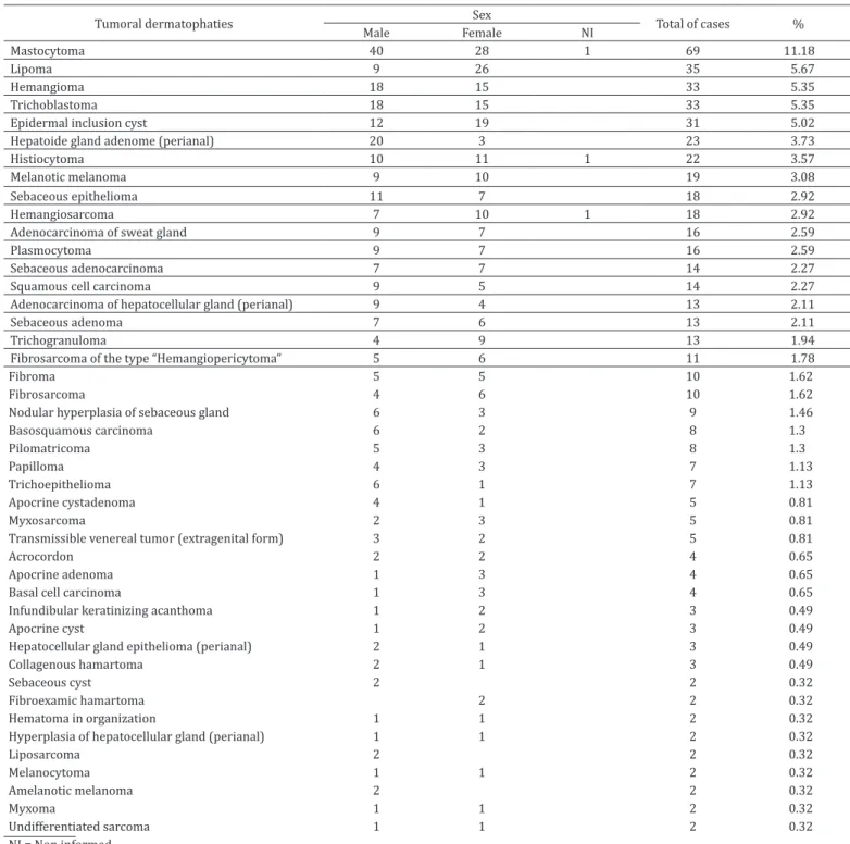
Table 1. Frequency of 546 tumoral dermatophaties diagnosed in 453 dogs at Laboratório de Patologia Veterinária, Universidade Federal da Bahia, 2007-2016
Tumoral dermatophaties Sex Total of cases %
Male Female NI
Mastocytoma 40 28 1 69 11.18
Lipoma 9 26 35 5.67
Hemangioma 18 15 33 5.35
Trichoblastoma 18 15 33 5.35
Epidermal inclusion cyst 12 19 31 5.02
Hepatoide gland adenome (perianal) 20 3 23 3.73
Histiocytoma 10 11 1 22 3.57
Melanotic melanoma 9 10 19 3.08
Sebaceous epithelioma 11 7 18 2.92
Hemangiosarcoma 7 10 1 18 2.92
Adenocarcinoma of sweat gland 9 7 16 2.59
Plasmocytoma 9 7 16 2.59
Sebaceous adenocarcinoma 7 7 14 2.27
Squamous cell carcinoma 9 5 14 2.27
Adenocarcinoma of hepatocellular gland (perianal) 9 4 13 2.11
Sebaceous adenoma 7 6 13 2.11
Trichogranuloma 4 9 13 1.94
Fibrosarcoma of the type “Hemangiopericytoma” 5 6 11 1.78
Fibroma 5 5 10 1.62
Fibrosarcoma 4 6 10 1.62
Nodular hyperplasia of sebaceous gland 6 3 9 1.46
Basosquamous carcinoma 6 2 8 1.3
Pilomatricoma 5 3 8 1.3
Papilloma 4 3 7 1.13
Trichoepithelioma 6 1 7 1.13
Apocrine cystadenoma 4 1 5 0.81
Myxosarcoma 2 3 5 0.81
Transmissible venereal tumor (extragenital form) 3 2 5 0.81
Acrocordon 2 2 4 0.65
Apocrine adenoma 1 3 4 0.65
Basal cell carcinoma 1 3 4 0.65
Infundibular keratinizing acanthoma 1 2 3 0.49
Apocrine cyst 1 2 3 0.49
Hepatocellular gland epithelioma (perianal) 2 1 3 0.49
Collagenous hamartoma 2 1 3 0.49
Sebaceous cyst 2 2 0.32
Fibroexamic hamartoma 2 2 0.32
Hematoma in organization 1 1 2 0.32
Hyperplasia of hepatocellular gland (perianal) 1 1 2 0.32
Liposarcoma 2 2 0.32
Melanocytoma 1 1 2 0.32
Amelanotic melanoma 2 2 0.32
Myxoma 1 1 2 0.32
Undifferentiated sarcoma 1 1 2 0.32
Tumoral dermatophaties Sex Total of cases %
Male Female NI
Malignant neoplasm of the peripheral nerve sheath 2 2 0.32
Apocrine adenocarcinoma of the anal sac 1 1 0.16
Cerumen adenocarcinoma of the gland 1 1 0.16
Apocrine gland adenoma of the anal sac 1 1 0.16
Nodular cutaneous amyloidosis 1 1 0.16
Meibomian gland carcinoma 1 1 0.16
Ceratogranuloma 1 1 0.16
Follicular cyst 1 1 0.16
Proliferative matrical cyst 1 1 0.16
Basosquamous epithelioma 1 1 0.16
Meibomian gland epithelioma 1 1 0.16
Granuloma associated with leishmaniasis 1 1 0.16
Fungal granuloma 1 1 0.16
Glandular epithelial hyperplasia 1 1 0.16
Metastasis of anaplastic carcinoma of the mammary gland 1 1 0.16
Metastasis of tubular carcinoma of the mammary gland 1 1 0.16
Malignant trichoepithelioma 1 1 0.16
Round cell tumor TOTAL 286 1 256 4 1 546 0.16 100 NI = Non informed.
Table 1. Continued...
Table 2. Fequency of breeds of 453 dogs with tumoral dermatopathies diagnosed at Laboratório de Patologia Veterinária, Universidade Federal da Bahia, 2007-2016
Breed Total os cases %
No defined breed
Poodle
American Pit Bull Terrier English Cocker Spaniel Siberian Husky Rottweiler Pinscher Dachshund Boxer Labrador Retriever Shih Tzu Brazilian Terrier Yorkshire Terrier
American Staffordshire Terrier Old English Sheepdog French Bulldog Schnauzer Beagle Golden Retriever Basset Hound Deutsche Dogge Pug Weimaraner Akita Chihuahua Chow Chow Brazilian Mastiff Pekingese
West Highland White Terrier English Bulldog Dogo Argentino Non informed German Sheperd St. Bernard 135 85 30 25 19 18 17 12 10 10 10 9 9 8 8 6 5 4 4 3 3 3 3 2 2 2 2 2 2 1 1 1 1 1 29.8 18.76 6.62 5.52 4.19 3.97 3.75 2.65 2.21 2.21 2.21 1.99 1.99 1.77 1.77 1.32 1.1 0.88 0.88 0.66 0.66 0.66 0.66 0.44 0.44 0.44 0.44 0.44 0.44 0.22 0.22 0.22 0.22 0.22
TOTAL 453 100%
this information. Of the 449 dogs whose sex was specified, 243 (53.64%) were males and 206 (45.47%) were females, that is, a male:female ratio of approximately 1.2.
A great diversity was observed with respect to the breeds of the 453 dogs affected by skin tumors (33 different breeds), with 135 (29.8%) mongrels and 317 (69.98%) dogs of defined breed, as shown in table 2. In one case (0.22%), the breed was not informed in the biopsy report. Of the 453 dogs with skin tumors evaluated, 26 (5.74%) did not have their age reported. Of the other 427 (94.26%) dogs that had their age described in the forms, 214 (50.12%) were classified as elderly, 200 (46.84%) were adults, and 13 (3.04%) were pups.
Comparative analysis between age group and skin tumor in the 546 diagnoses of dogs showed the following results: puppies presented higher incidence of histiocytoma (33.3%, 5/15), plasmacytoma (13.3%, 2/15), and mast cell tumor (13.3%, 2/15); adults had higher incidence of hemangioma (7.14%, 17/238), lipoma (6.72%, 16/238), sebaceous epithelioma (6.72%, 16/238), and trichoblastoma (6.72%, 16/238); elderly showed higher incidence of mast cell tumor (10.11%, 26/257), lipoma (7%, 18/257), hepatocellular adenoma (6.6%, 17/257), trichoblastoma (6.22%, 16/257), melanotic melanoma (5,83%, 15/257), and epidermal inclusion cyst (5.83%, 15/257).
13 (2.8%) on the back (hemangioma 15.4%); 10 (2.1%) in the scrotum (mast cell tumor 50% and plasmacytoma 20%); seven (1.5%) in the tail without predominance of neoplasm. Regarding non-neoplastic tumors, cysts accounted for 48.71% (38/78) of the total diagnoses. Among these, epidermal inclusion cysts (81.58%, 31/38) presented the highest incidence, and most of them were multicentric (25.8%, 8/31), followed by trichogranulomas (15.38%, 12/78), which occurred mainly in the neck, thoracic limb, or were multicentric with equal incidence in these sites (25% each 3/12), and nodular hyperplasia of the sebaceous gland (11.54%, 9/78), which was mostly multicentric (55.5%, 5/9).
DISCUSSION
There are several studies in the specific scientific literature that emphasize the fact that the skin is the organ most affected by diseases (Scott & Paradis 1990, Scott et al. 2001, Balda et al. 2004), in addition to being one of the main sites for the development of neoplasms (Goorman & Dobson 1995, Brønden et al. 2010). The findings of the present study corroborate the literature: among the 617 dermatopathies diagnosed, skin neoplasms presented the highest incidence (75.85%, 468/617) and were responsible for approximately ¾ of the cases in the time series investigated, which explains why this neoplasia has been one of the most studied in veterinary oncology over the years (Goldschmidt & Shofer 1992).
This high incidence of skin neoplasms is attributed, by some authors, to the fact that the skin is the largest organ of the body and constitutes an important barrier between the organism and the external environment (Jones et al. 2000) because of its exposure to oncogenic factors such as solar radiation (Medleau & Hnilica 2009), its cell renewal capacity, which increases the susceptibility of mutations, and of the increased attention and care provided by owners, which prolongs the life of domestic animals (Bonnett et al. 2005). In contrast, it is worth noting that the high incidence of neoplastic lesions found in this retrospective survey (2007-2016) may be justified by the fact that skin tumors are the main reason for skin biopsy, because other non-tumoral skin diseases are routinely diagnosed during clinical consultation or by other complementary tests.
Souza et al. (2006) conducted a national retrospective study of 40 years (1964-2003) that analyzed the diagnoses of 703 dog skin biopsy reports of the Laboratory of Veterinary Pathology of the Federal University of Santa Maria (LPV-UFSM) and found a mean of 17 skin biopsies per year, which was considered a significant number for the LPV-UFSM according to the authors. In the present study, 26% (503/1945) of the histopathological evaluations of samples from dogs were skin biopsies, that is, approximately ¼ of the cases at the LPV-UFBA in this period refers to dermatopathies, and nearly 50 histopathological examinations of dog skin were performed per year on average. This frequency of diagnoses in the LPV-UFBA routine is also quite significant, considering that most of the samples received come from the Hospital of Veterinary Medicine (Hospmev-UFBA).
With regard to the sex of dogs affected by tumor skin diseases, several studies have shown that males are more affected than females (Kaldrymidou et al. 2002, Bellei et al. 2006, Paranhos 2014); however, no statistically significant difference between the sexes of the dogs assessed was observed in
this study, corroborating the findings described by Souza (2005). In contrast, the literature reports that specific types of neoplasms, such as infundibular keratinizing acanthoma (Vail & Withrow 2007) and sebaceous gland carcinoma (Goldschmidt & Hendrick 2008) are more commonly found in male dogs. In the present study, however, females presented infundibular keratinizing acanthoma more often, but this comparison could not be performed significantly because of the small number of diagnosed cases (only three: one in a male dog and two in female dogs). As for sebaceous gland carcinoma, the same number of males (seven) and females (seven) was observed in this study. Regarding hepatocellular neoplasms (39 cases), hepatocellular adenoma presented the highest incidence (59%, 23/39), mainly in males (86.95%, 20/23), with a ratio of approximately 7:1. It is worth mentioning that this type of neoplasia presents marked predisposition in male dogs because of the influence of testosterone on tumor development (Wilson & Hayes Junior 1979, Turek & Withrow 2007, Daleck et al. 2009, North & Banks 2009); however, reproductive status (sexually intact or castrated) could not be assessed in the biopsy reports of this study.
Apparently, the high incidence of dogs with undefined breed (30.56%) with skin tumors found in this retrospective study may be associated with the fact that mongrels represent the majority of the canine population in Brazil (Fonseca 1999, Hataka 2004), as well as to the fact that the population assisted at the Hospmev-UFBA is, in general, of low-income. In contrast, it is known that, currently, many families with good standard of living have made an option to adopt mongrels, which also increases the population of these dogs even in upper-class neighborhoods. Similar findings have been reported in other studies that addressed the incidence of neoplastic lesions in mongrels (Pires et al. 2003, Bellei et al. 2006, Paranhos 2014). Some pertinent considerations on the age group of all the dogs included in this study are as follows: older dogs presented skin neoplasms more frequently compared with adult dogs, but no statistically significant difference was observed between these two age groups, with an elderly:adult ratio of only 1.07; however, the number of skin tumors in pups (15 cases) was directly influenced by the occurrence of histiocytomas (33.3%, 5/15), which corroborates the data reported by Bellei et al. (2006), considering that histiocytoma is a benign mesenchymal neoplasia more frequently found in dogs aged <2 years (Raskin & Meyer 2003, Cowell et al. 2009, Gross et al. 2009).
Among the neoplastic disorders observed (468), those with benign biological behavior were the most prevalent (230/468, 49.14%), in agreement with the findings described by Bellei et al. (2006) and Souza et al. (2006), but differing from the results found in a study conducted at the Hospital of Veterinary Clinics of the Federal University of Rio Grande do Sul (UFRGS) from January 2008 to December 2012 (Mazzocchin 2013), which described that 62.37% of the skin tumors diagnosed were of malignant biological behavior.
should be considered (Cotran et al. 2000), which reinforces the importance of adoption of early therapeutic measures for each lesion. In this study as a whole, the possibility of neoplasms of benign biological behavior (230) was similar to that of malignant neoplasms (215), with a ratio between them of only 1.07. The following ratios were observed when neoplasms were analyzed separately: 1.04 for benign and malignant mesenchymal tumors, 1.42 for benign and malignant epithelial tumors, and 3.17 for malignant and borderline tumors. As for melanocytic lesions, the occurrence of malignant tumors was 10.5 fold that of benign tumors. In comparison with the findings by Souza (2005), although the incidence of malignant tumors (359) was higher than that of benign tumors (314), the ratio between them was 1.14, a result similar to that observed in the present study; however, this ratio could have been even lower if the tumors had been classified as borderline as in this survey. Conversely, in the study conducted in the municipality of Santa Maria, Rio Grande do Sul state, prevalence of mesenchymal tumors of malignant biological behavior was higher than that of benign tumors, with a ratio of 1.6, whereas in relation to melanocytic tumors, the findings were similar for the higher incidence of malignant tumors, but in Santa Maria the malignant and benign ratio was only 2.5.
Of the 546 skin tumors (88.5%, 546/617) diagnosed in the 503 dogs of this research, 468 (85.7%) were neoplastic and 78 (14.3%) were non-neoplastic, that is, neoplastic proliferative lesions were six times more common than tumor-like lesions. This finding is quite similar to those observed in other retrospective studies (Bostock 1977, Finnie & Bostock 1979, Goldschmidt & Shofer 1992, Yager & Wilcock 1994, Souza 2005).
Of all the skin neoplasms evaluated in this survey (468), those of mesenchymal origin (243) were 1.2 and 10.5 times more common than those of epithelial and melanocytic origins, respectively, and incidence of epithelial tumors was 8.7 fold that of melanocytic lesions. These results corroborate those described by other authors (Bostock 1977, Finnie & Bostock 1979, Goldschmidt & Shofer 1992, Yager & Wilcock 1994, Souza 2005). In addition, melanocytic tumors were 11.5 times more frequent than skin metastases; however, the number of cases of metastatic skin neoplasms was extremely low in this study.
It is of uttermost importance to emphasize that in 87 of the 503 biopsy reports (17.3%) revised in this study more than one concurrent skin disease was observed, with one dog presenting five different dermatopathies. Thus, an extremely varied array of possible combinations (22 ratios) was observed within the classifications used in this study. Consequently, some dogs presented tumoral (neoplastic or not) and non-tumoral lesions at the same time, which resulted in diagnoses with prognoses that varied from good to unfavorable for the same dog. This finding stresses how careful clinicians and surgeons must be when collecting samples of skin lesions (skin biopsies) for histopathological diagnosis. It is essential that, in the presence of a dog with more than one skin tumor of macroscopic appearance similar to each other or not, common sense should always be used and, therefore, a significant number of fragments of different lesions should be collected and separately identified, considering that approximately one in six dogs presented more than one type of dermatopathy in this study.
This recommendation is also reinforced by the fact that mast cell tumor was the most prevalent skin tumor (11.18%) in this survey, considering that in dermatological veterinary clinical practice this tumor has been termed by many authors as “the great imitator” or “tumor of a thousand and one faces” (Murphy 2003), because of its extremely varied macroscopic appearance that can imitate numerous tumoral or non-tumoral skin lesions. Indeed, in the present study, four dogs presented multicentric cutaneous mast cell tumors with different macroscopic aspects, which were identified at histopathological examination as mastocytomas of different histological grades of differentiation. Souza (2005) described mast cell tumors with seven macroscopic patterns, namely, papule, nodule, plaque, mass, cyst, swelling, and “undeveloped feather aspect”.
A large number of cases of mast cell tumors were also reported in the research by Bellei et al. (2006). Moreover, Kiupel et al. (2004) stated that mastocytomas account for 7-21% of all skin tumors and Souza et al. (2006) concluded that they are the most prevalent skin tumor in the central region of Rio Grande do Sul state (20.9%, 158/761). In this study, mast cell tumors also presented high incidence: 1.9 fold that of lipoma - the second most frequent neoplasm. In comparison with other studies, mastocytoma was 3.8 times more frequent than the 18 cases reported by Andrade et al. (2012), 3.3 times more prevalent than the 21 cases described by Paranhos (2014), 2.22 fold the 31 cases observed by Mazzocchin (2013), and 1.7 times more frequent than the 39 cases found by De Nardi et al. (2002).
The 78 non-neoplastic tumoral skin diseases accounted for 14.3% of the diagnoses of skin tumors, which may be justified by the high incidence of cutaneous cysts (48.71%, 38/78). According to Pakhrin et al. (2007), epidermal inclusion cyst is one of the five most prevalent skin tumors in dogs in Korea. Fortunately, in spite of its high incidence, this lesion presents a favorable prognosis, and surgical excision is indicated only for esthetics or comfort reasons.
CONCLUSIONS
Skin tumors were observed in one-fourth of the dogs at the LPV-UFBA, with higher incidence of mesenchymal, epithelial and melanocytic neoplasms, in this order.
Mast cell tumor was the most frequent neoplasia in dogs, whereas epidermal inclusion cysts were the most common non-neoplastic skin tumors.
Distinct concurrent dermatological problems were frequently found in the dogs assessed in this study (one in six). Considering that these conditions can present with different cellular origin and biological behavior, it is crucial that histopathological evaluation be performed in fragments from the different cutaneous lesions.
Acknowledgements.- This study was financed in part by the Coordenação de Aperfeiçoamento de Pessoal de Nível Superior (CAPES), Brazil, Finance
Code 001.
REFERENCES
Andrade R.L.F.S., Oliveira D.M., Dantas A.F.M., Souza A.P., Nóbrega Neto P.I. & Riet-Correa F. 2012. Tumores de cães e gatos diagnosticados no semiárido da Paraíba. Pesq. Vet. Bras. 32(10):1037-1040. <http://dx.doi.org/10.1590/
Balda A.C., Larsson C.E., Otsuka M. & Gambale W. 2004. Estudo retrospectivo
de casuística das dermatofitoses em cães e gatos atendidos no Serviço
de Dermatologia da Faculdade de Medicina Veterinária e Zootecnia da Universidade de São Paulo. Acta Scient. Vet. 32(2):133-140.
Bastos R.S.C., Farias K.M., Lopes C.E.B., Pacheco A.C.L. & Viana D.A. 2017. Estudo retrospectivo de neoplasias cutâneas em cães da região metropolitana de Fortaleza. Revta Bras. Hig. San. Anim. 11(1):39-53. <http://dx.doi.
org/10.5935/1981-2965.20170005>
Bellei M.H.M., Neves D.S., Gava A., Liz P.P. & Pilati C. 2006. Prevalência de neoplasias cutâneas diagnosticadas em caninos no Estado de Santa Catarina, Brasil, no período de 1998 a 2002. Revta Ciênc. Agrovet. 5(1):73-79. Bonnett B.N., Egenvall A., Hedhammar A. & Olson P. 2005. Mortality in over
insured Swedish dogs from 1995 to 2000. Acta Scient. Vet. 46(3):105-120.
<http://dx.doi.org/10.1186/1751-0147-46-105> <PMid:16261924>
Bostock D.E. 1977. Neoplasia of the skin and mammary glands in dogs and cats, p.493-505. In: Kirk R.W. (Ed), Current Veterinary Therapy. W.B. Saunders, Philadelphia.
Brønden L.B., Eriksen T. & Kristensen A.T. 2010. Mast cell tumours and other skin neoplasia in Danish dogs: data from the Danish Veterinary Cancer Registry. Acta Vet. Scand. 52(1):6.
<http://dx.doi.org/10.1186/1751-0147-52-6> <PMid:20096110>
Cotran R.S., Kumar V. & Collins T. 2000. Neoplasias, p.233-295. In: Ibid. (Eds), Robbins Patologia Estrutural e Funcional. 6ª ed. Guanabara Koogan, Rio de Janeiro.
Cowell R.L., Tyler R.D. & Meinkoth J.H. 2009. Lesões cutâneas e subcutâneas, p.78-111. In: Cowell R.L., Tyler R.D., Meinkoth J.H. & Denicola D.B. (Eds), Diagnóstico Citológico e Hematologia de Cães e Gatos. 3ª ed. Medvet, São Paulo. Daleck C.R., Rodigheri S.M., De Nardi A.B. & Rodasaki S. 2009. Neoplasias
perianais, p.471-479. In: Daleck C.R., De Nardi A.B. & Rodaski S. (Eds), Oncologia em Cães e Gatos. Roca, São Paulo.
De Nardi A.B., Rodaski S., Sousa R.S., Costa T.A., Macedo T.R., Rodigheri S.M., Rios A. & Piekarz C.H. 2002. Prevalência de neoplasias e modalidades de tratamentos em cães, atendidos no Hospital Veterinário da Universidade Federal do Paraná. Arch. Vet. Sci. 7(2):15-26. <http://dx.doi.org/10.5380/
avs.v7i2.3977>
Finnie J.W. & Bostock D.E. 1979. Skin neoplasia in dogs. Aust. Vet. J. 55(12):602-604.
<http://dx.doi.org/10.1111/j.1751-0813.1979.tb07068.x> <PMid:231964> Fonseca C.S. 1999. Avaliação dos níveis séricos do estradiol e progesterona em cadelas portadoras de neoplasias mamárias. Dissertação de Mestrado,
Faculdade de Ciências Agrárias, Universidade Estadual Paulista, Jaboticabal, SP. 65p.
Goldschmidt M.H. & Shofer F.S. 1992. Skin Tumors of the Dog and Cat. Pergamon, Oxford. 316p.
Goldschmidt M.H. & Hendrick M.J. 2008. Tumors of the skin and soft tissues, p.44-117. In: Meuten D.J. (Ed), Tumors in Domestic Animals. 4th ed. Iowa
State Press, Ames. <http://dx.doi.org/10.1002/9780470376928.ch2>.
Goorman N.T. & Dobson J.M. 1995. The skin and associated tissues, p.187-200. In: White R.A.S. (Ed), Manual of Small Animal Oncology. British Small Animal, Shurdington.
Gross T.A., Ihrke P.J., Walder E.J. & Affolter V.K. 2009. Doenças de Pele do Cão e
do Gato: diagnóstico clínico e histopatológico. 2ª ed. Roca, São Paulo. 904p. Hataka A. 2004. Citologia aspirativa com agulha fina e histopatologia: valor e significado para diagnóstico e prognóstico do câncer de mama em cadelas. Tese de Doutorado, Faculdade de Medicina Veterinária e Zootecnia, Universidade Estadual Paulista, São Paulo. 68p.
Hill B.P., Lo A., Eden C.A., Huntley S., Morey V., Ramsey S., Richardson C., Smith D.J., Sutton C., Taylor M.D., Thorpe E., Tidmarsh R. & Williams V. 2006. Survey of the prevalence, diagnosis and treatment of dermatological conditions in small animals in general practice. Vet. Rec. 158(16):533-539. <http://dx.doi.
org/10.1136/vr.158.16.533> <PMid:16632525>
Jones T.C., Hunt R.D. & King N.W. 2000. Patologia Veterinária. 6ª ed. Manole, São Paulo. 1415p.
Kaldrymidou H., Leontides L., Koutinas A.F., Saridomichelakis M.N. & Karayannopoulou M. 2002. Prevalence, distribution and factors associated with the presence and potential for malignancy of cutaneous neoplasms in 174 dogs admitted to a clinic in Northern Greece. J. Am. Vet. Med. Assoc.
49(2):87-91. <http://dx.doi.org/10.1046/j.1439-0442.2002.jv408.x> <PMid:11958472>
Kiupel M., Webster J.D., Kaneene J.B., Miller R. & Yuzbasiyan-Gurkan V. 2004.
The use of Kit and Tryptase expression patterns as prognostic tools for canine cutaneous mast cell tumors. Vet. Pathol. 41(4):371-377. <http://
dx.doi.org/10.1354/vp.41-4-371> <PMid:15232137>
Mazzocchin R. 2013. Neoplasias cutâneas em pele. Monografia, Universidade Federal do Rio Grande do Sul, Porto Alegre. 64p.
Medleau L. & Hnilica K.A. 2009. Dermatologia de Pequenos Animais: atlas colorido e guia terapêutico. 2ª ed. Roca, São Paulo. 512p.
Meirelles A.E.W.B., Oliveira E.C., Rodrigues B.A., Costa G.R., Sonne L., Tesser E.S. & Driemeier D. 2010. Prevalência de neoplasmas cutâneos em cães da Região Metropolitana de Porto Alegre, RS: 1.017 casos (2002-2007). Pesq. Vet. Bras.
30(11):968-973. <http://dx.doi.org/10.1590/S0100-736X2010001100011>
Murphy S. 2003. Mast cell tumors, p.161-167. In: Dobson J.M. & Lascelles B.D.X. (Eds), BSAVA Manual of Canine and Feline Oncology. 2nd ed. BSAVA, Gloucester. North S. & Banks T. 2009. Tumors of skin and subcutaneous tissues, p.94-125. In: Ibid. (Eds), Introduction to Small Animal Oncology. Saunders Elsevier, London.
Pakhrin B., Kang M., Bae I., Park M., Jee H., You M., Kim J., Yoon B., Choi Y. &
Kim D. 2007. Retrospective study of canine cutaneous tumors in Korea.
J. Vet. Sci. 8(3):229-236. <http://dx.doi.org/10.4142/jvs.2007.8.3.229> <PMid:17679768>
Paranhos C.A. 2014. Neoplasias cutâneas caninas: um estudo descritivo de
quatro anos. Dissertação de Mestrado, Universidade de Trás-os-Montes e
Alto Douro, Vila Real. 77p.
Pires I., Oliveira J., Queiroga F. & Alves A. 2003. Doenças hístiocíticas do cão,
p.14-29. In: Ibid. (Eds), Série Didáctica. Universidade de Trás-os-Montes e Alto Douro, Vila Real.
Raskin R.E. & Meyer D.J. 2003. Atlas de Citologia de Cães e Gatos. Roca, São Paulo. 354p.
Scott D.W. & Paradis M. 1990. A survey of canine and feline skin disorders seen in a university practice: Small Animal Clinic, University of Montreal,
Saint-Hyacinthe, Quebec (1987-1988). Can. Vet. J. 31(12):830-835. <PMid:17423707>
Scott D.W., Miller D.H., Griffin C.E. & Muller K. 2001. Small Animal Dermatology. 6th ed. W.B. Saunders, Philadelphia. 1528p.
Sischo W.M., Ihrke P.J. & Franti C.E. 1989. Regional distribution of ten common
skin diseases in dogs. J. Am. Vet. Med. Assoc. 195(6):752-756. <PMid:2676928>
Souza T.M. 2005. Estudo retrospectivo de 761tumores cutâneos em cães.
Dissertação de Mestrado, Centro de Ciências Rurais, Universidade Federal
de Santa Maria, Santa Maria, RS. 296p.
Souza T.M., Fighera R.A., Irigoyen L.F. & Barros C.S.L. 2006. Estudo retrospectivo de 761 tumores cutâneos em cães. Ciência Rural 36(2):555-560. <http://
dx.doi.org/10.1590/S0103-84782006000200030>
Turek M.M. & Withrow S.J. 2007. Perianal tumors. p.503-508. In: Vail D.M. & Withrow S.J. (Eds), Small Animal Clinical Oncology. 4th ed. W.B. Saunders, Missouri.
Vail D.M. & Withrow S.J. 2007. Tumors of the skin and subcutaneous tissues, p.233-260. In: Withrow S.J. & Macewen E.G. (Eds), Small Animal Clinical Oncology. 4th ed. W.B. Saunders, Missouri. <http://dx.doi.org/10.1016/
B978-072160558-6.50021-6>.
Wilson G.P. & Hayes Junior H.M. 1979. Castration for treatment of perianal gland neoplasms in the dog. J. Am. Vet. Med. Assoc. 174(12):1301-1303.
<http://dx.doi.org/10.2460/javma.246.12.1301> <PMid:511727> Yager J.A. & Wilcock B.P. 1994. Color Atlas and Text of Surgical Pathology of the