Relationship between Canine Visceral Leishmaniosis
and the
Leishmania (Leishmania) chagasi
Burden in
Dermal Inflammatory Foci
R. C. Giunchetti
*,y, W. Mayrink
z, O. Genaro
z,y, C. M. Carneiro
*,O,
R. Corre
ˆa-Oliveira
y, O. A. Martins-Filho
z, M. J. Marques
z, W. L. Tafuri
*and A. B. Reis
*,y,O *LaboratoŁrio de Imunopatologia, NuŁcleo de Pesquisas em CieŒncias BioloŁgicas/NUPEB, Instituto de CieŒncias Exatas e BioloŁgicas, Universidade Federal de Ouro Preto, Ouro Preto, Minas Gerais,y
LaboratoŁrio de Imunologia Celulare Molecular, Centro de Pesquisas ReneŁ Rachou, Fundac-ao Oswaldo Cruz, Belo Horizonte, Minas Gerais,
z
LaboratoŁrio de Leishmanioses, Departamento de Parasitologia, Instituto de CieŒncias BioloŁgicas, Universidade Federal de Minas Gerais, Belo Horizonte, Minas Gerais,O
Departamento de AnaŁlises Cl|¤nicas, Escola de FarmaŁcia, Universidade Federal de Ouro Preto, Ouro Preto, Minas Gerais andz
LaboratoŁrio de Doenc-a de Chagas, Centro de Pesquisas ReneŁ Rachou, Fundac-ao Oswaldo Cruz, Belo Horizonte, Minas Gerais, Brasil
Summary
The skin is the ¢rst point of contact with organisms of the genusLeishmaniafrom sand £y vectors, and apparently normal skin of sick dogs harbours amastigote forms ofLeishmania chagasi. In relation to canine visceral leishma-niosis (CVL), the ear skin was examined in 10 uninfected dogs (UDs) and in 31 dogs dogs naturally infected with
L. chagasi. The infected animals consisted of 10 symptomless dogs (SLDs), 12 mildly a¡ected dogs (MADs) and nine a¡ected dogs (ADs). A higher parasite burden was demonstrated in ADs than in SLDs by anti-Leishmania
immunohistochemistry (Po0.01), and by Leishman Donivan Unit (LDU) indices (P¼0.0024) obtained from Giemsa-stained impression smears. Sections stained with haematoxylin and eosin demonstrated a higher intensity of in£ammatory changes in ADs than in SLDs (Po0.05), and in the latter group £ow cytometry demonstrated a correlation (P¼0.05/r¼0.7454) between the percentage of CD14+monocytes in peripheral blood and chronic
dermal in£ammation. Extracellular matrix assessment for reticular ¢bres by staining of sections with Masson trichrome and Gomori ammoniacal silver demonstrated a decrease in collagen type I and an increase in collagen type III as the clinical signs increased. The data on correlation between cellular phenotypes and histological changes seemed to re£ect cellular activation and migration from peripheral blood to the skin, mediated by anti-genic stimulation.The results suggested that chronic dermal in£ammation and cutaneous parasitism were directly related to the severity of clinical disease.
r2006 Elsevier Ltd. All rights reserved.
Keywords:dog; kala azar;Leishmania chagasi; parasitic infection
Introduction
Visceral leishmaniosis (VL; kala azar) is endemic in 87 countries, and approximately 90% of VL cases
re-corded worldwide occur in Bangladesh, Brazil, India and Sudan. Brazil is responsible for 90% of the VL re-cords from the American continent (Monteiro et al., 1994).
One of the ¢rst epidemiological surveys in Brazil was conducted by Chagas et al. (1938) in the region of AbaeteŁ, state of ParaŁ, where infection rates of 1.48% in man and 4.49% in dogs were found. Later,Deane and Deane (1962)reported the role of the dog and the fox www.elsevier.com/locate/jcpa
0021-9975/$ - see front matter r2006 Elsevier Ltd. All rights reserved. doi:10.1016/j.jcpa.2006.06.005
yIn memorium.
(Disicyon vetulus) as domestic and sylvatic reservoirs, re-spectively.
Earlier reports of canine visceral leishmaniosis (CVL) described various macroscopical skin lesions (e.g., desquamation, alopecia, pustular dermatitis, ul-cerative dermatoses and nodular disease, the type of which depended on the immune response (Adler and Theodor, 1932;Cunha, 1938;Torres, 1941;Ferreret al., 1988). The skin was considered by Abranches et al. (1991)to be an important reservoir compartment for parasites in healthy and sickLeishmania-infected dogs and the important role of dogs in VL transmission is supported by the high parasite loads found in the skin of infected animals (Deane and Deane,1962).
Histopathological changes in the skin ofLeishmania -infected dogs consist of variable degrees of focal or dif-fuse in£ammatory in¢ltrate in the dermis, and variable numbers of plasma cells, macrophages (parasitized or not by amastigotes ofLeishmania chagasi), lymphocytes and isolated neutrophils (Torres, 1941; Santos et al., 2004;Solano-Gallegoet al., 2004). Changes in the ex-tracellular matrix (ECM) inLeishmania amazonensis -in-fected mice are characterized by decreased collagen type I, increased collagen type III, and reduced ¢bro-nectin and laminin (Abreu-Silvaet al., 2004).
Histological tissue changes in CVL are probably triggered by the type of host immune response. Symp-tomless VL dogs are believed to produce a T-helper (Th) 1-mediated response. Clinically a¡ectedVL dogs, on the other hand, show a Th 2-mediated response, their peripheral blood mononuclear cells being unable to produce interferon (IFN)-gin the presence of para-site antigens (Pinelli et al., 1994). Bourdoiseau et al. (1997)reported the occurrence of immunosuppression associated with diminished numbers of CD4+T lym-phocytes and CD21+B lymphocytes.
Investigations on cutaneous immunopathology in CVL might contribute to a better understanding of events related to kala azar morbidity and to the human disease (Nietoet al.,1999;Moreno and Alvar, 2002).The present study was therefore designed to investigate the relationship between CVL and parasite burden as seen in the ear skin of dogs with di¡erent clinical forms ofL. chagasiinfection.
Materials and Methods
Animals
Thirty-one dogs naturally infected withL. chagasiand 10 uninfected dogs (UDs; controls) were obtained from the Zoonosis Control Centre, Belo Horizonte City Council. The dogs, of either sex, were aged 2^6 years. The UDs were con¢rmed as negative by parasitologi-cal examination and by an indirect £uorescent
antibody test (IFAT) foranti-LeishmaniaIgG (Bioman-guinhos Kit; FIOCRUZ-RJ, Brazil), titres ofo40 indi-cating freedom fromVL. The VL dogs, selected on the basis of IFAT titres of440, were classi¢ed clinically ac-cording to signs of infection (Manciantiet al.,1988) as: symptomless dogs (SLDs;n¼10); mildly a¡ected dogs (MADs; n¼12), with a maximum of three clinical signs; or a¡ected dogs (ADs; n¼9), with more than three clinical signs. The study was approved by the Ethical Committee for the use of Experimental Ani-mals, Universidade Federal de Minas Gerais.
Collection and Examination of Ear Skin Samples The dogs were euthanatized by an intravenous over-dose of barbiturate. Samples of ear skin were ¢xed in 10% neutral bu¡ered formalin for (1) routine histo-pathological examination of sections stained with hae-matoxylin and eosin (HE), Masson trichrome and Gomori ammoniacal silver, and (2) anti-Leishmania im-munohistochemistry.
Evaluation of parasite density in terms of the Leish-man Donovan Unit (LDU) index was carried out by light microscopy on Giemsa-stained impression smears prepared from fragments of ear skin, the LDU index being the number of Leishmaniaamastigotes per 1000 nucleated cells (Stauber,1956).
Parasite density was also evaluated immunohisto-chemically, as described byTafuriet al. (2004). Brie£y, serum from a dog naturally infected with L. chagasi
(IFAT titre41:40), diluted 1 in 100 in 0.01M phos-phate-bu¡ered saline (PBS), was applied as the pri-mary antibody. The slides were then incubated with biotinylated anti-mouse and anti-rabbit antibody (LSAB2 Kit; Dako, Carpinteria, CA, USA), which cross-reacts with canine serum immunoglobulins ( Ta-furiet al., 2004), and subsequently with the streptavi-din^peroxidase complex (LSAB2 Kit; Dako). The reaction was ‘‘visualized’’ with diaminobenzidine (DAB; Sigma, St Louis, MO, USA) and hydrogen per-oxide. Finally, the slides were dehydrated, cleared, counterstained with Harris’s haematoxylin, and mounted under coverslips.
The dermal in£ammatory pattern and the cell popu-lation were evaluated histologically on HE-stained sec-tions. The in£ammatory in¢ltrate was graded according to Solano-Gallego et al. (2004), as follows:
, no in£ammatory in¢ltrate; +, isolated foci of in-£ammatory cells; ++, isolated to coalescing areas of in£ammatory in¢ltrate; +++, di¡use areas of in£am-matory in¢ltrate.
with a modi¢cation of the methods described byRidley and Ridley (1983). The results were recorded as: , none; +, light density, (1^100); ++, moderate density (101^300); +++, high density (4300).
Extracellular matrix (ECM), assessed in sections stained with Masson trichrome (for collagen I) and Gomori ammoniacal silver (for collagen III), was clas-si¢ed as:^, normal distribution of collagen I and III; +, either slight reduction of collagen I or slight increase of collagen III; ++, either moderate reduction of collagen I or moderate increase of collagen III; and +++, strik-ing reduction of collagen I and strikstrik-ing increase of col-lagen III.
Flow Cytometry Analysis
Immunophenotyping analyses of peripheral blood by £ow cytometry was performed as described by Reis
et al. (2005). Brie£y, 1ml of whole blood with EDTA was subjected to pre-¢xation and erythrocyte lysis by the slow addition of 13 ml of lysis solution (FACS Lysing solution; Becton Dickinson) followed by incuba-tion for 10 min at room temperature (RT). After centri-fugation (450g, at RT for 10 min), the pellet was resuspended in 500ml PBS containing fetal bovine ser-um 10%.
In 96 well, ‘‘U’’ bottom plates (LIMBRO Biomedi-cals, Aurora, OH, USA), 30ml of pre-¢x leucocyte sus-pension were incubated at RT for 30 min in the dark with 30ml of anti-canine cell surface marker antibodies. These monoclonal antibodies (mAbs) which de¢ne ca-nine cell phenotypes, included puri¢ed rat anti-dog Thy-1 (Rat-IgG2b ^ Clone YKIX337.217), anti-dog CD5 (Rat-IgG2a ^ Clone YKIX322.3), anti-dog CD4 IgG2a ^ CloneYKIX302.9), anti-dog CD8 (Rat-IgG1 ^ Clone YCATE55.9), £uorescein isothiocyanate (FITC)- labelled mouse anti-human-CD21 (Mouse-IgG1çClone IOBla) and PE/Cy-5-conjugated mouse anti-human-CD14 (Mouse-IgG2a ^ Clone TUºK4), used in indirect and direct immuno£uorescence proce-dures.The unlabelled mAbs and anti-CD14 mAbs were purchased from SEROTEC (Oxford, UK), and anti-CD21 from Immunotech (Marseille, France).
When puri¢ed mAbs were used, the cells were also incubated, under the same conditions, with 60ml of previously diluted FITC-conjugated sheep anti-rat IgG antibody. Before £ow cytometric data collec-tion and analysis, labelled cells were ¢xed for 30 min with 200 ml of FACS FIX Solution (paraformal-dehyde 10.0 g/l, sodium cacodylate 10.2 g/l and sodium chloride 6.65 g/l, pH 7.2). The results were expressed as the percentage of positive cells within the selected lym-phocyte gate (Thy-1+, CD5+, CD4+, CD8+ and
CD21+) or of ungated leucocytes (CD14+). The latter
were also expressed as absolute counts taking into ac-count the white blood cell values for each animal.
Statistical Analysis
This was performed with the Prism 3.0 software pack-age (Prism Software, Irvine, CA, USA). The Kruskal-Wallis test was used to compare insensitivity between groups in both the immunohistochemical (anti-
Leish-mania) and histopathological approaches. w2 analysis
was used for the LDU index. The Spearman test was performed for strategy correlation between peripheral blood phenotype and histological/parasitological sta-tus.Po0.05 was considered signi¢cant.
Results
Clinical Evaluation
Table1shows the frequency of occurrence of the various clinical signs in the groups MADs and ADs. The most frequently observed signs were weight loss, onychogry-phosis, localized or di¡use ulcers, dry exfoliative der-matitis and hepatosplenomegaly.
Histological, Immunohistochemical and LDU Results These are shown inTable 2. In terms of frequency of oc-currence of chronic in£ammation of the dermis, SLDs, MADs and ADs all di¡ered signi¢cantly (Po0.05) from UDs (controls).Fig. 1(A, D, G and J) shows the normal appearance of ear skin, as seen in the UDs. In all infected animals, sparse cellular in¢ltrates in the super¢cial and deep dermis consisted mainly of plas-macytes, with smaller numbers of macrophages and lymphocytes. In this context, MADs and ADs (both groups clinically a¡ected) showed evident chronic
Table 1
Clinical signs recorded in mildly a¡ected dogs (MADs) and a¡ected dogs (ADs)
Clinical signs Number (and %) of dogs showing each stated sign
MADS (n¼12) ADs (n¼9)
Localized alopecia 5(45.4) 4(44.4)
Di¡use alopecia 0(0) 2(22.2)
Furfuraceous dermatitis 1(9.1) 3(33.3)
Opaque cornea 2(18.2) 1(11.1)
Localized ulcers 7(63.6) 3(33.3)
Di¡use ulcers 1(9.1) 5(55.5)
Paresis of limbs 0(0) 1(11.1)
Keratoconjunctivitis 2(18.2) 2(22.2) Loss of weight 2(18.2) 8(88.9) Onychogryphosis 6(54.5) 8(88.9) Hepatosplenomegaly 4(36.4) 6(66.7)
in£ammation. In ADs (Fig. 1C, F, I and L), the in£am-mation intensity was greater than in SLDs (Fig. 1B, E, H and K). As the clinical picture progressed, there was a reduction of collagen type I (Fig.1G, H and I) and an increase in collagen type III (Fig. 1J, K and L). Semi-quantitative histological analysis of sections stained by Masson trichrome or Gomori ammoniacal silver (Fig. 2) revealed that the percentage of ADs showing severe changes was higher than that of UDs and SLDs (Po0.05). Moreover, in terms of chronic dermal in-£ammation, the percentage of animals showing severe changes was higher in the MAD and AD groups than in the SLD and UD groups (Po0.05) (Fig. 2).
The anti-Leishmania immunohistochemical assay and LDU index showed that the percentage of animals with heavy parasite burdens in the ear skin was higher in the AD group than in the SLD group (Po0.05) ( Ta-ble 2). Assessment of the parasite burden by the LDU index revealed the following mean (and standard de-viation; SD) values: SLDs, 25.3 (57.8); MADs 196.9 (338.8); and ADs 1346.1 (2431.0). The LDU index re-vealed a higher parasitic burden in ADs than in SLDs (Po0.05). When the parasite burden was evaluated by anti-Leishmania immunohistochemical assay, higher parasite burdens were demonstrated in ADs than in SLDs (Po0.01) (Fig. 2).
Relationship between Chronic Dermal Inflammation and Immunophenotyping of Peripheral Blood
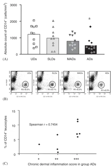
In ADs, the absolute counts of CD14+ cells were signi¢cantly lower than in UDs, and there was a posi-tive correlation between the low percentages of mono-cytes and chronic dermal in£ammation (P¼0.025/
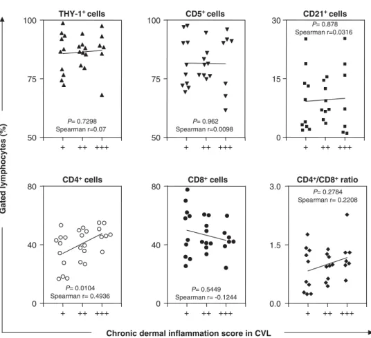
r¼0.7454) (Fig. 3A, B, C). There was no signi¢cant correlation between chronic dermal in£ammation and Thy-1+, CD5+, CD8+ and CD21+ cells, despite
a suggestion of increased numbers of CD8+
lympho-cytes in dogs with only a slight in£ammatory in¢ltrate (Fig. 4).
Discussion
The clinical outcome of CVL range from symptomless infection to classical kala azar with a full array of symptoms (Manciantiet al.,1988). In the present study, the main clinical signs observed were localized or dif-fuse ulcers, loss of weight, onychogryphosis and hepa-tosplenomegaly, signs similar to those reported by Limaet al. (2004). In VL endemic regions these signs are relevant to the di¡erential diagnosis of canine skin diseases.
Some authors (Adler and Theodor, 1932; Cunha, 1938,Tafuri et al., 2001) reported that the dermal in-£ammatory in¢ltrate in CVL consisted of mononuc-lear cells surrounding sebaceous follicles. In the present study, dogs naturally infected with L. chagasi
had plasmohistiocytic- and lymphocyte-like cellular in¢ltrates (data not shown), but no histiocytic granulo-ma such as those reported by Solano-Gallego et al. (2004)andSantoset al. (2004).
The observed reorganization of the ECM, with a re-duction of collagen type I and increase of collagen type III (reticular ¢bres), was related to the degree of tissue destruction produced by in£ammatory processes and to parasite burden, as prevously reported by Abreu-Sil-va et al. (2004). In the present study, the degree of in-£ammatory in¢ltration was related to the parasite burden in the skin.
The study showed di¡erences between the groups of infected dogs (SLDs, MADs and ADs) and the control group (UDs) in terms of in£ammatory in¢ltrate. The in¢ltrate was mainly distributed di¡usely in super¢cial and deep dermis but was sometimes concentrated in perifollicular sites. Solano-Gallego et al. (2004) re-ported a focal pattern of in£ammatory in¢ltrate Table 2
Assessment of chronic dermal in£ammation and heavy parasite burden in the ear skin of uninfected dogs (UDs), symptomless dogs (SLDs), mildly a¡ected dogs (MADs) and a¡ected dogs (ADs)
Sign of infection in ear skin Number (and %) of dogs showing each stated sign
UDs (n¼10) SLDs (n¼10) MADs (n¼12) ADs (n¼9)
Chronic dermal in£ammation 1(10) 7(70) 10 (83.3) 9(100)
Parasite burden
ALIH 0 3 (30) 6(50) 9(100)y
LDU 0 4 (40) 8(66.6)z 9(100)y
ALIH, as judged by anti-Leishmaniaimmunohistochemical assay.LDU, as judged by Leishman Donovan index
Signi¢cantly di¡erent from UDs (Po0.05). ySigni¢cantly di¡erent from SLDs (P
suggestive of perifollicular dermatitis. It should be em-phasized that although the frequency of occurrence of in£ammatory in¢ltrates was similar in SLDs and SDs, the latter group showed a higher in£ammatory density. Solano-Gallegoet al. (2004)observed a greater degree of cutaneous in£ammation in the nose of clinically in-fected dogs than in symptomless dogs; in the latter, the skin showed no signi¢cant changes.
Santoset al. (2004)reported that in most dogs with localized in£ammatory in¢ltrate it was not possible to
demonstrate cutaneous parasitism. The same study showed a correlation between parasitism, which ran-ged from moderate to dense, and di¡use and granulo-matous in£ammatory patterns. In the AD group, the present study revealed an intense, di¡use dermal in-£ammatory in¢ltrate, with a high parasite burden. (Figs 1 and 2). In addition, the in£ammatory in¢ltrates were more striking in ADs than in SLDs. Similar obser-vations were made by Mart|¤nez-Moreno et al. (1995), Tarantino et al. (2001) and Solano-Gallego et al.,
Fig.1. A^L Photomicroscopy of ear skin of uninfected dogs (A, D, G andJ), symptomless infected dogs (B, E, H and K) and clinically a¡ected dogs (C, F, I and L). HE staining (A, B and C). Anti-Leishmaniaimmunohistochemistry (D, E and F). Masson trichrome staining (G, H and I). Gomori ammoniacal silver staining (J, K and L). Bar, 50mm (applies to all photographs). As canine visceral leishmaniosis
(2004). The LDU index and anti-Leishmania immuno-histochemical assay for cutaneous parasitism gave si-milar results, suggesting that the former method
represents a reliable and inexpensive method for evalu-ating the cutaneous parasite burden.
As observed byPinelliet al. (1994,1995)and Bourdoi-seau et al. (1997), symptomless dogs would seem to be more resistant than clinically a¡ected dogs, probably as the result of more e⁄cient cell activation, resulting in limited in£ammatory in¢ltration.
In ADs, the correlation (r¼0.7454) observed between CD14+ monocytes and chronic dermal
- + ++ +++
Chronic dermal inflammation
a a, b
0% 20% 40% 60% 80% 100%
UDs SLDs MADs ADs
Anti-Leishmania immunohistochemical assay
0% 20% 40% 60% 80% 100%
0% 20% 40% 60% 80% 100%
a, b a
a
Masson trichrome and Gomori ammoniacal silver
UDs SLDs MADs ADs
UDs SLDs MADs ADs
b
Fig. 2. Chronic dermal in£ammation and parasite burden (immu-nohistochemical assay) in ear skin (white bar [], absent; grey bar [+], mild; dark bar [++], moderate; black bar [+++], intense. Masson and Gomori staining (white bar [-], normal distribution of collagen I and III; grey bar [+], slight reduction of collagen I and slight increase of collagen III; dark bar [++], moderate reduction of collagen I and moder-ate increase of collagen III; black bar [+++], striking reduc-tion of collagen I and striking increase of collagen III. ‘‘a’’, signi¢cantly di¡erent (Po0.05) from UDs; ‘‘b’’, signi¢cantly
di¡erent (Po0.05) from SLDs.
UDs
R1=8.0%
SLDs
R1=8.1%
MADs
R1=8.5%
ADs
R1=6.0% 0
1000 2000 3000
(A)
(B)
+ ++ +++ 0
5 10 15
(C)
Spearman r = 0.7454
a
MADs
SLDs ADs
UDs
Chronic dermal inflammation score in group ADs
Absolute count of CD14
+ (cells/mm 3)
% of CD14
+ leucocytes
Fig. 3. (A^C) Analysis of CD14+cells within peripheral blood leu-cocytes from dogs naturally infected with L. chagasi. (A) Data analysis of CD14+cells was performed on symptomless (SLDs,n), mildly a¡ected dogs (MADs,.), a¡ected dogs
(ADs,E) and uninfected dogs (UDs,&). Single £ow cyto-metry platform was used to determine the frequency and the absolute counts of CD14+cells expressed as scattering of individual values of CD14+ cells/mm3. (B) Representative side scatter (SSC) versus £uorescence type 3 (CD14Cy-5PE) dot plots illustrating the lower frequency of CD14+cells in ADs than in UDs (Po0.05). Region statistics were used for
in£ammation probably re£ected activation of such per-ipheral blood cells and their migration to the dermis, where they participated in the striking in£ammatory reaction (Fig. 3). However, this cell population prob-ably made little contribution to the resistance in view of the high morbidity and parasite burden shown by ADs.
Acknowledgments
This work was supported by the CNPq and FAPEMIG. We are also grateful to Mrs P. S. Eiras and Dr H. Black-more for assistance in preparing the manuscript.
References
Abranches, P., Silva-Pereira, M. C. D., Conceic-ao-Silva, F.,
Santos-Gomes, G. M. and Jans, J. G. (1991). Canine leish-maniasis: pathological and ecological factors in£uencing transmission of infection. Journal of Parasitology, 77,
577^581.
Abreu-Silva, A. L., Calabrese, K. S., Mortara, R. A., Tedes-co, R. C., Cardoso, F. O., Carvalho, L. O. P. and Gonc-^
alves da Costa, S. C. (2004). Extracellular matrix altera-tions in experimental murineLeishmania (L.) amazonensis
infection.Parasitology,128,385^390.
Adler, S. and Theodor, O. (1932). Investigations on Mediter-ranean kala-azar.VI ^ Canine visceral leishmaniasis. Pro-ceedings of the Royal Society, London,110,402^412.
Bourdoiseau, G., Bonnefont, C., Magnol, J. P., Saint-AnreŁ, I. and Chabanne, L. (1997). Lymphocyte subset abnormal-ities in canine leishmaniasis.Veterinary Immunology and Im-munopathology,56,345^351.
Chagas, E., Ferreira, L. C., Deane, G., Deane, L. and Gui-maraes, N. (1938). Leishmaniose visceral americana. II ^ Estudo epidemioloŁgico.MemoŁrias do Instituto Oswaldo Cruz,
33,138^206.
Cunha, A. M. (1938). Infecc-oes experimentais na
leishma-niose visceral americana. MemoŁrias do Instituto Oswaldo
Cruz,33,581^598.
Deane, L. M. and Deane, M. P. (1962).Visceral leishmaniasis in Brazil. Geographical distribution and transmission.
THY-1+ cells
P= 0.7298 Spearman r=0.07
+ ++ +++ 50
75
100 CD5
+ cells
P= 0.962 Spearman r=0.0098
50 75 100
+ ++ +++
CD21+ cells
0 15 30
P= 0.878 Spearman r=0.0316
+ ++ +++
CD8+cells
P= 0.5449 Spearman r= -0.1244
0 40 80
+ ++ +++
CD4+/CD8+ratio
P= 0.2784 Spearman r= 0.2208
0.0 1.5 3.0
+ ++ +++ CD4+cells
0 40 80
+ ++ +++
Gated lymphocytes (%)
Chronic dermal inflammation score in CVL P= 0.0104
Spearman r= 0.4936
Dermal Parasitism and Clinical Status in CVL
Fig. 4. Correlation between chronic dermal in£ammation score (+; ++; +++) with the percentage of Thy-1+(m), CD5+(.), CD21+(’), CD4+(
J), CD8+cells (K) and CD4/CD8 cell ratio (E), within gated peripheral blood lymphocytes fromL. chagasiinfected dogs. The results are expressed as scattering of individual values. Spearman correlation indices (r) atPo0.05 are shown on graphs.
Revista do Instituto de Medicina Tropical de Sao Paulo, 4,
149^212.
Ferrer, L., Rabanal, R., Fondevila, D., Ramos, J. A. and Domingo, M. (1988). Skin lesions in canine leishmaniasis.
Journal of Small Animal Practice,29,381^388.
Lima,W. G., Michalick, M. S. M., Melo, M. N.,Tafuri,W. L. andTafuri,Wg. L. (2004). Canine visceral leishmaniasis: a histopathological study of lymph nodes.Acta Tropica,92,
43^53.
Mancianti, F., Gramiccia, M., Gradoni, L. and Pieri, S. (1988). Studies on canine leishmaniasis control. I. Evolu-tion of infecEvolu-tion of di¡erent clinical forms of canine leish-maniasis following antimonial treatment.Transactions of the Royal Society ofTropical Medicine and Hygiene,82,566^567. Mart|¤nez-Moreno, A., Moreno, T., Mart|¤nez-Moreno, F. J.,
Acosta, I. and HernaŁndez, S. (1995). Humoral and cell-mediated immunity in natural and experimental canine leishmaniasis.Veterinary Immunology and Immunopathology,
48,209^220.
Monteiro, S. P., Lacerda, M. M. and Arias, J. R. (1994). Con-trole da leishmaniose visceral no Brasil.Revista da Socie-dade Brasileira de MedicinaTropical de Sao Paulo,27,67^72. Moreno, J. and Alvar, J. (2002). Canine leishmaniasis:
epide-miological risk and experimental model.Trends in Parasi-tology,18,399^405.
Nieto, C. G., Garc|¤a-Alonso, M., Requena, J. M., MiroŁn, C., Soto, M., Alonso, C. and Navarrete, I. (1999). Analysis of the humoral immune response against total and recombi-nant antigens ofLeishmania infantum: correlation with dis-ease progression in canine experimental leishmaniasis.
Veterinary Immunology and Immunopathology,67,117^130. Pinelli, E., Killick-Kendrick, R., Wagenaar, J., Bernadina,
W., Del Real, G. and Ruitenberg, J. (1994). Cellular and humoral immune responses in dogs experimentally and naturally infected withLeishmania infantum.Infection and Immunity,1,229^235.
Pinelli, E., Gonzalo, R. M., Boog, C. J. P., Rutten,V. P. M. G., Gebhard, D., Del Real, G. and Ruitenberg, E. J. (1995).
Leishmania infantum ^ speci¢c T cell lines derived from asymptomatic dogs that lyse infected macrophages in a major histocompatibility complex-restricted manner.
EuropeanJournal of Immunology,25,1594^1600.
Reis, A. B., Carneiro, C. M., Carvalho, M. G.,Teixeira-Car-valho, A., Giunchetti, R. C., Mayrink, W., Genaro, O., CorreŒa-Oliveira, R. and Martins-Filho, O. A. (2005). Es-tablishment of a microplate assay for £ow cytometric as-sessment and its use for the evaluation of age-related phenotypic changes in canine whole blood leukocytes. Ve-terinary Immunology and Immunopathology,103,173^185. Ridley, D. S. and Ridley, M. J. (1983). The evolution of the
lesions in cutaneous leishmaniasis. Journal of Pathology,
141,83^96.
Santos, W. L. C., BadaroŁ, J. D. R. and de-Freitas, L. A. R. (2004). Association between skin parasitism and a granu-lomatous in£ammatory pattern in canine visceral leish-maniasis.Parasitology Research,92,89^94.
Solano-Gallego, L., FernaŁndez-Bellon, H., Morell, P., Fonde-vila, P., Alberola, J., Ramis, A. and Ferrer, L. (2004). His-tological and immunohistochemical study of clinically normal skin ofLeishmania infantum-infected dogs.Journal of Comparative Pathology,130,7^12.
Stauber, L. A. (1956). Resistance to the Khartoum strain of
Leishmania donovani.The Rice Institute Pamphlet,45,80^96. Tafuri,Wg. L., Oliveira, M. R., Melo, M. N. andTafuri,W. L.
(2001). Canine visceral leishmaniosis: a remarkable histo-pathological picture of one case reported from Brazil. Ve-terinary Parasitology,96,203^212.
Tafuri, Wg. L., Santos, R. L., Arantes, R. M. E., Gonc-alves,
R., Melo, M. N., Michalik, M. S. M. and Tafuri, W. L. (2004). An alternative immunohistochemical method for detecting Leishmania amastigotes in para⁄n-embedded canine tissues.Journal of Immunological Methods,292,17^23. Tarantino, C., Rossi, G., Kramer, L. H., Perrucci, S., Cringo-li, G. and Macchioni, G. (2001).Leishmania infantumand
Neospora caninum simultaneous skin infection in a young dog in Italy.American Journal of Tropical Medicine and Hy-giene,102,77^83.
Torres, C. M. (1941). Alterac-oes cutaŒneas do cao no kala-azar
sul-americano. MemoŁrias do Instituto Oswaldo Cruz, 36,
37^67.
Received, June 6th, 2005 Accepted, June 9th, 2006