Diana Sofia Pereira dos Anjos
dspa.kvo@gmail.com
Orientador: Dr. António Pinheiro Vieira
Licenciado em Medicina
Assistente Hospitalar de Cardiologia do Hospital de Santo António
MESTRADO INTEGRADO EM MEDICINA
2010/2011
Ablation of Atrial Fibrillation
Artigo de Revisão Bibliográfica
1
Ab lat ion of At rial Fib rillat ionAbstract
IntroductionAtrial Fibrillation is an increasingly common and costly medical problem. Given the unsatisfactory efficacy and the side effects associated with pharmacological therapy, new treatment options are needed. The advancements of our understanding of the mechanisms of this arrhythmia, coupled with improvements in catheter ablation techniques, have impelled the development of catheter ablation from an experimental procedure to an increasingly important treatment alternative.
Objective
This essay will review the recent advances and outcomes of ablation of atrial fibrillation, in matters of patient selection, techniques, endpoints and complications of this procedure.
Development
Ablation of atrial fibrillation is possible because this dysrhythmia is frequently incited by focal triggers, many of which arise from the pulmonary veins. Current ablation techniques seek to eliminate or isolate these triggers from the rest of the atria in order to restore sinus rhythm. The mainstay of ablation remains in radiofrequency energy. Accurate imaging and mapping is important: the combination of intracardiac echocardiography, computed tomography, and magnetic resonance imaging with a three-dimensional electroanatomical mapping system is useful to prepare and perform these procedures and to identity future complications.
Conclusions
Catheter ablation is an important treatment in patients with atrial fibrillation. Multiple techniques and technologies presently exist, and it is hoped that their progress will lead to safer procedures and better outcomes. Randomized control trials with long-term follow-up periods are needed to improve the selection of patients who will most benefit from these treatments, and to safely establish ablation as a first line therapy.
Key Words
Atrial fibrillation, anti-arrhythmia agents, pulmonary veins, catheter ablation, treatment outcome, warfarin.
Abbreviations
2
Ab lat ion of At rial Fib rillat ionResumo
IntroduçãoA Fibrilhação Auricular é um problema médico dispendioso e cada vez mais comum na população. Dada a baixa eficácia e efeitos colaterais associados à terapia farmacológica, novas opções de tratamento são necessárias. O avanço no conhecimento dos mecanismos desta arritmia, juntamente com o progresso das técnicas ablativas, têm impulsionado o desenvolvimento da ablação por cateter como alternativa terapêutica importante.
Objectivo
Esta revisão bibliográfica propõe examinar os avanços e resultados mais recentes da ablação da fibrilhação auricular, no que diz respeito à selecção de pacientes, técnicas,
endpoints e complicações do procedimento.
Desenvolvimento
A ablação da fibrilhação auricular é possível porque esta arritmia é frequentemente despoletada por triggers focais, localizados nas veias pulmonares. As técnicas de ablação actuais procuram eliminar ou isolar esses triggers do resto das aurículas, a fim de restaurar o ritmo sinusal. Os procedimentos ablativos utilizam, maioritariamente, energia por radiofrequência. Imagiologia e mapeamento são fundamentais para o sucesso da ablação da fibrilhação auricular. A combinação da ecocardiografia intracardíaca, tomografia computadorizada e ressonância magnética com um sistema de mapeamento electroanatómico tridimensional é útil para preparar e executar este procedimento, além de identificar futuras complicações.
Conclusões
A ablação por cateter é um tratamento importante nos pacientes com fibrilhação auricular. Várias tecnologias estão disponíveis, e espera-se que o progresso conduza a procedimentos mais seguros e com melhores resultados. Ensaios clínicos randomizados, com períodos mais longos de seguimento, são necessários para melhorar a triagem dos pacientes que mais beneficiarão destes tratamentos, e para estabelecer a ablação como terapia de primeira linha.
3
Ab lat ion of At rial Fib rillat ionIntroduction
Atrial Fibrillation (AF) remains the most common sustained arrhythmia in clinical practice. 1 It is strongly age-dependent, affecting 4% of individuals older than 60 years
and 8% of persons older than 80 years. According to data from the Framingham and Rotterdam studies, approximately 25% of individuals aged 40 years and older will develop AF during their lifetime. 1
The prevalence of AF is 0.1% in persons younger than 55 years, 3.8% in persons 60 years or older, and 10% in persons 80 years or older. With the projected increase in the elderly population, the prevalence of AF is expected to more than double by the year 2050. 2 In 10-15% of cases of AF, the disease occurs in the absence of comorbidities (lone
atrial fibrillation). However, AF is often associated with other cardiovascular diseases, including hypertension; heart failure; diabetes-related heart disease; ischemic heart disease; and valvular, dilated, hypertrophic, restrictive, and congenital cardiomyopathies.
1
Atrial fibrillation constitutes a heavy burden on healthcare expenditure due to the high costs associated with AF-related hospitalization, evaluation, management, and loss of productivity. 3 It is imperative to promote coordinated efforts on behalf of cardiologists,
electrophysiologists, neurologists, and primary care providers to meet the increasing challenge of stroke prevention and rhythm management in the growing population with AF. 4
This paper aims at giving contemporary information on the constantly evolving indications, techniques, outcomes and technologies associated with ablation of AF.
4
Ab lat ion of At rial Fib rillat ionMaterial & Methods
The search strategy used the National Library of Medicine’s Medical Subject Headings (MeSH) keyword nomenclature developed for MEDLINE®. The searches were limited to
the English and Portuguese language. I searched the MEDLINE® databases from January,
2000 to January, 2011 for studies involving adult humans (19 years old or more, of both sexes) with atrial fibrillation who underwent ablation of atrial fibrillation. I combined search terms or MeSH terms for atrial fibrillation, anti-arrhythmia agents, pulmonary veins, catheter ablation, treatment outcome, warfarin. I included peer reviewed, clinical trials, meta-analysis, and randomized control trials. I excluded case reports and did not search systematically for unpublished data.
Results
The MEDLINE® database search yielded 516 citations. I identified 292 of these as
potentially relevant and retrieved the full-text articles for further evaluation. Of these, 240 did not meet eligibility criteria. A total of 52 studies were included in my analysis.
5
Ab lat ion of At rial Fib rillat ionAtrial Fibrillation Mechanisms
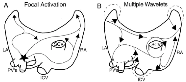
Understanding the pathophysiological mechanisms underlying the genesis and maintenance of AF is still a challenge. There are two main theories to explain it: the Focal Theory and the Multiple Wavelet Theory (Fig.1).
The Focal Theory was proposed by Sherf and colleagues (1948). It is based on the idea that all episodes of AF are preceded by atrial ectopic activity. Thus, AF can be induced by early extrasystoles, originating in most cases from ectopic foci in the pulmonary vein ostia. This theory is more appropriate in paroxysmal forms, in which simple ablation of the ectopic foci will lead to suppression of arrhythmic episodes. 5;6
Moe (1959) proposed the Multiple Wavelet Theory. According to this, the genesis and maintenance of AF depends on the existence of multiple reentrant circuits. The number of these circuits will depend on the atrial area involved and the refractory period and conduction velocity of the muscle fibers. Atrial dilatation promotes maintenance of AF, dispersing and shortening refractory periods and increasing intra-atrial conduction times.
7
Figure 1. (A) Focal Theory and (B) the Multiple Wavelet Theory.
LA= left atrium, RA= right atrium, PV’s= pulmonary veins, ICV= inferior cava vein, SCV= superior
6
Ab lat ion of At rial Fib rillat ionIn addition to these models for AF, important work has been done implicating the role of the local autonomic nervous system in the initiation and perpetuation of AF, consistent with the presence of vagal triggers for AF in some individuals. Parasympathetic ganglionated plexi are located near the pulmonary vein-left atrium junction and may be important targets for ablative therapy. 9;8
Despite these insights, the mechanisms of AF remain incompletely understood. It is now widely accepted that AF requires an initiating event and an anatomical substrate and that the pulmonary veins are intimately involved. There is evidence to support both Focal and Multiple Wavelet theories and therefore justification for different, or even stepwise, approaches to ablative strategies for different AF patients. In addition, multiple mechanisms may coexist based on underlying cardiac substrate. 3;10
7
Ab lat ion of At rial Fib rillat ionPatient Selection for Ablation of Atrial Fibrillation
The primary justification for catheter ablation is the presence of symptoms correlated with AF, with the goal of improving quality of life. It is also considered after failure of, at least, one Class I or Class III anti-arrhythmia agents, according to the Vaughn–Williams Classification, in patients suffering from recurrent paroxysmal AF. 3;11
Data show patient selection for catheter ablation evolving to include persistent AF and patients with heart failure and reduced ejection fraction. Small studies have shown improvement in left ventricular dysfunction and a decrease in left ventricular dimensions after AF catheter ablation. 3;12-14
Other considerations in patient selection include age, left atrium size, duration of AF. Ablation of atrial fibrillation requires high-intensity anticoagulation during the procedure with intravenous heparin. Warfarin is recommended, at least, short-term post procedure. Therefore, patients with major contraindications to anticoagulation are not candidates for ablation. 3
8
Ab lat ion of At rial Fib rillat ionTechniques and Endpoints for Ablation of Atrial Fibrillation
The goals of ablation of atrial fibrillation are to eliminate triggers and/or modify arrhythmogenic substrates.
Catheter ablation of AF has its roots in the surgical Maze procedure to cure AF developed by Dr. James Cox (Fig.2). 14 The Maze procedure consists of a series of incisions in the
right and left atria designed to develop anatomic barriers to conduction that would prevent maintenance of AF. This approach was patterned on the Multiple Wavelet Theory. Therefore, the surgical procedure erects "road blocks" designed to prevent perpetuation of these reentrant circuits. The Maze surgery is reasonably effective (the reported success rate reached above 95%, with perioperative mortality around 2%) but has not been accepted as a routine clinical technique because of its degree of difficulty and potential morbidity. 15
Figure 2. The Maze Procedure. (A) Right atrial lesions (black and white arrows). (B) The left atrial
lesions of the Maze procedure as shown with an open left atrium. The left upper arrow demonstrates the suture line that excludes the left atrial appendage to reduce the risk of thromboembolic events.
Haïssaguerre et al. (1998) demonstrated that the initiators of AF typically originate in the pulmonary veins, and isolation of these veins often prevents AF. This observation supports the Focal Theory of AF. 16 So, nowadays, the primary objective is to isolate the
pulmonary veins from the left atria.
The mainstay of ablation remains radiofrequency energy, although other energy sources are currently under investigation.
9
Ab lat ion of At rial Fib rillat ion Focal AblationFocal ablation within the pulmonary vein is guided by activation mapping, and the source of ectopy is identified by meticulous mapping, looking for the earliest "spike" electrical activity.
In 1998, Haïssaguerre and colleagues studied 45 patients with paroxysmal AF refractory to drug therapy. In the study, 94% of the points of AF origin were mapped to foci inside the pulmonary veins. They observed that elimination of local electrograms at these foci with radiofrequency energy rendered 62% of the patients free of AF recurrence over 8 months of follow-up. About 70% of these patients required more than one procedure. 16
The limited success, need for repeated procedures, and the relatively high incidence of pulmonary vein stenosis associated with focal ablation of AF led to a refinement in the technique.
Pulmonary Vein Isolation: Ablation at or near the Ostium of the Pulmonary vein (Veno - Atrial Junction)
Conduction areas from the left atrium to the pulmonary vein can be different and are recognized by analysis of electrograms on a circumferential mapping catheter positioned at the venous ostium. Radiofrequency energy is delivered at sites of earliest electrical activation to achieve a delay, change in activation pattern, or elimination of pulmonary vein potentials. 18 Ablation is continued until all pulmonary vein potentials are
10
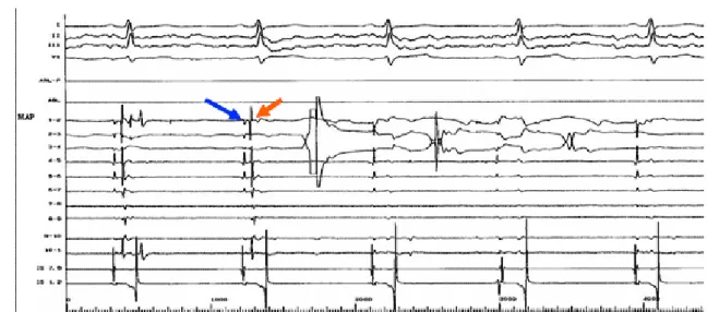
Ab lat ion of At rial Fib rillat ionFigure 3. Ablation at the ostium of the left superior pulmonary vein. A circumferential mapping
catheter positioned at the ostium records left atrial potentials (indicated by blue arrow) and pulmonary vein potentials (indicted by red arrow). Radiofrequency energy is delivered to the site of the earliest pulmonary vein potentials, resulting in elimination of these potentials on the third beat.
The success rate of this procedure ranges between 60-80% (mean follow-up of 4 ± 5 months). 18
Several limitations of this approach have been revealed. First, it appears to work predominantly in subjects with clear evidence of focus-triggered AF (i.e. subjects with multiple runs of self-terminating AF initiated by frequent premature ectopic beats) and less in patients with persistent or permanent AF. Second, there is a risk of pulmonary vein stenosis (1-3%). Third, the long-term success is impaired by very frequent recovery of conduction, and many subjects require repeated procedures. 19 This may reflect
minimalistic energy delivery in the proximal segments of the pulmonary vein in an attempt to eliminate risk of pulmonary venous stenosis.
Circumferential Ablation around the Pulmonary Vein O stia
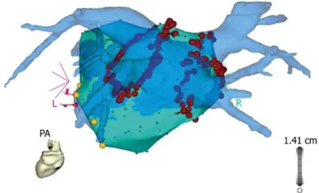
A circumferential anatomic approach guided by a nonfluoroscopic navigation system (CARTO; Biosense Webster; Diamond Bar, California) was described by Pappone and colleagues (Fig.4). 20
11
Ab lat ion of At rial Fib rillat ionFigure 4. The blue 3D anatomical shell of the left atrium and the pulmonary veins, as acquired by
pre-procedural computed tomography, is merged with the grey anatomical shell that is constructed with electro-anatomical mapping during the procedure (CARTO merge). The red ablation tags mark the circumferential ablation lesions around the pulmonary vein ostia.
A wide area of circumferential ablation is performed outside the pulmonary vein ostia. High power (100 W) and temperature (65°C) settings are used with an 8-mm tip ablation catheter. The power and temperature limits are reduced to 50 W and 55°C, respectively, in the posterior left atrial wall to avoid esophageal injury. 21 The ablation
catheter is dragged to create the circumferential lines, and an average of 10-15 seconds of radiofrequency energy is delivered at each site.
Local endpoints are bipolar electrogram reduction by 90% or to < 0.05 mV. A posterior line connecting the circumferential lines around the right and left-sided pulmonary vein is then performed to reduce the risk of developing macro-reentrant atrial arrhythmias. At sites eliciting a vagal reflex (sinus bradycardia, atrioventricular block, or hypotension), ablation is continued until the reflex is abolished. Of the 26 patients who underwent this procedure, at a mean follow-up of 9 ± 3 months, 85% were free of AF, including 62% not taking and 23% taking antiarrhythmic medications. 20 In a subsequent report, the
overall success was 80% (201 of 251) and only 13 of these patients were taking antiarrhythmic agents. 22
12
Ab lat ion of At rial Fib rillat ion Other TechniquesA method of ablating AF is to target both pulmonary vein and non-pulmonary vein triggers of AF. Non-pulmonary vein foci may originate from the superior vena cava, left atrium posterior wall, crista terminalis, coronary sinus, ligament of Marshall, or interatrial septum. 23 Atrium fibrillation triggers can be provoked, usually with high doses of
isoproterenol, and successfully ablated. 24;25 By eliminating both trigger sites, the
initiation of AF can potentially be prevented.
Complex Fractionated Electrogram Ablation is an approach, recently described, involving targeting complex fractionated electrograms for ablations and, at 1-year follow-up, 110 out of 121 (91%) patients undergoing ablation were free of AF. 26 Eighteen patients
required 2 procedures and 10 patients were receiving antiarrhythmic medications among those considered a success.
Another adjunctive (or possibly alternative) method of ablation of AF involves the intentional destruction of ganglionated plexi around the left atrium. Potential vagal target sites are identified during the procedure in ≥33% of patients. Vagal reflexes are considered sinus bradicardia (<40 beats per minute), asystole, atrioventricular block, or hypotension that occurs within a few seconds of the onset of radiofrequency application. If a reflex is elicited, radiofrequency energy is delivered until such reflexes are abolished for ≤30 seconds. The end point for ablation at these sites is termination of the reflex that is followed by sinus tachycardia or AF. Failure to reproduce the reflexes with repeat energy is considered confirmation of denervation. Complete local vagal denervation is confirmed by the abolition of all vagal reflexes. The most common sites are tagged on electroanatomic maps. 27
The premise that the mechanisms of AF may vary between patients, makes it necessary, in some patient subgroups, to combine different ablation techniques to achieve a successful outcome. This statement is exemplified by Weerasooriya et al., who studied one hundred patients that received catheter ablations from January 2001 to April 2002. They were followed to determine outcomes. Approximately one-third of these patients had persistent or longstanding persistent AF. After a single catheter ablation, the five-year freedom from arrhythmias was just 29%. But when measured from their last ablation, with a median of two procedures, 87% were free of arrhythmias at one year, 81% at two years, and 63% at five years. Those with valvular heart disease or cardiomyopathy were more likely to have a recurrence, and those with longstanding persistent AF were almost twice as likely as those with paroxysmal or persistent AF to have a recurrence. 28
13
Ab lat ion of At rial Fib rillat ionPost Procedure Considerations
Low-molecular-weight heparin or intravenous heparin is recommended as a bridge to therapeutic anticoagulation following ablation of AF. Warfarin is recommended for at least 3 to 6 months post ablation, regardless of anticoagulation status prior to the procedure. 37
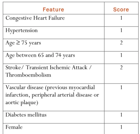
Discontinuation of warfarin therapy after a successful ablation of AF is also an unresolved issue. Several studies have shown that a significant number of asymptomatic patients after ablation still have episodes of unrecognized AF. However, if the AF burden, that is, frequency and duration of AF events, is markedly reduced, then the predilection to atrial thrombus development and subsequent stroke may be substantially reduced. This reasonable hypothesis has yet to be proved, and at present discontinuation of warfarin must be done with great caution, considering clinical indices such as CHADS2 or
CHA2DS2-VASc scores (Table I). 37
Table I. CHA2DS2-VASc score for stroke risk in atrial fibrillation. Developed on the same principles as
the CHADS2 score it considers additional stroke risk factors and gives age a higher weighing. This results
in better discrimination between high and low risk patients. 51
Feature Score
Congestive Heart Failure 1
Hypertension 1
Age ≥ 75 years 2
Age between 65 and 74 years 1
Stroke/ Transient Ischemic Attack /
Thromboembolism 2
Vascular disease (previous myocardial infarction, peripheral arterial disease or aortic plaque)
1
Diabetes mellitus 1
14
Ab lat ion of At rial Fib rillat ionComplications of Ablation of Atrial Fibrillation
Catheter ablation of AF has approximately a 6% major complication risk. 29 These
complications are a result of thromboembolism, direct injury to cardiac structures and thermal injury to adjacent viscera (Table II). 52
Pulmonary vein stenosis has a reported incidence of 1.5%-42.4%. Reasons for this large variation include: method of screening, differences in ablation technique and definition of stenosis. 3 Symptoms of pulmonary vein stenosis may include cough, hemoptysis,
dyspnea, chest pain and recurrent lung infections. 30; 31 However, the severity of the
stenosis does not always correlate with symptoms. Severe or even complete pulmonary vein occlusion may be asymptomatic due to the compensatory dilation of the ipsilateral vein. Post-procedure screening for pulmonary vein stenosis is performed either routinely or when potential symptoms develop. Imaging modalities include CT, MRI, transesophageal endoscopy and pulmonary venography. 3 Experience and improved
imaging have led to a reduction in the incidence of this complication.
Cardiac tamponade has an incidence of 1%-1.3%, which, if recognized early and treated appropriately, is usually completely reversible. 33 Systemic arterial monitoring, rapid
availability of echocardiography and intracardiac ecocardiography are recommended to rapidly identify cardiac tamponade. Tamponade can usually be managed with pericardiocentesis and reversal of anticoagulation with protamine. Access to emergent surgical support is mandatory on the rare occasion surgical drainage and/or repair is needed. 35
Thromboembolic events due to catheter ablation of AF have a true incidence that is 1.4%-2.6%. 3;33 They typically occur within the first 24 hours with a high-risk window
extending into the first 2 weeks post ablation. This evidence has led to more aggressive procedural anticoagulation protocols. 34
Phrenic nerve injury is a rare complication of ablation of AF, with a reported incidence of 0.11%. Symptoms include singultus, cough, dyspnea, atelectasis and/or thoracic pain. The diagnosis is usually made fluoroscopically, revealing unilateral diaphragmatic paralysis. Phrenic nerve recovery may occur between 1 day and > 1 year. 3
Atrioesophageal fistula is rare (estimated risk is <0.25%), but its occurrence is dramatic and devastating. 30 This complication usually presents 2 to 4 weeks post procedure with
15
Ab lat ion of At rial Fib rillat ionfever, chills and neurologic events. Monitoring of the esophagus to prevent injury during ablation is an important but challenging aspect of ablation of AF. Methods include MRI/CT imaging, electroanatomic tagging, intraesophageal temperature probe, ingestion of barium paste, intracardiac ecocardiography visualization and decreased power and duration of ablation applications. None of these methods have been shown to be effective at reducing clinically significant esophageal injuries, as they are all limited and the incidence of these complications is exceedingly uncommon. 3 The best
post-procedure diagnostic modalities are MRI or CT. Endoscopy should be avoided, as the insufflation of air into the esophagus has resulted in massive cerebrovascular events secondary to air embolus. 3
Additional rare but reported complications of AF ablation include gastric hypomotility or acute pyloric spasm as a result of injury to the periesophageal vagal plexus, injury to the recurrent laryngeal nerve, mitral valve damage secondary to trauma or catheter entrapment and air embolus. 3
The studies employ nonuniform definitions and assessments of adverse events, with sample sizes generally less than 100, and incomplete reporting. While there is no doubt that certain adverse events are uniquely associated with the use of radiofrequency ablation (e.g., atrioesophageal fistula), the limitations cited precluded accurate estimates of those adverse event rates. Furthermore, many of the studies had a mean follow-up of no more than 12 months, any long term events or delayed adverse effects from radiation exposure could not be properly assessed from these studies. 36
16
Ab lat ion of At rial Fib rillat ionTable II.Complications related ablation of atrial fibrillation and their relative incidence. 52
Pulmonary veins
Pulmonary vein stenosis (1.5%-42.4%) Pulmonary vein thrombosis*
Pulmonary vein dissection*
Lungs and pleura
Pulmonary hypertension (11%)
Pneumothorax (0.02%) Hemothorax (1.3%) Heart and pericardium
Pericarditis (3%-4.8%)
Hemopericardium, cardiac tamponade (1%-1.3%) ST-T wave changes (3%)
Coronary artery spasm*
Valvular damage (0.01%) Other
Stroke (0.28%)
Transient ischemic attack (0.66%)
Pain or discomfort during radiofrequency energy delivery*
Systemic thromboembolism (cerebral, retinal, or peripheral) (1.4%-2.6%) Permanent diaphragmatic paralysis (0.11%)
Hematoma at puncture site (13%) Cutaneous radiation damage*
Arteriovenous fistula (1%) Phrenic nerve injury (0-0.48%) Atrioesophageal fistula (<0.25%)
Indirect
Aspiration-induced pneumonia*
Sepsis (0.01%)
17
Ab lat ion of At rial Fib rillat ionAblation of Atrial Fibrillation: is it ready to become a First Line Therapy?
According to the guidelines, ablation is only considered “second-line” therapy for highly symptomatic patients who fail antiarrhythmic medications. But, the technique for ablation has become quite consistent and the outcomes better than those with drug therapy. The complication risk is also acceptably low. Current evidence suggests that AF ablation may not only be better than medical therapy, but may reduce both the morbidity and mortality associated with antiarrhythmic agents.
Recent publications of extraostial PV isolation show a consistent cure rate off drug therapy of 80.5% overall (Table III). 36-41 A further 10-20% becomes responsive to
previously ineffective antiarrhythmic drugs. 42
Table III. Success Rates of recent studies Employing Ablation of All Pulmonary Veins
Study Year n Age
(years) Parox Tool Endpoint AF-free (of
drugs)
Follow -up (days)
Mansour et al. 2004 40 55±10 80% CARTO PVI 75% 330
Ouyang et al. 2005 100 60±9 88% CARTO PVI 71%* 240
Hocini et al. 2005 90 55±9 100% NAVX PVI 87% 450
Oral et al. 2006 77 55±9 0% CARTO EGM ↓ 74% 365
Pappone et al. 2006 99 55±10 100% CARTO EGM ↓ 86% † 365
Kanj et al. 2007 180 59±9 86% ICE PVI 80% 270
Total 586 79,3%
Abbreviations: AF= atrial fibrillation, n= number of patients, Parox= Paroxysmal atrial fibrillation, CARTO= electroanatomical mapping system (Biosense Webster), NAVX= electroanatomical mapping system
(St Jude Medical), ICE= intracardiac echocardiography, PVI= pulmonary vein isolation, EGM ↓ = reduction of local EGM amplitude (usually >70%).
*Success was 95% off drugs after a second procedure. †Sucess was 93% off drugs after a second procedure.
18
Ab lat ion of At rial Fib rillat ionPatients with highly symptomatic paroxysmal or persistent AF and minimal structural heart disease experience considerable morbidity and mortality from AF. For these patients, the medication is not always effective and may be poorly tolerated. Therefore, if ablation is offered, it should be considered for those patients with symptomatic AF, mild-moderate structural heart disease, and paroxysmal or persistent AF. Ablation may particularly benefit younger patients with “lone AF,” for whom very long-term antiarrhythmic and anticoagulation drugs may pose potential risk and cost.
Currently, there are data that also show good results in patients with heart failure 43,
hypertrophic cardiomyopathy 44, moderate valvular heart disease 45, and advanced age. 46
Ablation is even more cost effective than medical therapy with the cost of ablation being offset by the higher cure rate. 47
However, there are patients who may not benefit from ablation. As an example, patients with extensive atrial scarring or severe left atrial enlargement (>55 mm) have lower success rates. 48
Initiatives are needed to help define the role of ablation of atrial fibrillation. The Cardiac
Ablation vs Antiarrhythmic Drug Therapy for Atrial Fibrillation (the CABANA) trial is a
multicenter randomized longitudinal study designed to determine whether ablation is more effective than drug therapy. Target enrollment is 3,000 patients. The National
Cardiovascular Data Registry is exploring the possibility of establishing a registry for
ablation of atrial fibrillation. This database could be used by physicians, hospitals, etc., to track overall outcomes of these complex procedures.
In spite of all these arguments, for now, antiarrhythmic drugs should remain the first line of treatment for atrial fibrillation, because cumulative evidence from additional randomized multicenter trials is needed. However, the threshold for deciding to do an ablation procedure is getting lower. It is reasonable to inform the patient, from the beginning of his arrhthymia, about ablation as an alternative to drug therapy.
19
Ab lat ion of At rial Fib rillat ionRemaining Affairs for Future Research
At present, there remains some key unanswered questions regarding who are the best candidates for ablation of AF and when is the optimal time, if ever, to discontinue warfarin therapy. 49
The best ablation technique to eliminate AF in an individual patient has yet to be defined. The ablation strategy may need to be tailored to the predominant mechanism responsible for AF in a given person. 49
Studies report different approaches to follow-up evaluations and treatments for recurrent AF. These differences limit the comparability and hamper the ability to assess the true effect of ablation of AF. Future studies should strive to adopt standardized monitoring modalities that would be more sensitive to asymptomatic recurrences of AF (e.g., event monitors, implantable loop recorders, or existing pacemakers). 49
Only one study, in the current literature, has a follow-up of five years. 28 Follow-up
durations longer than the typical 6 to 12 months are needed, before more reliable inferences could be made concerning longer-term efficacy of this procedure.50
To further understand why some patients benefit from ablation techniques and some do not, a uniform system of defining the various types of AF and conditions under which outcomes are evaluated should be implemented in future studies. 50 Whether the AF type
is predictive of a higher rate of AF recurrence after ablation is still unsettled. Data from a large registry of patients with uniformly defined AF types and AF recurrence outcomes may help improve future analyses examining this important question. 49
Even though major adverse events are uncommonly reported, serious and life-threatening complications (e.g., atrioesophageal fistula) do happen. These should be uniformly defined so that informative comparative analyses can be performed. 49
Further investigations are also needed on the effect of ablation of AF on quality of life, including in patient population under-represented in the current literature but often encountered in clinical practice (e.g., the elderly, women, those with very low ejection fraction or enlarged left atrium diameter, and patients with multiple comorbidities). 50
20
Ab lat ion of At rial Fib rillat ionConclusions
Catheter ablation is an important treatment for patients with atrial fibrillation. Multiple techniques and technologies currently exist, and it is expected that continued evolution of this therapy will lead to safer procedures and better outcomes. Ablation is struggling to establish itself as a first line therapy, but cumulative evidence is still lacking. Thus, rigorous randomized trials with long term follow-up are needed. These studies will, also, help the patient selection and the choice of the best catheter based treatment for each patient.
Acknowledgement
21
Ab lat ion of At rial Fib rillat ionReferences
1. Lloyd-Jones DM, Wang TJ, Leip EP, Larson MG, Levy D, Vasan RS, et al. Lifetime risk for development of atrial fibrillation: the Framingham Heart Study. Circulation Aug 31 2004; 110(9):1042-6
2. Abdel Latif A, Messinger-Rapport BJ. Should nursing home residents with atrial fibrillation be anticoagulated? Cleve Clin J Med Jan 2004; 71(1):40-4
3. Calkins H, Brugada J, Packer DL, et al. HRS/EHRA/ECAS expert Consensus Statement on catheter and surgical ablation of atrial fibrillation: recommendations for personnel, policy, procedures and follow-up. A report of the Heart Rhythm Society (HRS) Task Force on catheter and surgical ablation of atrial fibrillation. Heart Rhythm 2007; 4:816-861
4. Pappone C, Santinelli V. Atrial Fibrillation Ablation: State of the Art, Am J Cardiol 2005; 96 (suppl): 59L-64L
5. Bhatia A, Sra J. Atrial fibrillation: epidemiology, mechanisms and management. Indian Heart J. 2000 Mar-Apr; 52(2):129-64.
6. Olsson SB. Atrial fibrillation: where do we stand today? J Intern Med 2001; 250:19-28
7. Allessie MA, Boyden PA. Pathophysiology and prevention of atrial fibrillationCirculation. 2001 Feb 6; 103(5):769-77
8. Hou Y, Scherlag BJ, Lin J, et al. Ganglionated plexi modulate extrinsic cardiac autonomic nerve input: Effects on sinus rate, atrioventricular conduction, refractoriness, and inducibility of atrial fibrillation. J Am Coll Cardiol 2007; 50:61– 68
9. Hou Y, Scherlag BJ, Lin J, et al. Interactive atrial neural network: Determining the connections between ganglionated plexi. Heart Rhythm 2007; 4:56–63
10. Savelieva I, Camm J. Update on atrial fibrillation: Part I. Clin Cardiol 2008; 31:55– 62
11. Reynolds MR, Ellis E, Zimetbaum P. Quality of life in atrial fibrillation: Measurement tools and impact of interventions. J Cardiovasc Electrophysiol 2008; 19:762–768
22
Ab lat ion of At rial Fib rillat ion12. Hsu LF, Jais P, Sanders P, et al. Catheter ablation for atrial fibrillation in congestive heart failure. N Engl J Med 2004; 351:2373–2383
13. Khan MN, Jais P, Cummings J, et al. Pulmonary-vein isolation for atrial fibrillation in patients with heart failure. N Engl J Med 2008; 359:1778–1785
14. Prasad S, Maniar H, Camillo C, Schuessler R, Boineau J, Sundt T, Cox J, Damiano R. The Cox maze III procedure for atrial fibrillation: long-term efficacy in patients undergoing lone versus concomitant procedures. J Thorac Cardiovasc Surg 2003; 126 (6): 1822–8
15. Padanilam BJ, Prystowsky EN. Should ablation be first-line therapy and for whom: The antagonist position. Circulation 2005; 112:1223-1229
16. Shah AJ, Jadidi AS. Management of atrial fibrillation. Discov Med. 2010 Sep; 10(52):201-8
17. Haïssaguerre M, Shah DC, Jais P, et al. Electrophysiological breakthroughs from the left atrium to the pulmonary veins. Circulation 2000; 102:2463-2465
18. Haïssaguerre M, Shah DC, Jais P, et al. Mapping-guided ablation of pulmonary veins to cure atrial fibrillation. Am J Cardiol 2000; 86(suppl I)9K-19
19. Cappato R, Negroni S, Pecora D, et al. Prospective assessment of late conduction recurrence across radiofrequency lesions producing electrical disconnection at the pulmonary vein ostium in patients with atrial fibrillation. Circulation 2003; 108:1599-1604
20. Pappone C, Rosanio S, Oreto G, et al. Circumferential radiofrequency ablation of pulmonary vein ostia: A new anatomic approach for curing atrial fibrillation. Circulation 2000; 102:2619-2628
21. Pappone C, Santinelli V. The who, what, why and how-to guide for circumferential pulmonary vein ablation. J Cardiovasc Electrophysiol 2004; 15:1226-1230
22. Pappone C, Oreto G, Rosanio S, et al. Atrial electroanatomic remodeling after circumferential radiofrequency pulmonary vein ablation: efficacy of an anatomic approach in a large cohort of patients with atrial fibrillation. Circulation 2001; 104:2539-2544
23. Chen SA, Tai CT. Catheter ablation of atrial fibrillation originating from the non-pulmonary vein foci. J Cardiovasc Electrophysiol 2005; 16:229–232
23
Ab lat ion of At rial Fib rillat ion24. Lin WS, Tai CT, Hsieh MH, et al. Catheter ablation of paroxysmal atrial fibrillation initiated by nonpulmonary vein ectopy. Circulation 2003; 107:3176–3183
25. Tanner H, Hindricks G, Kobza R, et al. Trigger activity more than three years after left atrial linear ablation without pulmonary vein isolation in patients with atrial fibrillation. J Am Coll Cardiol 2005; 46:338–343
26. Nademanee K, McKenzie J, Kosar E, et al. A new approach for catheter ablation of atrial fibrillation: mapping of the electrophysiologic substrate. J Am Coll Card 2004; 43:2044-2053
27. Pappone C, Santinelli V, Manguso F, Vicedomini G, Gugliotta F, Augello G, Mazzone P, Tortoriello W, Landoni G, Zangrillo A, et al. PV denervation enhances long term benefit after circumferential ablation for paroxysmal AF. Circulation 2004; 109:327–334
28. Weerasooriya R, Khairy P, Litalien J, et al. Catheter ablation for atrial fibrillation. J
Am Coll Cardiol 2011; 57:160-166
29. Wazni O, Marrouche NF, Martin DO, et al. Randomized study comparing combined pulmonary vein left atrial junction disconnection and cavotricuspid isthmus ablation versus pulmonary vein-left atrial junction disconnection alone in patients presenting with typical atrial flutter and atrial fibrillation. Circulation 2003; 108:2479–2483 30. Pappone C, Oral H, Santinelli V, Vicedomini G, Lang CC, Manguso F, Torracca L,
Benussi S, Alfieri O, Hong R, et al. Atrio esophageal fistula as a complication of percutaneous transcatheter ablation of AF. Circulation 2004; 109:2724 – 2726
31. Dong J, Vasamreddy CR, Jayam V, et al. Incidence and predictors of pulmonary vein stenosis following catheter ablation of atrial fibrillation using the anatomic pulmonary vein ablation approach: Results from paired magnetic resonance imaging. J Cardiovasc Electrophysiol 2005; 16:845–852
32. Saad EB, Rossillo A, Saad CP, et al. Pulmonary vein stenosis after radiofrequency ablation of atrial fibrillation: Functional characterization, evolution, and influence of the ablation strategy. Circulation 2003; 108:3102–3107
33. Cappato R, Calkins H, Chen SA, et al. Worldwide survey on the methods, efficacy, and safety of catheter ablation for human atrial fibrillation. Circulation 2005; 111:1100–1105
24
Ab lat ion of At rial Fib rillat ion34. Oral H, Chugh A, Ozaydin M, et al. Risk of thromboembolic events after percutaneous left atrial radiofrequency ablation of atrial fibrillation. Circulation 2006; 114:759–765
35. Bunch TJ, Asirvatham SJ, Friedman PA, et al. Outcomes after cardiac perforation during radiofrequency ablation of the atrium. J Cardiovasc Electrophysiol 2005; 16:1172–1179
36. Mansour M, Ruskin J, Keane D. Efficacy and safety of segmental ostial versus circumferential extra-ostial pulmonary vein isolation for atrial fibrillation. J Cardiovasc Electrophysiol. 2004; 15(5):532-537
37. Ouyang F, Antz M, Ernst S, et al. Recovered pulmonary vein conduction as a dominant factor for recurrent atrial tachyarrhythmias after complete circular isolation of the pulmonary veins: lessons from double Lasso technique. Circulation. 2005; 111(2):127-135
38. Hocini M, Jais P, Sanders P, et al. Techniques, evaluation, and consequences of linear block at the left atrial roof in paroxysmal atrial fibrillation: a prospective randomized study. Circulation. 2005; 112(24):3688-3696
39. Oral H, Pappone C, Chugh A, et al. Circumferential pulmonary- vein ablation for chronic atrial fibrillation. N Engl J Med. 2006; 354(9):934-941
40. Pappone C, Augello G, Sala S, et al. A randomized trial of circumferential pulmonary vein ablation versus antiarrhythmic drug therapy in paroxysmal atrial fibrillation: the APAF Study. J Am Coll Cardiol. 2006; 48(11):2340-2347
41. Kanj MH, Wazni O, Fahmy T, et al. Pulmonary vein antral isolation using an open irrigation ablation catheter for the treatment of atrial fibrillation: a randomized pilot study. J Am Coll Cardiol. 2007; 49(15):1634-1641
42. Vasamreddy CR, Lickfett L, Jayam VK, et al. Predictors of recurrence following catheter ablation of atrial fibrillation using an irrigated-tip ablation catheter. J Cardiovasc Electrophysiol. 2004; 15(6):692-697
43. Chen MS, Marrouche NF, Khaykin Y, et al. Pulmonary vein isolation for the treatment of atrial fibrillation in patients with impaired systolic function. J Am Coll Cardiol. 2004 ;43(6):1004-1009
44. Kilicaslan F, Verma A, Saad E, et al. Efficacy of catheter ablation of atrial fibrillation in patients with hypertrophic obstructive cardiomyopathy. Heart Rhythm. 2006; 3(3):275-28
25
Ab lat ion of At rial Fib rillat ion45. Khaykin Y, Marrouche NF, Saliba W, et al. Pulmonary vein antrum isolation for treatment of atrial fibrillation in patients with valvular heart disease or prior open heart surgery. Heart Rhythm. 2004; 1(1):33-39.
46. Bhargava M, Marrouche NF, Martin DO, et al. Impact of age on the outcome of pulmonary vein isolation for atrial fibrillation using circular mapping technique and cooled-tip ablation catheter. J Cardiovasc Electrophysiol. 2004; 15(1):8- 3
47. Khaykin Y. Cost-effectiveness of catheter ablation for atrial fibrillation. Curr Opin Cardiol. 2007; 22(1):11-17
48. Verma A, Wazni OM, Marrouche NF, et al. Pre-existent left atrial scarring in patients undergoing pulmonary vein antrum isolation: an independent predictor of procedural failure. J Am Coll Cardiol. 2005; 45(2):285-292
49. Terasawa T, Balk E, et al. Comparative effectiveness of radiofrequency catheter ablation for atrial fibrillation. Ann Intern Med 2009; 151:191-202
50. Ernst S. The Future of Atrial Fibrillation Ablation: New Technologies and Indications. Heart 2009; 95:158-163
51. Olesen JB et. al. Validation of risk stratification schemes for predicting stroke and thromboembolism in patients with atrial fibrillation: nationwide cohort study. BMJ (Clinical research ed.). 2011; 342:d124
52. Sohns C, Vollmann D, et al. MDCT in the diagnostic algorithm in patients with symptomatic atrial fibrillation. World J Radiol. 2011 February 28; 3(2): 41-46
26
Ab lat ion of At rial Fib rillat ionResumo
IntroduçãoA fibrilhação auricular é a causa mais comum de arritmia, na prática clínica. É uma patologia dependente da idade, afectando 4% dos indivíduos com 60 ou mais anos e 8% das pessoas com mais de 80 anos de idade. De acordo com os estudos de Framingham e
Rotterdam, aproximadamente 25% dos indivíduos com 40 anos de idade ou mais
desenvolverão fibrilhação auricular no decorrer da sua vida.
A fibrilhação auricular constitui um problema médico dispendioso a níveis de diagnóstico, hospitalização, tratamento e perda de produtividade.
Dada a baixa eficácia e efeitos colaterais associados à terapêutica farmacológica, novas opções de tratamento são necessárias.
É imperativo estabelecer esforços conjuntos entre cardiologistas, electrofisiologistas, neurologistas e médicos assistentes de forma a controlar o ritmo cardíaco e prevenir os acidentes tromboembólicos.
O avanço no conhecimento dos mecanismos desta arritmia, juntamente com o progresso das técnicas ablativas, tem impulsionado o desenvolvimento da ablação por cateter como alternativa terapêutica importante.
Objectivos
Esta revisão bibliográfica propõe examinar os avanços e resultados mais recentes da ablação da fibrilhação auricular, no que diz respeito à selecção de pacientes, técnicas,
27
Ab lat ion of At rial Fib rillat ionMaterial & Métodos
A pesquisa usou a nomenclatura veiculada na National Library of Medicine’s Medical Subject
Headings (MeSh) desenvolvida para a MEDLINE®. A busca foi limitada às línguas
portuguesa e inglesa. Procurei na base de dados, no intervalo temporal de Janeiro de 2000 a Janeiro de 2011, por estudos que envolvessem adultos (19 anos ou mais, de ambos os sexos) que sofressem de fibrilhação auricular e tivessem sido submetidos a ablação. Utilizei como palavras-chave atrial fibrillation, anti-arrhytmia agents, pulmonary veins,
catheter ablation, treatment outcome, warfarin. Foram incluídos peer reviews, ensaios clínicos
randomizados e meta-análises. Excluí case reports e trabalhos não publicados em revistas de referência.
Resultados
A base de dados MEDLINE® apresentou 516 citações. Identifiquei 292 como potenciais
artigos de interesse, pelo que adquiri a versão completa para avaliação ulterior. Destes, 240 não possuíam critérios de elegibilidade. Um total de 52 estudos foi incluído na minha análise.
Desenvolvimento
Mecanismos da Fibrilhação Auricular
Diferentes teorias foram apresentadas nas últimas décadas, sendo que os possíveis mecanismos deram lugar a muita controvérsia. Existem duas teorias principais para explicar a sua génese e manutenção: a teoria focal e a teoria dos múltiplos circuitos de reentrada.
Estas duas teorias seriam mais ou menos relevantes consoante as alterações efectivas dos substratos anatómico e electrofisiológico auriculares e a modulação pelo sistema nervoso autónomo.
A teoria focal baseia-se no conceito de que todos os episódios de fibrilhação auricular são precedidos de actividade ectópica auricular. Assim, as extrasístoles muito precoces, provenientes na maioria das vezes de focos ectópicos localizados preferencialmente nos
28
Ab lat ion of At rial Fib rillat ionrelevante nas formas paroxísticas, podendo a simples ablação dos focos ectópicos conduzir à supressão dos episódios arrítmicos.
A teoria das múltiplas reentradas descrita por Moe et al. é proposta a partir de um modelo matemático em que a génese e persistência da FA depende da existência de múltiplos circuitos de reentrada. Este número dependeria da superfície auricular e do período refractário e velocidade de condução das fibras musculares envolvidas. A manutenção da fibrilhação auricular seria favorecida por aurículas dilatadas, com dispersão e encurtamento dos períodos refractários e aumento dos tempos de condução intra-auricular.
O sistema nervoso autónomo é um factor modulador que não pode ser ignorado e que está muitas vezes associado à génese de episódios de fibrilhação auricular, tanto nas formas vagotónicas como nas adrenérgicas.
Actualmente, acredita-se que tanto o mecanismo focal como o de reentrada estão envolvidos na fisiopatologia da fibrilhação auricular, tendo papel preponderante tanto no despoletar dos episódios como na sua perpetuação.
Selecção de pacientes para a ablação da fibrilhação auricular
A justificação primária para recurso a técnicas ablativas é a presença de fibrilhação auricular sintomática, tendo por objectivo melhorar a qualidade de vida. Este procedimento também é considerado após ineficácia terapêutica de, pelo menos, uma classe de agentes antiarrítmicos Classe I ou Classe II, de acordo com a classificação de Vaughn-Williams, em indivíduos com fibrilhação auricular paroxística recorrente.
A evolução dos conhecimentos nesta área permite que, hoje em dia, doentes com fibrilhação auricular e insuficiência cardíaca ou diminuição da fracção de ejecção concomitantes possam ser incluídos na selecção de pacientes.
Outras características a ter em consideração são a idade, tamanho da aurícula esquerda, duração da fibrilhação auricular.
Indivíduos com contra-indicação para terapêuticas anticoagulantes não poderão ser submetidos a ablação, dado que esta última requer, pelo menos, a toma de anticoagulantes após a sua realização.
29
Ab lat ion of At rial Fib rillat ionTécnicas e Endpoints da Ablação da Fibrilhação Auricular
Os objectivos da ablação da fibrilhação auricular são a eliminação dos triggers e/ou a modificação dos substratos arritmogénicos. A fonte de energia utilizada é a radiofrequência.
A ablação por cateter teve a sua origem com o procedimento cirúrgico Maze, desenvolvido pelo Dr. James Cox. Apesar das taxas de sucesso rondarem os 95%, a dificuldade técnica e a morbilidade potencial não a tornaram uma modalidade de rotina. A ablação focal dentro das veias pulmonares é orientado pelo mapeamento de activação, e a fonte de ectopia é identificada pelo mapeamento meticuloso, procurando o primeiro "pico" actividade eléctrica.
Na técnica por isolamento das veias pulmonares, energia por radiofrequência é administrada nos locais de actividade eléctrica precoce de forma a conseguir-se atraso, alteração no padrão de activação e eliminação dos potenciais das veias pulmonares.
Na ablação circunferencial ao redor do ostium da veia pulmonar, uma ampla área de ablação circunferencial é realizada fora dos ostia das veias pulmonares. Utilizam-se aparelhos de alta potência e temperatura. A potência e limites de temperatura são reduzidos na parede auricular posterior esquerda para evitar lesões esofágicas. A ablação cria linhas circunferenciais, e uma média de 10-15 segundos de energia de radiofrequência é administrada em cada local.
As novas técnicas de mapeamento e navegação (Carto, Ensite, Navx e Steriotaxis) permitem hoje efectuar ablações cada vez mais complexas, com segurança aumentada e taxa de sucesso crescentes. Para seleccionar a ablação mínima adaptada a cada doente será útil que estes sistemas venham a permitir actualizar facilmente os mapas da actividade eléctrica auricular, com retorno automático a zonas previamente mapeadas (já possível com estereotaxia), seleccionando as zonas de períodos refractários mais curtos em ritmo sinusal ou de actividade contínua em FA.
Considerações após ablação da fibrilhação auricular
É recomendada a toma de heparina de baixo peso molecular ou heparina endovenosa após a ablação da fibrilhação auricular. Varfarina está aconselhada por, pelo menos, 3 a 6 meses pós-ablação.
A descontinuação da varfarina, depois da uma ablação bem sucedida, ainda é controversa. Cabe ao profissional de saúde analisar cuidadosamente vários índices clínicos como a pontuação de CHADS2 ou CHA2DS2-VASc.
30
Ab lat ion of At rial Fib rillat ionComplicações da Fibrilhação auricular
A ablação por cateter tem um risco de 6% de complicações major. Estas complicações são resultado de tromboembolismo, lesão directa a estruturas cardíacas e lesão térmica das vísceras adjacentes.
A estenose da veia pulmonar tem uma incidência que varia dos 1.5% a 42.4%. A razão para esta discrepância prende-se com método de screening, diferentes técnicas ablativas e definições ambíguas de estenose. Os métodos de avaliação de estenose pulmonar podem ser realizados rotineiramente ou após o aparecimento dos sintomas. Esses métodos incluem tomografia computadorizada, ressonância magnética, endoscopia transesofágica e venografia pulmonar.
O tamponamento cardíaco tem uma incidência de 1% a 1.3%. Se reconhecido e tratado precocemente, é completamente reversível. Poderá recorrer-se a perocardiocentese e protamina.
Tromboembolismo tem uma incidência de 1.4%-2.6%. Ocorre, tipicamente, entre as primeiras 24 horas e duas semanas pós ablação.
A lesão do nervo frénico é uma complicação rara (incidência de 0.11%). O diagnóstico é normalmente feito por fluoroscopia, revelando paralisia diafragmática unilateral.
A fístula aurículoesofágica, apesar de rara (risco estimado e menor a 0.25%), tem consequências devastadoras. Apresenta-se, normalmente, 2 a 4 semanas após o procedimento. Métodos de imagem, como ressonância magnética, tomografia computadorizada, sonda térmica intraesofágica e ingestão de pasta baritada, são fundamentais para evitar esta complicação.
Outras complicações incluem: hipomotilidade gástrica, lesão do nervo laríngeo recorrente, lesão da válvula mitral, entre outras.
31
Ab lat ion of At rial Fib rillat ionAblação da Fibrilhação Auricular: estará pronta para se tornar uma terapia de primeira linha?
De acordo com as guidelines, a ablação da fibrilhação auricular é uma técnica de “segunda linha” para pacientes altamente sintomáticos e com falência dos anti-arrítmicos.
Mas, esta técnica está a tornar-se bastante consistente, apresentando resultados cada vez mais promissores. A taxa de complicações é consideravelmente baixa. A razão custo benefício também é favorável.
Publicações recentes sobre Isolamento das Veias Pulmonares mostram uma taxa de cura sem medicação de 80.5%. Dez a 20%, previamente refractários à terapêutica farmacológica, tornam-se responsivos aos fármacos.
Actualmente, existem dados que mostram benefício das técnicas ablativas em pacientes com insuficiência cardíaca, cardiomiopatia hipertrófica doença cardíaca valvular moderada e idade avançada.
No entanto, os dados disponíveis são ainda insuficientes para estabelecer a terapêutica ablativa como primeira linha de tratamento. Ensaios clínicos randomizados são, por conseguinte, iniciativas fundamentais para definir o papel da ablação da fibrilhação auricular.
Investigações Futuras
Presentemente, ainda existe muita controvérsia no que respeita à selecção de pacientes e ao período óptimo de descontinuação da terapia anticoagulante.
Serão necessários estudos que identifiquem as melhores técnicas ablativas para cada paciente individual, bem como o estabelecimento de definições claras e uniformizadas sobre todos os conceitos envolvidos, optimização das técnicas de detecção de complicações, maiores períodos de seguimento dos doentes (ao invés dos 6 a 12 meses), e a análise do efeito da ablação da fibrilhação auricular na qualidade de vida do doente.
32
Ab lat ion of At rial Fib rillat ion ConclusõesA ablação por cateter é um tratamento importante nos pacientes com fibrilhação auricular. Várias tecnologias estão disponíveis, e espera-se que o progresso conduza a procedimentos mais seguros e com melhores resultados. Ensaios clínicos randomizados, com períodos mais longos de seguimento, são necessários para melhorar a triagem dos pacientes que mais beneficiarão destes tratamentos, e para estabelecer a ablação como terapia de primeira linha.


