RESUMO.- [Características anatômicas e histológicas dos dentes na cutia (Dasyprocta prymnolopha Wagler, 1831).] A cutia espécie Dasyprocta prymnolopha (D. prym-nolopha) é um roedor de tamanho médio, diurno e carac-terístico do Nordeste do Brasil, sul da Amazônia. Vários estudos têm sido feitos sobre estes roedores. No entanto,
há uma carência de estudos do sistema estomatognático, em particular, a morfologia dos dentes. Assim, esta pesqui-sa procura descrever aspectos anatômicos e histológicos dos dentes cutia. Para isto, nós utilizamos cutias adultas, em que as mensurações e as descrições dos dentes e dos tecidos dentais foram feitas. Observou-se que a arcada dentária de D. prymnolopha é composta por vinte dentes, distribuídas uniformemente no arco superior e inferior, sendo os dentes inferiores, maiores do que os seus corres-pondentes superiores. Os incisivos são maiores, e entre os pré-molares e molares posteriores, existe um aumento gra-dual no comprimento do arco anterior-posterior. No exame microscópico, uma forma prismática foi observada o que consiste de prismas de esmalte dispostos em diferentes
Anatomical and histological characteristics of teeth in agouti
(
Dasyprocta prymnolopha
Wagler, 1831)
1Daiane C. Baia da Silva2, Nathália C.F. Fagundes2, Francisco B. Teixeira2,
Nelson E.A. da Penha2, Luana N. da Silva Santana2, Ana Cristina Mendes-Oliveira2
and Rafael Rodrigues Lima3*
ABSTRACT.- Silva D.C.B., Fagundes N.C.F., Teixeira F.B., Penha N.E.A., Santana L.N.S., Oliveira A.C.M. & Lima R.R. 2013. Anatomical and histological characteristics of teeth in agouti (Dasyprocta prymnolopha Wagler, 1831). Pesquisa Veterinária Brasileira 33(Supl.1):51-57. Laboratório de Biologia Estrutural e Funcional, Instituto de Ciências Biológicas, Uni-versidade Federal do Pará, Rua Augusto Correa 1, Guamá, Belém, PA 66075-900, Brazil. E-mail: rafalima@ufpa.br
The agouti species Dasyprocta prymnolopha (D. prymnolopha) is a medium-sized ro-dent, diurnal, and characteristic of northeastern Brazil, south of the Amazon. Several stud-ies have been made on these rodents. However, there is a lack of analysis of masticatory system, in particular morphology of the teeth. Thus, this research seeks to describe ana-tomical and histological aspects of the agouti teeth. For this purpose, we used adult agouti, in which measurements and descriptions of teeth and dental tissues were made. It was ob-served that the dental arch of D. prymnolopha comprises of twenty teeth, evenly distributed in the upper and lower arch, being inferior teeth larger than their corresponding higher. The incisors are larger, and between the posterior premolars and molars, there is a gradual increase in length in the anterior-posterior arch. In microscopic examination, a prismatic appearance was observed consisting of enamel prisms arranged in different directions, be-hind the enamel and dentin with standard tubular dentinal tubules with variable diameter and far between, also showing a sinuous path from the inner portion to the junction with more superficial enamel. Morphological analysis of dental tissues showed that an enamel with structural organization adapted to the act of chewing and high impact dentin com-patible with standard tubular function resilience and mechanical damping of masticatory forces, as found in larger animals, confirming the understanding of eating habits that define much of its ecological functions within the ecosystem they inhabit.
INDEX TERMS: Agouti, tooth, morphology, Dasyprocta prymnolopha.
1 Received on July 29, 2013.
Accepted for publication on November 27, 2013.
2 Instituto de Ciências Biológicas, Universidade Federal do Pará (UFPA),
Belém, PA, Brazil.
3 Laboratório de Biologia Estrutural e Funcional, Instituto de Ciências
direções, atrás do esmalte e dentina com túbulos dentiná-rios com padrão tubular de diâmetro variável e distantes entre si, mostrando também um caminho sinuoso a partir da parte interna da junção com o esmalte mais superficial. A análise morfológica dos tecidos dentários mostrou um esmalte com a organização estrutural adaptada para o ato de mastigar e dentina de alto impacto compatível com a função do padrão tubular de resiliência e amortecimento mecânico de forças mastigatórias, como encontrado em animais maiores, confirmando o entendimento de hábitos alimentares que definem muito das suas funções ecológi -cas dentro do ecossistema em que vivem.
TERMOS DE INDEXAÇÃO: Cutia, dente, morfologia, Dasyprocta prymnolopha.
INTRODUCTION
The agouti species Dasyprocta prymnolopha (D. prymnolo-pha) is a mammal of the order Rodentia endemic to Sou-th America (Eisenberg & Redford 1999, Hosken & Silveira 2001). This beast is a medium-sized terrestrial and burro-wing rodent (about ten times as heavy as the rat); an adult can weigh of 1400-8500g (Ximenes 1999). This phenotype has a coat with reddish flanks with a contrasting black rump (Bonvicino & Oliveira 2006, Lee et al. 2006), their legs are long and slender, with the larger and later adapted to run (Tirira 1999, Lange 2003).
The genus Dasyprocta has a wide geographical distri-bution and occurs in virtually all biomes. This genus inha-bits the upland and floodplain forests, open areas of cer -rado (vasttropicalsavannaecoregionofBrazil) and caatinga (is a type of vegetation, and anecoregioncharacterized by this vegetation in the northeastern part of Brazil), and is associated with streams. This species occurs preferably in northeastern Brazil, south of the Amazon (Eisenberg & Redford 1999). The type locality of this species is Belém, capital of Pará.
These animals are diurnal, principally frugivores that not only consume fruit (pulp and seeds), but also leaves, fibers, flowers, and even invertebrates in smaller quan -ties, usually manipulating seeds with high dexterity with their forelimbs (Henry 1999). The animals of this genus are able to open hard inner layer of the pericarp of many fruits (endocarps) sitting on their hind legs and holding the food, breaking or peeling fruit (Henry 1999). These eating behaviors combined with tooth structure typical of these animals characterize their eating habits and define much of their ecological functions within the ecosystem (Mendes--Oliveira et al. 2012).
Like other rodent species, D. prymnolopha presents tee-th heterodont, hypsodont, witee-th continuous growtee-th (Eurell & Frappier 2006). The incisors with apical foramen perma-nently open and continuously grow in the apical end throu-ghout life. According to Eisenberg & Redford (1999), the mechanism of this compensatory growth is continuous over the great wear on the teeth during animal development as a result of eating hard foods. The pattern of tooth wear in these rodents is also directly related to their eating habits. The dental formula of the genus Dasyprocta is presented as:
incisors 1/1, canines 0/0, premolars 1/1, molars 3/3. The absence of canines and tooth structure, as different patterns of cusps of teeth are directly linked to the type of food spe-cies of the order Rodentia (Janis & Fortelius 1988).
Macroscopic pattern of dentition of medium-sized ro-dents are described in the literature (Pough et al. 2003, Bonvicino & Oliveira 2006, Oliveira et al. 2006); however, little information is available on the microscopic pattern (Oliveira et al. 2007). Thus, the objective of this study was to investigate aspects of dental morphology of D. Prymnolo-pha, both anatomical and histological findings, in order to present a better understanding of the correlation between dental morphology of these animals, the adaptation of the-se bodies to habits and their functional role in the ecosys-tem (Mendes-Oliveira et al. 2012).
MATERIALS AND METHODS
Animals
For this study, eight adult male agoutis with average body mass of 2.25 kg (±0.28 kg) were used, which were obtained from the animal house of the Universidade Federal do Pará. All experi-mental procedures were performed in accordance with the stan-dards established by the Ethics Committee on Experimental Ani-mals of the Federal University of Pará (CEPAE-UFPA opinion No. BIO001-10). Moreover, other organs of these animals were also used for other investigations conducted in UFPA, which are not the subject of this work.
Perfusion obtaining samples
Animals were deeply anesthetized with an intramuscular injec-tion of a mixture of ketamine hydrochloride (90 mg/kg) and xylazi-ne hydrochloride (10mg/kg). After extinction of the corxylazi-neal reflex, animals were perfused transcardially with heparinized 0.9% saline solution followed by 4% paraformaldehyde (Sigma Company, USA) in 0.1 M phosphate buffer (PB) (Freire et al. 2008). Their disarticu-lated jaws were then decapitated and dissected from their skulls and were finally photographed for subsequent descriptions.
Macroscopic analysis of the teeth
All teeth were removed, cleaned with curettes for removing the periodontal tissue adhering to the roots, were immersed in 1% sodium hypochlorite (NaOCl, Sultan Healthcare, Inc., USA) to remove the rest of the organic material for 10 min, and then wa-shed in distilled water in an ultrasonic bath.
Since then, the teeth were measured in their long axes, in the most extreme apical occluded with a digital caliper (Mitutoyo Ab-solute Digimatic Series 500, accuracy ±0.02mm, Aurora, IL, USA) and tabulated with mean and standard deviation.
After measurement, the teeth were evaluated for their exter-nal and coronary morphology and number of roots.
Microscopic examination of the teeth
For scanning electron microscopy (SEM), the samples were used to observe the external faces of the teeth, and the sectioned teeth were used to visualize the internal faces (face sectioning).
All samples prepared for SEM were immersed in a solution containing 1% NaOCl for 5 min so as to remove any remains of organic matter, followed by immersion in ultrasonic bath with distilled water for 30 sec. The cleaning process continued with immersion in 17% EDTA solution for 10 sec to remove residue fragmentation, and again rinsed with distilled water in ultrasonic bath (Santana et al. 2013).
The samples were dehydrated using ethyl alcohol of increa-sing solutions and dried at room temperature. Next, the samples were mounted, metalized and evaluated in SEM, model LEO-1430 (Zeiss Inc., Thornwood, NY, USA). Digital micrographs of various regions of the teeth were captured under different magnifications.
These processes made it possible to make a detailed descrip-tion of the morphology of the incisors, premolars and molars of Dasyprocta prymnolopha.
RESULTS
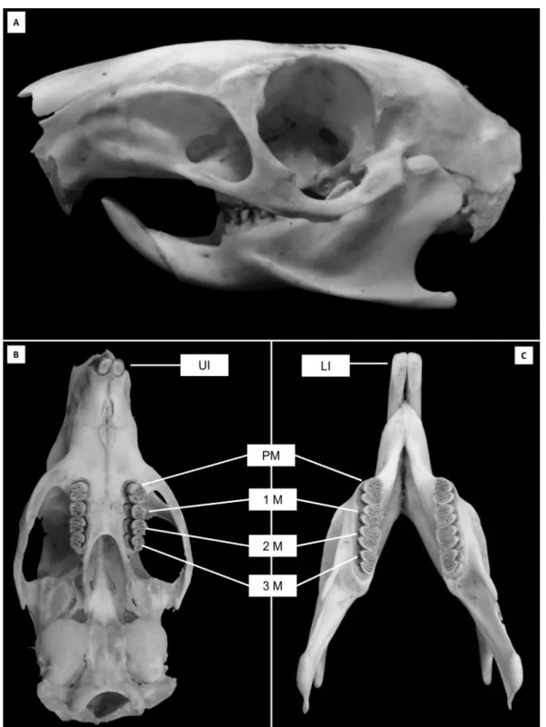
The dental formula of Dasyprocta prymnolopha was as follows: incisors 1/1, canines 0/0, premolars 1/1, molars 3/3. With this structure, the dental formula for an adult animal was obtained by an arc (Fig.1).
While measuring the tooth length, it was observed that the lower jaw with teeth showed greater axial length compared to their counterparts in the upper arch; the upper incisors ran-ged from 30.04-30.46mm, while those presented below were of 53.10-56.68mm, while in other groups, the dental axial length of the remaining teeth ranged from 8.87-11.24mm between the upper and lower in the range of 9.80-12.78mm. The values found for dental elements are shown in Table 1.
The morphology of crown teeth are shown in Table 2. While analyzing the root portion, it was observed that the upper posterior teeth had, on average, four roots. Palatal root exhibited a slight curvature buccally. In contrast, the lower posterior teeth showed two to four roots, where the roots vestibular had a curvature directed towards the inner portion of the mandibular arch towards the lingual area (Table 3 and 4).
LM showed a pattern of tubular dentines with the tu-bules winding, traversing the entire thickness of the dentin from the pulp cavity to the enamel. The enamel was shown as a compacted surface layer covering the dentin along its entire length, except the incisal edge of incisors and some occlusal regions of the premolars and molars (Fig.2).
The low magnification photomicrographs obtained by SEM showed a prismatic enamel on the tooth surface (Fig.3A,B), and a tubular orthodentine underlying layer of enamel (Fig.3C,D).
The analysis at higher magnification showed a characte -ristic dentin pattern classified as orthodentine. This struc -ture can be defined as mineralized cloagenous tissue which circle pulp cavity and presents certain morphologic varia-tions on the direction and dentin tubules diameters. These which are coated with a peritubular dentin and immersed in a layer of intertubular dentin (Miles 1967, Baume 1980). This characteristic pattern of dentin is found in all mam-mals and some reptiles, amphibians and fishes (Miles 1967, Baume 1980) (Fig.3C,D).
The enamel was shown to be organized into individu-al prismatic rods with an interprismatic layer between the deep portion adjacent to the dentino-enamel junction and the surface layer (Fig.3E,F). The prisms in turn, were clearly individualized by interprismatic enamel, arranged irregularly, and assumed a sinuous waved form (Fig.3E,F).
The occlusal surface of the analyzed elements, dentin were exposed in some regions (Fig.3A). Furthermore, we observed the presence of enamel amid the exposed dentin surrounding deep grooves, suggesting that this finding is not attributable to normal morphology of the tooth, but the consequent physiological wears their eating habits (Fig.1).
DISCUSSION
The description of the morphology of the teeth on a micros-copic scale of Dasyprocta prymnolopha supports the adap-tation of this animal for eating hard foods. Although this is a common feature among members of the order Rodentia, agoutis have specific habits and behaviors that enable them to use hard fruits with higher efficiency than other rodents.
Several studies have been done on D. prymnolopha (Lo-pez et al. 2004, Braz et al. 2006, Bonvicino & Oliveira 2006). However, few data exist regarding the stomatognatic sys-tem of rodents. Therefore, this study sought to describe the anatomical and histological aspects of their teeth.
Agouti (D. prymnolopha), paca (Agouti paca) and mocó (Kerodon rock) present a pair of incisors in general oran-ge color (color is related to the habit of chewing in these animals), no canines, a pair of premolars, and six molars in each jaw (Eisenberg & Redford 1999, Thomaz et al. 2006). Furthermore, the lower incisors’ elements are more pro-nounced in length, as in paca (Oliveira et al. 2006).
It was observed that the lower arch of teeth showed a greater axial length compared with their counterparts in the upper arch, contrasting data obtained by Oliveira et al. (2006) in their study of paca (Agouti paca). Between the incisors and the pre-molar, a space is found, which is des-cribed by Pough et al. (2003) as a diastema.
The incisors had an open apex, a characteristic morpho-logical finding of teeth in the process of root formation or continued growth, as found in other rodent incisors (Pou-gh, Janis and Heiser, 2003; Oliveira et al., 2006). In the oc-clusal surfaces of the elements, molariforms of Agouti was observed (the same as that found in pacas); the presence of depressions were circumscribed by a layer of enamel, pro-viding the chewing surface a rather uneven look like ele-ments molariformes equine (St Clair 1986, Dyce et al. 1997, Konig et al. 2004).
The organization of dental tissues observed by SEM and LM showed many similarities compared to other mammals previously described in the literature (Forssell-Ahlberg et al. 1975, Kroon et al. 1986, Pough et al. 2003, Oliveira et al. 2006, Santana et al. 2013).
Fig.1. Macrographs skull and jaws of agouti (Dasyprocta prymnolopha). (A) Side view of the skull. (B) Occlusal view of the upper dental arch, (C) Occlusal view of the lower arch. Note the wear of dental structures in their occlusal portion (premolars and molars) and incisal (incisors). Upper incisors (UI), Pre-molars (PM), first molar (1M), second molar (2M), third molar (3M), Lower incisors (LI).
imbrication are more adapted to act as masticatory dental elements, which features a small enamel prismless layer temporary on the tooth surface, or even mice, which have a thin layer of enamel surface (Bishop 1995, Koussoulakou 2009, Ohazama et al. 2010).
The underlying dentin enamel tubules showed a pattern in which the tubules are interleaved by a more compacted dentin, called intertubular dentin, and this pattern is found in other mammals, even in humans (Forssell-Ahlberg et al.
Table 1. Average length of teeth (mm): Mean and standard deviation (in the bracket) of the length of the teeth of the
species Dasyprocta prymnolopha
Teeth Upper Jaw Lower Jaw
1975, Brännström & Garberoglio 1972, Muylle et al. 2001). This tubular structure, filled with fluid and collagenous materials found in several species reported in the literatu-re, confer resilience and cushioning to the impacts applied on the enamel (Forssell-Ahlberg et al. 1975, Brännström & Garberoglio 1972, Muylle et al. 2001). In this research, our methodology did not allow SEM observation to look inside these tubules, but the findings suggest the same internal or -ganization as found in other animals (Forssell-Ahlberg et al. 1975, Brännström & Garberoglio 1972, Muylle et al. 2001).
The tubular pattern found with individual tubules is characteristic of a pattern of dentin termed orthodentine found in the dentin of some mammals, as depicted through Santana et al. (2013). Unlike other types of dentin such as vasodentina, this dentin has some morphological variations with respect to the direction of the dentinal tubules (some-times it can present itself very much branched or deformed in some species), the most common among mammals.
The findings from LM and SEM revealed that the tubules showed a direction between the root canal pulp cavity to the surface with enamel cementum, which assumes a
per-Fig.2. Light microscopy of the enamel and dentin of the agouti (Dasyprocta prymnolopha). (A) Signal-ing in enamel (white asterisk) and dentin (black asterisk). (B) Black asterisk indicates enamel and white asterisk indicates dentin. Arrow indicates the bottom of a sulcus occlusal.
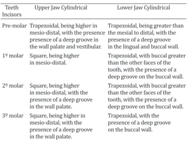
Table 2. Morphology of the dental crown Dasyprocta prymnolopha
Teeth Upper Jaw Cylindrical Lower Jaw Cylindrical Incisors
Pre-molar Trapezoidal, being higher in Trapezoidal, being greater than mesio-distal, with the presence the mesial to distal, with the presence of a deep groove in presence of a deep groove the wall palate and vestibular. in the lingual and buccal wall. 1º molar Square, being higher Trapezoidal, with buccal greater in mesio-distal. than the other faces of the
tooth, with the presence of a
deep groove on the buccal wall.
2º molar Square, being higher Trapezoidal, with buccal greater in mesio-distal, with the than the other faces of the presence of a deep groove tooth, with the presence of a in the wall palate. deep groove on the buccal wall. 3º molar Square, being higher in Trapezoidal, with the mesio-distal, with the presence of a deep groove presence of a deep groove on the buccal wall. in the wall palate.
Table 4. Lower Jaw: Morphology of the lower jaw dental root
Dasyprocta prymnolopha
Teeth Number Shape Direction of root Incisors of roots
One Taper and bulky Bend lingually Pre-molar Three Distal root bulky and Root mesiolingual long. Root mesial inclined lingually. buccal and mesial buccal roots and disposed lingual bulky and short along the distal
axis of the tooth.
1º molar Four Mesiobuccal root bulkier Root it with buccal than mesiolingual. buccal inclination. Lingual root of this The others are bigger and bulkier arranged along the
than the other tooth axis
2º molar Four Distal root bulky and Mesial root curvature long. Root mesial buccal for lingual and and mesial lingual distal root straight
bulky and short
3º molar Two Mesial root tapered distal Mesial root curvature bulky. Root bit long for lingual and
and has a flattening distal root straight
in the mesio-distal
Table 3. Upper Jaw: Morphology of the upper jaw dental root
Dasyprocta prymnolopha
Teeth Number Shape Direction of root Incisors of roots
One Taper and bulky Bend to the palate Pre-molar Three Root is distolingual, thin. Root Distobuccal directed to is distobuccal, long and curved palatal, and mesial to the lingual. Mesial root is distolingual arranged bulky for the buccal groove. along the axis of the
tooth.
1º molar Three Root is long and voluminous Palatal root curvature to palate, palatal root mesio- the vestibular root buccal and palatal root of this palatina distopalatina smaller volume and size and arranged along
the axis of the tooth.
2º molar Three Root long lingual and buccal Palatal root curvature to curvature. Mesiopalatina the vestibular root root and have similar palatina distopalatina lengths distopalatina and arranged along
the axis of the tooth
3º molar Three Lingual root bulky (large) and Lingual root curvature long with buckle to school; buccally; mesial buccal mesial buccal root curvature root straight and with conical short buccal and distobuccal root with lingual root of this cone with a slight curvature in diameter smaller than the buccal
Fig.3. Scanning electron microscopy of enamel and dentin of the agouti (Dasyprocta prymnolopha). (A) Occlusal view of a molar with enamel surface (black asterisk) bypassing the dentin (white asterisk). Arrow heads indicate grooves occlusal enamel coated. (B) Sample worn along a molar, where you can see the layout of the enamel (black asterisk), dentin (white asterisk) and blind grooves (arrow heads). (C,D) Black asterisk indicates enamel, white asterisk indicates dentin, arrow indicates dentino-enamel junction, ar-row heads indicate dentinal tubules and crucifix in intertubular dentin. (E,F) Arrows indicate arrangement of enamel prisms.
meable fabric and an important means of communication with the pulp tissue as described in humans and other ani-mals (Brännström & Garberoglio 1972, Vongsavan & Mat-thews 1991, Muylle et al. 2001, Robb et al. 2007).
Thus, the dental morphology found in Dasyprocta
REFERENCES
Baume L.J. 1980. The biology of pulp and dentine (Monogr.). Oral Sci., Ba-sel, 8:1-183.
Bishop M.A. 1995. Is rabbit dentine innervated? A fine-structural study of
the pulpal innervation in the cheek teeth of the rabbit. J. Anat. 186:365-372.
Brannstrom M. & Garberoglio R. 1972. The dentinal tubules and the odon-toblast process. Acta. Odontol. Scand. 30:291-311.
Braz D.C., Pinheiro A.M.V., Moura W.L. & Carvalho M.A. 2006. Descrição his-tológica dos incisivos da cutia Dasyprocta prymnolopha (Wagler, 1831). Ciênc. Anim. Bras. 7:177-185.
Dyce K.M., Sack W.O. & Wensing C.J.G. 1997. Tratado de Anatomia Veteri-nária. Guanabara Koogan, Rio de Janeiro, 872p.
Eisenberg J.F. & Redford K.H. 1999.Mammals of the Neotropics - The Cen-tral Neotropics: Ecuador, Peru, Bolivia, Brazil.Univ. Chicago Press, Chi-cago. 624.
Eurell J.A. & Frappier B.L. 2006. Dellmann’s Textbook of Veterinary Histo- logy. 6th ed. Blackwell Publishing, Ames, IA. 416p.
Forssell-Ahlberg K., Brannstrom M. & Edwall L. 1975. The Diameter and number of dentinal tubules in rat, cat, dog and monkey: a comparative scanning electron microscopic study. Acta. Odontol. Scand. 33:243-250.
Freire M.A.M., Tourinhoc S.C., Guimarães J.S., Oliveira J.L.F., Picanço-Diniz C.W., Gomes-Leal W. & Pereira Jr A. 2008. Histochemical characteriza-tion, distribution and morphometric analysis of NADPH diaphorase neurons in the spinal cord of the agouti. Front. Neuroanat. 2:2-9. Henry O. 1999. Frugivory and the importance of seeds in the diet of the
orange-rumped agouti (Dasyprocta leporina) in French Guiana. J. Trop. Ecol. 15:291-300.
Hosken F.M. & Silveira A.C. 2001. Criação de Cutias. Aprenda Fácil Editora, Viçosa, MG.
König H.E., Sautet J. & Liebich H.G. 2004: Anatomia dos Animais Domésti-cos. 4ª ed. Artmed, Porto Alegre. 787p.
Koussoulakou D.S., Margarits L.H. & Koussoulakos I. 2009. A Curriculum vitae of teeth: evolution, generation, regeneration. Int. J. Biol. Sci. 5:226-243.
Kroon J., Grossman E.S. & Cleaton-Jones P. 1986. A scanning electron mi-croscope study of dentinal tubule continuity in monkey tooth sections. Endod. Dent. Traumatol. 2:39-42.
Lange R.R., Abilhôa V., Margarido T.C.C. & Monteiro-Filho E.L.A. 2003. Relação entre peso e comprimento total em ninhadas de Dasyprocta azarae Lichtenstein, 1823 em cativeiro. Arq. Ciênc. Vet. Zool. Unipar 6:101-104.
Lee T.E., Hartline-Jr H.B. & Barnes B.M. 2006. Mammalian species ( Dasy-procta ruatanica). American Society of Mammalogists 800:1-3. Lopes J.B., Cavalcante R.R., Almeida M.M., Carvalho M.A.M., Moura S.G., Fi-
lho L.A.D. & Conceição W.L.F. 2004. Desempenho de cutias (Dasyprocta
prymnolopha) criadas em cativeiro do nascimento até o desmame em Teresina, Piauí. Revta Bras. Zootec. 6:2318-2322.
Mendes-Oliveira A.C., Santos P.G.P., Carvalho Júnior O., Lima R.S., Montag L.F.A., Maria S.L. & Rossi R.V. 2012. Edge effects and the impact of
wild-fires on populations of small non-volant mammals in the forest-savanna
transition zone in Southern Amazonia. Biota Neotrop. 12:12-29. Miles A.E.W. 1967. Structural and Chemical Organization of Teeth.
Aca-demic Press, New York.
Muylle S., Simoens P. & Lauwers H. 2001. The distribution of intratubular dentine in equine incisors: a scanning electron microscopic study. Equi-ne Vet. J. 33:65-69.
Oliveira F.S., Canola J.C., Oliveira P.T., Pecora J.D. & Capelli A. 2006. Anato-moradiographic description of the teeth of pacas bred in captivity (Agouti paca Linnaeus, 1766). Anat. Histol. Embryol. 35:316-318. Oliveira F.S., Canola J.C., Oliveira P.T., Pecora J.D. & Capelli A. 2007.
Micro-scopic characterization of teeth of pacas bred in captivity (Agouti paca, Linnaeus, 1766). Anat. Histol. Embryol. 36:371–374.
Oliveira J.A. & Bonvicino C.R. 2006. Ordem Rodentia, p. 351-411. In: Reis N.R., Peracchi A.L., Pedro W.A. & Lima I.P. (Eds), Mamíferos do Brasil. Edifurb, Londrina.
Ohazama A., Blackburn J., Porntaveetus T., Otab M.S., Choic H.Y. & Johnson E.B. 2010. A role for suppressed incisor cuspal morphogenesis in the evolution of mammalian heterodont dentition. Proc. Natl Acad. Sci. USA 107:92-97.
Pough F.H., Janis C.M. & Heiser J.B. 2003. A Vida dos Vertebrados. 4ª ed. Atheneu Editora, São Paulo. 750p.
Robb L., Marx J., Steenkamp G., Van Heerden W.F., Pretorius E. & Boy S.C. 2007. Scanning electron microscopic study of the dentinal tubules in dog canine teeth. J. Vet. Dent. 24:86-89.
Santana L.N.S., Barbosa L.V.M., Teixeira F.B., Costa A.M.P., Fernandes L.M.P. & Lima R.R. 2013. Morphology of the Dentin Structure of Sloths Bradypus tridactylus: a light and scanning electron microscopy investigation. Anat. Histol. Embryol. 14. Doi: 10.1111/ahe.12029. [Epub ahead of print] St Clair L.E. 1986. Anatomia dos Animais Domésticos. Guanabara Koogan,
Rio de Janeiro. 324p.
Thomaz J.M., Carvalho A., Miglino M.A., Maçanares C.A.F., Ambrósio C.E. & Oliveira M.F. 2006. Teeth morphologic characterization of rock ca-vyKerodon rupestris: Mammalia: Rodentia. Braz. J. Vet. Res. Anim. Sci. 43:702-707.
Tirira D.S. 1999. Mamíferos del Ecuador. Museo de Zoologia, Centro de
Biodiversidade y Ambiente, Ponitificia Universidade Católica del Equa -dor, Sociedad para la Investigación y Monitoreo de la Biodiversidae, Quito. 245p.
Vongsavan N. & Matthews B. 1991. The permeability ofcatdentine in vivo and in vitro. Arch. Oral Biol. 36:641-646.