Marcia Regina Ramalho da Silva Bardauil(a)
Cacio de Moura Netto(b)
Abílio Albuquerque Maranhão de Moura(b)
(a) PhD, Department of Endodontics, Dental School, University Paulista, São Paulo, SP, Brazil.
(b) PhD, Department of Endodontics, Dental School, University of São Paulo, São Paulo, SP, Brazil.
Corresponding author:
Marcia Regina Ramalho da Silva Bardauil Universidade Paulista
Rua Helena, 218 - cj 212 SãoPaulo - SP - Brazil CEP: 04552-050
E-mail: [email protected]
Received for publication on Jan 11, 2010 Accepted for publication on May 31, 2010
Evaluation of the maxillary premolar
roots dissociation using radiographic
holders with conventional and digital
radiography
Abstract: This in vivo study evaluated the dissociation quality of maxil-lary premolar roots combining variations of vertical and horizontal an-gulations by using X-ray holders (Rinn - XCP), and made a comparison between two types of intraoral radiography systems - conventional ilm (Kodak Insight, Rochester, USA) and digital radiography (Kodak RVG 6100, Kodak, Rochester, USA). The study sample was comprised of 20 patients with a total of 20 maxillary premolars that were radiographed, using the paralleling angle technique (GP), with a 20° variation of the horizontal angle (GM) and 25° variation of the horizontal angle com-bined with 15° vertical angle (GMV). Each image was independently analyzed by two experienced examiners. These examiners assigned a score to the diagnostic capability of root dissociation and the measure-ment of the distance between the apexes. Statistical data was derived using the Wilcoxon Signed Rank test, Friedman and T test. The means of the measured distances between buccal and lingual root apexes were greater for the GMV, which ranged from 2.3 mm to 3.3 mm. A statis-tically signiicant difference was found between GM and GMV when compared to GP with p < 0.01. An established best diagnostic dissocia-tion roots image was found in the GMV. These results support the use of the anterior X-ray holders which offer a better combined deviation (GMV) to dissociate maxillary premolar roots in both radiography sys-tems.
Descriptors: Radiography, dental; Radiography, dental, digital; Endodontics; Radiographic image enhancement.
Introduction
The complexity of the root canal system demands extra care from the endodontist in the morphological analysis when treating a tooth, requir-ing information related to the number and shape of roots and canals, in order to plan and carry out a satisfactory endodontic treatment. Ra-diographic investigations are routinely used as part of the assessment of new endodontic treatment and these radiographic investigations provide tangible beneits to the endodontist when planning a treatment.1,2,3
vari-ety of anatomic conditions throughout the coronal - radicular structures. The parallelism radiography technique has the advantage of a standardized pro-cedure that can be used during the entire endodon-tic treatment.4,5
Although, in certain tooth groups, several dif-ferent cone angulations are necessary in order to be able to overcome the superimposition of roots which relect the shape of the tooth in third dimension. For example, the maxillary premolars, according to lit-erature, may have two roots and two canals (70%6,7
to 98%8) and may nearly always have the
appear-ance of a single root. The roots of maxillary pre-molars are often superimposed in the buccolingual direction, which is the same direction of the X-ray beam.
Previously, in such cases, a variation of 20 de-grees mesially in a horizontal angulation has been suggested in order to dissociate the superimposed roots.9 In many cases, the deviation overcomes the
superimposition, but by contrast, can bring the disadvantage of producing geometric distortions by elongating or foreshortening the apparent root length of the premolars.4
In endodontic clinics, the lowest possible number of radiographies can minimize the amount of radia-tion exposure for both the professional and the pa-tient, following the principle of ALARA (As Low As Reasonably Achievable).1
The intraoral digital radiographic system has acquired great signiicance as a diagnostic tool, be-cause of the improvement of sensor spatial resolu-tion and the reducresolu-tion of exposure time by up to 90% for images similar to those of radiographic ilms.10,11,12,13,14
In relation to endodontics, many studies have analyzed and reported the diagnostic capabilities of digital images,15,16,17,18,19,20 for endodontic
pro-cedures12,13,21 and follow ups of endodontic
treat-ment,17,21 which clearly demonstrates the eficacy
and safety of the digital system.
It is, therefore, important that all intraoral dental exposures are taken with the corresponding hold-ers and the paralleling technique.22 However, some
clinicians have reported their dificulty to properly make deviation, sometimes making additional
radi-ographies.
In this context, the present in vivo study aimed to use radiographic holders to correctly place the ilm or sensor in the oral cavity in order to evaluate the dissociation of the maxillary premolars roots.
Material and Methods
After the approval of an Ethics Committee, twen-ty patients were randomly selected from patients screened for Endodontic treatment at the Clinics of the School of Dentistry, Paulista University, and in-vited to participate in this study. Only patients that had at least one maxillary premolar, left or right, with two roots and without periapical lesion, indi-cated for endodontic treatment were considered for participation in the study. If the patient did not ful-ill these conditions or the patient’s tooth presented anatomic complications, like severe curvatures, api-cal resorptions or differences of root length greater than 5 mm, the candidate would be treated but ex-cluded from the study. All patients were informed about the study, about its risks and beneits, and signed an agreement concerning their participation.
Six images were made of each patient, 3 conven-tional (CR) and 3 digital radiographs (DR) in three periapical radiographic techniques. The three peri-apical radiographic techniques were: Parallelism (GP), Parallelism with a variation of 20° horizon-tal angulation mesially (GM) and Parallelism with a variation of the mesio-vertical angle of 25° and 15°
respectively (GMV). A total of 120 images were ob-tained, divided into 6 groups of 20 images, to rep-resent the three techniques associated with the two systems (CR and DR) (table 1).
All periapical images (CR and DR) were exposed using conventional X-ray equipment, 70 kV, 8 mA (Trophy Radiologie, Vincennes, France) with a fo-cal distance of 30 cm. Kodak Insightilm (Eastman Kodak Co., Rochester, USA) was used with an ex-posure time of 0.32 s and RVG 6100 (Kodak, Roch-ester, USA), sensor #1, with an exposure time of 0.18 s.
the angulations of 15° vertical and 25° horizontal for the GMV. Another guide ring was confectioned and placed 20° to the posterior ring, thereby repro-ducing 20° mesially. The bite block was always po-sitioned on the patients’ irst premolar. In order to standardize and customize X-ray projections, an im-pression of the patient’s bite was taken with heavy body silicone (Zetaplus - ZhermackSpA, Badia Pole-sine RO, Italy) in order to maintain the support in the same position for all 3 techniques.
The CR images were processed with an automat-ic Periomat devautomat-ice (Dürr Dental, Bietigheim-Bissin-gen, Germany) and DR images were stored in TIFF format and each image was identiied (only in ile name) by patient and the corresponding group tech-nique angulation, GP, GM or GMV.
All images were evaluated by two expert exam-iners, a dental radiologist and an endodontist. Ob-servations were individually made by the examiners, in a dark room during two different sessions (irst one for conventional and the other for digital ra-diographs) with 3 weeks between the two intervals, in order to avoid the tendency of remembering the measurement from the irst session and allowing that measurement to inluence the same case dur-ing the second session. CR images were evaluated using a light box (Rinn Co. Abbott Drive Elgin,
IL, USA) and a magniier lens (2 X). Measurements were made with a lexible and calibrated transpar-ent acrylic ruler, graduated in millimeters and half millimeters (Trident Desetec, São Paulo, Brazil). DR images were analyzed on a monitor screen using a digital ruler tool from the RVG’s software system (RVG 6100, Kodak, Rochester, USA). The examin-ers measured the distance between the center of buc-cal (B) and lingual (L) root apexes of the premolars in all 120 images. The mean value of each image was compared among the three techniques by Analysis of Variance and Tukey’s test. They also chose which technique, in both systems (CR and DR), showed the best diagnostic capability of root dissociation with the best distinguishable apexes with the lowest amount of distortion. The outcome variable in this case was dichotomous, good or bad quality of the radiograph. For these comparisons among the three groups, the Cochrane Q test was run.
Results
In order to validate the calibration made by the two examiners, all data was analyzed with the Kappa test, which showed a good level of agreement among the examiners (Kappa = 0.62).
Table 2 shows the means and standard deviations of the distance between buccal and palatal apexes.
Table 1 - Experimental groups.
Technique Angulation Groups
Conventional (CR) Digital (DR)
Parallelism none GPC GPDR
Parallelism with mesial angulation 20° horizontal mesial GMC GMDR
Parallelism with mesial and vertical angulation 25° horizontal mesial / 15° vertical GMVC GMVDR
Table 2 - Mean of the distance (mm) between apexes and standard deviations.
Technique Groups Comparison
CR X DR Conventional (CR) Digital (DR)
Parallelism (GP) 1.72 ± 1.00a 1.66 ± 0.51* n.s.
Parallelism with mesial angulation (GM) 2.67 ± 0.86b 2.26 ± 0.69† n.s.
Parallelism with mesial and vertical angulation (GMV) 2.92 ± 0.85b 2.47 ± 0.66† n.s.
Comparison between techniques p < 0.01 p < 0.01
The measurements of root apexes distance showed a statistically signiicant difference between GP and the other groups, regardless of the X-ray system ac-cording to one-way ANOVA test (p < 0.01). Such difference, in comparison to the parallelism tech-nique, certiies that variation of angulation can have an inluence on the identiication of apexes. When comparing pairs of systems (CR and DR), no sig-niicant difference was found. The GMV groups showed the greatest distances between apexes in both radiographic systems (CR and DR).
Finally, the three techniques were compared in terms of quality (good or bad) using the Cochrane Q test for matched groups (Table 3). The Parallel-ism technique with 25° mesial and 15° vertical an-gulations (GMV) was the most chosen method for quality root apexes dissociation (80% in DR and 85% in CR). An increase in mesial and vertical angles improved the identiication of 2 root apexes and caused less distortion of apical area, making the apex limits easy to identify. The Cochrane Q test showed signiicant statistical differences when comparing GMV with GP and GM groups, in both X-ray systems (p < 0.05).
Discussion
The Parallelism Technique22 is commonly used
for the diagnosis radiographs, because the images are sharp and of suficient quality to plan an end-odontic treatment. Since radiographs are two dimen-sional images of 3 forms, some images are usually superimposed making it dificult to distinguish the number of roots that may be found in multirooted teeth. For this reason, different radiographic tech-niques, specially varying horizontal angles9,23 are
used to maximize the information about the studied
region. This is the best way to minimize diagnostic shortcomings, during transoperative procedures and also in the post-treatment analysis.
While Walton9 concluded that a variation of 20°
for the horizontal angle mesially to the premolar al-lows the dissociation of buccal and lingual roots, Martinez-Lozano et al.23 found that 40° yielded
the best results. However, the present study found that an alteration in horizontal angle of 25° mesi-ally may offer a better identiication of the roots and apexes without twisted or blurred image and with-out superimposition of other anatomic structures, like the maxillary sinus or canine root, which could occur with 40°. Contrary to Martinez-Lozano et al.23 who found that vertical inclination of the
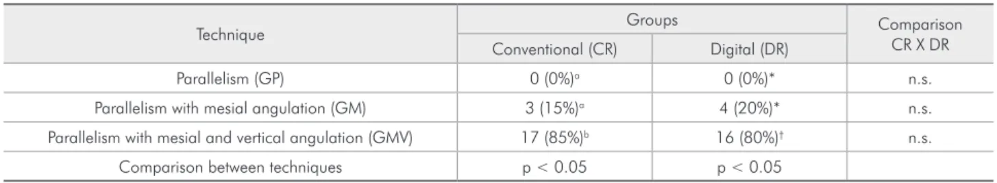
X-ray tube did not affect the identiication of roots, this study found that a variation of 15° vertically, with the central beam directed towards the canine, maximized the dissociation of the root apexes in a vertical plane. Therefore, using the anterior tooth ilm holder instead of the posterior one, the apexes of maxillary premolars can be easily identiied (Fig-ures 1 and 2).
Table 2 shows that the mean distances using the GMV were 2.92 mm and 2.47 mm for CR and DR, larger distances than those for GM and GP. Thus, in both systems, the GMV was the most reliably utilized for the dissociation of roots. This table also indicated that CR produced higher values than the DR in all of the techniques observed. This differ-ence may be explained by the fact that DR readings are made with a more precise digital ruler when compared with CR readings which are measured manually.
Since some authors17,19,24,25,26,27 do not consider
digital image manipulation as a possible strategy,
Table 3 - Dissociation quality according to the examiner’s choice (number of events and percentage).
Technique Groups Comparison
CR X DR Conventional (CR) Digital (DR)
Parallelism (GP) 0 (0%)a 0 (0%)* n.s.
Parallelism with mesial angulation (GM) 3 (15%)a 4 (20%)* n.s.
Parallelism with mesial and vertical angulation (GMV) 17 (85%)b 16 (80%)† n.s.
Comparison between techniques p < 0.05 p < 0.05
Figure 1 - Analysis of conventional radiographs using different techniques: (A) Parallelism; (B) Mesially 20°; (C) Mesially 25° and vertical 15°.
A B C
B A
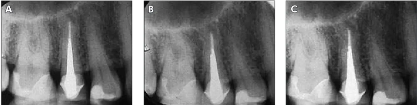
C Figure 2 - Digital images measurements with RVG 6100
software system: (A) Parallelism; (B) Mesially 20°; (C) Mesially 25°and vertical 15°.
accepting only the enlargement associated to the negative – positive image inversion that allows for a better visualization of anatomic structures,27 these
strategies manipulations were not used in this study. Further studies are expected to provide more infor-mation on the use of digital manipulation of imag-es.
Results showed that the proposed technique,
systems, there were no statistically signiicant differ-ences between CR and DR system.
Conclusions
According to the results, we can conclude that
the use of anterior teeth ilm holder improved dis-sociation of buccal and lingual root apexes in both systems, CR and DR, producing sharper and clearer images to better plan the endodontic treatment.
References
1. Langland OE, Langlais RP. Early pioneers of oral and max-illofacial radiology. Oral Surg Oral Med Oral Pathol Oral Radiol Endod. 1995 May;80(5):496-511.
2. Wu DM, Wu YN, Guo W, Sameer S. Accuracy of direct digital radiography in the study of the root canal type. Dentomaxil-lofac Radiol. 2006 Apr;35(4):263-5.
3. Omer OE, Al Shalabi RM, Jennings M, Glennon J, Claffey NM. A comparison between clearing and radiographic tech-niques in the study of root-canal anatomy of maxillary first and second molars. Int Endod J. 2004 May;37(5):291-6. 4. Kaffe I, Gratt BM. Variations in the radiographic interpretation
of the periapical dental region. J Endod. 1988 Jul;14(7):330-5.
5. Gelfand M, Sunderman EJ, Goldman M. Reliability of radio-graphical interpretations. J Endod. 1983 Feb;9(2):71-75. 6. Vertucci JF, Gegauff A. Root canal morphology of the first
premolar. J Am Dent Assoc. 1979 Aug;99(2):194-8. 7. Kartal N, Özçelik B, Cimilli H. Root canal morphology of
maxillary premolars. J Endod. 1998 Jun;24(6):417-9. 8. Green D. Double canals in single roots. Oral Surg Oral Med
Oral Pathol. 1973 May;35(5):689-96.
9. Walton RE. Endodontic radiographic technics. Dent Radiogr Photogr. 1973 Mar;46(3): 51-9.
10. Ferreira RI, Haiter-Neto F, Tabchoury CPM, Paiva GAN, Bóscolo FN. Assessment of enamel demineralization using conventional, digital, and digitized radiography. Braz Oral Res. 2006 Apr-Jun;20(2):114-9.
11. Mouyen F, Benz C, Sonnabend E, Lodter JP. Presentation and physical evaluation of RadioVisioGraphy. Oral Surg Oral Med Oral Pathol. 1989 Aug 68(2):238-42.
12. Burger CL, Mork TO, Hutter JW, Nocoll B. Direct digital radiography versus conventional radiography for estimation of canal length in curved canals. J Endod. 1999 Apr;25(4):260-3.
13. Vandre RH, Pajak JC, Abdel-Nabi H, Farman TT, Farman AG. Comparison of observer performance in determining the position of endodontics files with physical measures in the evaluation of dental X-ray imaging systems. Dentomaxillofac Radiol. 2000 July;29(4):216-22.
14. Wenzel A, Grondahl HG. Direct digital radiography in the dental office. Int Dent J. 1995 Feb;45(1): 27–34.
15. Gundappa M, Ng SY, Whaites EJ. Comparison of ultrasound, digital and conventional radiography in differentiating
peri-apical area. Dentomaxillofac Radiol. 2006 Sep;35(5):326-33.
16. Folk RB, Thorpe JR, Mc Clanahan SB, Johnson JD, Strother JM. Comparison of two different direct digital radiography systems for the ability to detect artificially prepared periapical lesions. J Endod. 2005 Apr;31(4):304-6.
17. Versteeg KH, Sanderink GCH, Van Ginkel FC, Van Der Stelt PF. Estimating distances on direct digital images and conven-tional radiographs. J Am Dent Assoc. 1997 Apr;128(4):439– 43.
18. Westphalen VP, Gomes de Moraes I, Westphalen FH. Efficacy of conventional and digital radiographic imaging methods for diagnosis of simulated external root resorption. J Appl Oral Sci. 2004 Apr-Jun;12(2):108-12.
19. Sullivan JR, Di Fiore PM, Koerber A. Radiovisiography in detection of periapical lesions. J Endod. 2000 Jan;26(1):32-5.
20. Constante IGT, DavidowiczH, Barletta FB, Moura AAM. Study of the areas and thicknesses of mesiobucal root canals prepared by three endodontic techniques. Braz Oral Res. 2007 Apr-Jun;21(2):118-26.
21. Fuhrmann AW. Current practice in conventional and digital intraoral radiography: Problems and solutions. Int J Comput Dent. 2006 Jan;9:(1)61-8.
22. Fitzgerald GM. Dental roentgenography I: an investigation in adumbration or the factors that control geometric unsharp-ness. J Am Dent Assoc. 1947 Jan;34(1):1-20.
23. Martinez-Lozano MA, Forner-Navarro L, Sánchez-Cortés JL. Analysis of radiologic factors in determinig premolar root canal systems. Oral Surg Oral Med Oral Pathol Oral Radiol Endod. 1999 Jun;88(6):719-22.
24. Welander U, Yoshiura K, Li G, Sallstrom P, McDavid WD. Correction for attenuation and visual response in digital radi-ography. Dentomaxillofac Radiol. 2002 Mar;31(2):117-25. 25. Naoum HJ, Chandler NP, Love RM. Conventional versus
storage phosphor-plate digital images to visualize the root canal system contrasted with a radiopaque medium. J Endod 2003 May; 29(5):349–52.
26. Farman AG, Farman TT. A comparison of 18 different x-ray detectors currently used in dentistry. Oral Surg Oral Med Oral Pathol Oral Radiol Endod. 2005 Apr;99(4):485–9.