Abstract
Submitted: April 27, 2016 0RGL¿FDWLRQ-XO\ Accepted: July 19, 2016
canal shaping capabilities of the
ProTaper Universal and ProTaper Gold
rotary instrument systems
Conventional NiTi (ProTaper Universal PTU) and Controlled Memory NiTi (ProTaper
Gold PTG) instrument systems on the quality of root canal preparation. Material and Methods: Twelve mandibular molars with separate mesial canals were
and PTG instruments were used to shape twelve mesial canals each. The canals
were scanned after preparation with F2 and F3 instruments of the PTU and PTG
the apical and cervical levels, root canal volume and untouched canal walls. Data
comparison of data between groups, the Mann-Whitney test was used. Results:
between the groups in terms of the area and volume of root canals (P>.05).
respect to root canal volume after use of the F2 and F3 instruments. There was
before and after instrumentation for both systems. At the 3 cervical levels, the
PTG maintained centralization of the preparation on the transition between
the F2 and F3 instruments, which did not occur with the PTU. Conclusion The Conventional NiTi (PTU) and Controlled Memory NiTi (PTG) instruments
displayed comparable capabilities for shaping the straight mesial root canals of
mandibular molars, although the PTG was better than the PTU at maintaining
the centralization of the shape in the cervical portion.
Ke y w or ds:
Root canal preparation. Jussaro Alves DUQUE1
Rodrigo Ricci VIVAN1
Bruno Cavalini CAVENAGO2
Pablo Andrés AMOROSO-SILVA1
Ricardo Affonso BERNARDES3
Bruno Carvalho de VASCONCELOS4
Marco Antonio Hungaro DUARTE1
1Universidade de São Paulo, Faculdade de Odontologia de Bauru, Departamento de
Dentística, Endodontia e Materiais Odontológicos, Bauru, SP, Brasil.
2Universidade Federal do Paraná, Departamento de Odontologia, Curitiba, PR, Brasil. 3Associação Brasileira de Odontologia, Departamento de Endodontia, Taguatinga, DF,
Brasil.
4Universidade Federal do Ceará, Departamento de Odontologia, Fortaleza, CE, Brasil.
Introduction
Mechanical preparation of the root canal is an
important step in endodontic treatment. The aim of this step is to remove vital or necrotic pulp tissue while
simultaneously increasing the root canal volume to
facilitate the decontamination of the root canal system
by irrigants and medicaments14.
Several types of instruments and techniques for
root canal preparations have been described6,11,16-18.
deviations in the root canal preparation and excessive
have been widely used in endodontic practice due
compared to hand-held instruments24
and provide higher quality in the preparation of root
5 and higher fracture
resistance to cyclic and torsional fatigue25.
in fabrication, including different types of thermic treatments that have been employed during the
manufacturing process, which are designed to optimize
physical characteristics that favor greater elasticity as
20. Among
highlighted in the present study.
According to the manufacturers, CM wire
weight) than other NiTi instruments10,22. Additionally,
because of the thermic treatments used during the
manufacturing process, CM wire instruments do not bounce (rebound) to their original shape after being
instrument fracture, and perforations15.
Among the NiTi systems, the PTU (Dentsply
Maillefer, Ballaigues, Switzerland) is a rotary system of conventional NiTi wire that has been widely used and
studied1,6,11,24. It has a variable taper along the length
of the instrument, a convex triangular cross-section,
and a sharp tip7,13. Another system, the PTG (Dentsply
Maillefer, Ballaigues, Switzerland), was recently
are generally similar to those of the PTU, except that
the PTG uses CM wire, while the PTU uses conventional
NiTi wire. Because of this distinction, the PTG exhibits
than the PTU system11,13.
The aim of the present study was to evaluate the
on the quality of root canal preparation, as assessed by microcomputed tomography (micro CT). The null
hypothesis evaluated was that the type of NiTi wire
Material and methods
Selection of the teeth
One hundred extracted human mandibular molars with complete apexes were selected. The teeth were
Twelve teeth were selected based on the following
type IV23, with distinct main foramens, and maximum
curvature of 5°19.
Micro CT scanning procedures
The teeth were scanned as follows: 19 μm voxel
μ
using a 1024x1304 resolution. The obtained images
were reconstructed with dedicated software (NRecon
in BMP format. The images were used to observe the
Root canal preparation
A single operator performed all procedures. The
access cavities were conventionally performed,
and the root canal lengths were determined by the
Switzerland) until visualization in the apical foramen
with a stereomicroscope (Carl Zeiss Micro-imaging,
established as being 1 mm shorter than the real root
canal length (RL). The #10 and #15 K-Files (Dentsply
Maillefer, Ballaigues, Switzerland) were used in the WL
before instrumentation with the rotary systems. Next, 6 mesio-buccal and 6 mesio-lingual canals in different
teeth (n=12) were instrumented using the PTU system
as follows: S1 (18/.02) and SX (19/.035) instruments
were used in the WL, according to the manufacturer’s
instructions.
The remaining root canals (n=12) were instrumented using the PTG system in the same
sequence as the one used for the PTU group.
Subsequently, the roots were scanned again as in
the initial evaluation. After scanning, the root canals were prepared with the F3 (30/.09) instrument of
each system. The X-Smart Plus motor (Dentsply
Maillefer, Ballaigues, Switzerland) was used with a
accordance with the manufacturer’s instructions, and
the instrumentation was performed with in-and-out
procedure, a 2.5% sodium hypochlorite solution was
used; the root canals were irrigated before, during,
and after instrumentation. Two milliliters of solution
irrigation was made with 3 mL of 17% EDTA for 3
minutes, and then with 3 mL of saline solution. Next, a
new scan of the roots was performed using previously
set parameters.
Micro-CT measurements and evaluations
used to measure the canal volumes as well as the
the cervical and apical portions. For pre- and
post-operative measurements, images of 3 different slices
was 1 mm below the level of bifurcation, and the other 2 slices followed apically at intervals of 1 mm.
mm from the apical foramen and the other 2 slices
followed cervically at 1 mm intervals. The minimal
root canals was measured in millimeters. The CTAn
v.1.12 software was used to measure the canal volume
between the 1 mm apical level and at 1mm below the root furcation.
For analysis of the un-instrumented area, 3D
models were created with color codes, and the pre- and
post-operative images were recorded using automatic image registration. Prepared areas and stripes of
the canals were compared using the CTVol v. 2.2.1
of the canal was determined based on the number
position on the surface of the root canal before and
after instrumentation11. The data was then converted
and presented as: (a) percentage (%) of dentinal
removal, (b) un-instrumented area, and (c) relative
volume increase between groups.
of the non-parametric data. Results of the volume
areas were compared using the Friedmann and Dunn’s
tests for the intra-group analyses. For analysis of the percentage of dentinal wear, un-instrumented areas,
and volume increase between the groups, the
Mann-established at 5% (P<0.05).
Results
In the pre-operative analysis, there were no
volume of root canals between the PTU and PTG
groups (P>0.05), indicating adequate pairing of the
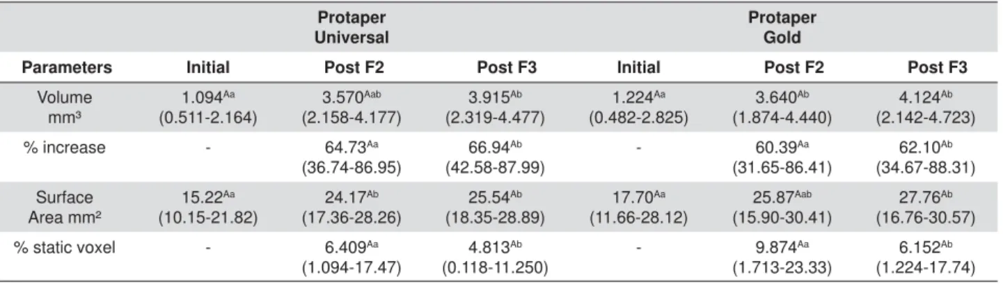
root canals. Table 1 shows the median, minimum, and maximum values of the root canal volume (mm³) and
Protaper Universal
Protaper Gold
Parameters Initial Post F2 Post F3 Initial Post F2 Post F3
Volume mm³
1.094Aa
(0.511-2.164)
3.570Aab
(2.158-4.177)
3.915Ab
(2.319-4.477)
1.224Aa
(0.482-2.825)
3.640Ab
(1.874-4.440)
4.124Ab
(2.142-4.723)
% increase - 64.73Aa
(36.74-86.95)
66.94Ab
(42.58-87.99)
- 60.39Aa
(31.65-86.41)
62.10Ab
(34.67-88.31)
Surface Area mm²
15.22Aa
(10.15-21.82)
24.17Ab
(17.36-28.26)
25.54Ab
(18.35-28.89)
17.70Aa
(11.66-28.12)
25.87Aab
(15.90-30.41)
27.76Ab
(16.76-30.57)
% static voxel - 6.409Aa
(1.094-17.47)
4.813Ab
(0.118-11.250)
- 9.874Aa
(1.713-23.33)
6.152Ab
(1.224-17.74)
'LIIHUHQWXSSHUFDVHOHWWHUVFRUUHVSRQGWRGLIIHUHQWYDOXHVZLWKVWDWLVWLFDOGLIIHUHQFHVEHWZHHQJURXSVDQGWKHVDPHLQVWUXPHQW3 'LIIHUHQWORZHUFDVHOHWWHUVFRUUHVSRQGWRYDOXHVZLWKLQWUDJURXSVWDWLVWLFDOGLIIHUHQFHV3
surface area (mm²) before and after use of the F2
and F3 instruments. Also presented in Table 1 are the
percentage of volume increase and the percentage of static voxel after use of the F2 and F3 instruments.
between the PTU and PTG systems with respect to
the root canal volumes obtained after use of the F2 and F3 instruments (P>0.05). However, upon
intra-group analysis of the PTU group, a statistically
significant difference was observed between the
pre-operative and post-operative volumes, but only after use of the F3 instrument (P<0.05). For the PTG
between the volumes before and after use of the F2
and F3 instruments (P<0.05). Figure 1 depicts the three-dimensional reconstructions of mesial root
Figure 1- Three-dimensional reconstructions of mesial root canals of a mandibular molar: (A) before instrumentation (green), (B) after LQVWUXPHQWDWLRQZLWKWKH3UR7DSHU*ROG)OLJKWUHGDQGWKH3UR7DSHU8QLYHUVDO)¿OHOLJKWEOXH&6KRZVWKHPHVLDOFDQDOVDIWHU LQVWUXPHQWDWLRQZLWK3UR7DSHU*ROG)GDUNUHGDQGWKH3UR7DSHU8QLYHUVDO)¿OHGDUNEOXH
canals before instrumentation (Figure 1A), and after
instrumentation with F2 (Figure 1B) and F3 (Figure 1C) instruments.
Table 2 shows the median, minimum and maximum
values (presented as percentages) of dentin wear
in the mesial and distal walls (mm) at the 3 apical and 3 cervical levels, before and after use of the
F2 and F3 instruments for the PTU and PTG groups,
respectively. There was no statistical difference
apical level before and after instrumentation for both
systems (P>0.05). At the 3 cervical levels, the PTG
system maintained centralization of the preparation
on transition between the F2 and F3 instruments, in contrast to the PTU system where this did not occur.
For all segments that were evaluated, a dentin
of the instrument and system used (Figure 2).
Discussion
that was targeted for analysis in the present study,
the data supports the null hypothesis because there
PTU and PTG instruments with respect to root canal
enlargement or un-instrumented areas.
After a micro-CT evaluation, the mandibular molars
to be examined were selected based on whether they:
(a) had two canals in the mesial roots, (b) presented
distinct foramens and (c) had no isthmuses related to
23. This experimental
model allowed us to compare the capabilities of two
instrument systems based on the same anatomical
condition18.
Several methodologies such as histologic sections,
serial axial sections, and scanning electron microscopy
have been used to evaluate root canal preparations2,4,8;
however, these methods compromise the integrity of the samples because they require the cutting of
the specimens, which limits specimen use to a single
analysis. By contrast, micro-CT is a nondestructive
method that allows for the progressive evaluation and observation of the preparation instruments with
18. The evaluations conducted in the
present study were made after the use of the F2 and
F3 instruments.
13,14. With
technological advancement, however, new generations
ProTaper Gold Protaper Gold
Level Dentin wall
Post F2 Post F3 P mesial x distal
Level Dentin wall
Post F2 Post F3 P mesial x distal Apical 1 Mesial 4.129
(0-15.020)
0.930 (0-9.332)
Ini/F2 NS
Apical 1 Mesial 6.648
(2.172-12.830)
2.043 (0-8.989)
Ini/F2 NS
Apical Distal 5.481
(0.838-20.570)
2.515 (0-11.090)
F2/F3 NS
Apical Distal 3.448
(0-10.490)
0 (0-12.940)
F2/F3 NS Apical 2 Mesial 8.011
(2.558-27.960)
1.754 (0-12.490)
Ini/F2 NS
Apical 2 Mesial 7.576
(2.243-20.980)
4.441 (0-16.640)
Ini/F2 S
Apical Distal 6.106
(2.775-23.770)
2.563 (0-14.800)
F2/F3 NS
Apical Distal 3.729
(2.727-10.450)
2.107 (0-9.365)
F2/F3 S Apical 3 Mesial 8.662
(3.092-24.960)
3.614 (0-13.880)
Ini/F2 NS
Apical 3 Mesial 8.518
(3.980-19.980)
3.478 (0-12.580)
Ini/F2 NS
Apical Distal 6.751
(2.049-22.690)
3.373 (0-8.549)
F2/F3 NS
Apical Distal 5.948
(0-13.320)
3.348 (0-9.365)
F2/F3 NS Cervical 1 Mesial 6.259
(0-14.490)
0.518 (0-5.286)
Ini/F2
S Cervical 1 Mesial (1.816-21.950)3.673
1.927 (0-6.041)
Ini/F2 S Cervical Distal 31.890
(10.440-42.950)
4.728 (2.035-17.010)
F2/F3
S Cervical Distal (8.824-51.870)25.920
3.211 (0-15.930)
F2/F3 NS Cervical 2 Mesial 10.550
(0-15.770)
0.297
Ini/F2 S Cervical 2 Mesial (1.852-13.890)5.369
2.332
Ini/F2 S
Cervical Distal 37.100 (7.697-52.450)
6.186 (1.031-10.510)
F2/F3
S Cervical Distal (10.370-63.450)30.840
4.855 (0-12.680)
F2/F3 NS Cervical 3 Mesial 10.500
(1.289-20.710)
1.324 (0-6.693)
Ini/F2
S Cervical 3 Mesial (1.149-25.240)5.878
2.327 (0-5.891)
Ini/F2 S Cervical Distal 24.070
(5.411-42.460)
6.452 (3.699-17.360)
F2/F3
S Cervical Distal (12.810-53.480)22.280
5.648 (0-14.970)
F2/F3 NS
VWDWLVWLFDOO\VLJQL¿FDQWGLIIHUHQFHS
Table 2-0HGLDQPLQLPXPDQGPD[LPXPSHUFHQWDJHRIPHVLDODQGGLVWDOZDOOZHDUDWDSLFDODQGFHUYLFDOOHYHOVSURSRUWLRQHGE\WKH
of instruments have been developed with different
cross sections, diameters, tapers, and blades that have
various cutting angles. Interestingly, the most recent advances have been directed towards improvements of
the NiTi wire, whereby subtle changes in the proportion
of metals, and in thermomechanical treatments have
enabled the development of instruments with greater
their previous generations4,10,13. For instance, the CM
wire is widely used in rotary instruments because it has
10,15,21,22.
The CM wire is manufactured using a complex heat
treatment, with variations in wire composition; i.e., it
the most commonly used wire, which is composed of
21. The PTU and PTG
systems share a similar design, but they vary in the
composition of the NiTi wire. One study assessed the shaping ability of these two systems, and found that
the PTG produced less transportation and maintained
more dentin than the PTU11.
Apical preparations with larger diameters have been suggested based on studies that showed that
apical region3, better infection control and improved
9. A previous report
showed that, in the mesial roots of mandibular molars,
the increase of apical preparation before use of the
18. In the present study,
the apical preparation was performed until use of the
increase in the root canal volume. In both groups the preparation was centralized at the last apical
millimeter; however, in the 3 cervical levels evaluated,
the PTG system maintained centralized shaping on transition between the F2 and F3 instruments, which
did not occur with the PTU. Nonetheless, both systems
Another study by Gagliardi, et al.11 (2015) showed
that the PTG was associated with less deviation than
the PTU, which could be explained by the curvature of
the specimens, as that study used moderately curved (25°–35°) mesial canals of mandibular molars, in
contrast to the present study that used straight root
canals. Therefore, the degree of curvature of the
performance of the NiTi CM wire.
For both groups, un-instrumented areas of the root
canals were observed in all specimens, demonstrating that the PTU and the PTG instruments are not as capable
of performing a complete mechanical cleaning of the
dentin walls as other NiTi rotary instruments11,12,26. The
post-preparation percentage of un-instrumented areas prior to use of the F2 instrument was comparable to
that obtained in a previous study that employed similar
instruments and methodologies11; however, in the
present study, the root canals were enlarged before
reduction in un-instrumented areas. This increase in
diameter after preparation may be associated with
use of the F3 instrument in the PTU group, and the
However, after use of the F3 instrument, the dentin
was, on average, larger than 0.4 mm in both groups.
Conclusions
The ProTaper Universal and ProTaper Gold systems
exhibited similar capabilities for shaping the straight mesial root canals of mandibular molars. In the
preparation with the F3 instrument, regardless of the
system, enhanced the root canal volume and reduced the un-instrumented area. In conclusion, theControlled
Memory NiTi ProTaper Gold PTG system was better at
maintaining the centralization of shape in the cervical
portion of the straight mesial root canals of mandibular molars than the conventional NiTi ProTaper Universal
PTU system.
References
1- Arias A, Singh R, Peters OA. Torque and force induced by ProTaper universal and ProTaper next during shaping of large and small root canals in extracted teeth. J Endod. 2014;40:973-6.
2- Bramante CM, Berbert A, Borges RP. A methodology for evaluation of root canal instrumentation. J Endod. 1987;13:243-5.
3- Brunson M, Heilborn C, Johnson DJ, Cohenca N. Effect of apical preparation size and preparation taper on irrigant volume delivered by using negative pressure irrigation system. J Endod. 2010;36:721-4. 4- Burroughs JR, Bergeron BE, Roberts MD, Hagan JL, Himel VT. Shaping
S-shaped root canals. J Endod. 2012;38:1618-21.
in severely curved root canals. J Endod. 2014;40:852-6.
computed tomography evaluation of apical root canal transportation with the use of ProTaper, RaCe and Safesider systems in human teeth. Aust Endod J. 2014;40:12-6.
8- De-Deus G, Barino B, Zamolyi RQ, Souza E, Fonseca A Jr, Fidel S,
ProTaper technique in oval-shaped canals. J Endod. 2010;36:1897-900.
enlarged apical preparation. Oral Surg Oral Med Oral Pathol Oral Radiol Endod. 2009;108:e141-6.
11- Gagliardi J, Versiani MA, Sousa-Neto MD, Plazas-Garzon A, Basrani B. Evaluation of the shaping characteristics of ProTaper Gold, ProTaper NEXT, and ProTaper Universal in curved canals. J Endod. 2015;41:1718-24.
12- Gergi R, Osta N, Bourbouze G, Zgheib C, Arbab-Chirani R, Naaman
geometry assessed by micro-computed tomography. Int Endod J. 2015;48:162-70.
13- Hieawy A, Haapasalo M, Zhou H, Wang ZJ, Shen Y. Phase transformation behavior and resistance to bending and cyclic fatigue of ProTaper Gold and ProTaper Universal instruments. J Endod. 2015;41:1134-8.
of root canals: shaping goals, techniques and means. Endod Topics. 2005;10:30-76.
15- Ninan E, Berzins DW. Torsion and bending properties of shape
2013;39:101-4.
16- Paqué F, Peters OA. Micro-computed tomography evaluation of the preparation of long oval root canals in mandibular molars with the
analysed by micro CT. Int Endod J. 2003;36:86-92.
18- Sant'Anna Júnior A, Cavenago BC, Ordinola-Zapata R, De-Deus G, Bramante CM, Duarte MA. The effect of larger apical preparations in the danger zone of lower molars prepared using the Mtwo and Reciproc systems. J Endod. 2014;40:1855-9.
19- Schneider SW. A comparison of canal preparations in straight and curved root canals. Oral Surg Oral Med Oral Pathol. 1971;32:271-5. 20- Shen Y, Zhou HM, Zheng YF, Peng B, Haapasalo M. Current
titanium instruments. J Endod. 2013;39:163-72.
21- Testarelli L, Plotino G, Al-Sudani D, Vincenzi V, Giansiracusa A,
in cyclic fatigue resistance among ProTaper Gold, ProTaper Next and ProTaper Universal instruments at different levels. Int Endod J. 201;49:494-9.
23- Vertucci FJ. Root canal anatomy of the human permanent teeth. Oral Surg Oral Med Oral Pathol. 1984;58:589-99.
24- Wu H, Peng C, Bai Y, Hu X, Wang L, Li C. Shaping ability of ProTaper Universal, WaveOne and ProTaper Next in simulated L-shaped and S-shaped root canals. BMC Oral Health. 2015;15:27.
shape memory alloy used for endodontic rotary instruments during low-cycle fatigue. J Endod. 2012;38:105-7.
26- Zhao D, Shen Y, Peng B, Haapasalo M. Root canal preparation of