RESUMO.- [Avaliação das concentrações séricas de pa-ratormônio intacto em gatos com doença renal crônica.] A doença renal crônica (DRC) em gatos é frequentemente observada e caracteriza-se como alteração multissistêmi-ca, causada por alterações metabólicas, e o hiperparati-reoidismo secundário renal (HPTSR) seria o mais comum e usualmente está associada com progressão da doença renal e mau prognóstico. Esse estudo teve como objetivo determinar a frequência do HPTSR, e discutir os possíveis
Serum intact parathyroid hormone levels in cats with
chronic kidney disease
1Luciano H. Giovaninni2*, Marcia M. Kogika2, Marcio D. Lustoza2, Archivaldo Reche Junior2, Vera A.B.F. Wirthl2, Denise M.N. Simões2 and Bruna M. Coelho2
ABSTRACT.- Giovaninni L.H., Kogika M.M., Lustoza M.D., Reche Jr A., Wirthl V.A.B.F., Simões D.M.N. & Coelho B.M. 2013. Serum intact parathyroid hormone levels in cats with chronic kidney disease.Pesquisa Veterinária Brasileira 33(2):229-235. Departamento de Clínica Mé-dica, Faculdade de Medicina Veterinária e Zootecnia, Universidade de São Paulo, Av. Prof. Dr. Orlando Marques de Paiva 87, São Paulo, SP 05508-270, Brazil. E-mail: [email protected]
Chronic kidney disease (CKD) is frequently observed in cats and it is characterized as a multisystemic illness, caused by several underlying metabolic changes, and secondary renal hyperparathyroidism (SRHPT) is relatively common; usually it is associated with the progression of renal disease and poor prognosis. This study aimed at determining the frequency of SRHPT, and discussing possible mechanisms that could contribute to the de-velopment of SRHPT in cats at different stages of CKD through the evaluation of calcium and phosphorus metabolism, as well as acid-base status. Forty owned cats with CKD were
included and divided into three groups, according to the stages of the disease, classified
according to the International Renal Interest Society (IRIS) as Stage II (n=12), Stage III (n=22) and Stage IV (n=6). Control group was composed of 21 clinically healthy cats. Incre-ased serum intact parathyroid hormone (iPTH) concentrations were observed in most CKD cats in all stages, and mainly in Stage IV, which hyperphosphatemia and ionized hypocal-cemia were detected and associated to the cause for the development of SRHPT. In Stages II and III, however, ionized hypercalcemia was noticed suggesting that the development of SRHPT might be associated with other factors, and metabolic acidosis could be involved to the increase of serum ionized calcium. Therefore, causes for the development of SRHPT seem to be multifactorial and they must be further investigated, mainly in the early stages of CKD in cats, as hyperphosphatemia and ionized hypocalcemia could not be the only fac-tors involved.
INDEX TERMS: Renal disease in cats, ionized calcium, hyperphosphatemia, secondary renal hyperpa-rathyroidism, metabolic acidosis.
1 Received on August 8, 2012.
Accepted for publication on November 22, 2012.
2 Departamento de Clínica Médica, Faculdade de Medicina Veterinária e
Zootecnia (FMVZ), Universidade de São Paulo (USP), Av. Prof. Dr. Orlando Marques de Paiva 87, São Paulo, SP 05508-270, Brazil. *Corresponding au-thor: [email protected]
mecanismos que podem contribuir para o desenvolvimen-to de SRHPT em gadesenvolvimen-tos em diferentes estágios de DRC, pela avaliação do metabolismo do cálcio e fósforo, bem como do equilíbrio ácido-base. Quarenta gatos com DRC foram
di-vididos em três subgrupos, de acordo com a classificação
associado a outros fatores, e a acidose metabólica pode estar envolvida com o desenvolvimento de hipercalcemia ionizada. Assim, outros fatores, além da hiperfosfatemia e da hipocalcemia ionizada, possam estar envolvidos com o desenvolvimento do HPTSR, principalmente nos estágios iniciais da DRC. Futuros estudos são necessários para uma
melhor compreensão da fisiopatologia do HPTSR em gatos. TERMOS DE INDEXAÇÃO: Doença renal em gatos, cálcio iônico, hiperfosfatemia, hiperparatireoidismo secundário renal, acidose metabólica.
INTRODUCTION
Chronic kidney disease (CKD) is often diagnosed in cats (DiBartola et al. 1987, Lulich et al. 1992, Polzin et al. 1997, Elliott & Barber 1998) and it is characterized by the pre-sence of irreversible renal lesions, resulting in progressive impairment of renal function (Polzin & Osborne 1995, Pol-zin et al. 1997), which leads to metabolic changes (Elliott & Barber 1998, Polzin et al. 2009a). Cats with CKD may be asymptomatic, usually in the early stages, or may have a variety of clinical presentation during the progression of
the disease (Elliott & Barber 1998). Thus, identification of
risk factors is important to provide earlier treatment and longer and better quality of life (Brown et al. 1997, Bar-ber & Elliott 1998). Secondary renal hyperparathyroidism (SRHPT) is an important metabolic change that causes or-ganic disorders such as soft tissue mineralization (inclu-ding kidneys), lea(inclu-ding to nephron loss (Brown et al. 1997, Polzin et al. 1997, Barber & Elliott 1998). Increased serum levels of intact parathyroid hormone (iPTH) are associated to poor prognosis and severity of feline CKD (Slatopolsky et al. 1980, Nagode et al. 1996, Barber & Elliott 1998).
The pathophysiology of SRHPT is multifactorial and complex (Kidder & Chew 2009). In the early stages (I and II) of CKD, according to the International Renal Interest
Society (IRIS) classification, reduced renal excretion of
phosphorus results in phosphorus retention, but phos-phorus serum levels are usually within the normal range because of the compensatory mechanisms responsible for enhancing phosphaturia (Slatopolsky et al. 1980, Kidder & Chew 2009, Wesseling-Perry K. 2010, Finch et al. 2011, Geddes et al. 2011). However, in the late stages (III and IV), those compensatory mechanisms could fail to prevent hyperphosphatemia, leading to inhibition of calcitriol synthesis that promotes reduction in serum ionized cal-cium and, in turn, stimulation in iPTH synthesis and se-cretion, and development of SRHPT (Krueger & Osborne 1995, Goodman et al. 1996, Polzin et al. 2009b). According to the “trade off” hypothesis proposed by Slatopolsky et al. (1971) and studies in cats with CKD (Barber & Elliott 1998, Pusoonthornthum et al. 2010),in advanced stages of the disease, the abnormalities observed in calcium and phosphorus homeostasis are enough to explain the pa-thophysiology of SRHPT. However, the papa-thophysiology of SRHPT in cats with CKD at early stages of the disease remains unclear.
Recent studies (Oliveira & Moysés 2010, Wesseling-Per-ry 2010, Finch et al. 2011, Geddes et al. 2011, Williams et
al. 2011) proposed a possible mechanism for the
pathoge-nesis of SRHPT, involving fibroblast growth factor 23
(FGF-23), a phosphotonin that is secreted by osteocytes and osteoblasts that plays an important physiological role in the regulation of phosphorous and vitamin D metabolism. FGF-23 acts by enhancing phosphaturia and inhibiting
1α-hydroxylase in the presence of hyperphosphatemia,
and in humans and cats, it has been observed that FGF-23 is already elevated in the early course of CKD, increasing even more as renal function declines, before any apparent abnormalities in serum calcium, phosphorus or iPTH are observed.
The role of phosphate in the etiopathogenesis of SRHPT
has been confirmed in cats, dogs and humans (Krueger &
Osborne 1995, Goodman et al. 1996, Nagode et al. 1996, Plo-tnick 2007, Polzin et al. 2009b). In cats with CKD, the increa-se of one unit in increa-serum phosphorus concentration has been associated with the elevation of 11.8% in the risk of death (Kidder & Chew 2009), and serum phosphate above 7 mg/ dL could decrease serum iCa fraction and, in turn, increasing iPTH synthesis (Plotnick 2007). In humans with CKD, it is re-commended to maintain serum phosphorus levels between 2.5 and 5.5 mg/dL in order to minimize the risk of mortality
resulting from calcification of coronary arteries and renal
tissue (Mendonça et al. 2002, Sesso & Ferraz 2003, Rasouli & Kiasari 2006). For cats with CKD, serum phosphorus levels should range from 2.7 mg/dL to 4.5 mg/dL, and to 5 mg/dL, or to 6 mg/dL in Stages II, III and IV, respectively (Elliott & Watson 2009, Polzin et al. 2009a). Thus, hyperphosphate-mia is regarded as one of the main factors responsible for the development of SRHPT, because it also may, per se, incre-ase PTH RNA messenger synthesis (Ramasamy 2006).
Parathyroid hormone promotes bone calcium reabsorption and demineralization (Ramasamy 2006). In humans with CKD in the early stage, serum iPTH levels twice higher than the reference value is associated with bone lesions and early cardiovascular disease (Mendonça et al. 2002, Gomes et al. 2005, Rasouli & Kiasari 2006). In dogs with SRHPT, other metabolic changes can also be seen, i.e. erythropoiesis inhibition, increased erythrocyte osmotic fragility (Murphy 1989) and decreased appetite (Weller 1985). Therefore, SRHPT contributes to CKD progression, leading to several changes, besides increasing morbidity and mortality rates (Slatopolsky et al. 1980, Barber & Elliott1998, Mendonça et al. 2002, Barber 2004, Notomi et al. 2006, Rasouli & Kiasari 2006).
The aim of this study was to determine the frequency of SRHPT in cats at different stages of CKD by means of eva-luation of calcium and phosphorus metabolism, as well as acid-base status, and to discuss possible mechanisms invol-ved in the development of SRHPT.
MATERIALS AND METHODS
Animals
This study was approved by the Animal Care and Use Com-mittee of the School of Veterinary Medicine and Animal Science of University of São Paulo (Protocol #1353/2008).
to 21 year-old; from different breeds (short-haired domestic and also mongrel or crossbreed); female or male; spayed or neutered]. They were referred to Veterinary Teaching Hospital, School of Ve-terinary Medicine and Animal Science, University of São Paulo. Diagnosis of CKD was based on chronic or persistent renal azo-temia (longer than 3 months), associated with clinical signs such as loss of appetite, emesis, weight loss, polyuria and polydipsia.
Cats with CKD were classified into three stages according to the
IRIS staging system, based on serum creatinine concentrations in patients in normovolemia (Elliott & Watson 2009) as CKD in Stage II (n=12; creatinine within the range of 1.6 to 2.8mg/dL), CKD in Stage III (n=22; creatinine between 2.9 to 5.0mg/dL), and CKD in Stage IV (n=6; creatinine above 5.0mg/dL). Cats with concurrent diseases, or those treated with vitamin D or calcium supplemen-tation, were excluded.
The control group was composed of 21 clinically healthy cats (median age of 4.3 year-old, ranging from 0.7 to 11.3 year-old) that they were selected from 61 cats of a wide range of ages and asymptomatic, according to clinical history and physical exam; however, among them, only 21 cats had normal values of CBC and serum biochemistry (creatinine, urea, sodium, potassium and al-bumin). Although the effort to compose an age-matched control group, most of the older cats had laboratorial abnormalities.
Laboratory data
Venous blood was collected anaerobically after 8 to 10 hrs fasting at morning period. For iPTH measurements, blood was collected into vacuum serum tube and fractionated in a
refrigera-ted centrifuge (Sorvall RT7). Serum samples were stored at -80°C for further analyses. Intact PTH was determined by immunora-diometric assay (Diagnostic Systems Limited Inc.), according to a previously protocol validated for serum PTH measurements in cats (Barber et al. 1993, Bolliger et al. 2008, Pusoonthornthum et al. 2010). All samples were assayed in duplicates.
For biochemical analyses, standard autoanalyzer techniques (Automatic analyzer RA-100, Technicon - Bayer) were used for total calcium (Sarkar & Chauhan 1967) and phosphorus (Berti 1987) measurements. Ionized calcium (iCa) was measured (Chew & Meuten 1982) using ion-selective electrode method (AVL OMNI 4 - Roche). Venous blood samples were collected anaerobically in a lithium heparin syringe for determination of blood pH, gases and bicarbonate levels (AVL OMNI 4 - Roche), and measurements were performed within 30 minutes after blood collection.
Statistical analysis
Kolmogorov-Smirnov test was performed in order to evaluate the normality of data distribution. Nonparametric statistical tests, such as (a) Mann-Whitney (to investigate differences between control group and CKD group), and (b) Kruskal-Wallis, followed by Dunn test (to investigate differences among control group and CKD subsets of Stages II, III and IV) were used. Only for blood pH data, Student t test and ANOVA were performed.
Reference ranges of all variables were defined from control group and expressed as the 90% confidence interval bounded
by 5th and 95th percentiles. Since data had normal distribution in these animals; these ranges were used to determine the
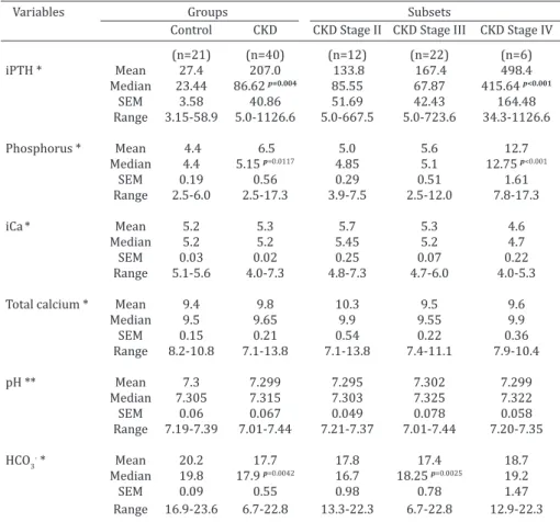
fre-Table 1. Serum intact parathyroid hormone (iPTH), phosphorus, ionized calcium (iCa) and total calcium levels, as well as acid-base status of cats with chronic
kid-ney disease (CKD) and clinically normal cats (control group)
Variables Groups Subsets
Control CKD CKD Stage II CKD Stage III CKD Stage IV
(n=21) (n=40) (n=12) (n=22) (n=6)
iPTH * Mean 27.4 207.0 133.8 167.4 498.4 Median 23.44 86.62 p=0.004 85.55 67.87 415.64 p<0.001
SEM 3.58 40.86 51.69 42.43 164.48
Range 3.15-58.9 5.0-1126.6 5.0-667.5 5.0-723.6 34.3-1126.6
Phosphorus * Mean 4.4 6.5 5.0 5.6 12.7
Median 4.4 5.15 p=0.0117 4.85 5.1 12.75 p<0.001
SEM 0.19 0.56 0.29 0.51 1.61
Range 2.5-6.0 2.5-17.3 3.9-7.5 2.5-12.0 7.8-17.3
iCa* Mean 5.2 5.3 5.7 5.3 4.6
Median 5.2 5.2 5.45 5.2 4.7
SEM 0.03 0.02 0.25 0.07 0.22
Range 5.1-5.6 4.0-7.3 4.8-7.3 4.7-6.0 4.0-5.3 Total calcium * Mean 9.4 9.8 10.3 9.5 9.6
Median 9.5 9.65 9.9 9.55 9.9
SEM 0.15 0.21 0.54 0.22 0.36
Range 8.2-10.8 7.1-13.8 7.1-13.8 7.4-11.1 7.9-10.4
pH ** Mean 7.3 7.299 7.295 7.302 7.299
Median 7.305 7.315 7.303 7.325 7.322
SEM 0.06 0.067 0.049 0.078 0.058
Range 7.19-7.39 7.01-7.44 7.21-7.37 7.01-7.44 7.20-7.35 HCO3- * Mean 20.2 17.7 17.8 17.4 18.7
Median 19.8 17.9 p=0.0042 16.7 18.25 p=0.0025 19.2
SEM 0.09 0.55 0.98 0.78 1.47
Range 16.9-23.6 6.7-22.8 13.3-22.3 6.7-22.8 12.9-22.3 * Mann-Whitney and Kruskal-Wallis tests, **student t test and analysis of variance (ANOVA), SEM =
quency of laboratorial variables as increased or decreased values in all cats with CKD, as well as at different stages of the disease.
According to the normality of distribution of analyzed varia-bles, Spearman or Pearson ranking tests were used and
correla-tion coefficients calculated. Significance was defined as p < 0.05.
RESULTS
Serum iPTH and phosphorus levels were significantly in -creased in the group with CKD. However, comparing the
different stages of cats with CKD, significant difference was
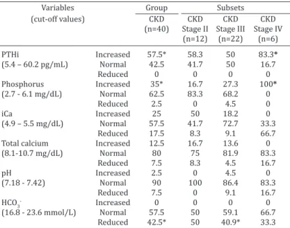
noticed only in Stage IV (Table 1). Increased serum iPTH was detected in 57.5% of the cats in the group with CKD (including Stages II, III and IV). With regard to different sta-ges (II, III and IV) of CKD, increased serum concentrations of iPTH were detected in 58.3%, 50% and 83.3%, respecti-vely (Table 2). Hyperphosphatemia was observed in 35% of all cats with CKD, and in 16.7%, 27.3% and 100% in Sta-ges II, III and IV, respectively (Table 2).
Serum iCa was not significantly different between con -trol group and CKD group, as well as among the stages of CKD (Table 1). Although no statistical difference was de-tected regarding iCa, in Stage IV, 66.7% of CKD cats had ionized hypocalcemia, and 33.3% had ionized normocal-cemia (Table 2). On the other hand, ionized hypercalnormocal-cemia was detected in Stages II and III, corresponding to 50% and 18.2%, and ionized hypocalcemia in 8.3% and 9.1% respec-tively (Table 2).
No significant differences were noticed regarding se
-rum total calcium levels and blood pH (Table 1). Signifi -cant decreased serum bicarbonate (HCO3-) concentrations
(metabolic acidosis) were detected in cats of Stage III, and observed metabolic acidosis frequencies were 50%, 40.9% and 33.3% in Stages II, III and IV, respectively (Ta-bles 1 and 2). In addition, metabolic acidosis was noticed
in 5 out of 10 cats presenting ionized hypercalcemia in Stages II and III.
Spearman (for serum concentration of iPTH, phospho-rus, total calcium and iCa) or Pearson (for serum bicarbo-nate and iCa) ranking tests were used to determine the
correlation coefficients. Positive correlation (moderate de-gree) was observed between serum iPTH and phosphorus (r=0.45; p=0.0032; Spearman)in the group with all stages of CKD cats. Three cats from Stage II (3 of 12) and six cats from Stage III (6 of 22) showed increased iPTH and con-current normophosphatemia and ionized normocalcemia. Two CKD cats of Stage IV had simultaneously the highest levels of serum phosphorus (17.3 mg/dL and 15.6mg/dL) and the lowest levels of ionized calcium (4.0 mg/dL in both cats), as well as increased of iPTH in both animals. Negative correlation (moderatedegree) was detected between se-rum iPTH and iCa in all cats with CKD (r=-0.48; p=0.0019; Spearman), as well as in CKD cats of Stage III (r=-0.6; p=0.003; Pearson).
DISCUSSION
Increased serum iPTH concentrations, observed in 57.5% of the cats with CKD, were similar to those reported in other studies in cats (Barber & Elliott, 1998, Pusoonthorn-thum et al. 2010) and dogs (Lazaretti et al. 2006), however, those reports in cats had not measured iPTH at different
stages of CKD, according to the IRIS classification. In a stu -dy conducted by Barber & Elliott (1998) involving 80 cats with CKD, the animals were subjectively categorized accor-ding to the severity of clinical signs; SRHPT was detected in 47% of CKD cats that were categorized as asymptoma-tic (which CKD was only evidenced by laboratorial exams), and in 100% of CKD cats categorized as presenting more
Table 2. Frequency (%) of normal, increased, and reduced levels of serum intact parathyroid hormone (iPTH), phosphorus, ionized
calcium (iCa), total calcium, blood pH and bicarbonate (HCO3-) of
cats with chronic kidney disease (CKD)
Variables Group Subsets
(cut-off values) CKD CKD CKD CKD (n=40) Stage II Stage III Stage IV
(n=12) (n=22) (n=6)
PTHi Increased 57.5* 58.3 50 83.3*
(5.4 – 60.2 pg/mL) Normal 42.5 41.7 50 16.7
Reduced 0 0 0 0
Phosphorus Increased 35* 16.7 27.3 100*
(2.7 - 6.1 mg/dL) Normal 62.5 83.3 68.2 0
Reduced 2.5 0 4.5 0
iCa Increased 25 50 18.2 0
(4.9 – 5.5 mg/dL) Normal 57.5 41.7 72.7 33.3
Reduced 17.5 8.3 9.1 66.7
Total calcium Increased 12.5 16.7 13.6 0 (8.1-10.7 mg/dL) Normal 80 75 81.9 83.3
Reduced 7.5 8.3 4.5 16.7
pH Increased 2.5 0 4.5 0
(7.18 - 7.42) Normal 90 100 86.4 83.3
Reduced 7.5 0 9.1 16.7
HCO3- Increased 0 0 0 0
(16.8 - 23.6 mmol/L) Normal 57.5 50 59.1 66.7
Reduced 42.5* 50 40.9* 33.3
severe clinical signs (end-stage renal failure). Similar fin -dings were observed herein: cats with CKD in advanced
stage of the disease (Stage IV) had also shown significant
increase in serum iPTH levels (83.3%). Pusoonthornthu et
al. (2010) also showed that iPTH levels were significantly
increased in cats with end-stage CKD, which contributed to the decrease in survival rate.
In the present study, hyperphosphatemia was detec-ted in 35% of all cats with CKD, and it could be the trig-ger for the development of SRHPT, as previously proposed by Slatopolsky et al. (1971) in the “trade off”’ hypothesis, that involves the abnormalities in calcium and phospho-rus homeostasis to explain the pathophysiology of SRHPT. Hyperphosphatemia detected in 100% of CKD cats of
Sta-ge IV may also have directly influenced iPTH synthesis.
Furthermore, hyperphosphatemia may cause ionized hy-pocalcemia, as a result of the increase in other fraction of serum calcium that is chelated (law of mass equation) to phosphorus (Barber & Elliott 1998, Greco & Stabenfeldt 1999). In CKD cats of Stage IV, the decrease in free fraction of serum calcium (iCa) was observed in 66.7%, and
althou-gh this change did not have significant difference between
control group and CKD cats of Stage IV, it is known that io-nized hypocalcemia is also an important component in the regulation of synthesis and secretion of iPTH (Galbraith & Quarles 1996, Barber & Elliott 1998, Greco & Stabenfeldt 1999, Ramasamy 2006, Schenck et al. 2006). A review pa-per about calcium metabolism described that 2% decrease in serum iCa levels could increase iPTH synthesis by 400% (Galbraith & Quarles 1996).
In accordance with the hypothesis previously mentio-ned, two cats of Stage IV revealed the highest values of serum phosphorus (17.3mg/dL and 15.6mg/dL) and the lowest values of ionized calcium (4.0mg/dL in both cats), as well as an increased of serum iPTH and a relevant
im-pairment of their clinical condition. These findings were
similar to those reported by Elliot & Barber (1998) that ob-served hyperphosphatemia, increased serum levels of iPTH as well as ionized hypocalcemia associated with poor clini-cal condition in cats categorized subjectively as end-stage of CKD. In addition, in an analogous study in dogs with CKD, hyperphosphatemia and increased iPTH were detected in 100% (n=8) of the animals in Stage IV, as well as ionized hypocalcemia was observed in 87.5% (Cortadellas et al. 2010). Thus, in this present study, hyperphosphatemia see-med to be the main contributing factor for the development of SRHPT in CKD cats of Stage IV.
Cats with CKD in Stages II and III had no significant di -fference observed in iPTH, phosphorus and iCa (Table 1). Even though these statistical difference were not detected, ionized hypercalcemia was noticed in Stage II and III, and
these findings may be considered for discussion because
different mechanisms, other than the “trade off” hypothe-sis proposed by Slatopolsky et al. (1971), may be involved in the development of SRHPT in CKD cats of Stages II and III, in which elevated serum concentrations of iPTH was observed in 58.3% and 50%, respectively. Ionized hyper-calcemia plays an important role in the development of
metastatic calcification, including the kidneys (Barber &
Elliott 1998). Serum levels of iCa may also be affected by acid-base disorders; thus, metabolic acidosis may increase serum iCa levels by decreasing calcium fraction bound to protein or albumin (Chew & DiBartola 1986, Rosol & Capen 1996). In the present study, decreased blood HCO3-
(meta-bolic acidosis) was noticed in 50% and 40.9% of the cats of Stages II and III, respectively; therefore, ionized hyper-calcemia, observed in those cats, could have been partially attributed to metabolic acidosis, mainly in cats of Stage III, since HCO3- was significantly decreased in this subset. In
addition, metabolic acidosis may also enhance osteoclastic
iPHT activity and bone calcium efflux to the bloodstream,
as well as may reduce directly serum levels of calcitriol and, therefore, contribute to SRHPT progression (Gomes et al. 2005). In humans with CKD, even in the early stages of the disease, metabolic acidosis and SRHPT have been descri-bed in conjunction with bone abnormalities (Gomes et al. 2005).
In ten cats with CKD of Stages II and III that had ioni-zed hypercalcemia, 50% of them had metabolic acidosis, thus, other mechanisms, and not only acid-base imbalance, should be considered in the development of ionized hyper-calcemia in these animals. Possible mechanisms involved could be related to (a) decreased renal excretion of calcium and iPTH, (b) increased bone reabsorption mediated by iPTH, (c) lack of parathyroid glands feedback response to serum calcium levels, (d) increased intestinal calcium ab-sorption (Chew & Meuten 1982, Krueger et al. 1996, Polzin et al 1997), and even (e) autonomous parathyroid secre-tion (Chew & DiBartola 1986, Feldman 1995, Galbraith & Quarles 1996, Barber & Elliott 1998, Barber 2004, Rama-samy 2006).
In cats with CKD in Stage II, hyperphosphatemia was detected in only 16.7% and increased serum iPTH in 58.3% (Table 2). Similar frequency of those results was also repor-ted in a study in cats during the early stages of CKD, in whi-ch hyperphosphatemia was detected in 20% and increased
iPTH in 47% (Barber & Elliott 1998). Therefore, these fin -dings may suggest that other mechanisms, besides hyper-phosphatemia, could lead to the increase in iPTH synthesis.
Thus, further identification of these mechanisms involved
could provide a better understanding of SRHPT pathophy-siology, as well as improve the therapeutic management.
One of the limitations of the present study was the low number of animals included, as well as serum FGF-23 and calcitriol concentrations that could not be measured in or-der to investigate other factors that could be involved. Fur-ther studies should be directed toward the investigation of the role of FGF-23 in the development of SRHPT at different stages of CKD in cats, however, validation of the methodo-logy must be performed previously. In a study in cats with
CKD, serum calcitriol was significantly decreased in 80%
in dogs with CKD, having scientific support for use in stages
III or IV and even in the cases in which serum iPTH levels are within normal ranges (Polzin 2007, Polzin et al. 2009b, Roudebush et al. 2010). However, for cats with CKD in Sta-ges III or IV, the use of calcitriol seems to have a weak
scien-tific support, requiring further investigation (Hostutler et
al. 2006, Polzin et al. 2009a).
In short, metabolic acidosis could have contributed to ionized hypercalcemia observed in CKD cats in Stages II and III, even though no statistical difference in iCa was detected among the Stages of CKD, and it did not seem to be directly affected by SRHPT. In cats with CKD in stage IV, hyperphosphatemia and ionized hypocalcemia may have had an important role to increase serum iPTH, however in Stages II and III, other factors, besides hyperphosphatemia and ionized hypocalcemia, seem to be involved. Further in-vestigations are needed to provide better understanding of the pathophysiology of SRHPT, mainly in the early stages of CKD in cats.
Acknowledgements.- For the grant from the Research Support
Founda-tion of São Paulo State - FAPESP (Fundação de Amparo a Pesquisa do Es-tado de São Paulo).
REFERENCES
Barber P.J., Elliott J. & Torrance A.G. 1993. Measurament of feline intact parathyroid hormone: assay validation and sample handling studies. J. Small Anim. Pract. 4:614-620.
Barber P.J. & Elliott J. 1998. Feline chronic renal failure: calcium homeosta-sis in 80 cases diagnosed between 1992 and 1995. J. Small Anim. Pract. 39:108-116.
Barber P.J. 2004. Disorders of the parathyroid glands. J. Fel. Med. Surg. 6:259-69.
Berti G., Fossati P., Eril G.V.M., Tarengghi G. & Musetellir C. 1987. Enzimatic colorimetric assay of inorganic phosphate. Clin. Chem. 33:312.
Bolliger A.P., Graham P.A., Richard V., Rosol T.J., Nachreiner R.F. & Refsal K.R. 2008. Detection of parathyroid hormone-related protein in cats with humoral hypercalcemia of malignancy. Vet. Clin. Pathol. 31:3-8. Brown S.A., Crowell W.A., Brown C.A., Barsanti J.A. & Finco D.R. 1997.
Pa-thophysiology and management of progressive renal disease. Vet. Jour-nal 154:93-109.
Chew D.J. & Meuten D.J. 1982. Disorders of calcium and phosphorus meta-bolism. Vet. Clin. North Am. Small Anim. Pract. 12:411-438.
Chew D.J. & DiBartola S.P. 1986. Manual of Small Animal Nephrology and Urology. 3rd ed. Churchill Livingstone, New York, 526p.
Cortadellas O., Palacio M.J.F., Tavalera J. & Bayo´n A. 2010. Calcium and phosphorus homeostasis in dogs with spontaneous chronic kidney dise-ase at different stages of severity. J. Vet. Intern. Med. 24:73-79.
DiBartola S.P., Rutgers H.C., Zack P.M. & Tarr M.J. 1987. Clinicopathologic
findings associated with chronic renal disease in cats: 74 cases
(1973-1984). J. Am. Vet. Med. Assoc. 190:1196-1202.
Elliott J. & Barber P.J. 1998. Feline chronic renal failure: clinical findings
in 80 cases diagnosed between 1992 and 1995. J. Small Anim. Pract. 39:78-85.
Elliott J. & Watson A.D.J. 2009. Chronic kidney disease: staging and mana-gement, p.883-891. In: Bonagura J.D. & Twedt D.C. (Eds), Kirk´s Current Veterinary Therapy XIV. Saunders Elsevier, St Louis, Missouri.
Feldman E.C. 1995. Disorders of the parathyroid glands, p.1437-1461. In: Ettinger S.J. & Feldman E.C. (Eds), Textbook of Veterinary Internal Medi-cine. Vol.3. 4th ed. W.B. Saunders, Philadelphia.
Finch N.C., Geddes R.F., Syme H. & Elliott J. 2011. FGF-23 - Mediator of
re-nal secondary hyperparathyroidism or a marker of glomerular filtration
rate (GRF) in cats? Proc. ACVIM Forum, Denver, USA. J. Vet. Intern. Med. 25:632-767.
Galbraith S.C. & Quarles L.D. 1996. Tertiary hyperparathyroidism and refractory secondary hyperparathyroidism, p.193-198. In: Favus M.J. (Ed.), Primer on the Metabolic Bone Disease and Disorders of Mineral Metabolism. 3rd ed. Lippincott-Raven, Philadelphia.
Geddes R.F., Finch N.C., Elliott J. & Syme H.M. 2011. Fibroblast growth fac-tor 23 (FGF-23) in feline chronic kidney disease. Proc. ACVIM Forum, Denver, USA. J. Vet. Intern. Med. 25:632-767.
Gomes C.P., Silva M.I.B., Duarte M.E.L., Dorigo D., Lemos C.C.S. & Bregman R. 2005. Bone disease in patients with chronic kidney disease under conservative management. São Paulo Med. J. 123:83-87.
Goodman W.G., Coburn J.W., Slatopolsky E. & Salusky I.B. 1996. Renal oste-odistrophy in adults and children, p.341-360. In: Favus M.J. (Ed.), Primer on the Metabolic Bone Disease and Disorders of Mineral Metabolism. 3rd
ed. Lippincott-Raven, Philadelphia.
Greco D. & Stabenfeldt G.H. 1999. Endocrinologia, p.345-359. In: Cunnin-gham J.G. (Ed.), Tratado de Fisiologia Veterinária. 2nd ed. Guanabara
Koo-gan, Rio de Janeiro.
Hostutler R.A., DiBartola S.P., Chew D.J., Nagode L.A., Schenck P.A., Rajala--Schultz P.J. & Drost W.T. 2006. Comparison of the effects of daily and intermittent-dose calcitriol on serum parathyroid hormone and ionized calcium concentrations in normal cats and cats with chronic renal failu-re. J. Vet. Intern. Med. 20:1307-1313.
Kidder A.C. & Chew D. 2009. Treatment options for hyperphosphatemia in feline CKD: What’s out there? J. Feline Med. Surg. 11:213-224.
Krueger J.M. & Osborne C.A. 1995. Canine and feline hypercalcemic ne-phropathy, p.416-440. In: Osborne C.A. & Finco D.R. (Eds), Canine and Feline Nephrology and Urology. Willians and Wilkins, Philadelphia. Krueger J.M., Osborne C.A., Nachreiner R.F. & Refsal K.R. 1996.
Hypercalce-mia and renal failure, etilogy, pathophysiology, diagnosis and treatment. Vet. Clin. North Am., Small Anim. Pract. 26:1417-1445.
Lazaretti P., Kogika M.M., Hagiwara M.K., Lustoza M.D. & Mirandola R.M.S.
2006. Concentração sérica de paratormônio intacto em cães com insufi
-ciência renal crônica. Arq. Bras. Med. Vet. Zootec. 58:489-494.
Lulich J.P., Osborne C.A., O´brien T.D. & Polzin D.J. 1992. Feline renal failure: Questions, answers, questions. Compend. Contin. Educ. Vet. 14:127-152. Mendonça D.U., Lobão R.R.S. & Carvalho A.B. 2002. Secondary hyperpara-thyroidism: An updated view of pathogenic and clinical aspects. J. Bras. Nefrol. 24:48-55.
Murphy M.B.P., Pierce K.R., Lowry S.R. & Fisher J.W. 1989. Role of para-thyroid hormone in the anemia of chronic terminal renal dysfunction in dogs. Am. J. Vet. Res. 50:1898-1905.
Nagode L.A., Chew D.J. & Podell M. 1996. Benefits of calcitriol therapy and
serum phosphorus control in dogs and cats with chronic renal failure. Vet. Clin. North Am., Small Anim. Pract. 26:1293-1330.
Notomi M.K., Kogika M.M., Ikesaki J.Y.H., Monteiro P.R.G. & Marquesi M.L. 2006. Retrospective study of chronic renal failure cases in dogs between 1999 a 2002. Braz. J. Vet. Res. Anim. Sci. 43(Suppl.):12-22.
Oliveira R.B. & Moysés R.M.A. 2010. FGF-23: State of the art. J. Bras. Nefrol. 32(3):323-331.
Plotnick A. 2007. Feline chronic renal failure: long-term medical manage-ment. Compend. Contin. Educ. Vet. 29:342-351.
Polzin D.J. & Osborne C.A. 1995. Pathophysiology of renal failure and ure-mia, p.335-367. In: Osborne C.A. & Finco D.R. (Eds), Canine and Feline Nephrology and Urology. Willians and Wilkins, Philadelphia.
Polzin D.J., Osborne C.A., Bartges J.W., James K.M. & Churchill J.A. 1997.
Insu-ficiência renal crônica, p.2394-2431. In: Ettinger S.J. & Feldman E.C. (Eds),
Tratado de Medicina Interna Veterinária. Vol.4. 4ª ed. Manole, São Paulo. Polzin D.J. 2007. 11 Guidelines for conservatively treating chronic kidney
disease. Vet. Med. 102:788-799.
Polzin D.J., Ross S. & Osborne C.A. 2009b. Calcitriol, p.892-895. In: Bonagu-ra J.D. & Twedt D.C. (Eds), Kirk´s Current Veterinary TheBonagu-rapy. XIV. Saun-ders Elsevier, St Louis, Missouri.
Pusoonthornthum R., Pusoonthornthu P. & Krishnamra N. 2010. Calcium--phosphorus homeostasis and changes in parathyroid hormone secre-tion in cats with various stages of spontaneous chronic renal failure. Compend. Clin. Pathol. 19:287-293.
Ramasamy I. 2006. Recent advances in physiological calcium homeostasis. Clin. Chem. Lab. Med. 44:237-273.
Rasouli M. & Kiasari AM. 2006. Serum calcium and phosphorus associate with the occurrence and severity of angiographically documented coro-nary heart disease, possibly through correlation with atherogenic (apo) lipoproteins. Clin. Chem. Lab. Med. 4:43-50.
Rosol T.J. & Capen C.C. 1996. Pathophysiology of calcium, phosphorus, and magnesium metabolism in animals. Vet. Clin. North Am., Small Anim. Pract. 26:1155-1181.
Roudebush P., Polzin D.J., Adams L.G., Towell T.L. & Forrester S.D. 2010. An evidence-based review of therapies for canine chronic kidney disease. J. Small Anim. Pract. 51:244-252.
Sarkar B.C. & Chauhan U.P.S. 1967. A new method for determining micro quantities of calcium in biological material. Anal. Biochem. 20:155-165.
Schenck P.A., Chew D.J., Nagode L.A. & Rosol T.J. 2006. Disorders of cal-cium: hypercalcemia and hypocalcemia, p.122-194. In: DiBartola S.P. (Ed.), Fluid, Electrolyte, and Acid-base Disorders in Small Animal Practi-ce. 3rd ed. W.B. Saunders, St Louis.
Sesso R. & Ferraz M.B. 2003. Critical appraisal of sevelamer for the treat-ment of hyperphosphatemia in patients with chronic renal failure. Revta Assoc. Med. Bras. 49:103-108.
Slatopolsky E., Cagla S., Pennell J.P., Taggart D.D. & Canterbury J.M. 1971. On the pathogenesis of hyperparathyroidism in chronic experimental
renal insufficiency in the dog. J. Clin. Investig. 50:492-499.
Slatopolsky E., Martin K. & Hruska K. 1980. Parathyroid hormone metabo-lism and its potential as a uremic toxin. Am. J. Phys. 239:1-12.
Weller R.J., Cullen J. & Dagle G.E. 1985. Hyperparathyroid disorder in dog: primary, secondary and cancer-associated (pseudo). J. Small Anim. Pract. 26:329-341.
Wesseling-Perry K. 2010. FGF-23 in bone biology. Pediatr. Nephrol. 25: 603-608.