Cop
yright
© ABE&M t
odos os dir
eit
os r
eser
vados
.
583
case report
Arq Bras Endocrinol Metab. 2014;58/5
Normocalcemic primary
hyperparathyroidism: long-term follow-up
associated with multiple adenomas
Hiperparatireoidismo primário normocalcêmico associado a múltiplos adenomas
Larissa Pimentel1, Sirley Portela1, Alyne Loureiro1, Francisco Bandeira1
SUMMARY
Normocalcemic primary hyperparathyroidism (NPHPT) is a condition characterized by elevation of the parathyroid hormone (PTH) in the presence of normal serum calcium and the absence of secondary causes. The case described illustrates the long-term follow-up of a postmenopau-sal woman with NPHPT patient who progressed with multiple adenomas. This case reports a 77-year-old female who has chronic generalized pain and osteoporosis. Her initial serum PTH was 105 pg/mL, with total serum calcium of 9.6 mg/dL, albumin 4.79 g/dL, phosphorus 2.8 mg/dL, and 25OHD after supplementation was 34.6 ng/mL. The bone densitometry (BMD) results were as follows: lumbar spine: T-score -3.0, femoral neck: T-score -2.6 and distal radius: -4.2. Other causes of secondary hyperparathyroidism were ruled out and cervical ultrasound and Tc-99-Sestamibi scan were negative. She used oral alendronate and three infusions of zoledronic acid for treat-ment of osteoporosis. In the 10th year of follow-up, after successive negative cervical imaging,
ultrasound showed a nodule suggestive of an enlarged right inferior parathyroid gland. PTH levels in luid which was obtained during ine-needle aspiration (FNA) were over 5,000 pg/mL and a Sestamibi scan was negative. The patient underwent parathyroidectomy, and a histological examination conirmed parathyroid adenoma. Post-operatively serum PTH remained elevated in the presence of normal serum calcium levels. A follow-up cervical ultrasound showed a new solid nodule suggestive of an enlarged right superior parathyroid gland. PTH levels in the aspira-tion luid were remarkably high. A second parathyroidectomy was performed, with the excision of a histologically conirmed parathyroid adenoma. In conclusion, this is an unusual presentation of NPHPT and highlights the long-term complications. Arq Bras Endocrinol Metab. 2014;58(5):583-6
SUMÁRIO
Hiperparatiroidismo primário normocalcêmico (NPHPT) caracteriza-se pela elevação do hormô-nio da paratiroide (PTH), na ausência da elevação dos níveis séricos de cálcio e exclusão de causas secundárias. O caso descrito ilustra o seguimento de uma mulher na pós-menopausa com NPHPT que evoluiu com múltiplos adenomas. Este caso relata uma paciente de 77 anos de idade que tem dor generalizada crônica e osteoporose. O PTH inicial foi elevado com níveis séricos de cálcio, albumina, fósforo e 25OH vitamina D normais. A densitometria óssea (DMO) evidenciou um T-SCORE da coluna lombar: -3.0, colo do fêmur: -2.6 e rádio distal: -4.2. Outras causas de hiperparatireoidismo secundário foram descartadas e a ultrassonograia cervical e varredura com Sestamibi foram negativos. Fez uso de alendronato e três infusões de ácido zole-drônico para o tratamento da osteoporose. No décimo ano de seguimento, depois de sucessivas imagens negativas, a ultrassonograia evidenciou um nódulo sugestivo de adenoma de parati-reoide inferior direita. A paciente foi submetida à paratiparati-reoidectomia, e um exame histológico conirmou a hipótese. A elevação dos níveis séricos de PTH no pós-operatório se manteve com níveis normais de cálcio. A nova ultrassonograia cervical evidenciou outro nódulo sugestivo de adenoma de paratireoide superior direita. Uma segunda paratireoidectomia foi realizada, cujo histológico conirmou outro adenoma de paratireoide. Conclui-se que essa é uma apresentação incomum de NPHPT e destaca as complicações a longo prazo. Arq Bras Endocrinol Metab. 2014;58(5):583-6
1 Division of Endocrinology,
Diabetes and Bone Diseases, Agamenon Magalhaes Hospital, Brazilian Ministry of Health, University of Pernambuco (UPE) Medical School, Recife, PE, Brazil
Correspondence to:
Francisco Bandeira Disciplina de Endocrinologia, Faculdade de Ciências Médicas, Universidade de Pernambuco Av. Rui Barbosa, 1435 52050-450 – Recife, PE, Brazil [email protected]
Received on Mar/19/2014 Accepted on May/26/2014
Cop
yright
© ABE&M t
odos os dir
eit
os r
eser
vados
.
584 Arq Bras Endocrinol Metab. 2014;58/5
INTRODUCTION
N
ormocalcemic primary hyperparathyroidism (NPHPT) is a condition characterized by eleva-tion of the parathyroid hormone (PTH) in the presence of normal serum calcium and the absence of secondary causes, such as renal insuficiency, vitamin D deiciency, use of medications such as hydrochlorothiazide and li-thium, as well as hypercalciuria and malabsorption sta-tes. NPHPT was only recognized as a distinct condition at the Third Workshop on Management of Asymptoma-tic Primary Hyperparathyroidism held in 2008. Its natu-ral history and management are not yet fully established, with few studies and case reports in the literature (1,2). Such cases usually originate in referral centers for bone diseases (3).The epidemiology of this disease is not well deined, due to the different exclusion criteria of secon-dary causes. In MrOS (The Osteoporotic Fractures in Men Study), the prevalence of normocalcemic hyperpa-rathyroidism was 0.4% in a population of 2,364 males, while in the DHS (Dallas Heart Study), in a population of 3,450 individuals of males and females, it was 3.1% (4). As in most cases of primary hyperparathyroidism, the elevation of PTH is the result of a single parathyroid adenoma, which is removed by surgery, leading to clini-cal and laboratory resolution (3,5). The case described below illustrates the long-term follow-up of a postme-nopausal woman with NPHPT patient who progressed with multiple adenomas.CASE REPORT
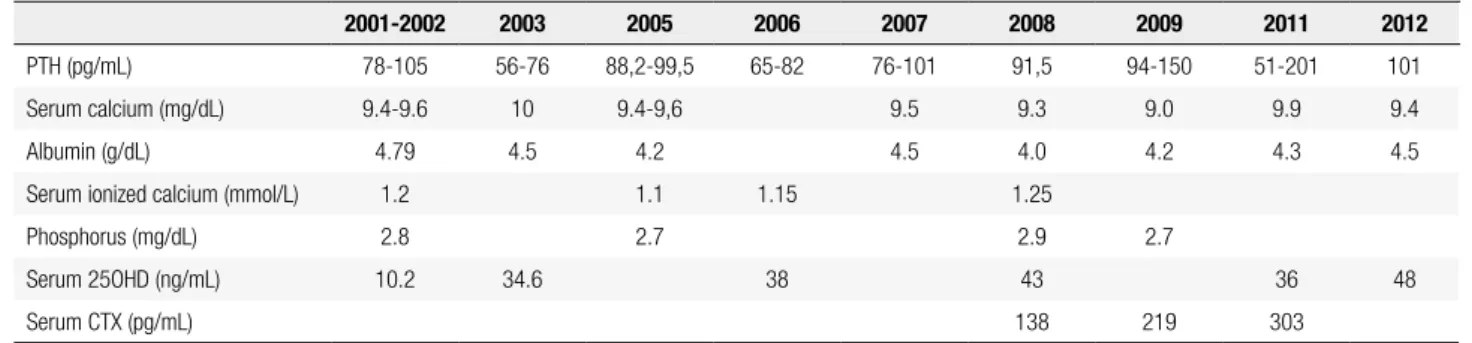
A 77-year-old female was referred to the endocrine clinic for evaluation of chronic generalized pain and osteoporosis. She presented a history of surgical meno-pause at the age of 45. Initial serum PTH was 105 pg/ mL, with total serum calcium of 9.6 mg/dL, albumin 4.79 g/dL, phosphorus 2.8 mg/dL, and 25OHD of
10.2 ng/mL (Table 1). Vitamin D supplementation was started and by the end of the irst year of follow-up serum 25OHD concentrations were 34.6 ng/mL, which remained adequate during the course of treat-ment. Bone densitometry (BMD) results were as fol-lows: lumbar spine 0.862 (T-score -3.0), femoral neck 0.721 (T-score -2.6) and distal radius -4.2 (Table 2). Serum PTH remained elevated despite adequate serum 25OHD levels. Other causes of secondary hyperpar-athyroidism were ruled out and cervical ultrasound and Tc-99-Sestamibi scan were negative. Oral alendronate was started.
A follow-up BMD two years later showed no change at the lumbar spine and a 7% gain at the femoral neck. The patient continued to exhibit elevated serum PTH levels in the presence of normal serum calcium and 25OHD concentrations (Table 1). Cervical imaging continued to be negative for parathyroid enlargement.
Owing to gastrointestinal complaints oral alen-dronate was replaced by zoledronic acid and the pa-tient continued to exhibit improvements in BMD: 6.9% gain at the lumbar spine and no change at the femoral neck (Table 2). After the second infusion, further im-provements were observed: 3.9% gain in lumbar spine and 3.3% in femoral neck and no gain in femoral neck, BMD 0.765, T-score (-1.8) (Table 2). After the third infusion of zoledronic acid, the laboratory values were as follows: serum ionized calcium 1.25 mmol/L, PTH 91.5 pg/mL, 25OHD 43 ng/mL and C-terminal telo-peptide (CTX) 138 pg/mL (Table 1).
In the 10th year of follow-up, after successive
nega-tive cervical imaging, ultrasound showed a 0.6 cm-solid nodule suggestive of an enlarged right inferior parathy-roid gland. PTH levels in luid which was obtained during ine-needle aspiration (FNA) were over 5,000 pg/mL and a Sestamibi scan was negative. The patient under-went parathyroidectomy, and a histological
examina-Table 1. Laboratory data during follow up
2001-2002 2003 2005 2006 2007 2008 2009 2011 2012
PTH (pg/mL) 78-105 56-76 88,2-99,5 65-82 76-101 91,5 94-150 51-201 101
Serum calcium (mg/dL) 9.4-9.6 10 9.4-9,6 9.5 9.3 9.0 9.9 9.4
Albumin (g/dL) 4.79 4.5 4.2 4.5 4.0 4.2 4.3 4.5
Serum ionized calcium (mmol/L) 1.2 1.1 1.15 1.25
Phosphorus (mg/dL) 2.8 2.7 2.9 2.7
Serum 25OHD (ng/mL) 10.2 34.6 38 43 36 48
Serum CTX (pg/mL) 138 219 303
PTH: parathyroid hormone; 25OHD: 25-hydroxi-vitamin D; CTX: C-telopeptide. References: PTH: 15-65 pg/mL, serum calcium: 8,5-10,2 mg/dL, albumin: 3,5-5,5 g/dL, serum ionized calcium: 1,05-1,30 mmol/L, phosphorus: 2,5-4,5 mg/dL, serum 25OHD: > 30 ng/mL, serum CTX: 60-480 pg/mL.
Cop
yright
© ABE&M t
odos os dir
eit
os r
eser
vados
.
585 Arq Bras Endocrinol Metab. 2014;58/5
years of follow-up the disease progressed without the development of hypercalcemia, suggesting that it may be an independent condition.
There are few data available regarding complications and the natural history of NPHPT. Lowe and cols. re-ported a follow-up of 37 patients, of whom 29 were postmenopausal women, 6 premenopausal women and 2 males, with a mean age of 58 years with normal renal function. They considered serum 25OHD above 20 ng/mL as an inclusion criterion (65% had levels abo-ve 30 ng/mL). In the follow up analysis, there were 4 fragility fractures, 14% had nephrolithiasis and 57% had osteoporosis at diagnosis. Parathyroidectomy was performed in 10 subjects, in whom the presence of two parathyroid adenomas and hyperplasia was observed in 2 cases, while the others showed a single parathyroid adenoma (6).
In our institution, a study on 33 patients compa-red data on NPHPT with asymptomatic hypercalcemic PHPT (5). The following inclusion criteria for the diag-nosis of NPHPT were used: serum 25OHD levels abo-ve 30 ng/mL, no use of bisphosphonates, thiazide diu-retics, anticonvulsants or lithium, estimated glomerular iltration rate above 60 mL/min (MDRD-Modiication of Diet in Renal Disease), and absence of hypercalciu-ria and malabsorption diseases. Mean age was 64 years, and 79% were female. There was a high prevalence of nephrolithiasis (18%), suggesting that normocalcaemia does not preclude clinical manifestations. It was also observed that bone mineral density at the distal radius was better preserved in the group with normal calcium serum than in the group with hypercalcemia. In our case, the patient presented predominantly with osteo-porosis, which initially showed some response to bis-phosphonates but subsequently progressed to a more rapid bone loss without the development of hypercal-cemia. There was no evidence of nephrolithiasis during follow-up.
Other studies have evaluated the presence of osteo-porosis and nephrolithiasis in NPHPT. Tordjman and cols., evaluated a cohort comprising 32 patients with normocalcemic hyperparathyroidism and found a high prevalence of osteoporosis (36%), with 9% presenting nephrolithiasis (7). Cakir and cols., in their investigation of 18 patients with normocalcemic hyperparathyroidism and a mean age of 50 years (47% female) found that 47% had osteoporosis and 11% nephrolithiasis (8).
Wade and cols. presented a cohort of 93 patients with primary hyperparathyroidism, of whom 58
pa-Table 2. Bone mineral density (T-score) during follow up previously to parathyroidectomy
Baseline T-score
LS -3.0
FN -2.6
DR -4.2
Year 2
LS -2.75
FN -1.79
Year 5
LS -2.2
FN -1.8
Year 10
LS -2.5
FN -2.4
DR -3.8
LS: lumbar spine; FN: femoral neck; DR: distal radius.
tion conirmed a right inferior parathyroid adenoma of 0.7 x 0.6 cm, the nodule wasn’t intrathyroid and the predominant cell type was oxyphil. Post-operatively serum PTH remained elevated in the presence of nor-mal serum calcium levels. At that time abdominal ultra-sound showed no evidence of kidney stones.
During the irst year following parathyroidectomy, BMD unexpectedly worsened at the lumbar spine (-7%) and at the distal radius (-8%), excluding other factors that could interfere in BMD (Table 2). At this time, se-rum PTH was 201 pg/mL, and a follow-up cervical ul-trasound showed a 0.6 cm new solid nodule suggestive of an enlarged right superior parathyroid gland, again with a negative Tc-99-Sestamibi scan. PTH levels in the aspiration luid were remarkably high (19,600 pg/mL) suggesting a new parathyroid adenoma as a cause of the elevated PTH levels. A second parathyroidectomy was performed, with the excision of a histologically con-irmed parathyroid adenoma.
DISCUSSION
Elevation of serum PTH may be a inding during eva-luation of reduced bone mass and, when associated with the absence of hypercalcemia, causes of seconda-ry hyperparathyroidism should be excluded in order to conirm the diagnosis of normocalcemic primary hyperparathyroidism. There is uncertainty regarding NPHPT as an incipient presentation of classic primary hyperparathyroidism, but in the present case over 10
Cop
yright
© ABE&M t
odos os dir
eit
os r
eser
vados
.
586 Arq Bras Endocrinol Metab. 2014;58/5
tients (62% of the sample) had serum ionized calcium measured preoperatively. Eighty percent had elevated levels and 14% normal levels. The mean age was 60 years, 63% were female, 13% had a history of bone fragility fracture and 25% had nephrolithiasis. Of the patients with normal serum ionized calcium, 5 (62%) subjects had a single adenoma at surgery and 3 (28%) had mul-tiple adenomas (9). Our patient always had normal se-rum ionized calcium concentrations.
Cervical ultrasonography was effective in localizing the parathyroid lesions in the present case although it took almost a decade of follow-up and repeated nor-mal ultrasound examinations for those lesions become apparent. The Tc-99-Sestamibi scan was less sensitive, failing to localize either adenoma. The sensitivity of ul-trasound is highly operator-dependent, but its accuracy may be as high as 88% in asymptomatic hypercalcemic patients (10). When it detects intra-thyroid nodules, FNA with measurement of PTH in the aspiration luid is very useful as thyroid nodules may cause false-positi-ve Sestamibi scan results (11).In the present case both adenomas had very high PTH levels in their aspiration luid. Multiphase computed tomography of the cervical region (4D-CT) has became the preferred localizing method for those patients in whom the parathyroid le-sion is not detected by ultrasound or scintigraphy, as its sensitivity is high for both adenoma and hyperpla-sia (12). Until the issue of long-term complications of NPHPT is not completely understood, it seems reaso-nable to consider surgery for those patients in whom the parathyroid lesion is localized by imaging procedu-res or traditional operation (neck exploration) for tho-se who have osteoporosis or renal stone ditho-seatho-se with negative imaging.
In conclusion, this case illustrates an unusual pre-sentation of NPHPT and highlights the long-term
complications, irrespective of the development of hypercalcemia.
Disclosure: no potential conlict of interest relevant to this article was reported.
REFERENCES
1. Bilezikian JP, Khan AA, Potts JT. Guidelines for the management of asymptomatic primary hyperparathyroidism: summary state-ment from the Third International Workshop. J Clin Endocrinol Metab. 2009;94(2):335-9.
2. Bilezikian JP, Silverberg SJ. Normocalcemic primary hyperpara-thyroidism. Arq Bras Endocrinol Metabol. 2010;54(2):106-9. 3. Cusano NE, Silverberg SJ, Bilezikian JP. Normocalcemic primary
hyperparathyroidism. J Clin Densit. 2013;16(1):33-9.
4. Cusano NE, Naim MM, Wang PY, Zhang C, Cremers SC, Haney EM, et al. Normocalcemic hyperparathyroidism and hypopara-thyroidism in two community-based nonreferral populations. J Clin Endocrinol Metab. 2013;98(7):2734-41.
5. Amaral LM, Queiroz DC, Marques TF, Mendes M, Bandeira F. Nor-mocalcemic versus hypercalcemic primary hyperparathyroidism: more stone than bone? J Osteoporos. 2012;3:128-352.
6. Lowe H, McMahon DJ, Rubin MR. Normocalcemic primary hyper-parathyroidism: further characterization of a new clinical pheno-type. J Clin Endocrinol Metab. 2007;92(8):3001-5.
7. Tordjman KM, Greenman Y, Osher E. Characterization of normocal-cemic primary hyperparathyroidism. Am J Med. 2004;117:861-3. 8. Cakir I, Unluhizarci K, Tanriverdi F. Investigation of insulin
resis-tance in patients with normocalcemic primary hyperparathyroi-dism. Endoc. 2012;42:419-22.
9. Wade TJ, Yen TWF, Amin AL, Wang TS. Surgical management of normocalcemic primary hyperparathyroidism. World J Surg. 2012;36:761-6.
10. Bandeira F, Griz L, Chaves N, Carvalho NC, Borges LM, Castro ML, et al. Diagnosis and management of primary hyperparathyroi-dism – A scientiic statement from the Department of Bone Meta-bolism, the Brazilian Society for Endocrinology and Metabolism. Arq Bras Endocrinol Metab. 2013;57(6):406-24.
11. Khang AR, Kim EK, Namm EY, Byeon SJ, Kim JH, Ohn JH, et al. Primary hyperaparathyroidism due to cystic parathyroid ade-noma not detected on 99mTc-Sestamibi scan. Endocrinol Metab.
2012;27:83-8.
12. Chazen JL, Gupta A, Dunning A, Phillips CD. Diagnostic accuracy of 4D-CT for parathyroid adenomas and hyperplasia. Am J Neu-roradiol. 2012;33(3):429-33.