Response to an oral calcium load in
nephrolithiasis patients with fluctuating
parathyroid hormone and ionized
calcium levels
Departamentos de 1Nefrologia and 2Endocrinologia,
Universidade Federal de São Paulo, São Paulo, SP, Brasil S.A. Gomes1, A. Lage2,
M. Lazaretti-Castro2,
J.G.H. Vieira2
and I.P. Heilberg1
Abstract
The response to an oral calcium load test was assessed in 17 hypercal-ciuric nephrolithiasis patients who presented elevated parathyroid hormone (PTH) irrespective of the ionized calcium (sCa2+) levels.
Blood samples were collected at baseline (0 min) and at 60 and 180 min after 1 g calcium load for serum PTH, total calcium, sCa2+, and
1.25(OH)2D3 determinations. According to the sCa2+ level at baseline,
patients were classified as normocalcemic (N = 9) or hypercalcemic (N = 8). Six healthy subjects were also evaluated as controls. Bone mineral density was reduced in 14/17 patients. In the normocalcemic group, mean PTH levels at 0, 60 and 180 min (95 ± 76, 56 ± 40, 57 ± 45 pg/ml, respectively) did not differ from the hypercalcemic group (130 ± 75, 68 ± 35, 80 ± 33 pg/ml) but were significantly higher compared to healthy subjects despite a similar elevation in sCa2+ after
60 and 180 min vs baseline in all 3 groups. Mean total calcium and
1.25(OH)2D3 were similar in the 3 groups. Additionally, we observed
that 5 of 9 normocalcemic patients presented a significantly higher concentration-time curve for serum PTH (AUC0',60',180') than the other
4 patients and the healthy subjects, suggesting a primary parathyroid dysfunction. These data suggest that the individual response to an oral calcium load test may be a valuable dynamic tool to disclose a subtle primary hyperparathyroidism in patients with high PTH and fluctuat-ing sCa2+ levels, avoiding repeated measurements of both parameters.
Correspondence
I.P. Heilberg
Departamento de Nefrologia Universidade Federal de São Paulo Rua Botucatu, 740
04023-900 São Paulo, SP Brasil
Fax: +55-11-5573-9652 E-mail: [email protected]
Part of this study was presented at the 35th Annual Meeting of the American Society of Nephrology, Philadelphia, PA, USA, October 2002.
Research supported by CNPq and Fundação Oswaldo Ramos.
Publication supported by FAPESP.
Received September 29, 2003 Accepted May 25, 2004
Key words
•Parathyroid hormone •Calcium
•Hyperparathyroidism •Hypercalciuria •Nephrolithiasis •Bone mineral density
Introduction
Primary hyperparathyroidism is charac-terized by inappropriately increased secre-tion of parathyroid hormone (PTH) in rela-tion to the serum calcium level of the patient. Clinically the disorder is accompanied by hypercalcemia, serum levels of intact PTH
within or above the upper normal range and an increased parathyroid cell mass (1).
total serum calcium in hyperparathyroidism is not always demonstrable and several meas-urements may be needed to ensure that hy-percalcemia is present. Although hypercal-cemia is a key feature in establishing the diagnosis of primary hyperparathyroidism, the possibility of the existence of hyperpara-thyroidism with intermittent or no elevation in total serum calcium has been raised by several investigators since the first descrip-tion of normocalcemic hyperparathyroidism in 1953 (4-8).
On the other hand, the suspicion of nor-mocalcemic primary hyperparathyroidism may be raised on the basis of a history of recurrent renal calculi and increased urine calcium excretion. Primary hyperparathyroid-ism is observed in 1 to 5% of all calcium stone formers (9). However, about 50% of patients with renal calculi have idiopathic hypercalciuria, a condition associated with normocalcemia as determined by total se-rum calcium. Despite initial evidence of sec-ondary hyperparathyroidism due to chronic renal tubular calcium leakage, many investi-gators have not confirmed it (10-14).
It has been reported that the measure-ment of ionized calcium is more sensitive than total serum calcium to indicate hyper-parathyroidism (5,15).
Levels of intact PTH even when deter-mined by optimal two-site immunoassays may reflect measurement of molecules with no bioactivity (16). Thus, the overestimation of PTH impairs the diagnosis of subtle pri-mary hyperparathyroidism. In addition, there is an intrinsic physiologic variation in serum PTH levels. Therefore, several measurements of both PTH and ionized calcium are needed to diagnose parathyroid dysfunction in pa-tients with intermittent or no elevation of total serum calcium.
Although there are distinct pathophysi-ologic mechanisms, failure to separate the rare patient with normocalcemic primary hyperparathyroidism from the idiopathic hypercalciuria patients with eventual
sec-ondary hyperparathyroidism has led to inap-propriate neck exploration (17).
We hypothesized that an oral calcium load test could detect a dynamic response by the parathyroid glands, providing better evaluation of parathyroid dysfunction, rather than multiple determinations of PTH and ionized calcium.
Patients and Methods
Informed written consent to participate was obtained from each patient and the study was approved by the Ethics Committee of Universidade Federal de São Paulo.
Seventeen outpatients (14 women and 3 men; mean age 48 ± 13 years) from the Nephrology or Endocrinology Divisions pre-senting high serum PTH levels irrespective of total and ionized calcium levels, hypercal-ciuria and or nephrolithiasis, were included in the study.
Patients with chronic renal failure, endo-crinological disorders such as hyperthyroid-ism, acromegaly, sarcoidosis, diabetes mel-litus, or neoplasias, or taking drugs which could not be withdrawn during the protocol (corticosteroid, diuretics, oral contraceptives, etc.) were excluded. All patients were in-structed by a dietitian to follow the same dietary recommendations (i.e., calcium in-take of 500 mg/day and sodium chloride intake of 140 mEq/day) 2 weeks before the test was performed. A control group matched for calcium intake, consisting of 6 healthy volunteers (4 women and 2 men; mean age: 26 ± 4 years) was studied.
Urinary calcium
A 24-h urine sample was obtained from 15 patients for calcium determination. Hypercal-ciuria was defined as urinary calcium >250 mg/24 h for females, >300 mg/24 h for males and/or ≥4 mg kg-1 24 h-1 for either sex (18).
Acute calcium load test. All patients and
diet before the test. Blood and urine samples consecutively obtained during fasting and after the oral calcium load were analyzed. The patients started fasting soon after dinner (19:00 h) during the evening preceding the test except for 300 ml of distilled water which they drank at 21:00 h and at midnight. On the test day, the patients drank 300 ml of distilled water at 6:00 h. At 7:00 h a blood sample was drawn for measurements of total and ionized calcium, intact PTH, creatinine, 1.25(OH)2D3, and phosphate. Basal urine was collected over a period of 2 h from 7:00 to 9:00 h for calcium, creatinine and phos-phate determinations. An oral calcium load of 1 g (as gluconolactate and carbonate; Calcium Sandoz F®, Basel, Switzerland) was then given. The second period began imme-diately after calcium administration. Another blood sample was collected at 60 and 180 min following the calcium load for total and ionized calcium, phosphate and intact PTH determinations. Ionized calcium was drawn under vacuum venipuncture and immedi-ately centrifuged. In the second urine sample collected 180 min after the calcium load, calcium, creatinine and phosphate were again determined.
Intact serum parathyroid hormone
Intact serum PTH was determined by an in-house immunofluorometric assay (normal range: 4-70 pg/ml) based on a chicken egg yolk-derived amino-terminal antibody (PTH-1-34) bound to a microtiter plate by anti-chicken Ig monoclonal antibody. A Eu-ropium-labeled carboxyl-terminal specific monoclonal antibody produced from a mouse immunized with a PTH-(53-84)-BSA conju-gate was employed as the tracer antibody. The intra-assay error was 5% and the inter-assay error was 13.4% (19).
Ionized serum calcium
Ionized serum calcium was determined
by ion selective electrode (AVL Model 9180,
Roswell, GA, USA); normal range: ≤1.32
mmol/l. Total serum calcium was determined by atomic absorption spectrophotometry (Perkin-Elmer Atomic Spectrophotometer 290B, Norwalk, CT, USA); normal range: 8.5 to 10.5 mg/dl. Inorganic phosphate was determined by the Fiske and Subbarow method (20) and creatinine was measured by the alkaline picrate Jaffe reaction (21).
1.25(OH)2D3 determination
Serum levels of 1.25(OH)2D3 were deter-mined by radioimmunoassay (123I RIA kit; DiaSorin, Stillwater, MN, USA); normal range: 15.9 to 55.6 pg/ml.
Bone mineral density
Bone mineral density (BMD) was as-sessed by dual-energy X-ray absorptiometry at lumbar spine and femoral sites using a DPX-L apparatus (Lunar Radiation Corp., Madison, WI, USA). The criterion for the definition of osteopenia was a hip or spine BMD value >1.0 SD below the mean BMD for the young adult population (T score) according to WHO criteria (22).
Parathyroid scintigraphy
The procedure was carried out using a double-phase study with Tc-99 sestamibi (APEX SPX, Haifa, Israel) (23).
Statistical analysis
Results
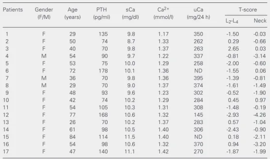
Patients’ characteristics on admission in-cluding individual serum PTH, total and ion-ized calcium values, urinary calcium, and T-score are shown in Table 1. Hypercalciuria was detected in 14 of 15 of the 24-h urine samples obtained from all patients. Osteope-nia was detected in 14/17 patients, but no fractures were observed. Only 2 patients (numbers 12 and 15) had hyperfunctioning nodules identified by parathyroid scintigra-phy (data not shown in Table 1).
Acute calcium load test
Patients were classified as normocalce-mic (N = 9) or hypercalcenormocalce-mic (N = 8) accord-ing to the level of ionized calcium deter-mined at baseline (0 min). The normocalce-mic group presented mean ionized calcium similar to the control group at baseline (1.29 ± 0.04 vs 1.30 ± 0.01 mmol/l) but signifi-cantly lower than the hypercalcemic group
(1.29 ± 0.04 vs 1.39 ± 0.05 mmol/l). A
significant difference between the
normo-calcemic and hypernormo-calcemic groups was also observed at 60 (1.35 ± 0.05 vs 1.47 ± 0.10 mmol/l) and 180 min (1.39 ± 0.06 vs 1.51 ± 0.10 mmol/l) but the mean ionized calcium of normocalcemic subjects remained similar to control at 60 and 180 min. When com-pared to baseline, all 3 groups presented higher mean ionized calcium levels at 60 and 180 min after the load, as shown in Figure 1. In contrast, mean total serum calcium was similar among the normocalcemic, hyper-calcemic and control groups at baseline (9.7 ± 0.4 vs 9.7 ± 0.5 and 9.9 ± 0.4 mg/dl), at 60 min (10.0 ± 0.2 vs 10.4 ± 0.7 mg/dl and 10.1 ± 0.5 mg/dl) and 180 min (10.3 ± 0.5 vs 10.3 ± 0.7 and 10.3 ± 0.2 mg/dl), respectively.
As shown in Figure 2, the mean PTH of the normocalcemic group was significantly higher than control at baseline (95 ± 76 vs 30 ± 14 pg/ ml), after 60 min of calcium load (56 ± 40 vs 17 ± 13 pg/ml) and also after 180 min (57 ± 45 vs 17 ± 11 pg/ml). However, mean PTH values did not differ between the normocalcemic and hypercalcemic groups at 0, 60 or 180 min (95 ± 76 vs 130 ± 75, 56 ± 40 vs 68 ± 35 and 57 ± 45 vs 80 ± 33 pg/ml, respectively). There was
Table 1. Patient characteristics at admission.
Patients Gender Age PTH sCa Ca2+ uCa T-score
(F/M) (years) (pg/ml) (mg/dl) (mmol/l) (mg/24 h)
L2-L4 Neck
1 F 29 135 9.8 1.17 350 -1.50 -0.03
2 F 50 74 8.7 1.33 262 0.29 -0.66
3 F 40 70 9.8 1.37 263 2.65 0.03
4 M 54 90 9.7 1.22 337 -0.81 -3.14
5 F 53 75 10.0 1.29 258 -2.00 -0.60
6 F 72 178 10.1 1.36 ND -1.55 0.06
7 M 36 70 9.8 1.36 395 -1.39 -0.81
8 M 29 70 9.0 1.37 374 -1.61 -1.49
9 F 48 93 9.6 1.23 302 -0.52 -1.90
10 F 42 74 10.2 1.29 284 0.45 0.97
11 F 54 105 10.3 1.31 308 -1.48 -0.19
12 F 77 168 10.6 1.32 145 -2.93 -4.26
13 F 26 70 10.2 1.37 283 0.57 -1.04
14 F 61 98 10.5 1.40 306 -2.43 -0.90
15 F 84 114 11.5 1.40 ND 0.18 -2.11
16 F 54 98 10.6 1.32 370 0.94 -3.20
17 F 47 140 11.1 1.42 270 -1.87 -1.99
no difference among the normocalcemic, hy-percalcemic and control groups in regard to percent PTH suppression from baseline after 60 min (-40 ± 11, -46 ± 17 and -54 ± 20%, respectively) or after 180 min (-40 ± 12, -38 ± 15 and -47 ± 30%).
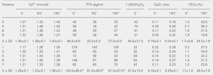
The serum and urinary parameters ob-tained during the calcium load test in the normocalcemic group are presented in Table 2. The table shows that the mean PTH value of patients 1, 3, 4, 6, and 7 was significantly higher at 0 min and after 60 and 180 min, respectively (134 ± 85, 81 ± 38 and 81 ± 57 pg/ml) when compared to that of patients 2, 5, 8, and 9 (46 ± 9, 25 ± 5 and 27 ± 5 pg/ml) and of healthy subjects (30 ± 14, 17 ± 13 and 17 ± 11 pg/ml). The mean PTH value for patients 2, 5, 8, and 9 did not differ from that of the healthy subjects. The concentration-time curve of serum PTH (AUC0',60',180') for patients 1, 3, 4, 6, and 7 (16,171 ± 9005 pg ml-1 min-1) was significantly higher than for patients 2, 5, 8, and 9 (5280 ± 967 pg ml-1 min-1, P = 0.016) and also than the curve for healthy subjects (3492 ± 202, P = 0.004). No differences were found in mean urinary Ca/ Cr, or FeCa% between patients 1, 3, 4, and 7 and patients 2, 5, 8, and 9, as shown in Table 2. The mean urinary Ca/Cr ratio (data not shown
Figure 1. Mean serum ionized calcium levels (Ca2+) at base-line (0 min) and at 60 and 180 min after oral calcium load in hypercalcemic (squares), nor-mocalcemic (circles) and con-trol (triangles) subjects. *P < 0.05 vs hypercalcemic patients (Mann-Whitney test).
Figure 2. Mean serum parathy-roid hormone (PTH) levels at baseline (0 min) and at 60 and 180 min after oral calcium load in hypercalcemic (squares) nor-mocalcemic (circles) and con-trol (triangles) subjects. *P < 0.05 vs control subjects (Mann-Whitney test).
Ionized calcium (mM)
1.56
* *
*
0 60 180
Time (min) 1.51
1.46
1.41
1.36
1.31
1.26
1.21
1.16
1.11
PTH (pg/ml)
280
210
140
70
0
0 60 180
Time (min)
* *
*
Table 2. Individual values of serum parathyroid hormone (PTH), 1.25(OH)2D3, urinary Ca/Cr ratio, and fractional excretion of calcium (FECa%) during a calcium load test in normocalcemic patients.
Patients Ca2+ (mmol/l) PTH (pg/ml) 1.25(OH)
2D3 Ca/Cr ratio FECa (%)
0 ' 6 0 ' 1 8 0 ' 0 ' 6 0 ' 1 8 0 ' 0 ' 0 ' 1 8 0 ' 0 ' 1 8 0 '
2 1.27 1.33 1.40 40 26 25 42 0.11 0.18 1.0 20.6
5 1.31 1.48 1.50 38 18 22 74 0.35 0.36 2.7 36.3
8 1.31 1.36 1.43 48 29 27 41 0.11 0.22 1.0 21.5
9 1.32 1.36 1.37 59 28 34 21 0.20 0.22 1.9 19.6
X ± SD 1.30±0.1 1.38±0.1 1.43±0.1 46.0±9.0 25.0±5.0 27.0±5.0 45.0±21.0 0.19±0.1 0.24±0.1 1.7±0.8 24.5±7.9
1 1.17 1.26 1.26 279 142 159 32 0.32 0.38 3.3 37.5
3 1.30 1.33 1.41 93 50 33 35 0.14 0.24 1.1 24.6
4 1.31 1.34 1.38 72 56 43 59 0.14 0.22 1.3 22.2
6 1.31 1.36 1.39 146 97 99 64 0.18 0.37 1.4 37.3
7 1.31 1.35 1.36 82 60 70 42 0.11 0.23 1.0 22.6
X ± SD 1.28±0.1 1.33±0.1 1.36±0.1 134.0±85.0* 81.0±38.0* 81.0±57.0* 47.0±14.0 0.18±0.1 0.29±0.1 17±1.0 29.0±7.9
in tables) was significantly higher vs base-line after 180 min in the normocalcemic (0.27 ± 0.1 vs 0.18 ± 0.1), hypercalcemic (0.43 ± 0.2 vs 0.19 ± 0.1) and control (0.24 ±
0.1 vs 0.11 ± 0.1) groups. Although the
hypercalcemic group tended to show a higher urinary Ca/Cr value after 180 min in com-parison to the other 2 groups, the difference did not reach statistical significance.
Mean fractional calcium excretion was significantly elevated after 180 min of oral calcium load in the normocalcemic (27.0 ± 7.7 vs 1.7 ± 0.8%), hypercalcemic (44.0 ± 25.0 vs 1.6 ± 0.5%) and control (23.0 ± 0.11
vs 0.9 ± 0.1%) groups, but again did not
differ among the 3 groups.
Mean fractional phosphate excretion 180 min after the oral calcium load was only significantly lower than baseline in the con-trol group (3.3 ± 2.9 vs 9.8 ± 3.2%) but not in the normocalcemic or hypercalcemic groups (10.2 ± 5.3 vs 10.3 ± 6.3% and 10.5 ± 9.9 vs 12.7 ± 7.0%; data not shown in tables).
The mean values of 1.25(OH)2D3 also did not differ between the normocalcemic, hypercalcemic and control groups (46 ± 17 vs 48 ± 24 vs 36 ± 12 pg/ml). The individual
and mean values are presented in Figure 3.
Discussion
The diagnosis of classic primary hyper-parathyroidism is easily established by in-creased levels of PTH in the presence of hypercalcemia. On the other hand, patients
with primary hyperparathyroidism may pres-ent significant symptoms and minimal or no elevation in total serum calcium levels (24). Normocalcemic hyperparathyroidism de-fined as completely normal total serum val-ues is a rare entity that must be searched for in patients with symptoms or complications of hyperparathyroidism, such as renal cal-culi and bone mineral loss (6). However, the existence of normocalcemic hyperparathy-roidism is still controversial (4-7). Another possible explanation for the finding of high levels of PTH with normal serum calcium values is the putative secondary hyperpara-thyroidism due to renal tubular leakage of calcium in patients with idiopathic hypercal-ciuria (10-12). Nevertheless, several investi-gators have reported normal parathyroid func-tion in idiopathic hypercalciuria (13,14,25). Lundgren et al. (7) found abnormal para-thyroid tissue (75% adenomas and 25% hy-perplasias) in 16/57 (28%) postmenopausal women with high PTH levels and normal ionized and total serum calcium values, sug-gesting the presence of normocalcemic pri-mary hyperparathyroidism.
The reason why total serum calcium is not increased in patients with primary hyper-parathyroidism in the presence of normal serum values of albumin, magnesium, phos-phate, and 1.25(OH)2D3 is not clear. While some investigators have attributed a normal total calcium level in patients with normo-calcemic hyperparathyroidism to an in-creased ratio of ionized and ultrafiltrable calcium to total calcium in these patients compared to normal subjects, others did not confirm it (15,26,27). Resistance to the ef-fect of PTH in increasing renal tubular reab-sorption of calcium resulting in higher uri-nary calcium has also been postulated (28). Another possibility could be the presence of non-1-84 PTH circulating molecules such as a 7-84 PTH fragment, blocking the calcemic effect of 1-84 PTH and preventing hypercal-cemia (16,29).
In the present study, we hypothesized
1.25(OH)
2
D3
(pg/ml)
100 90 80 70 60 50 40 30 20 10 0
Normocalcemic Hypercalcemic Control Figure 3. Individual levels of
that an acute oral calcium load test would help to better evaluate the dynamic response of the parathyroid gland to fluctuations of
serum calcium in patients withhigh serum
PTH. The reproducibility of the test was good since the test was easy to perform in an outpatient setting (3 blood collections over a period of 180 min), as already suggested by other investigators (30,31), with a sensitivity and specificity of 100 and 87% (31), respec-tively. Calcium administration was well tol-erated by all patients.
In response to the oral calcium load, ion-ized calcium levels but not total serum cal-cium levels were increased in normocalce-mic, hypercalcemic and control patients. The higher levels of ionized calcium were main-tained even after 180 min in all 3 groups, including the control. These data suggest that ionized calcium is more sensitive than total serum calcium in reflecting acute changes in serum calcium regulation, as also reported by others (5,15).
We observed a higher mean PTH in the normocalcemic group compared to control despite the similar elevation of ionized cal-cium presented by both. The mean PTH in this normocalcemic group was not signifi-cantly different from that presented by the hypercalcemic group, suggesting a distur-bance in calcium set point in the former.
A significant difference in the percent-age of mean PTH suppression from baseline among the 3 groups was not evidenced in the present study. These findings contrast with those of Monchik et al. (30), who observed a clear-cut separation between normal sub-jects and patients with high PTH levels and normal total serum calcium, whose PTH sup-pression achieved values higher and lower than 30%, respectively.
Although the mean percent PTH sup-pression of the normocalcemic group did not differ from control, we observed that 5 of 9 normocalcemic patients presented a concentration-time curve of serum PTH (AUC0',60',180') significantly higher not only
than control but also than the remaining 4 normocalcemic patients. It could be argued that the minimal reduction of PTH in these 5 patients in response to the calcium load might have reflected the detection of inactive long half-life fragments of PTH. Nevertheless, the different response among control pa-tients using the same assay (19), which pre-sents a good correlation (r = 0.938) with a commercial immunoradiometric assay (PTH-IRMA; Nichols Institute, San Juan Capis-trano, CA, USA), does not suggest this hypo-thesis. On the other hand, according to Sil-verberg et al. (29), inactive 7-84 PTH frag-ments may be produced by parathyroid ad-enomas. It is important to emphasize that although second-generation 1-84 PTH as-says present a better diagnostic sensitivity in primary hyperparathyroidism (16,29), they are still not widely available and their diag-nostic value is especially important among patients with chronic renal failure.
Our findings suggested a disturbance in calcium set point in the normocalcemic pa-tients when compared to controls. It is note-worthy that even the hypercalcemic group was able to suppress PTH on a percent basis as did the control group but, again, at levels much higher than the normal range.
Recurrent stone formation was the main clinical manifestation and hypercalciuria could be detected in all but one of the normo-calcemic patients (8/9).
Following the oral calcium load, a reduc-tion in fracreduc-tional phosphate excrereduc-tion was observed only in the control group, reflect-ing an appropriate PTH suppression in this group. This finding emphasizes the lack of a physiological response of phosphate reab-sorption due to the persistent elevation of PTH in both the normocalcemic and hyper-calcemic groups.
In the present series, serum 1.25(OH)2D3 was similar among the 3 groups, in agree-ment with Monchik et al. (30), who did not observe an increased 1.25(OH)2D3 in hyperparathyroidism patients. Conversely, some investigators observed mild elevations of 1.25(OH)2D3 levels in classic hyperpara-thyroidism (9,32). Although the elevation of 1.25(OH)2D3 in idiopathic hypercalciu-ric patients has been reported by some investigators, others have pointed out high normal values or completely normal values, so that this is still a matter of controversy (32-34).
An elevated frequency of bone mass loss, mainly cortical bone, is often found in pri-mary hyperparathyroidism, as well as in nor-mocalcemic calcium stone formers with idiopathic hypercalciuria, mostly in trabecu-lar bone (33,35). Some investigators have hypothesized that a primary increase in bone resorption due to a monocyte disorder may result in hypercalciuria in these patients (36,37). In our study, we observed osteope-nia in 14 of 17 patients with similar distribu-tion of affected sites, lumbar spine or femo-ral neck.
Parathyroid scintigraphy was not helpful in the diagnosis of hyperparathyroidism in the present study, as also pointed out by other investigators (38).
Although primary hyperparathyroidism patients with the most marked elevation in serum calcium levels are thought to present the most marked symptoms, many investiga-tors have reported that they may present significant symptoms despite a minimal or no elevation in their serum calcium level
(4-7,24). In a series of 142 patients with hyper-calcemia, intermittent hypercalcemia or normocalcemia, Siperstein et al. (24) ob-served a similar frequency of preoperative symptoms and surprisingly the same degree of symptom amelioration after surgery, sug-gesting that factors other than elevated se-rum calcium levels are responsible for symp-toms of hyperparathyroidism. These investi-gators also did not find statistical differences in the frequency of single adenomas, hyper-plasia or multiple adenomas among these 3 subgroups.
This poses the problem of difficulties regarding the diagnosis of hyperparathyroid-ism, mainly if the patient is normocalcemic, especially when one is dealing with ionized calcium (39). As we could not easily estab-lish the diagnosis of hyperparathyroidism in these normocalcemic patients despite the high PTH levels, we tested the dynamic re-sponse of the parathyroid gland to an oral calcium load. The response to the oral cal-cium load test among normocalcemic pa-tients showed heterogeneity with respect to PTH levels, irrespective of the clinical manifestation. The maintenance of higher PTH levels, higher than the threshold of 31 pg/ml in 5 patients, differing statistically from control, was probably due to an altered calcium set point and suggests that a primary dysfunction of the gland must be present. The concentration-time curve of serum PTH (AUC) of the remaining 4 patients was slightly higher, although not significantly different from control (5280 ± 967 vs 3492 ± 202). On the basis of this response, those 4 patients could be either considered as ad-equate responders or as presenting second-ary hyperparathyroidism. The use of thiaz-ide drugs particularly in these patients in the future might help us to further confirm such hypothesis.
PTH and fluctuating ionized calcium levels, avoiding numerous repeated PTH and serum calcium determinations leading to inconclu-sive results. The test also provides a faster diagnosis determining an earlier therapeutic intervention.
Acknowledgments
The authors thank Prof. Omar M. Hauache MD, PhD, for fruitful discussions, Ilda S. Kunii, for the determination of serum PTH, and Silvia R. Moreira, for technical assistance.
References
1. Grimelius L, Akerstrom G, Johansson H, Juhlin C & Rastad J (1991). The parathyroid glands. In: Kovaks K & Asa SL (Editors), Functional Endocrine Pathology. Blackwell Scientific, Oxford, Oxford Shire, England, 375-395.
2. Deaconson TG, Wilson SD & Lemann JM (1987). The effect of parathyroidectomy on the recurrence of nephrolithiasis. Surgery, 102: 910-913.
3. Mundy GR, Coue DIT & Fisken R (1980). Primary hyperparathyroid-ism: changes in the pattern of clinical presentation. Lancet, 1: 1317-1320.
4. Johannson H, Thoren L & Werner L (1975). Normocalcemic hyper-parathyroidism, kidney stones, and idiopathic hypercalciuria. Sur-gery, 77: 691-696.
5. Forster J, Monchik JM & Martin HF (1988). A comparative study of serum ultrafiltrable, ionized, and total calcium in the diagnosis of primary hyperparathyroidism in patients with intermittent or no elevation in total calcium. Surgery, 104: 1137-1142.
6. Monchik JM (1995). Normocalcemic hyperparathyroidism. Surgery, 118: 917-923.
7. Lundgren E, Ridefelt P, Akerström G, Ljunghall S & Rastad J (1996). Parathyroid tissue in normocalcemic and hypercalcemic primary hyperparathyroidism recruited by health screening. World Journal of Surgery, 20: 727-735.
8. Mather HG (1953). Hyperparathyroidism with normal serum cal-cium. British Medical Journal, 2: 424-425.
9. Broadus AE, Horst RL & Lang R (1980). The importance of circulat-ing 1.25 dihydroxyvitamin D in the pathogenesis of hypercalciuria and renal-stone formation in primary hyperparathyroidism. New England Journal of Medicine, 302: 421-426.
10. Coe FL, Canterbury JM, Firpo JJ & Reiss E (1973). Evidence for secondary hyperparathyroidism in idiopathic hypercalciuria. Journal of Clinical Investigation, 52: 134-142.
11. Pak CYC, Kaplan R, Bone H, Towsend J & Waters O (1975). A simple test for the diagnosis of absorptive, resorptive and renal hypercalciurias. New England Journal of Medicine, 292: 497-500. 12. Broadus AE, Lang R & Kliger AS (1981). The influence of calcium
intake and the status of intestinal calcium absorption on the diag-nostic utility of measurements of 24-hour cyclic adenosine 3',5'-monophosphate excretion. Journal of Clinical Endocrinology and Metabolism, 52: 1085-1089.
13. Olmer M, Berland Y & Argemi B (1983). Absence of secondary hyperparathyroidism in most patients with renal hypercalciuria. Kid-ney International, 24: 175-179.
14. Coe FL, Favus MJ, Crokett T, Strauss AL, Parks JH, Porat A, Gant C & Sherwood LM (1982). Effects of low calcium diet on urine calcium excretion, parathyroid function and serum 1.25(OH)2D3 levels in patients with idiopathic hypercalciuria and normal subjects. Ameri-can Journal of Medicine, 72: 25-32.
15. Monchik JM & Martin HF (1980). Ionized calcium in the diagnosis of primary hyperparathyroidism. Surgery, 82: 185-192.
16. Gao P, Scheibel S, D’Amour P, John MR, Rao SD, Schmidt-Gayk H & Cantor TL (2001). Development of a novel immunoradiometric assay exclusively for biologically active whole parathyroid hormone 1-84: implications for improvement of accurate assessment of para-thyroid function. Journal of Bone and Mineral Research, 16: 605-614.
17. Poole GV, Albertson DA & Myers RT (1983). Normocalcemic hyper-parathyroidism revisited. American Journal of Surgery, 49: 668-671. 18. Hodgkinison A & Pyrah LN (1958). The urinary excretion of calcium and inorganic phosphate in 344 patients with calcium stone renal origin. British Journal of Surgery, 46: 10-18.
19. Vieira JGH, Nishida SK, Kasamatsu TS, Amarante EC & Kunii IS (1994). Development and clinical application of an immunofluoro-metric assay for intact parathyroid hormone. Brazilian Journal of Medical and Biological Research, 27: 2379-2382.
20. Fiske CH & Subbarow Y (1988). Inorganic phosphate. Journal of Biological Chemistry, 63: 375-400.
21. McFate RP, Cohn C, Eichelberger L & Cooper JA (1954). Sympo-sium on azotemia. American Journal of Clinical Pathology, 24: 511-571.
22. Kanis JA, Melton III LJ, Christiansen C, Johnston CC & Khaltaev N (1994). The diagnosis of osteoporosis. Journal of Bone and Mineral Research, 9: 1137-1141.
23. Taillefer R, Boucher Y, Potvin C & Lambert R (1992). Detection and localization of parathyroid adenomas in patients with hyperparathy-roidism using a single radionuclide imaging procedure with techne-tium 99 sestamibi (double phase study). Journal of Nuclear Medi-cine, 33: 1801-1807.
24. Siperstein AE, Shen W, Chan AK, Duh QY & Clark OH (1992). Normocalcemic hyperparathyroidism. Archives of Surgery, 127: 1157-1163.
25. Lilienfeld-Toal HV, Bach D, Hesse A, Franck H & Issa S (1982). Parathyroid hormone is normal in renal stone patients with idio-pathic hypercalciuria and high fasting urinary calcium. Urology Re-search, 10: 205-207.
26. Muldoney FP, Freaney R & McMullin JP (1976). Serum ionized calcium and parathyroid hormone in renal stone disease. Quarterly Journal of Medicine, 45: 75-86.
27. Strott CA & Nugent CA (1968). Laboratory test in the diagnosis of hyperparathyroidism in hypercalcemic patients. Annals of Internal Medicine, 68: 188-202.
28. Gardin JP & Paillard M (1984). Normocalcemic hyperparathyroidism resistance to PTH effect on tubular reabsorption of calcium. Mineral and Electrolyte Metabolism, 10: 301-308.
hormone (1-84) in primary hyperparathyroidism. Journal of Clinical Endocrinology and Metabolism, 88: 4725-4730.
30. Monchik JM, Lamberton RP & Roth U (1992). Role of the oral calcium-loading test with measurement of intact parathyroid hor-mone in the diagnosis of symptomatic subtle primary hyperparathy-roidism. Surgery, 112: 1103-1110.
31. Hagag P, Revet-Zak I, Hod N, Horne T, Rapoport MJ & Weiss M (2003). Diagnosis of normocalcemic hyperparathyroidism by oral calcium load. Journal of Endocrinology Investigation, 26: 227-232. 32. Locascio V, Adami S & Galvanini G (1985). Substrate-product
rela-tion of 1-hydroxylase activity in primary hyperparathyroidism. New England Journal of Medicine, 313: 1123-1130.
33. Heilberg IP, Martini LA, Szejnfeld VL, Carvalho AB, Draibe SA, Ajzen H, Ramos OL & Schor N (1994). Bone disease in calcium stone forming patients. Clinical Nephrology, 42: 175-182.
34. Bushinsky DA, Johnston RB, Nalbantian CE & Favus MJ (1989). Increased calcium absorption and retention, without elevated se-rum 1.25(OH)2D3 in genetically hypercalciuric rats. Kidney
Interna-tional, 33: 336 (Abstract).
35. Rossini M, Gatti D, Isaia G, Sartori L, Braga V & Adami S (2001).
Effects of oral alendronate in elderly patients with osteoporosis and mild primary hyperparathyroidism. Journal of Bone and Mineral Research, 16: 113-119.
36. Weisinger JR (1996). New insights into the pathogenesis of idio-pathic hypercalciuria: The role of bone. Kidney International, 49: 1507-1518.
37. Pacifici R, Rothstein M, Rifas L, Lau Kw, Baylink DJ, Avioli LV & Hruska K (1991). Increased monocyte interleukin-1 activity and de-creased vertebral bone density in patients with fasting idiopathic hypercalciuria. Journal of Clinical Endocrinology and Metabolism, 71: 138-145.
38. Castellani M, Reschini E, Longari V, Paracchi A, Corbetta S, Marottha G & Gerundini P (2001). Role of Tc-99m sestamibi scintigraphy in the diagnosis and surgical decision-making process in primary hy-perparathyroid disease. Clinical Nuclear Medicine, 26: 139-144. 39. Bilezikian PJ, Potts JTJ, Fuleihan GEH, Kleerekoper M, Neer R,
Peacock M, Rastad J, Silverberg SJ, Uldesman R & Wells AS (2002). Summary statement from a workshop on asymptomatic primary hyperparathyroidism: A perspective for the 21st century.