UNIVERSIDADE DA BEIRA INTERIOR
Ciências
Histamine modulates nitric oxide release by
microglia and dopaminergic neuronal survival
Joel Pereira Pires
Dissertação para obtenção do Grau de Mestre em
Bioquímica
(2º ciclo de estudos)
Orientador: Prof. Doutora Liliana Bernardino
Co-orientador: Prof. Doutora Graça Baltazar
iii
Agradecimentos
Quero agradecer em primeiro lugar a minha família, em especial aos meus pais, pelo vosso amor, carinho e compreensão ao longo da minha vida. Obrigado pelo apoio e por acreditarem em min, sem vocês não teria realizado muitos dos meus sonhos. OBRIGADO por TUDO.
À minha orientadora científica Professora Doutora Liliana Bernardino pela oportunidade de desenvolver este projeto, pelo apoio e dedicação, pela disponibilidade constante e por todos os conhecimentos transmitidos.
À Professora Doutora Graça Baltazar, a minha co-orientadora científica e à Professora Doutora Carla Fonseca pela disponibilidade, ajuda e simpatia que demostraram sempre que preciso.
Um muito obrigado à Sandra Rocha pela disponibilidade, paciência e grande ajuda no desenvolvimento deste trabalho. Obrigado a todas as colegas de laboratório, Susana Gama, Rita Videira, Diana Rodrigues e Ana pelo apoio, amizade e pela pronta disponibilidade em ajudar sempre que preciso.
Aos funcionários e técnicos do CICS pela colaboração, que tornaram a realização deste trabalho possível.
A todos os meus amigos, em especial ao Rui e ao Francisco, por todos os bons momentos e grande apoio nos momentos mais difíceis, estarão sempre no meu coração.
v
Resumo
As células microgliais, células imunitárias residentes no cérebro, desempenham um papel crítico na etiologia e progressão de várias doenças neurodegenerativas. A doença de Parkinson (DP) é uma doença neurodegenerativa caraterizada por uma grande perda dos neurónios dopaminérgicos na Substantia Nigra (SN), diminuição dos níveis de dopamina no estriado e complicações motoras. Várias evidências clínicas e experimentais sugerem que a neuroinflamação tem um papel crítico na patogénese da DP através da ativação das células microgliais e consequente produção de mediadores inflamatórios, incluindo o óxido nítrico (ON). A Histamina (HIS), uma amina que atua como neurotransmissor e mediador inflamatório, tem sido descrita como tendo um importante papel na patogénese da DP. Alterações nas inervações histaminérgicas no estriado e SN bem como um aumento das concentrações de histamina no sangue, estriado e SN foram observadas em pacientes com DP. Com base nestes dados, o nosso objetivo foi avaliar o efeito da histamina nas células microgliais obtidas da Substantia Nigra de ratos Wistar e seguidamente avaliar de que forma fatores solúveis libertados pela microglia previamente estimulada com histamina podem modular a sobrevivência neuronal dopaminérgica. Inicialmente foram utilizadas culturas de células microgliais para estudar o efeito da histamina e os seus recetores na produção de ON, o qual foi quantificado pelo teste de Griess. Demostramos que a HIS provoca um aumento da produção de ON quando comparado com o controlo, um efeito mediado pela ativação do recetor 4 da histamina (H4R). Contudo, num contexto inflamatório induzido pelo Lipopolissacarídeo (LPS), a HIS inibe a produção de ON induzida pelo LPS não só pelo R4H, mas também possivelmente através da ativação do recetor 1 da histamina (R1H). Em seguida, recolhemos o meio condicionado das células microgliais (MCM) tratado com HIS e/ou LPS para avaliar o seu efeito na viabilidade dos neurónios dopaminérgicos presentes em co-culturas de neurónios e astrócitos isoladas do mesencéfalo. De facto, o meio condicionado obtido das células microgliais expostas ao LPS ou à HIS levaram a uma diminuição do número de neurónios positivos para a Tirosina Hidroxilase; sendo este efeito anulado quando o MCM é obtido das células microglias tratadas com a HIS mais LPS. Curiosamente, o mesmo efeito foi observado quando a HIS e/ou LPS foram adicionados diretamente nas co-culturas de neurónios e astrócitos. Assim, estes resultados sugerem que a HIS por si só atua como um mediador pró-inflamatório, enquanto, num contexto pró-inflamatório, a HIS tem supostamente um efeito anti-inflamatório promovendo desta forma a sobrevivência dos neurónios dopaminérgicos.
vi
Palavras-chave
vii
Abstract
Microglia cells, the resident immune cells in the brain, play a critical role in the development and progression of several neurodegenerative diseases. Parkinson's disease (PD) is a neurodegenerative disorder characterized by a dramatic loss of dopaminergic neurons (DA) in the substantia nigra (SN), striatal dopamine depletion and motor impairments. Accumulating clinical and experimental evidences suggest that neuroinflammation plays a critical role in the pathogenesis of PD through the activation of microglia cells and the subsequent production of a vast array of inflammatory mediators, including nitric oxide (NO). Histamine (HIS), an amine that acts as a neurotransmitter and inflammatory mediator, has been reported to play a role in the pathogenesis of PD. Indeed, alterations in the histaminergic innervations in the striatum and SN and increased histamine concentrations in the blood, striatum and SN were found in PD patients.
Based on these data, our aim was to uncover the effects of histamine on microglia cells derived from the SN of Wistar rats and then evaluate whether soluble factors released by microglia previously stimulated with histamine could modulate dopaminergic neuronal survival. Firstly, microglia cell cultures were used to study the effects of HIS and its receptors on NO production, which was measured by the Griess assay. We demonstrated that HIS triggered an increase of NO production as compared with control, an effect mediated by histamine H4 receptor (H4R) activation. Interestingly, in the presence of an inflammatory context, mimicked by lipopolysaccharide (LPS), HIS inhibited LPS-induced NO production not only by H4R but, possibly through histamine H1 receptor (H1R) activation. Then, conditioned medium derived from microglia cells (MCM) challenged with HIS and/or LPS was collected to evaluate its effects on the viability of DA neurons present in neuron-astrocyte midbrain co-cultures. In fact, conditioned medium derived from microglia cells exposed to LPS or HIS induced a decrease in the number of Tyrosine Hydroxylase positive neurons; whereas this noxious effect was abolished when MCM obtained from microglia challenged with HIS plus LPS was used. Curiously, the same effects were observed when HIS and/or LPS were added directly on neuron-astrocyte midbrain co-cultures. Together, our results suggest that HIS per se acts as a pro-inflammatory mediator, whereas, in an inflammatory context, HIS has a putative anti-inflammatory profile that can protect dopaminergic neurons.
viii
Keywords
ix
Index
LIST OF FIGURES... xi
LIST OF TABLES... xii
LIST OF ABBREVIATIONS... xiii
CHAPTER 1 – INTRODUCTION... 1
1.1 NEUROINFLAMMATION... 1
1.1.1 MICROGLIA IN HEALTH AND DISEASE... 1
1.1.2 CONTRIBUTION OF NEUROINFLAMMATION TO EXCITOTOXICITY... 4
1.2 ETIOLOGY AND PATHOGENESIS OF PARKINSON’S DISEASE... 5
1.2.1 ROLE OF MICROGLIAL CELLS IN PARKINSON’S DISEASE... 6
1.2.2 LIPOPOLYSACCHARIDE-INDUCED PARKINSON’S DISEASE ANIMAL MODEL... 8
1.3 HISTAMINE... 10
1.3.1 ROLE OF THE HISTAMINERGIC SYSTEM IN PARKINSON’S DISEASE... 12
CHAPTER 2 – OBJECTIVES... 13
CHAPTER 3 – MATERIALS AND METHODS... 14
3.1 IN VITRO CELL CULTURES... 14
3.1.1 N9 MICROGLIA CELL LINE CULTURE... 14
3.1.2 PRIMARY MICROGLIA CELL CULTURES FROM SUBSTANTIA NIGRA... 14
3.1.3 NEURON-ASTROCYTE MIDBRAIN CO-CULTURES... 15
3.2 CELL TREATMENTS... 16
3.3 EVALUATION OF CELL DEATH ASSAY... 16
3.3.1 PROPIDIUM IODIDE UPTAKE... 16
3.3.2 TUNEL LABELING... 17
3.4 MEASUREMENT OF NITRIC OXIDE (NO) RELEASE... 17
3.5 IMMUNOCYTOCHEMISTRY... 18
x
CHAPTER 4 – RESULTS... 19
4.1 CHARACTERIZATION OF PRIMARY MICROGLIA CELL CULTURES DERIVED FROM SN... 19
4.2 EXPRESSION OF HISTAMINE H4 RECEPTOR IN MICROGLIA CELLS DERIVED FROM SN... 20
4.3 EFFECT OF HISTAMINE ON THE PRODUCTION OF NO... 21
4.4 H4R ACTIVATION MEDIATES THE PRODUCTION OF NO TRIGGERED BY HISTAMINE... 22
4.5 EFFECT OF HISTAMINE ON NO PRODUCTIONIN IN AN INFLAMMATORY CONTEXT INDUCED BY LPS... 23
4.6 EFFECT OF HISTAMINE ON CELLULAR VIABILITY... 25
4.7 HISTAMINE H4 AND H1 RECEPTORS ACTIVATION MODULATES LPS-INDUCED NO PRODUCTION... 26
4.8 EFFECT ON CONDITIONED MEDIUM DERIVED FROM MICROGLIAL CELLS TREATED WITH LPS AND/OR HISTAMINE IN THE VIABILITY OF DOPAMINERGIC NEURONS... 27
4.9 EFFECT OF HISTAMINE AND/OR LPS PER SE ON CELLULAR VIABILITY OF DOPAMINERGIC NEURONS... 29
CHAPTER 5 – DISCUSSION... 30
CHAPTER 6 – CONCLUSIONS... 34
xi
List of Figures
Figure 1 - Microglia activation by endogenous and/or exogenous stimuli... 3 Figure 2 - Possible mechanisms of glutamate induced neuronal death... 5 Figure 3 - Relationship between microglia activation and dopaminergic neuronal
damage... 8 Figure 4 - Simplified schematic representation of the link between LPS-induced microglial
activation and dopaminergic neurodegeneration... 10 Figure 5 - Immunocytochemical stainings of a primary microglia cell culture
derived from SN of the Wistar rats... 19 Figure 6 - Microglia cells from SN express histamine H4 receptor... 20 Figure 7 - Histamine-induced NO release from microglia cells... 22 Figure 8 - Histamine induces NO release by microglia cells via H4 receptor
activation... 23 Figure 9 - LPS-induced NO release is inhibited by Histamine... 24 Figure 10 - Microglia viability after Histamine exposure... 25 Figure 11 – LPS-induced NO release is inhibited by histamine, via H1 and H4 receptor
activation... 26 Figure 12 - Microglia protects dopaminergic neurons from LPS injury when
co-stimulated with histamine... 28 Figure 13 -Effects of Histamine and/or LPS per se on dopaminergic
xii
List of Tables
Table 1 - Expression, Function, and Signaling of Histamine Receptors and
the G Proteins Involved... 11 Table 2 - Primary and secondary antibodies used for immunocytochemistry... 18
xiii
Abbreviations
Ag H4R Agonist of histamine 4 receptor (4-methylhistamine dihydrochloride)
Ant H1R Antagonist of Histamine 1 receptor (mepyramine maleate)
Ant H2R Antagonist of Histamine 2 receptor (cimetidine)
Ant H3R Antagonist of Histamine 3 receptor (carcinine ditrifluoroacetate)
Ant H4R Antagonist of Histamine 4 receptor (JNJ7777120)
ATP Adenosine 5’-triphosphate
Bax Bcl-2–associated X protein
BSA Bovine Serum Albumin
CD11b cluster of differentiation molecule 11b
CNS Central nervous system
COX-2 Cyclo-oxygenase 2
DMEM Dubecco’s modified eagle medium
FBS Fetal bovine serum
H1R Histamine 1 receptor H2R Histamine 2 receptor H3R Histamine 3 receptor H4R Histamine 4 receptor HIS Histamine IFN-γ Interferon-γ
IL-1β Interleukin-1 beta
iNOS Inducible Nitric oxide synthases
LPS Lipopolysaccharide
MAP2 Microtubule-associated protein 2 MAPK Mitogen-activated protein kinase
MCM Conditioned medium derived from microglial cells MPT Mitochondrial permeability transition
NBM Neurobasal medium
NMDA N-Methyl-D-aspartate
NO Nitric oxide
xiv
PD Parkinson’ disease
PFA Paraformaldehyde
PGE Prostaglandin E2
PI Propidium iodide
RNS Reactive nitrogen species
ROS Reactive oxygen species
RPMI Roswell Park Memorial Institute media
RT Room temperature
SN Substantia nigra pars compacta
TH Tyrosine hydroxylase
TNFR-1 TNF-α receptor 1
TNF-α Tumor necrosis factor-alpha
TUNEL Terminal deoxynucleotidyl transferase (TdT)-mediated dUTP nick-end belling
Chapter 1
INTRODUCTION
1.1. NEUROINFLAMMATION
In the past, the central nervous system (CNS) was considered an immune-privileged site. Nowadays, it is well established that the activation of the immune cells present in the CNS to infection, trauma, toxins, among other stimuli plays a crucial role in the development and progression of neurodegenerative and neuropsychiatric diseases, including Alzheimer’s disease (AD), Parkinson’s disease (PD), Huntington’s disease (HD), bipolar disorder (BD), schizophrenia (SZ) and depression. The inflammatory responses in the brain, also known as neuroinflammation, are a complex combination of acute and chronic responses of several types of cells, including neurons, microglia, astrocytes and infiltrating leukocytes. The acute inflammatory responses are believed to be beneficial, since it tends to minimize further injury and contributes to repair of damaged tissue. On the other hand, chronic neuroinflammation produces long-lasting and self-perpetuating neuroinflammatory mediators that remain after the initial neuroinflammatory insult has passed (Frank-Cannon et al., 2009; Kraft and Harry, 2011; Rao et al., 2012).
1.1.1 MICROGLIA IN HEALTH AND DISEASE
Microglia are the resident immune-competent cells of the CNS and have a role in monitoring the brain for immune insults and invading pathogens. Ramon and Cajal considered microglia to be part of the ‘third element’ of the CNS, being neither neuronal nor astrocytic (Long-Smith et al., 2009).
The origin of microglia still remains highly debated. The hypothesis most accepted is the ‘‘myeloid-monocytic hypothesis’’, which states that resident microglia, as well as the other tissue resident macrophages, are derived from circulating blood monocytes, during the late embryonic life and post-natally (Flügel et al., 2001; Kaur et al., 2001; Polazzi and Monti, 2010;).
Microglia cells are distributed throughout the CNS, represent around 5–20% of the total adult brain cells, depending on the species, and constitute approximately 20% of the glial cell population. Interestingly, the density and the morphology of microglia are
region-2
specific. This strongly suggests that these differences might be related to a microglial functional heterogeneity (Lawson et al., 1990; Polazzi and Monti, 2010).Major features of microglia are their highly ramified morphology and plasticity that allow them to supervise the extracellular CNS parenchyma and to be quickly activated in response to pathological conditions, thus exerting typical macrophagic functions, such as phagocytosis, secretion of proinflammatory cytokines and antigen presentation (Gehrmann et al., 1995; Stence et al., 2001; Ladeby et al., 2005;).
It was presumed for many years that under normal physiological conditions microglial cells are quiescent and in a resting state. But in vivo two-photon microscopy studies in living mice showed that microglial processes are substantially motile, and survey their local surroundings thought formation of random filopodia-like protrusions, extensions and withdrawal of bulbus endings. This state of high motility facilitates the microglial processes to perceive the status of their microenvironment, to endocytose nutrients and to clear debris and apoptotic cellular material (Nimmerjahn et al., 2005; Napoli and Neumann, 2009). They are also actively involved in the determination of cell fate (elimination/survival) of developing neurons by enforcing the programmed elimination of neural cells or enhance neuronal survival through the release of trophic and anti-inflammatory factors. In addition, in the mature brain, microglia facilitate brain repair through the guided migration of stem cells to the site of inflammation and injury, and might be involved in neurogenesis (Marín-Teva et al., 2004; Ekdahl, 2012). Microglia are potentially also promoters of the migration, axonal growth, and terminal differentiation of different neuronal subsets, through the release of extracellular matrix components, soluble factors and direct cell–cell contact. Moreover, the cross–talk with neurons is believed to be an important factor in guarding microglia cells in a quiescent state. For example, interaction of the neuronal membrane protein CD200 with the myeloid cell receptor CD200R dampens microglial activation. Mice deficient in CD200 show morphological and molecular signs of microglia activation in the resting CNS, and the microglial response to different forms of experimental brain injury is excessive (Polazzi and Contestabile, 2002; Streit, 2002). The interaction between microglia and other glial cells namely astrocytes is also complicated due the reciprocal interaction, both in health and unhealthy brain, and like microglia cells, astrocytes play diverse functions in the brain, both harmful and beneficial. For example, it is known that activated microglia facilitates astrocytic activation and activated astrocytes in turn regulate microglial activities and also promote microglial activation. Astrocytes play a dual role in CNS inflammatory diseases, not only having the ability to enhance immune responses and postpone restoration, but also limiting CNS inflammation and being neuroprotective (inhibitory effect on activated microglia).Therefore an important question is how these two totally opposite effects coexist because the degree of inflammation is crucial (De Keyser et al., 2008; Liu et al., 2011; Rocha et al., 2012). Clearly, much more remains to be learned about the intricate functional inter-relationships that exist between microglia and astrocytes, as well as their meaning for neuronal regeneration and degeneration.
3
Apart from these important roles of microglial cells in healthy brain, microglia also plays an important role in unhealthy brain since it is exquisitely sensitive to disturbance of their microenvironment. Microglia detect the changes in its environment through the expression of a great number of cellular surface receptors and nuclear receptors that play a critical role in the initiation and/or modulation of its immunitary responses (Hanisch, 2002; Block, 2007).Virtually, every neurological disorder leads to inflammation, with activation of resident microglia, accompanied by an increase in number and change in phenotype of glial cells, a phenomenon generally termed ‘‘reactive gliosis’’. Acute neurodegenerative diseases, such as stroke, hypoxia, and trauma, compromise neuronal survival and indirectly trigger neuroinflammation, as microglia become activated in response to the insult itself, thus adopting a phagocytic phenotype and releasing inflammatory mediators, mainly cytokines and chemokines. This acute neuroinflammatory response is generally beneficial to the CNS, since it tends to minimize further injury and it contributes to repair of damaged tissues.
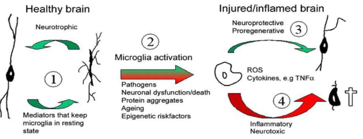
In contrast, chronic neurodegenerative diseases, including AD and PD, are known to be associated with chronic neuroinflammation, even if several differences have been identified among these pathologies. Chronic neuroinflammation is a long-standing and often self-perpetuating response that persists long after an initial injury or insult either genetical or environmental in nature. It is generally characterized by a long-standing activation of microglia and subsequent sustained release of inflammatory mediators leading to increased oxidative and nitrosative stress. This, in turn, works to perpetuate the inflammatory cycle, activating additional microglia, promoting their proliferation and resulting in a further release of inflammatory factors (Fig. 1). Besides playing a protective role as acute neuroinflammation does, chronic neuroinflammation is most often considered detrimental and damaging to nervous tissue. Thus, whether neuroinflammation has beneficial or harmful outcomes in the brain may depend critically on the duration of the inflammatory response and on the kind of microglial activation (Frank-Cannon et al., 2009; Polazzi et al., 2010).
Figure 1: Microglia activation by endogenous and/or exogenous stimuli. (1) In the healthy brain microglia support neuronal well-being, and in turn receives cues from neurons and glial cells to remain in the resting state. (2) In response to a wide array of noxious stimuli microglia undergo activation.
4
Activation may be beneficial to the host (3) when reactive oxygen spices (ROS) and secreted cytokines are kept at low and/or transient levels. In this instance these proinflammatory mediators are neuroprotective. However, when they surpass a certain level of host tolerance (4) these mechanisms become neurotoxic and result in neuronal dysfunction and cell death, which may further contribute to microglial activation (from Vilhardt, 2005).
1.1.2 CONTRIBUTION OF NEUROINFLAMMATION TO EXCITOTOXICITY
Neuroinflammation may play a critical role in the modulation of excitotoxicity that occurs in several neurodegenerative diseases. Excitotoxicity refers to a process of neuronal death caused by excessive or prolonged activation of receptors for the excitatory amino acid neurotransmitter glutamic acid (Zimmer et al., 2000).Glutamate-induced death of neurons can be mediated by: (a) activation of the N-Methyl-D-aspartate (NMDA) subtype of glutamate receptor, resulting in Ca2+ and/or Na+ overload of the neuron; (b) activation of α-amino-3-hydroxy-5-methyl-4-isoxazolepropionic acid receptor (AMPA) or (c) glutamate inhibition of cystine uptake, resulting in oxidative stress/death of the neuron. Calcium elevation may: (a) stimulate calcineurin causing Bcl-2-associated death promoter (Bad) and Bcl-2–associated X protein (Bax) activation; (b) stimulate mitochondrial oxidant production and mitochondrial permeability transition (MPT); and (c) stimulate neuronal Nitric Oxide Synthases (nNOS) production of NO and oxidants. Activation of Bax pores and/or MPT may result in release of apoptosis inducing factor (AIF) and cytochrome c, and/or cause ATP depletion (Fig. 2) (Brown and Bal-Price, 2003; Ankarcrona et al., 1995; Doble, 1999).
Microglial cells express both ionotropic and metabotropic glutamatergic receptors, that when overactivated in pathological conditions induce microglia activation and subsequent release of pro-inflammatory cytokines (Noda et al., 2000 and Taylor et al., 2005). In addition to glutamate, adenosine triphosphate (ATP) is a co-transmitter also released by injuried or dying neurons following brain insults that can modulate microglial activation, via interaction with both metabotropic (P2YR) and ianotropic (P2XR) receptors (Davalos et al., 2005). All these mechanisms lead to neuronal cell death and therefore, we can’t consider a single mechanism involved in chronic neurodegenerative disease, but a set of mechanisms that may act in synergy, where microglial cells play a central role.
5
Figure 2: Possible mechanisms of glutamate induced neuronal death. Arrows indicate movement of reaction/production; thunderbolts indicate activation (from Brown et al., 2003).
1.1.3 ROLE OF NO IN NEUROINFLAMMATION
Several mechanisms underlying the activation of the microglia, such as oxidative stress and mitochondrial dysfunction may be involved in neuronal cell death (Doble, 1999; Andersen, 2004; Witte et al., 2010). The NO released from microglial cells, play an important link between microglia activation and these mechanisms that lead to neuronal cell death (Duncan and Heales, 2005). NO is an important second messenger, having a crucial role in intercellular communication and in intracellular signaling in many tissues (Moncada et al., 1989; Kerwin et al., 1995), including the brain (Garthwaite et al. 1988). NO can be produced by three nitric oxide synthase (NOS) genetically different isoforms: the neuronal NOS (nNOS), the endothelial isoform (eNOS) and, the inducible NOS (iNOS). The iNOS is expressed in microglia and cells from the immune system, leading to a production of large amounts of NO that may be cytotoxic. For example, NO itself can causes rapid, selective, potent but reversible inhibition of cytochrome oxidase that leads to mitochondrial respiration inhibition and consequently ATP depletion, ATP depletion causing failure of the sodium pump, resulting in plasma-membrane depolarization and removal of the Mg2+ block of the NMDA channel with glutamate release (Brownand Bal-Price, 2003).
Certainly the actions of NO in neuroinflammation are complex and varied, thus future research to cover the regulatory and signalling effects of NO on cell death are necessary.
6
1.2. ETIOLOGY AND PATHOGENESIS OF PARKINSON’S DISEASE
PD was first described in 1817 by Dr. James Parkinson in his monography entitled “An essay on the Shaking Palsy”. The median age of onset is 60 years and the mean duration of the disease from diagnosis to death is 15 years, with a mortality ratio of 2 to 1. Evidence exists that men are about 1.5 times more likely than women to develop PD (Andrew, et al.,
2009; Trimmer and Bennett, 2009). PD is characterized by cardinal motor features such tremor, rigidity, slowed body movements (bradykinesia), unstable posture and difficulty in walking (characterized by the patient’s shuffling gait). Although non-motor symptoms are also typically observed in patients with PD including neuropsychiatric symptoms, sleep disturbances, autonomic impairments, and sensory dysfunctions (Singh et al., 2007; Kim et al., 2009). Yet, as there are no specific markers to identify the onset of PD or any of the stages of disease progression, the diagnosis is based on clinical signs and symptoms.
The causes of PD are unknown but considerable evidences suggest a multifactorial etiology involving genetic and environmental factors
,
neuronal injury such as traumatic brain injury or stroke, bacterial or viral infections and age-related factors (Collins et al., 2012). Pathological hallmarks comprise the loss of dopaminergic neurons in the Substantia Nigra (SN) that results in the loss of dopaminergic neurotransmission in the striatum and by the presence of insoluble protein inclusions termed Lewy bodies and Lewy neurites, located in either the neuronal cell body or neuronal processes, respectively (Dunning et al., 2012). Therefore, cerebrospinal fluid (CSF) profiles of dopamine and its metabolites are potential neurochemical biomarkers and together with the 6-[18F]fluorodopa positron emission tomographic (PET) scanning, can help in diagnosis of PD (Goldstein et al., 2008; Vernon, 2008; Andrew et al., 2009).PD is still an incurable progressive disease, but treatment substantially improves quality of life and functional capacity. Dopamine replacement with Levedopa remains the gold standard regarding symptomatic efficacy. However, long-term treatment with Levedopa is often complicated by the development of various types of motor response oscillations over the day. Dopamine agonists as early treatment have been reported to reduce the risk of motor fluctuations. Deprenyl, a monoamina oxidase-B (MAO-B) inhibitor was found to be effective in parkinsonian patients (Caraceni and Musicco, 2001; Poewe et al., 2010). Deep brain stimulation of the subthalamic nucleus (STN-DBS) is an established therapy for advanced PD patients with motor complications (Weaver et al., 2009). Furthermore several neuroprotective agents have been study in order to delay the progression of disease. Antiapoptotic agents, antioxidants, glutamate antagonists, neurotrophic factors and nonaspirin nonsteroidal anti-inflammatory drug (NSADs) are some examples (Bornebroek et al., 2007; Löhle and Reichmann, 2010). Other promising therapeutics is the intrabody technology as a novel tool to modulate the function of intracellular proteins such as alpha-synuclein (α-syn) or the use of stem cells which enables the replacement of dopaminergic
7
neurons and others systems that degenerate in PD patients (Svendsen, 2008; Zhou and Przedborski, 2009). However, there is still much to learn in order to design a fully effective therapy to cure PD.1.2.1 ROLE OF MICROGLIAL CELLS IN PARKINSON’S DISEASE
Even if the causes and underlying mechanisms of PD remain uncertain, recent studies suggest that neuroinflammation and microglia activation play important roles in PD pathogenesis (Tansey and Goldberg, 2010; Collins et al., 2012). Activated microglial cells might contribute to dopaminergic cell death by releasing cytotoxic inflammatory compounds such as proinflammatory cytokines (TNF-α, IL-1β, and interferon γ (IFN-γ)) (Fig. 3). Among these cytokines, TNF-α might have a direct damaging effect on dopaminergic neurons by activating an intracellular death pathway coupled with TNF-α receptor 1 (TNFR-1) expressed on the cell surface of these neurons (Mogi et al., 2000; Long-Smith et al., 2010). Pathways transduced by activation of TNFR-1 are linked to the induced expression of cyclo-oxygenase 2 (COX2) within dopaminergic neurons. COX-2-positive neurons release prostaglandin E2 (PGE2), which promotes the production of microglial-derived mediators, which, in turn, help in killing neurons (Teismann et al., 2003; Sánchez-Pernaute et al., 2004 Hewet et al., 2006). These cytokines might also stimulate the expression of iNOS within microglial cells (Sheng et al., 2011). This process might lead to the production of toxic amounts of NO free radicals (Hunot et al., 1996).In turn, these free radicals could potentiate the expression and release of TNF-α by adjacent microglial cells, thereby amplifying further the inflammatory reaction (McCoy et al., 2006; Hirsch and Hunot, 2009).
Besides these proinflammatory factors, others cytotoxic factors, such glutamate, eicosanoids, reactive oxygen species (ROS) and others reactive nitrogen species (RNS) are released by activated microglia (Smith et al., 2012). In addition, several cytokines released from microglia can increase the blood–brain-barrier (BBB) permeability and enhanced movement of leukocytes into the CNS by increasing expression of cell adhesion molecules essential for extravasation (e.g., intracellular adhesion molecule 1 [ICAM-1], vascular cell adhesion molecule 1 [VCAM-1]) and trafficking. For example, activated CD4+ T cells might express and release several inflammatory factors, such as TNF-α, IFN-γ, and Fas ligand. In fact, Fas ligand-derived CD4+ T cells might have a deleterious effect on dopaminergic neurons directly (by activating an intracellular death pathway coupled with Fas receptor expressed on the cell surface of dopaminergic neurons) or indirectly (by activating Fas receptor expressed on activated microglial and reactive astrocytic glial cells), thereby stimulating their activation and the release of additional inflammatory factors (Hirsch and Hunot, 2009).
8
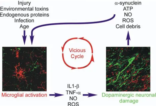
Figure 3: Relationship between microglia activation and dopaminergic neuronal damage. Schematic
representation of the impact of microglial activation on dopaminergic neuronal survival, and the consequent effects of substances released from dying dopaminergic neurons on microglial activation (from Collins et al., 2012).
1.2.2 LIPOPOLYSACCHARIDE-INDUCED PARKINSON’S DISEASE
ANIMAL MODEL
As stated above, PD is characterized by a selective and gradual loss of dopaminergic innervations from the SN to the striatum of the basal ganglia. Findings from epidemiological studies and analysis of postmortem PD brains and animal PD models have provided increasing evidence to support a role for inflammation and microglia activation in the pathogenesis of PD. Due to the role of inflammation in PD, the need for purely inflammation-driven animal models has emerged. An animal model widely used is the lipopolysaccharide PD animal model (Gao et al., 2002).
LPS, an endotoxin found in the Gram-negative bacteria cell wall, is a potent inducer of inflammation, a powerful activator of microglia cells. LPS associates with the soluble LPS binding protein (LBP) and CD14 which is anchored in the outer leaflet of the plasma membrane. Signal transduction across the plasma membrane is made possible through the interaction of the LPS CD14 complex with the transmembrane Toll-like receptor-4 (TLR-4) and the extracellular accessory protein MD-2 (Dutta et al., 2008). Activation of this receptor triggers major intracellular signaling pathways such as the mitogen-activated protein kinase
9
(MAPK) pathway. MAPK are serine-theronine kinases that mediate intracellular signaling and leads to a variety of cellular responses including cell proliferation, differentiation, survival and cell death. There are three main members of the MAPK family, extracellular signal-regulated kinase (ERK), c-Jun N-terminal kinase (JNK) and p38 MAPK, each member exerts different biological functions. For instance, activated ERK1/2 pathway is involved in proliferation and survival whereas JNK and p38 MAPK are associated with apoptosis (Svensson et al., 2011). Downstream the activation of MAPK leads to activation of transcription factors, such as, nuclear factor (NF)-kB and activator protein 1 (AP-1). These transcription factors may be thus involved in the expression of genes involved in pro-inflammatory processes (Kim and Kim, 2005).Besides these signaling pathways activated by LPS that leads to inflammation, various brain regions are differentially susceptible to LPS-induced degeneration. Neurons in the Substantia Nigra (SN) are the most sensitive region to bacterial endotoxin LPS-induced neurotoxicity, whereas neurons in hippocampus or cortex remain insensitive to this treatment, even with higher concentrations of LPS. In the SN, LPS induce a rapid activation of microglia followed by a delayed, progressive and selective destruction of nigral dopaminergic neurons (Gao et al., 2002). The region-specific susceptibility to LPS-induced degeneration is most likely attributable to the abundance of microglia in that region and consequently a high concentration of the inflammation-related factors produced by these cells, such as TNF-α and NO (Pintado et al., 2001; Kim et al., 2000).Selective degeneration of nigral dopaminergic neurons can be also related with the particular vulnerability of these neurons to oxidative stress as they operate under high oxidant conditions due to reduced levels of the anti-oxidant glutathione and increased nigral iron content. Moreover dopamine can generates redox metabolites including semiquinone, quinone, zwitterionic 5,6-hydroxyindoles, and possibly oxygen free radicals that increases the susceptibility of dopaminergic neurons to oxidative stress and consequently contributes to LPS-induced degeneration (De Pablos et al., 2005; Machado et al., 2011).
10
Figure 4: Simplified schematic representation of the link between LPS-induced microglial activation and dopaminergic neurodegeneration. LPS activates microglia cells by binding to its intermediate
receptor CD14, in concert with TLR4 and the accessory adaptor protein MD2. This complex triggers the activation of the MyD88-dependent cascade which initiates NFκB activation, leading to the upregulated expression of pro-inflammatory cytokines (TNFα, IL-1β) and increased production of other inflammatory mediators (NO and PGE2, synthesized by iNOS and COX-2, respectively). These soluble mediators collectively damage nigral dopaminergic neurons. Conversely, MMP-3 and alpha-Synuclein (α-SYN)
released by stressed neurons may aggravate microglial activation and, ultimately, exacerbate
dopaminergic degeneration(from Tufekci et al., 2011).
1.3 HISTAMINE
Histamine (4-imidazolyl-2-ethylamine) is a biogenic amine present as a normal constituent of the body with multiple effects in several organs (Fernández-Novoa and Cacabelos, 2001).
Histamine is mostly stored in the granules of mast cells and basophils. Other sources of histamine include T cells, dendritic cells, platelets and gastric enterochromaffin like cells, to name a few (Schneider et al., 2002). In the CNS, there are three main types of histamine-producing cells: neurons (where it acts as a neurotransmitter), mast cells and microglial cells (Katoh et al., 2002). Histamine exerts its effects by activating four types of receptors, namely: H1R, H2R, H3R, and H4R. All of these histamine receptors belong to the G protein–
11
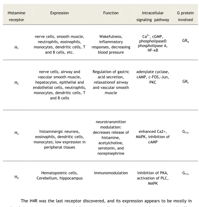
coupled receptorfamily (Marson, 2011). Depending on the type of receptor, histamine plays multiple functions (Table 1).Table 1:Expression, Function, and Signaling of Histamine Receptors and the G Proteins Involved
(Adapted from: Jadidi-Niaragh and Mirshafiey, 2010; Marson, 2011).
The H4R was the last receptor discovered, and its expression appears to be mostly in cells of the immune system, especially in mast cells, lymphocytes, and dendritic cells. This receptor is linked to a chemotactic effect on mast cells and eosinophils (Marson, 2011). However, the expression of H4R in the brain has remained controversial. Several groups could not detect any H4R mRNA in the brain, while a few labs reported their expression by RT-PCR in various parts of the CNS, including amygdala, cerebellum, hippocampus, caudate nucleus, substantia nigra, thalamus and hypothalamus (Strakhova et al., 2009). Interestingly, a recent report demonstrated that all known HRs are expressed in a N9 microglia cell line. Moreover,
Histamine receptor
Expression Function Intracellular
signaling pathway
G protein involved
H1
nerve cells, smooth muscle, neutrophils, eosinophils, monocytes, dendritic cells, T
and B cells, etc.
Wakefulness, inflammatory responses, decreasing blood pressure Ca2+, cGMP, phospholipaseD phospholipase A, NF-κB GRq H2
nerve cells, airway and vascular smooth muscle, hepatocytes, epithelial and endothelial cells, neutrophils,
monocytes, dendritic cells, T and B cells
Regulation of gastric acid secretion, relaxationof airway and vascular smooth
muscle
adenylate cyclase, cAMP, c-FOS,-Jun,
PKC GRs
H3 histaminergic neurons,
eosinophils, dendritic cells, monocytes; low expression in
peripheral tissues neurotransmitter modulation: decreases release of histamine, acetylcholine, serotonin, and norepinephrine enhanced Ca2+, MAPK, inhibition of cAMP Gi/o H4 Hematopoietic cells, Cerebellum, hippocampus
immunomodulation Inhibition of PKA,
activation of PLC, MAPK
12
was also demonstrated the expression of H4R in microglia from the cortex. The same authors show that histamine per se stimulates microglia motility and most interestingly they saw that in a LPS-induced inflammatory context histamine plays an inhibitory action on microglia migration and in the release of IL-1β (Ferreira et al., 2012).These findings could provide a new approach for the treatment of CNS pathologies or neurodegenerative disorders which are commonly accompanied by inflammation.1.3.1 ROLE OF THE HISTAMINERGIC SYSTEM IN PARKINSON’S
DISEASE
Histaminergic neurons are located exclusively in the tuberomammillary nucleus (TM) of the hypothalamus, from where they project to practically all brain regions including SN (Lee et al., 2008). The dopaminergic and histaminergic systems interact extensively, but little is known about the role of the histaminergic system in diseases affecting the dopaminergic neurons (Anichtchik et al., 2000). However, it is known that several functions regulated by the histaminergic system including the sleep-wake cycle, sensory and motor adjustment, cognition, attention, learning and memory are altered in PD (Shan et al., 2012).
In post-morten brain from PD patients, has been reported a dramatic increase of histaminergic innervations and histamine concentration in the SN (Anichtchik et al., 2000; Rinne et al., 2002). Moreover, a Thr105Ile polymorphism of histamine methyltransferase (HMT), the main enzyme breaking down histamine, was observed to be associated with PD, suggesting that a changed histamine homeostasis in the CNS is associated with the risk for PD (Palada et al., 2012). Besides, histamine is able to produce a specific degeneration of dopaminergic neurons in SN along with a highly inflammatory process (Vizuete et al., 2000).
Based on these studies, it has been proposed that histamine may play a role in the pathogenesis of PD. However, it is still unclear the exact role of histamine in the degeneration of mesencephalic dopaminergic neurons in the context of PD.
13
Chapter 2
OBJECTIVES
Recently, Ferreira and collaborators showed that microglial cells from the cortex express the H4R and that histamine may trigger dual effects (in the presence or absence of LPS) in microglia migration and cytokines release (Ferreira et al., 2012). However, there is no information regarding the effects of histamine in microglia derived from the SN, a brain region with a high density of these cells and highly susceptible to dopaminergic neuronal loss present in Parkinson’s disease. With this in mind, we proposed to:
Analyze the expression of histamine H4 receptor in primary microglia cell cultures from SN;
Study the effects of histamine on the production of NO by microglial cells derived from SN, in the presence or absence of an inflammatory context mimicked by LPS;
Spell out which histamine receptors are involved on the production of NO, both in the presence or absence of an inflammatory stimulus;
Evaluate the effects of conditioned medium derived from microglial cells previously treated with LPS and/or histamine in cellular viability of dopaminergic neurons;
Evaluate the effects of histamine and/or LPS per se in cellular viability of dopaminergic neurons.
14
Chapter 3
MATERIALS AND METHODS
3.1. IN VITRO CELL CULTURES
3.1.1. N9 MICROGLIA CELL LINE CULTURE
Murine N9 microglia cell line (kind gift from Prof. Claudia Verderio, CNR Institute of Neuroscience, Cellular and Molecular Pharmacology, Milan, Italy)was grown in RPMI medium supplemented with 30 mM glucose (Sigma, St. Louis, MO, USA), 100 U/ml penicillin and 100 μg/ml streptomycin (GIBCO, Invitrogen, Barcelona, Spain). Cells were kept at 37ºC in a 95% atmospheric air and 5% CO2 humidified atmosphere. When cells reached an approximately 70% confluence was carried out the passage of the cells with a trypsin solution (Sigma, St. Louis, USA). Number of viable cells was evaluated counting trypan blue-excluding cells which were then plated at a density of 2×10⁴ cells per well in 24-well trays. Cell treatments included the following incubation setup: histamine dihydrochloride (1-100 μM, Sigma) or LPS (100 ng/ml, Sigma) for 24h.
3.1.2. PRIMARY MICROGLIA CELL CULTURES FROM SUBSTANTIA NIGRA
All animals were handled in accordance with the national ethical requirements for animal research, and with the European Convention for the Protection of Vertebrate Animals Used for Experimental and Other Scientific Purposes (2010/63/EU).
Briefly, the ventral midbrain of postnatal day 2 or 3 Wistar rat pups was dissected, carefully stripped of the meninges, and put in iced phosphate buffer saline (PBS: NaCl 140 mM, KCl 2.7 mM, KHPO4 1.5 mM and Na2HPO4 8.1 mM, pH 7.4). The tissue was then digested in cysteine solution (1.9 mM CaCl2, 1.3 mM cysteine) and H&B solution (116 mM NaCl, 5.4 mM KCl, 26 mM NaHCO3, 12 mM NaH2PO4.H2O, 1 mM MgSO4.7H2O, 0.5 mM EDTA, 25 mM glicose, pH 7.3) supplemented with 20 U/ml papain and 0.001% phenol red. The average time for digestion was 4 min at 37ºC. After the digestion, the tissue was removed to a sterile tube and washed 3 times with 5 mL of warmed Dulbecco’s modified Eagle’s medium (DMEM, Life
15
Technologies) with 10% Fetal Bovine Serum (FBS, Biochrom AG), and 100 U/ml penicillin plus 100 µg/ml streptomycin (Sigma). The tissue was then mechanically dissociated with a 5 mL pipette, followed by further 5-10 sequential passes with techtips. Finally, the tissue was pelleted by centrifugation (3K18C Bioblock Scientific; Sigma Laboratory Centrifuges) for 3 min at 405 g and then resuspended in DMEM. Number of viable cells was evaluated counting trypan blue-excluding cells which were then plated at a density of 0.233 x 10⁶ cells per well in 48-well trays (NO release) and 0.402 x 10⁶ cells per well in 24-well trays in slides coated with poly-D-lysine (Sigma-Aldrich, St. Louis, USA) (immunocytochemistry). The cultures were kept at 37oC under a 5% CO2 and 95% air atmosphere. The medium was changed every 7 days. After 20-21 days in vitro, the microglia were obtained by trypsinization of astrocytes with a trypsin solution (Sigma) diluted 1:3 in DMEM (without FBS, penicillin and streptomycin) for 40 min. Microglia were kept in DMEM with 10% FBS, and 100 U/ml penicillin plus 100 µg/ml streptomycin at 37 ºC in a 5% CO2, 95% air atmosphere for further 5 days.
3.1.3 NEURON-ASTROCYTE MIDBRAIN CO-CULTURES
The embryos of Wistar pregnant females with 15 or 16 days of gestation were removed and the ventral midbrain was dissected, carefully stripped of the meninges, and put in phosphate buffer saline (PBS: NaCl 140 mM, KCl 2.7 mM, KH2PO4 1.5 mM and Na2HPO4 8.1 mM, pH 7.4). The tissue was then dissociated by enzymatic digestion (Tripsin 4.5 mg/ml and DNAse 2.5 mg/ml diluted in PBS) and incubated at 37º C for 5 min. The cells were pelleted by centrifugation (3K18C Bioblock Scientific; Sigma Laboratory Centrifuges) for 1 minute at 88 g. A solution containing PBS with 10% Fetal Bovine Serum (FBS) heat-inactivated (Biochrom, Holliston, USA) was used to stop the enzymatic digestion and the pellet was then centrifugated for 1 min at 88 g. After discarding the supernatant, cells were rinsed with the PBS solution and then mechanically dissociated with a 5 mL pipette, followed by further 5-10 sequential passes with techtips. Cell suspension was then collected by centrifugation for 3 minutes at 405 g and then resuspended in Neurobasal Medium (Gibco, Paisley, Scotland, UK) supplemented with B27 2%, glutamate 25 M/mL, glutamine 0.5 mM/mL and gentamicine 120
g/mL. Viable cells were counted by the trypan blue exclusion method and were plated at a density of 0.8x10⁶ cells per well in 24-well trays in slides coated with poly-D-lysine
(Sigma-Aldrich, St. Louis, USA). The cultures were kept at 37º C in a 5% CO2 and 95% air atmosphere during 5-6 days. After cells reach confluence, a 5- fluorodeoxyuridine solution (FDU: uridine 16.5 µg/mL and 5-Fluoro-5′-deoxyuridine 6.7 µg/mL) (Sigma-Aldrich, St. Louis, USA) was added to inhibit further cell culture growth.
16
3.2. CELL TREATMENTS
Cell treatments include the following incubation setup: histamine dihydrochloride (100 μM, Sigma) and/or LPS (100 ng/ml, Sigma) for 24h (cell death assays); histamine dihydrochloride (1-100 μM, Sigma), LPS (100 ng/ml, Sigma), H1 receptor antagonist, 2-((2-(dimethylamino)ethyl)(p-methoxybenzyl)amino)-pyridine maleate (mepyramine maleate, 1 μM), H2 receptor antagonist, N-cyano-N’-methyl-N’’-[2-[(5-methyl-1Himidazol- 4-yl)methyl]thio]ethyl]guanidine (cimetidine, 5 μM), H3 receptor antagonist 3-amino-N-[2-(1H-imidazol-4-yl)ethyl]propanamide ditrifluoroacetate (carcinine ditrifluoroacetate, 5 μM), H4 receptor antagonist, 1-[(5-chloro-1H-indol-2-yl)carbonyl]-4-methylpiperazine (JNJ7777120, 1 μM) and H4 receptor agonist, 5-(2-aminoethyl)-4-8methylimidazole dihydrochloride (4-methylhistamine dihydrochloride, 20 μM), (all from Tocris, MO, USA) for 24h (NO release). All histamine receptor antagonists were added 40 min prior to cell treatments.
To assess the effects of soluble mediators released by microglia on dopaminergic neuronal viability, primary microglia cell cultures were exposed for 24h with HIS 100 µM and/or LPS 100 ng/ml and the resulting conditioned medium (MCM) was collected and stored at -80 ºC. The MCM was then added to neuron-astrocyte midbrain co-cultures for further 24h and the viability of TH-neurons was evaluated. In another set of experiments, HIS 100 µM and/or LPS 100 ng/ml were added directly to neuron-astrocyte midbrain co-cultures for 24h and the viability of dopaminergic neurons was then evaluated.
3.3. EVALUATION OF CELL DEATH ASSAYS
3.3.1. PROPIDIUM IODIDE UPTAKE
The propidium iodide (PI; 3,8-diamino-5-(3-(diethylmethylamino)propyl)-6-phenyl phenanthridinium diiodide) is a stable fluorescent dye absorbing blue-green light (493 nm) and emitting red fluorescence (630 nm). As a polar substance it only enters dead or dying cells with a damaged or leaky cell membrane, interacting with DNA to yield a bright red fluorescence. PI is non-toxic to cells and has been used as an indicator for cellular membrane integrity and cell damage. After cell exposure to histamine and/or LPS for 24h, 3 µg/ml of PI was added for further 40 min at 37º C in a 5% CO2 and 95% air atmosphere. Then, the cells were fixed for 30 min in PFA 4% at room temperature (RT). For nuclear labeling, cell preparations were counterstained with Hoechst (2 μg/ml) (Molecular Probes) in PBS, for 5 min at RT and mounted with a fluorescent mounting medium (DAKO, Glostrup, Denmark). The cellular uptake of PI was recorder by fluorescence microscopy (Zeiss Axio imaging Microscope (Axiobserver Z1, Zeiss) using a 63x lens).
17
3.3.2. TUNEL LABELING
Cell apoptosis in microglial cells was evaluated by the terminal deoxynucleotidyl transferase (TdT)-mediated dUTP nick-end labeling (TUNEL). This method in based on the specific binding of TdT, which attaches nucleotides (dUTP), to 3’-OH ends of the DNA generated during apoptotic-induced DNA fragmentation. Incorporation of biotinylated dUTPs allows the detection of cell apoptosis by immunocytochemistry procedures. At the end of each Histamine and/or LPS incubation (24h), microglia cells were fixed for 30 min in PFA 4% at RT and rinsed in PBS. Then, cells were incubated in a humidified atmosphere with a TUNEL reaction mix (Roche kit, REF: 11684795910) for 60 min at 37ºC. For nuclear labeling, cell preparations were counterstained with Hoechst (2 μg/ml) (Molecular Probes) in PBS, for 5 min at RT and mounted with a fluorescent mounting medium (DAKO, Glostrup, Denmark). TUNEL labeling were assessed using fluorescence microscopy (Zeiss Axio imaging Microscope (Axiobserver Z1, Zeiss) using a 63x lens).
3.4. MEASUREMENT OF NITRIC OXIDE (NO) RELEASE
Nitric oxide concentration was determined by measuring the total amount of nitrite (NaNO2, including nitrate that is converted to nitrite by the Griess reagent), one end product of NO oxidation that is released to the culture medium. This assay relies on a diazotization reaction that was originally described by Griess in 1879, and is based on the chemical reaction which uses sulfanilamide and NED (N-1-napthylethylenediamine dihydrochloride) under acidic conditions. The amount of NO formed was determined from the accumulation of the stable NO metabolite (nitrite) in the supernatant after 24h of stimulation. Supernatants (50 μl) were collected, transferred to a 96-well plate, and mixed with an equal volume of the Griess reagent (sulfanilamide plus NED). The mixture was incubated in the dark for 10 min at RT, and the absorbance was read at 540 nm. To ensure accuracy of the nitrite quantification, a reference curve was prepared using as a matrix DMEM. The concentration of nitrite in the samples was determined from a sodium nitrite (NaNO2) standard curve.
3.5. IMMUNOCYTOCHEMISTRY
Cells were fixed in 4% PFA for 20 min at RT. After washing with PBS, unspecific binding was prevented by incubating cells in a PBS solution with 3% Bovine Serum Albumin (BSA) and 0.5% Triton X-100 for 30 min, at RT. The cells were then incubated overnight at 4°C with the primary antibodies diluted in a PBS solution with 0.3% BSA and 0.1% Triton X-100, then washed with PBS the following day, and incubated for 1h at RT with the corresponding
18
secondary antibodies diluted in PBS. Antibodies were used as listed on Table 1. For nuclear labeling, cell preparation were stained with Hoechst (2 μg/ml) (Molecular Probes) for 5 min at RT and mounted with a fluorescent mounting medium (DAKO, Glostrup, Denmark). Fluorescent images were acquired using a fluorescence microscopy (Zeiss Axio imaging Microscope (Axiobserver Z1, Zeiss) using a 63x lens).Table 2. Primary and secondary antibodies used for immunocytochemistry. Primary
antibody Target Dilution Company
Secondary
antibody Dilution Company
Mouse anti CD11b Microglia 1:600 Chemicon Goat anti mouse 488 1:200 Invitrogene Mouse anti TH Dopaminergic neurons 1:1000 Abcam Goat anti mouse 488 1:1000 Invitrogene Rabbit anti
MAP2 Neurons 1:200 Chemicon
Goat anti rabbit 594 1:1000 Invitrogene Goat anti H4R Histamine H4 receptor 1:100 Santa Cruz Biotechonology Rat anti Goat 488 1:200 Molecular probes
Legend: CD11b, cluster of differentiation molecule 11B; TH, tyrosin hydroxylase; MAP2, Microtubule-associated protein 2; H4R, histamine H4 receptor.
3.6 DATA ANALYSIS
Data are expressed as percentages of values obtained in control conditions or as percentages of the total number of cells, and are presented as mean ± S.E.M. of at least three independent experiments, performed in triplicate. Statistical analysis was performed using one-way ANOVA followed by the Dunnett’s test. Values of P<0.05 were considered significant. All statistical procedures were performed using GraphPad Prism 5 Demo (GraphPad Sotware Inc., San Diego, CA).
19
Chapter 4
RESULTS
4.1 Characterization of primary microglia cell cultures
derived from the SN.
The cell culture methodology for purification of microglia from the SN was an indirect method starting from a mixed primary co-culture of astrocytes and microglial cells which grow for 21 days. The microglia cells were then purified by the removal of astrocytes using a mild trypsinization protocol (see section 3.1.2). To assess the purity of these primary microglia cell cultures, we performed an immunocytochemical staining for the alpha chain of αMβ2-integrin, CD11b, a well known surface marker for microglia, whose over-expression is associated to microglial activation (Fig. 5). We found that about 95% of cells were CD11b positive microglial cells (n=3 independent cell cultures). However some residual astrocytic
cells could be also found on our culture, especially at the border of the glass slides (Fig. 5B).
Figure 5: Immunocytochemical stainings of a primary microglia cell culture derived from SN of the Wistar neonatal rats. Representative photomicrographs taken at the center of slide (A) and in periphery
(B). The microglia cells were stained with an anti-CD11b antibody (green) and for nuclear labeling, cell preparations were counterstained with Hoechst 3342 (blue). White arrows depict some cells that do not stain for CD11b.
A) B)
Hoescht
CD11b
20
4.2 Expression of histamine H4 receptor in microglial cells
derived from SN.
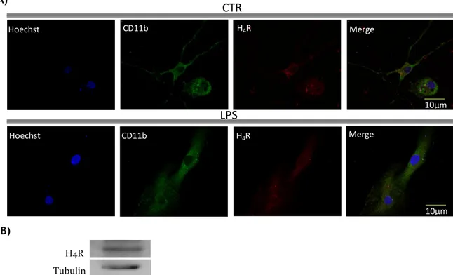
Recently, it was reported by Ferreira and collaborators (Ferreira et al. 2012) that both the N9 microglia cell line as well as primary microglia cells isolated from the cortex of rats express the H4R. In this project, we analyzed the expression of this receptor in primary microglia cell cultures derived from SN, a region with a density of microglia 4-5 times higher than in other brain regions. The results show that SN-derived microglial cells indeed express H4R, as detected by immunocytochemistry (Fig. 6A) and western blot (Fig. 6B). Furthermore, to determine whether differences regarding the pattern of receptor expression existed in an inflammatory context, we stimulated the microglial cells with LPS (100 ng/ml) for 24h and then immunocytochemistry against H4R was also performed. As shown in Fig.6A, no differences in H4R protein expression were found between the control and the LPS-treated cells. Negative controls were performed to confirm the specificity of the primary antibody used for the detection of H4 receptors.
A)
B)
Figure 6: Microglia cells from SN express the histamine H4 receptor. A) Immunocytochemical analysis
of histamine H4 receptor expression on untreated microglial cells (CTR) and treated with 100 ng/mL LPS for 24h. Photomicrographs depict staining for nuclei (Hoechst, blue), microglial cells (CD11b, green) and histamine H4 receptor (H4R, red). B) Histamine receptor expression analysis by western blot showed that microglial cells express the histamine H4 receptor (H4R: 44 KDa; Tubulin: 55 KDa).
H4R Tubulin
CTR
LPS
CD11b CD11b Hoechst Hoechst H4R H4R Merge Merge 10µm 10µm21
4.3 Effect of histamine on the production of NO.
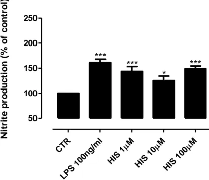
A feature of brain inflammation is the release of inflammatory mediators, such as NO, by activated microglial cells (Stence et al., 2001; Gibbons and Dragunow, 2006).It is known that histamine can induce microglia mobility and IL-1β release (Ferreira et al., 2012) but their effect on NO production is unknown. To study this effect, we measured the amount of nitrite (a stable metabolite of NO) released by an N9 microglia cell line culture after 24h of treatment with different histamine concentrations (1 µM; 10 µM and 100 µM). The results showed that histamine stimulation significantly increased the NO release, and this increase is directly proportional with the histamine concentration (meanHIS1µM = 144.3±6.3; meanHIS1OµM = 150.6±8.3; meanHIS100µM = 174.3±12.4, n=7-11) (Fig 7A). As we expected, LPS stimulation (100 ng/ml; positive control) leads to an increase of NO production (meanLPS100ng/ml= 218.6±19.1, n=11) (Fig 7A). The same experimental conditions were also applied to a primary microglia cell culture from SN, since it mimics the physiological condition better than a N9 microglia cell line. Similarly to the in vitro cell line model, both the LPS and the histamine treated cells showed higher levels of NO release as compared to the control condition (meanHIS1µM = 143.7±9.8; meanHIS1OµM = 125.0±9.4; meanHIS100µM = 149.0±5.6; meanLPS100ng/ml = 161.3±6.7, n=4-9) (Fig 7B). Based on these results, we then decided to use primary microglia cell cultures to disclose the receptor involved in histamine-induced NO release.
A) CTR LPS 100n g/ml M HIS 1 M HIS 10 M HIS 100 50 100 150 200 250
***
*
*
***
N
it
ri
te
p
ro
d
u
ct
io
n
(
%
o
f
co
n
tr
o
l)
22
B)C
TR
LP
S
1
00
ng
/ml
M
H
IS
1
M
H
IS
1
0
M
H
IS
1
00
50
100
150
200
250
***
***
*
***
N
it
ri
te
p
ro
d
u
c
ti
o
n
(
%
o
f
c
o
n
tr
o
l)
Figure 7: Histamine-induced NO release from microglia cells. Histamine at 1 µM, 10 µM and 100 µM
triggered an increase of NO release in (A) N9 microglia cell line culture and (B) primary microglia cell culture derived from the SN of neonatal rats. LPS (100 ng/ml) was used as a positive control and also increased significantly the production of NO in both cell cultures. Data are expressed as mean ± SEM. Statistical analysis was performed using one-way ANOVA with Dunnett’s correction (*P<0.05; ***P<0.001 as compared with control). The control was set to 100%.
4.4 H4R activation mediates the production of NO triggered by
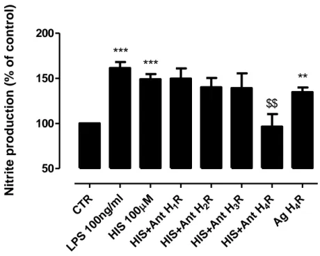
histamine
To uncover which histamine receptor was involved in the modulation of NO production by microglial cells, we then pretreated primary microglia cells culture for 40 min with all histamine receptors antagonists individually (Ant H1R, 1 µM; Ant H2R, 2.5 µM; Ant H3R, 5 µM; Ant H4R, 1µM) followed by an co-incubation for 24 h with histamine (100 µM). As shown in Fig. 8, histamine induces NO production via H4R activation, since in the presence of a H4R antagonist (JNJ7777120, 5 μM), the stimulatory effect on NO production is reversed to values near to control, and the same is not observed for all others antagonists (meanHIS100µM = 149.0±5.6; meanHIS+H1RAnt = 149.7±11.2; meanHIS+H2RAnt = 140.0±10.4; meanHIS+H3RAnt = 139.0±16.5;
23
meanHIS+H4RAnt = 96.4±13.9, n=4) (Fig. 8). Furthermore, when microglial cells were treated with a H4R agonist (4-methylhistamine dihydrochloride, 20 µM) for 24h, the NO production was similar to the levels induced by histamine per se (meanHIS100µM = 149.0±5.6; meanH4RAg = 134.7±5.0)(Fig. 8). These data suggest that histamine per se induced NO release by microglia via H4R activation.C
TR
LP
S
1
00
ng
/ml
M
H
IS
1
00
R
1H
IS
+An
t H
R
2H
IS
+An
t H
R
3H
IS
+An
t H
R
4H
IS
+An
t H
R
4A
g
H
50
100
150
200
$$
***
***
**
Nitrit
e
pro
duc
tion
(% o
f con
tro
l)
Figure 8: Histamine induces NO release by microglia cells via H4 receptor activation. NO production
by microglia cells derived from the SN was increased when cells were treated with 100 ng/mL LPS, 100 µM histamine or 20 µM of an H4R agonist (4-methylhistamine dihydrochloride). Furthermore, histamine induced NO production is abolished in the presence of an H4R antagonist (JNJ7777120; 5 μM). Data are expressed as mean ± SEM. Statistical analysis was performed using one-way ANOVA with Dunnett’s
correction (**P<0.01; ***P<0.001 as compared with control and $$P<0.01 as compared with histamine).
The control was set to 100%.
4.5 Effect of Histamine on NO production in an inflammatory
context induced by LPS
Since histamine per se can induce NO release by microglial cells (Fig. 7), next we evaluated the role of histamine in an inflammatory context induced by LPS. For this, we treated N9 microglia cell line cultures with histamine (100 µM), concentration at which there was a higher increase of NO production (Fig. 7) together with LPS (100 ng/ml) for 24h. Surprisingly, upon LPS and histamine co-administration, NO release induced by LPS alone was significantly inhibited to levels similar to control cultures (meanLPS100ng/ml = 218.6±19.1;
24
meanHIS+LPS = 129.8±11.6, n=7-11) (Fig. 9A). We later explored the role of histamine upon an inflammatory challenge triggered by LPS (100 ng/mL) in a more complex biological model by using a primary microglia cell culture derived from the SN of neonatal rats. Similarly to the in vitro cell line model, we observed that the co-administration of LPS and histamine prevented NO release triggered by LPS per se (meanLPS100ng/ml = 161.3±6.7; meanHIS+LPS = 105.7±9.9, n=7-8) (Fig. 9B).A) CTR LPS 100n g/ml M HIS 100 HIS +LPS 50 100 150 200 250 *** *** ++ N it ri te p ro d u c ti o n ( % o f c o n tr o l)
B) CTR LPS 100 ng/ml M HIS 100 HIS +LP S 50 100 150 200
***
***
+++ N it ri te p ro d u c ti o n ( % o f c o n tr o l)Figure 9: LPS-induced NO release is inhibited by Histamine. LPS and histamine individually increased
NO release while co-administration abolished this effect, both in a (A) N9 microglia cell line culture and in (B) primary microglia cell cultures derived from the SN. Data are expressed as mean ± SEM. Statistical analysis was performed using one-way ANOVA with Dunnett’s correction (***P<0.001 as compared with