online | memorias.ioc.fiocruz.br
Malaria continues to be one of the world’s most dev-astating diseases and is caused by parasites of the genus
Plasmodium. In 2010, there were an estimated 219 million cases of malaria resulting in around one million fatali-ties, mostly in children under five years old (Murray et al. 2012, WHO 2012). Increased awareness of the devastat-ing impacts of malaria has led to a significant reduction of malaria cases and fatalities in the recent past. How-ever, these achievements are threatened by a reduction in the distribution of insecticide treated nets, the resistance of mosquitoes to insecticides, parasite resistance to anti-malarial drugs (including the 1st-line drug artemisinin) together with the levelling of funding for malaria control efforts (WHO 2012). For these reasons, there is an ur-gent need to improve current malaria control efforts and, importantly, to develop new strategies to eliminate and eventually eradicate the disease. Different from the other two major infectious disease killers, human immunodefi-ciency virus and tuberculosis, malaria is unique because it requires a mosquito vector for transmission to occur. Thus, the mosquito stages of the malaria parasite devel-opment have the potential to provide important targets for the control of transmission.
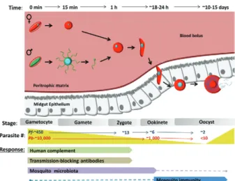
Parasite development in its vector starts when a mosquito ingests an infected blood meal containing
Plasmodium sexual forms, known as gametocytes (Fig. 1). Within ~15 min, gametocytes round-up (in case of
Plasmodium falciparum), egress from the red blood cell (RBC) and differentiate into gametes. Male gametocytes undergo a drastic transformation known as
exflagella-tion by which the DNA replicates to 8N followed by the formation of eight haploid microgametes. Microgametes detach from the exflagellation centre and actively search for female gametes to fertilise. Fertilisation gives rise to a diploid zygote that subsequently undergoes one round of DNA replication to become tetraploid. Zygotes dif-ferentiate into motile ookinetes that migrate in the blood bolus to invade and traverse the mosquito midgut epithe-lium. The ookinete may traverse multiple epithelial cells before emerging from the basal side facing the haemo-coel, where it lodges beneath the basal lamina and dif-ferentiates into a round oocyst. Within the next 10-14 days each oocyst grows in size and undergoes sporogony to produce thousands of sporozoites. Upon oocyst mat-uration, sporozoites are released into the haemolymph where they circulate with the haemolymph and specifi-cally invade the salivary glands. Following invasion, sporozoites lodge in the lumen of the salivary gland. When an infected mosquito feeds on a human host, sporozoites are released with the saliva and deposited in the skin, thus closing the transmission cycle.
Malaria parasites undergo dramatic losses during their development in the mosquito vector (Fig. 1). Popu-lation reduction occurs at each developmental step, from the formation of gametocytes in the human host to oo-cyst formation, resulting in very low parasite numbers. In fact, even in high transmission areas, the majority of the mosquitoes are not infected by the parasite (Chege & Beier 1990, Mbogo et al. 1993, Mendis et al. 2000, Gouagna et al. 2010). This reduction in numbers in the mosquito midgut is mediated in part by the transition of the parasite from an intracellular (RBC) to extracellular forms, thus exposing the parasites to both human and mosquito components that are deleterious to the para-site (Figs 1-3). It has been estimated that out of the thou-sands of gametocytes that a female Anopheles mosquito typically ingests in a blood meal, only 50-100 develop into ookinetes and only around five survive to form oo-cysts (Gouagna et al. 1998, Sinden & Billingsley 2001, Whitten et al. 2006). A study performed in Anopheles gambiae mosquitoes with blood from P. falciparum -infected patients showed that on average, from 433.5
doi: 10.1590/0074-0276130597
Financial support: NIAID USA (AI031478)
Additional support was provided by a JHMRI Post-doctoral ship (to RCS) and a Calvin S and Helen H Lang Post-doctoral fellow-ship (to JV-R). RCS and JV-R contributed equally to this work. + Corresponding author: [email protected]
Received 26 December 2013 Accepted 14 March 2014
The
Plasmodium
bottleneck:
malaria parasite losses in the mosquito vector
Ryan C Smith, Joel Vega-Rodríguez, Marcelo Jacobs-Lorena/+
Department of Molecular Microbiology, Malaria Research Institute,
Johns Hopkins Bloomberg School of Public Health and Immunology, Baltimore, MD, USA
Nearly one million people are killed every year by the malaria parasite Plasmodium. Although the disease-caus-ing forms of the parasite exist only in the human blood, mosquitoes of the genus Anopheles are the obligate vector for transmission. Here, we review the parasite life cycle in the vector and highlight the human and mosquito contribu-tions that limit malaria parasite development in the mosquito host. We address parasite killing in its mosquito host and bottlenecks in parasite numbers that might guide intervention strategies to prevent transmission.
gametocytes detected in patient blood, only 12.6 round forms, 5.5 ookinetes, 1.8 early oocysts and two mid-size oocyst were detected in the mosquito (Gouagna et al. 1998) (Fig. 1). More importantly, this study also shows that prevalence (the proportion of mosquitoes that carry at least 1 parasite) was only 38%, meaning that 62% of the mosquitoes that fed on the infected blood never got infected (Gouagna et al. 1998). In the entire Plasmodium
life cycle (in both human and mosquito hosts), parasite numbers are lowest during the oocyst stage and then quickly expand when each oocyst releases thousands of sporozoites. For this reason, the midgut stages of para-site development constitute prime targets for strategies aiming to block malaria transmission.
Here we discuss the multiple mechanisms that limit parasite survival in the mosquito, starting with events occurring in the human host, and then in the mosquito midgut lumen (Fig. 2), and finally during invasion of the mosquito midgut epithelium (Fig. 3). In addition, we de-scribe the mechanisms that the parasite has evolved to evade some of these antiplasmodial responses. Finally, we address strategies that are under consideration to target
Plasmodium development in the mosquito and discuss fu-ture challenges that need to be overcome in order to suc-ceed in any malaria transmission-blocking (TB) strategy.
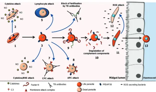
Human factors - Cytokines and reactive nitrogen spe-cies (RNS) - Evidence suggests that cytokines produced during malaria infections can mediate the killing of
gametocytes circulating in the blood stream of the host (Naotunne et al. 1991, Karunaweera et al. 1992) (Fig. 2). Infections of Plasmodium cynomolgi in toque monkeys or Plasmodium vivax in humans cause a period known as “crisis” or clinical paroxysms that are characterised by acute fever preceded by chills and rigor that coincide with the release of asexual parasites from RBCs (Naotunne et al. 1991, Karunaweera et al. 2003). During this period,
Plasmodium gametocytes lose infectivity to mosquitoes due to an increase of the pro-inflammatory cytokines tumour necrosis factor (TNF)-α and interferon (IFN)-γ produced by the host immune system (Naotunne et al. 1991, Karunaweera et al. 1992). Although not completely understood, two proposed mechanisms by which TNF-α and IFN-γ could affect Plasmodium gametocytes in the human host are through the induction of phagocytosis or by increased nitric oxide (NO) production by leukocytes (Naotunne et al. 1993, Muniz-Junqueira et al. 2001).
Similar to the effects of pro-inflammatory cytokines, the levels of RNS have also been shown to increase dur-ing the paroxysm periods of P. vivax infections (Nao-tunne et al. 1993, Cao et al. 1998). Induced by unknown parasite factors, the activation of leukocytes results in an up-regulation of NO synthase (NOS) expression and increased NO production (Naotunne et al. 1993). NO is a highly reactive molecule and its reaction products (RNS) induce damage to DNA, proteins and lipids that ultimately result in cell death. Supporting the role of NO, Naotunne et al. (1993) demonstrate that TNF-α-mediated gametocyte inactivation is dependent on leukocyte acti-vation and the production of NO. The inhibition of NO synthesis restored the infectivity of P. falciparum and
P. vivax gametocytes to mosquitoes (Motard et al. 1993, Naotunne et al. 1993). Similarly, in mouse infections with Plasmodium yoelii, gametocyte infectivity to mos-quitoes is highly impaired when mice experience crisis (4-5 days after infection) (Cao et al. 1998). During this time, treatment of the mice with the NOS inhibitor L-NMMA partially restored gametocyte infectivity sug-gesting that RNS are involved in this inhibitory effect.
One important aspect of these studies is that serum from semi-immune patients, which present mild clinical symptoms, did not affect P. vivax gametocyte infectivity (Karunaweera et al. 1992). This would suggest that clinical immunity is an adaptation in which parasite-killing fac-tors (e.g., TNF-α) are reduced in order to diminish disease pathology (Karunaweera et al. 1992), where lower levels of circulating TNF-α would have little effect on circulating gametocytes. This is in contrast to the high TNF-α levels and increased NO production by a non-immune malaria-infected host (e.g., a child under 5 years old), which re-duces gametocyte infectivity of the mosquito (Mshana et al. 1991, Othoro et al. 1999, Lyke et al. 2004).
In summary, these data suggest that human factors that limit gametocyte infectivity are probably the first contributors to the reduction of malaria parasite num-bers in the mosquito.
Mosquito midgut lumen - Cytokines - In addition to their effects in the human host, antimalarial blood stream components can also target the parasite in the
mosquito midgut lumen (Fig. 2). Blood components, in-cluding white blood cells (WBCs), cytokines, comple-ment proteins, RNS and other factors, remain active for several hours in the mosquito midgut after blood inges-tion (Lensen et al. 1997, Margos et al. 2001, Simon et al. 2013). TNF-α was shown to reduce the formation of Plasmodium berghei ookinetes through the RNS-medi-ated reduction of exflagellating males and subsequent ookinete formation (Ramiro et al. 2011). Although the mechanism by which TNF-α enhances RNS inhibition of male gamete exflagellation is unknown, it has been proposed that the reduction in ookinete numbers could be the result of TNF-α-induction of leukocyte phago -cytosis of sexual stage parasites (Muniz-Junqueira et al. 2001). In support of this theory, phagocytosis of P. falciparum and P. berghei gametocytes/gametes by lymphocytes was shown to occur in vitro and in vivo in the midgut of An. gambiae mosquitoes after inges-tion of an infected blood meal (Sinden & Smalley 1976, Lensen et al. 1997).
RNS - RNS have been shown to affect the develop-ment of the malaria parasite inside the mosquito midgut lumen (Fig. 2). Pre-incubation of P. yoelii gametocytes with NOC5, a NO donor, inhibits gametogenesis and zygote formation (Cao et al. 1998). In addition, it has been reported that up to 50% of P. berghei ookinetes
developing inside the midgut of Anopheles stephensi
mosquitoes show markers of apoptosis. It was hypoth-esised that these killing effects could be achieved in newly formed ookinetes when exposed to RNS or reac-tive oxygen species (ROS) donors (Ali et al. 2010). Sup-porting this theory, the removal of WBCs or treatment with the NOS inhibitor L-NAME significantly reduced the number of apoptotic ookinetes (Ali et al. 2010). In addition, Ramiro et al. (2011) showed that RNS can af-fect P. berghei male gamete exflagellation, fertilisation, as well as ookinete development.
Activated WBCs are believed to be one of the pri-mary sources of NO that affect the sexual stages of the parasite in the human host and in the mosquito midgut. However, additional sources of NO and RNS come into play after a mosquito takes a blood meal.
Upon ingestion of a malaria infected blood meal, mosquito midgut levels of NO and nitrates significantly increase (Luckhart et al. 2003, Peterson et al. 2007). Di-gestion of RBCs by mosquito proteases release haemo-globin which comprises ~90% of the total ingested blood mass (Briegel & Rezzonico 1985). Oxyhaemoglobin and haeme which persist throughout blood digestion in the mosquito midgut can react with NO and ROS to produce toxic NO metabolites which in turn can affect malaria parasite development (Peterson et al. 2007).
In addition, the mosquito midgut epithelium can also be a source of NO. The up-regulation of NOS has been observed in midguts of An. gambiae and An. stephensi
mosquitoes upon ingestion of a malaria infected blood meal (Dimopoulos et al. 1998, Luckhart et al. 1998). Blood components have also been shown to increase mosquito midgut NOS expression. The cytokine trans-forming growth factor-β1 (TGF-β1), a component of the human serum, at low concentrations induces An. ste-phensi NOS and reduces the mosquito parasite burden (Luckhart et al. 2003). In addition, P. falciparum gly-cosylphosphatidylinositol and parasite-derived haemo-zoin pigments also appear to induce An. stephensi NOS expression in the mosquito midgut epithelium (Lim et al. 2005, Akman-Anderson et al. 2007). However, the impact that each of these NO sources (especially mid- gut-derived NO) have on each parasite stage in the mid- gut lumen remains to be determined.
The two main antioxidant systems of the malaria parasite, the thioredoxin and the glutathione (GSH) re-dox systems, play a protective role during the parasite development in the mosquito. Interruption of the GSH redox pathway by disruption of the gamma-glutamyl-cysteine synthetase gene (the rate limiting enzyme in GSH biosynthesis) or alternatively, the GSH reductase gene, results in the formation of fewer ookinetes and stunted oocysts that fail to fully develop in An. stephensi
mosquitoes (Vega-Rodríguez et al. 2009, Pastrana-Mena et al. 2011). Likewise, P. berghei oocysts lacking thioredoxin-dependent 2-Cys peroxidase 1 expression, which is part of the thioredoxin redox system, produce fewer sporozoites (Yano et al. 2008). Moreover, P. ber-ghei ookinetes increase the expression of antioxidant peroxiredoxins after exposure to increased levels of oxidative stress (Turturice et al. 2013). It is conceivable that the malaria parasite regulates its antioxidant de-fense systems to overcome the damage cause by RNS in the mosquito midgut lumen. The mosquito peritrophic matrix (PM), a cellular layer that completely surrounds the blood meal and is secreted by the midgut epithelium in response to feeding, may constitute another protec-tive mechanism from RNS damage. Proteins from the PM have been shown to bind and consequently remove haeme produced by haemoglobin digestion, thus reduc-ing its capacity to form additional RNS (Pascoa et al. 2002, Devenport et al. 2006). The protective properties of the PM have yet to be determined, but presumably this structure confers some level of protection for parasites located at the periphery of the blood bolus where active digestion is taking place.
Host complement system - The complement system is part of the innate immune defense in vertebrates and is a first-line defense against pathogens including bacteria, fungi and protozoans. After a foreign organism is de-tected by the vertebrate host, the complement system is activated within seconds through either the classical, lec-tin or alternative pathways [reviewed in Walport (2001)]. This complex system is made up of around 30 abundant proteins in the blood plasma. Once complement has been activated, ultimate destruction of the foreign organism
can be achieved by several mechanisms including inges-tion by phagocytes or by the assembly of a membrane attack complex on the pathogen surface that ultimately results in its killing.
Infection of the human host by malaria parasites activates both the classical and alternative complement pathways (ACP) (Adam et al. 1981, Wenisch et al. 1997, Goka et al. 2001, Roestenberg et al. 2007). By compar-ing the infectivity of Plasmodium parasites to mosqui-toes by feeding gametocytes in either native or in heat-inactivated serum (heat destroys complement activity), parasites in inactivated serum produced significantly more oocysts than those in native serum (Grotendorst et al. 1986, Tsuboi et al. 1995, Margos et al. 2001, Simon et al. 2013). Furthermore, Grotendorst et al. (1986) dem-onstrated that the ACP is responsible for this reduction in parasite numbers. When factor B (a specific compo-nent of the alternative pathway) is removed from serum, parasite viability is increased. Similar experiments, in which components specific to the classical pathway were also removed from serum, did not alter parasite viability. Interestingly, the early mosquito stages of the parasite (gametocytes, gametes and early zygotes) are more pro-tected from complement attack than later stages (late zy-gotes and ookinetes) (Grotendorst et al. 1986, Tsuboi et al. 1995, Margos et al. 2001, Simon et al. 2013), suggest-ing that malaria parasites may have evolved a mecha-nism of protection during their most vulnerable stages.
Immediately after the female mosquito feeds on blood, the midgut starts secreting digestive enzymes that degrade proteins from the serum and the RBCs. Host complement proteins are also exposed to mosquito proteases which raise the question of how long does the complement system from the human serum remain ac-tive after the mosquito has taken a blood meal? Two dif-ferent studies addressed this question by measuring the products of C3 activation, which results in the formation of C3a (Simon et al. 2013) and C3b (Margos et al. 2001). Both reports show that the peak of complement activity in the midgut is during the first hour post-blood-feeding and remains active up to 6 h after feeding. This loss of complement activity in the mosquito blood bolus also coincides with the transition of the parasite from com-plement-resistant stages (gametocytes, gametes and ear-ly zygotes) to those that are complement-sensitive (late zygotes and ookinetes) (Fig. 2) (Grotendorst et al. 1986, Tsuboi et al. 1995, Margos et al. 2001, Simon et al. 2013). However, digestion of the blood bolus is not uniform and occurs from the outside in. Therefore, it can be predicted that the complement molecules closer to the periphery of the blood bolus will be inactivated faster and conse-quently that parasites located in these areas will be pro-tected from complement attack much earlier.
proteins. There is evidence to suggest that Plasmodium
has evolved to acquire a complement regulatory protein needed to evade complement attack during its develop-ment in the mosquito blood bolus (Fig. 2).
Parasites became sensitive to complement attack fol-lowing treatment of Plasmodium gallinaceum gametes with trypsin, implying that a parasite surface protein is responsible for the protection from complement (Groten-dorst et al. 1986). This was further examined in a recent report showing that Plasmodium gametes and zygotes bind on their surface factor H (FH) from the host serum. FH regulates ACP activation by binding to C3b thus pre-venting the formation of the C3 convertase and acceler-ating its decay [reviewed in de Córdoba et al. (2004)]. When mosquitoes were fed with P. falciparum gameto-cytes in native human serum together with anti-FH an-tibodies, mosquitoes were rendered resistant as parasite numbers were effectively reduced to zero (Simon et al. 2013). Co-immunoprecipitation assays with anti-FH an-tibodies on protein extracts from activated gametes in native human serum identified PfGAP50 as a parasite receptor for FH (Simon et al. 2013). However, the au-thors suggest that there are additional unknown FH re-ceptors on the surface of the gametes as anti-PfGAP50 antibodies only reduced the infectivity to mosquitoes by 38-60%. This hypothesis is plausible as other pathogens including Streptococcus pyogenes and Borrelia burg-dorferi use more than one surface protein to capture FH from the host and evade the ACP response [reviewed in Zipfel et al. (2007)].
TB antibodies - Naturally acquired immunity against malaria asexual stages can confer partial protection against the disease [reviewed in Doolan et al. (2009)]. In addition to the asexual stages, gametocytes also cir-culate in the blood. If a gametocyte is not ingested by a mosquito during blood-feeding, it decays and evidence suggests that it is eventually removed from circulation by the host immune system resulting in the production of gametocyte-specific antibodies (Baird et al. 1991, Taylor & Read 1997, Saeed et al. 2008). Such antibodies would also be ingested by the mosquito and could inter-fere with progression of the developmental cycle in the midgut lumen (Fig. 2).
Immune sera of individuals living in malaria endem-ic regions have TB activity against the mosquito midgut stages of both P. falciparum and P. vivax (Baird et al. 1991, Mulder et al. 1994, Lensen et al. 1998, Arévalo-Herrera et al. 2005, Bousema et al. 2011). Specifically, antibodies against the gametocyte/gamete proteins Pfs48/45 and Pfs230 show a negative correlation be-tween antibody activity and parasite development in the mosquito (Healer et al. 1999, Drakeley et al. 2004, van der Kolk et al. 2006, Bousema et al. 2007, 2010). Both Pfs48/45 and Pfs230 participate in gamete fertilisation and are members of a 6-cysteine protein family contain-ing adhesive domains (van Dijk et al. 2001, 2010, Eksi et al. 2006). These proteins are stored in the parasito-phorous vacuole membrane of the gametocyte and relo-cate to the surface of the gamete during activation in the mosquito midgut (Williamson et al. 1996). Gene
disrup-tion of P48/45 and P230 in P. berghei and Pfs230 in P. falciparum drastically reduces infectivity to mosquitoes by interfering with fertilisation (van Dijk et al. 2001, 2010, Eksi et al. 2006).
It appears that anti-Pfs230 antibodies hinder fer-tilisation by a mechanism different from interference with gamete adhesion. Early TB studies reported that antibodies against Pfs48/45, but not Pfs230 inhibited P. falciparum development in the mosquito, a finding that discouraged the use of Pfs230 as a potential TB vaccine antigen (Vermeulen et al. 1985). Later studies showed that anti-Pfs230 antibodies inhibit P. falciparum development in the mosquito in the presence of active serum comple-ment (Quakyi et al. 1987, Read et al. 1994, Healer et al. 1997). These antibodies activate the classical complement pathway resulting in the formation of the membrane-at-tack complex causing lysis of the parasite (Fig. 2).
In summary, these studies highlight the possible ap-plications of an antibody-based TB vaccine to reduce parasite development in its mosquito host and could have a profound influence on the transmission of malaria in endemic countries.
Midgut microbiota - Mosquitoes, as all higher organ-isms including humans, carry an intestinal microbiota that is mostly composed of bacteria and yeast [reviewed in Dillon and Dillon (2004)]. There is evidence to sug-gest that the symbiotic relationship between bacteria and the mosquito confers to the latter some protection against invading pathogens like malaria. Several studies using laboratory-reared and field-captured Anopheles
mosquitoes have shown that midgut bacteria (prima-rily Gram-negative bacteria) have a negative effect on
P. falciparum and P. vivax development in the mosquito (Seitz et al. 1987, Pumpuni et al. 1993, 1996, Straif et al. 1998, Gonzalez-Ceron et al. 2003, Dong et al. 2009, Cirimotich et al. 2011). For instance, infections after re-moval of endogenous microbiota by antibiotic treatment lead to higher parasite numbers when compared with infections of untreated mosquitoes (Beier et al. 1994, Dong et al. 2009, Meister et al. 2009). In addition, when antisera raised against An. gambiae midgut lysates were fed to An. gambiae mosquitoes together with P. falci-parum cultured gametocytes, there was an increase in the number of parasites that developed into oocysts rela-tive to controls and this effect was attributed to antibac-teria antibodies in the serum (Noden et al. 2011). These results are in contrast to previous reports where similar experiments resulted into a reduced parasite load due to antibodies targeting mosquito proteins required for parasite development (Srikrishnaraj et al. 1995, Lal et al. 2001, Suneja et al. 2003, Chugh et al. 2011). The reasons for these discrepancies are unclear and require further investigation.
Recent studies have shown that certain variants of
al. 2013). The authors hypothesise that a physical barrier imposed by the flagella may contribute to the decreased success of ookinetes to invade the midgut epithelium.
Recent evidence has also emerged that bacteria may influence Plasmodium development directly through the production of antimalarial compounds (Fig. 2). Ci-rimotich et al. (2011) isolated an Enterobacter bacte-rium (Esp_ Z) from Zambian populations of Anopheles arabiensis mosquitoes. Co-feeding of Esp_ Z and P. falciparum-infected blood to mosquitoes reduced the parasite burden in a dose-dependent manner. The an-tiparasitic effect of this bacterium, which is observed at the zygote to ookinete transition, can be rescued by addi-tion of the antioxidant vitamin C to the infectious blood meal. This result suggests that ROS secreted by Esp_ Z
bacteria is responsible for the reduction in parasite num-bers (Cirimotich et al. 2011). These findings have im-portant implications as a naturally occurring bacterium like Esp_ Z could be exploited to develop new malaria TB alternatives. However, as mentioned above, the para-site’s antioxidant systems are highly active during devel-opment in the mosquito and can be regulated according to the redox state of the environment were the parasite is developing (Yano et al. 2008, Vega-Rodríguez et al. 2009, Pastrana-Mena et al. 2011).
Additional evidence suggests that the midgut micro-biota play a direct role in the activation of the mosquito immune response. Shortly after blood ingestion, the resi-dent microbiota undergo dramatic proliferation (about 2 orders of magnitude) peaking at about 24 h (Pumpuni et al. 1996). This strong bacterial proliferation is likely to result in an immune response independent of parasite presence. Dong et al. (2009) reported that the mosquito midgut bacteria induce a basal level of immunity that enhances the expression of antimicrobial immune genes that also have antiplasmodial activity. This is true for the induction of SRPN6 in response to Enterobacter infec-tion, which in turn contributes to the anti-Plasmodium
response of the mosquito (Eappen et al. 2013). Moreover, Rodrigues et al. (2010) reported that bacteria are neces-sary for immune priming.
A delicate balance exists between the commensal gut microbiota and its mosquito host to limit bacterial over-proliferation and the subsequent immune response that may have negative effects towards mosquito fitness. To reduce hyperactivation of the mosquito immune system by the midgut bacteria, the mosquito is thought to form a dityrosine network that restricts contact of bacteria with the midgut epithelium, thus reducing the antimicrobial response (Kumar et al. 2010). As a result, proliferation of the normal gut flora is not impaired and malaria parasite development is unhindered (Kumar et al. 2010). In ad-dition, dual oxidase expression by the mosquito midgut epithelium likely results in ROS production, playing a similar role to that in Drosophila of limiting bacterial numbers (Ha et al. 2009). Moreover, it was proposed that when Plasmodium ookinetes breach the PM and the pro-tective dityrosine network lining the luminal surface of the midgut epithelium, bacteria “leak” through the open spaces thus activating an antibacterial immune response that is also harmful to invading ookinetes (Kumar et al. 2010, Rodrigues et al. 2010).
Physical barriers to Plasmodium development - The PM - The distension of the mosquito midgut by a blood meal induces midgut epithelial cells to secrete compo-nents of an extracellular layer, known as the PM, which completely surrounds the ingested blood. The PM is composed of proteins, glycoproteins and proteoglycans that are structurally linked by chitin (Shao et al. 2001, Dinglasan et al. 2009). The PM constitutes a physical barrier that prevents direct contact of commensal bacte-ria and of components of the blood meal with the midgut epithelium. Initially soft and fragile, the PM polymer-ises and gradually thickens reaching maximal rigidity at about 24 h after the blood meal (Shao et al. 2001). As the PM completely surrounds the blood meal, it consti-tutes the first significant physical barrier to Plasmodium
development in the mosquito. As ookinetes mature (ap-proximately 16-20 h after blood ingestion) they migrate to the periphery of the blood bolus, presumably guided by environmental or sensory cues. Upon contact with the PM, the ookinete secretes a chitinase (and possibly other proteases) from its micronemes to locally disrupt and penetrate the chitinous PM. Inactivation of the chitinase genes dramatically reduces the ability of the ookinete to traverse the PM (Dessens et al. 2001, Tsai et al. 2001). In some parasite species, ookinetes secrete a chitinase pre-cursor (or zymogen) that is activated by mosquito mid-gut proteases (Shahabuddin et al. 1993, Shahabuddin & Kaslow 1994), indicating that the parasite has adapted to the protease-rich environment of the mosquito midgut to facilitate its own development.
The midgut epithelium - After traversal of the PM it is believed that ookinetes display extensive gliding motility along the lumenal surface of the midgut epithelium. This movement may be important to initiate midgut invasion (Zieler & Dvorak 2000), possibly through the interac-tion with one or more of the numerous glycoproteins that comprise the glycocalyx of the midgut epithelium (Shen et al. 1999, Wilkins & Billingsley 2001, Dinglasan et al. 2007a). Carbohydrate moieties on the midgut epithelium seem to play an important role in ookinete binding to the midgut (Zieler et al. 1999, Zieler & Dvorak 2000). Moreover, it appears that the interaction between mos-quito sugars and parasite lectins are required to establish invasion (Zieler & Dvorak 2000, Dinglasan et al. 2007a, b). However, very little is known regarding the specific protein-protein interactions that mediate this process since these carbohydrate moieties are post-translational modifications of yet unknown proteins. Given the impli-cations as possible TB vaccine targets, identifying these specific interactions is a high priority area of research.
(its conformation resembles) of Plasmodium enolase, a protein secreted onto the surface of the ookinete where it acts as an invasion ligand (Ghosh et al. 2011). Ookinete surface enolase also interacts with plasminogen from the blood serum to locally promote its conversion into the proteolytically active plasmin that in turn, is required for midgut invasion (Ghosh et al. 2011). Recently, the mosquito midgut receptor for SM1 and ookinete surface enolase was identified as enolase binding protein (EBP) (Vega-Rodríguez et al. 2014). EBP is expressed on the lumenal surface of the mosquito midgut and is required for P. berghei ookinete midgut invasion through its in-teraction with surface enolase. This inin-teraction can be disrupted through competition with excess SM1, thus inhibiting ookinete midgut invasion (Vega-Rodríguez et al. 2014). However, some ookinetes still invade the mosquito midgut in the presence of excess SM1 and this served as the basis for the selection of SM1-resistant parasites that do not require EBP for midgut invasion. This is the first evidence demonstrating that mosquito midgut invasion by Plasmodium ookinetes, similar to merozoite invasion of the RBC, can occur through mul-tiple invasion pathways (Vega-Rodríguez et al. 2014). While further work is needed to characterise additional midgut receptor(s), these findings suggest that the proc-ess of ookinete midgut invasion is complex and involves multiple parasite-mosquito interactions. Characterisa-tion of these interacCharacterisa-tions remains a major goal for future research and has the potential to lead to novel interven-tion strategies.
The process of ookinete invasion is thought to produce severe damage to the midgut as the ookinete traverses multiple epithelial cells during its journey to the basal lamina. Invaded cells undergo dramatic cytoskeletal changes and increase the production of reactive oxygen and nitrogen species that trigger apoptosis and cell death (Han et al. 2000). During this time ookinetes are likely exposed to a highly toxic cellular environment. To en-sure its survival, the ookinete must rapidly escape be-fore it is damaged or bebe-fore the damaged cell is extruded from the midgut epithelium. This evasion process occurs by invasion of neighbouring naïve cells or by exiting the epithelium to rest at its final extracellular destination between the basal side of the epithelium and the basal lamina. While it remains unclear how ookinete invasion triggers programmed cell death, this is likely a general response to remove damaged epithelial cells and may not be specific to parasite invasion (Baton & Ranford-Cartwright 2005, Okuda et al. 2007).
Recent evidence further supports the concept that midgut traversal is a critical step in the mosquito
anti-Plasmodium response. Invaded cells produce high levels of NOS and peroxidases that in turn increase the levels of midgut nitration (Kumar et al. 2004). Further nitra-tion is increased by the activanitra-tion of the haeme peroxi-dase 2 (HPX2) and the nicotinamide adenine dinucle-otide phosphate (NADPH) oxidase 5 (NOX5), processes that are regulated by the c-Jun N-terminal kinase (JNK) pathway (Oliveira et al. 2012, Garver et al. 2013). This epithelial nitration response is believed to modify ooki-netes, thus “marking” them for immune recognition by
the mosquito complement system (Oliveira et al. 2012, Garver et al. 2013). Thus, midgut nitration appears to be a major determinant of Plasmodium survival, yet many questions remain regarding how nitration mediates im-mune recognition. What proteins on the surface of the ookinete undergo modifications as a result of increased nitration? How are these modifications subsequently recognised by the mosquito complement system?
Components of the anti-Plasmodium immune re-sponse - A great deal of effort has been invested in the identification of components that contribute to parasite developmental success in the mosquito in the hope that this knowledge can be translated to the development of al-ternative strategies for parasite killing. Through the use of dsRNA-mediated gene-silencing, several genes have been described that positively or negatively influence parasite development in the mosquito (Blandin et al. 2008). While more research is required to fully understand the contri-butions of individual mosquito genes to anti-Plasmodium
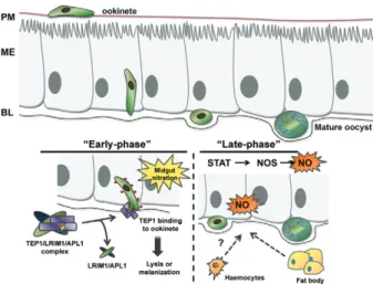
immunity, evidence suggests that parasite killing after traversal of the midgut epithelium occurs primarily dur-ing two separate stages or phases of parasite development (Fig. 3). The first phase, or “early-phase”, occurs during the ookinete to oocyst transition immediately after exit-ing the midgut epithelium. A second, or “late-phase”, is thought to act on developing oocysts.
The “early-phase” immune response - Approximate-ly 18-24 h after blood-feeding, Plasmodium ookinetes invade the lumenal side of the midgut epithelium and within minutes reach the basal surface that is believed to be the major site of ookinete killing (Shiao et al. 2006). At this point, ookinetes are exposed to the haemocoel and to the complement-like soluble immune proteins that circulate in the haemolymph (Blandin et al. 2004, Shiao et al. 2006).
system is able to distinguish their targets, it is reasonable to assume that members of the APL1 gene family have important functions in parasite recognition as part of the mosquito complement-like pathway.
Upon binding to the ookinete, TEP1 is thought to promote parasite killing through lysis or melanisation (Blandin et al. 2004). Recent evidence has identified that a non-catalytic protease, SPCLIP1, may function to amplify complement activation by binding to TEP1 on the pathogen surface and promote the recruitment of ad-ditional TEP1 (Povelones et al. 2013). A similar ampli-fication of the complement response is also seen in the vertebrate complement system where C3b, the cleavage product of C3 (equivalent to TEP1), binds factor B on the surface of the pathogen. This complex is then activated by factor D into the C3 convertase that in turn mediates the recruitment of additional C3b molecules to the path-ogen surface [reviewed in Walport (2001)]. However, our knowledge of this process in the mosquito is incomplete and additional proteins that promote parasite killing may be recruited to the ookinete surface, as is the case for the vertebrate complement system. Multiple proteins and co-factors are known to assemble onto pathogen surfac-es as part of the mammalian complement pathways and
further investigation of the components of the mosquito complement-like pathway remains an important goal for future investigation.
Furthermore, many questions remain regarding the mechanisms involved in directing TEP1 binding to the ookinete surface. As mentioned previously, midgut ni-tration appears to be a critical determinant of parasite recognition by the mosquito complement-like pathway (Oliveira et al. 2012). However, it is unclear if TEP1 directly recognises these protein modifications on the parasite surface or if this is mediated by other support-ing molecules.
“Late-phase” immunity - Ookinetes that survive the process of midgut invasion and the early immune re-sponses are subjected to a second or “late-phase” im-mune response that further limits oocyst numbers. First proposed by Gupta et al.(2009), the signal transducer and activator of transcription (STAT)-A or STAT-B sig--A or STAT-B sig-nificantly increases parasite survival without altering the number of early oocysts. Further experiments de-termined that STAT-A or STAT-B silencing increased oocyst survival through decreased production of NOS (Gupta et al. 2009).
In An. gambiae, STAT-B regulates stat-A mRNA expression and STAT-A mediates the transcriptional activation of nos in response to infection (Gupta et al. 2009). This transcriptional cascade can be manipulated through suppressor of cytokine signalling-3 (SOCS-3)-silencing (an inhibitor of STAT-A) resulting in the con-stitutive activation of STAT-A signalling and increased NOS expression (Gupta et al. 2009). This results in a near refractory phenotype that elevates NOS expression in the mosquito midgut and carcass (Gupta et al. 2009).
In addition to this study, there is a great deal of evi-dence suggesting that NOS and subsequent NO produc-tion are important determinants of oocyst development (Luckhart et al. 1998, Bahia et al. 2011, Vijay et al. 2011). NOS expression appears to be induced throughout the entire mosquito in response to Plasmodium infection (Luckhart et al. 1998, Gupta et al. 2009) suggesting that NOS activation is a generalised, rather than local, epithe-lial response to infection (Gupta et al. 2009). Much work remains to be done to better define the mechanisms of late-phase immune response and how it interferes with oocyst development.
Immune pathways that limit parasite development - Much of our knowledge of the mosquito innate immune response stems from Drosophila research and from oth-er insect systems. Orthologous immune signalling path-ways have been described in mosquitoes that respond to a variety of pathogens and regulate anti-Plasmodium
immunity. However, compared to Drosophila, there have been rapid expansions of mosquito immune gene fami-lies, suggesting a functional broadening of the mosquito defense systems (Waterhouse et al. 2007). As a result, the mosquito represents a unique model to study host-parasite interactions and the innate immune response.
In Drosophila, the Toll and immunodeficiency (IMD) pathways have traditionally been associated with the production of antimicrobial peptides (such as cecropin
and defensin) in response to bacterial infection, while the JAK-STAT pathway has been implicated in antiviral immunity (Lemaitre & Hoffmann 2007). While these pathways certainly exist in mosquitoes [reviewed by Cirimotich et al. (2010)], it appears that these immune pathways also mediate anti-Plasmodium defenses. How-ever, given current technology that relies primarily on systemic gene-silencing, the contributions of individual mosquito tissues (midgut, haemocytes, fat body) to the immune defenses has been difficult to ascertain. Three major insect immune pathways have been described: Toll, IMD and JAK-STAT.
The Toll pathway - In Drosophila, the involvement of the Toll pathway has been well described in the host re-sponse to Gram-positive bacteria, fungi and viruses (Le-maitre & Hoffmann 2007). Similar experiments in Anophe- les suggest that this pathway is evolutionarily conserved in mosquitoes (Frolet et al. 2006, Garver et al. 2009).
REL1, originally described as Gambif1, is an anophe-line nuclear factor kappa B-like transcription factor or-nuclear factor kappa B-like transcription factor or--like transcription factor or-thologous to Drosophila Dorsal (Barillas-Mury et al. 1996). Upon immune activation, Toll signalling results in the directed degradation of Cactus, a negative regula-tor of REL1, thus allowing translocation of REL1 to the nucleus and the expression of its downstream effector genes (Frolet et al. 2006). Silencing cactus expression by dsRNA injection results in the constitutive activa-tion of the Toll pathway, even without immune challenge (Frolet et al. 2006). Frolet et al. (2006) demonstrate that Toll activation significantly impairs P. berghei develop-ment, although the exact mechanism is not well under-stood. Recent evidence implicates mosquito haemocytes as critical mediators of this anti-Plasmodium response (Ramirez et al. 2014).
Further studies suggest that Toll activation may be more efficient at limiting P. berghei (a rodent parasite) development than P. falciparum (the human malaria par-asite) in multiple mosquito vectors (Garver et al. 2009). This would imply that immune recognition of rodent and human malaria parasites may occur through different mechanisms (Dong et al. 2006, Garver et al. 2009). How the mosquito distinguishes these two parasites is a very interesting question that remains to be answered.
The IMD pathway - The IMD pathway of mosqui-toes is analogous to the TNF signalling pathway in mammals. Pathogen recognition is mediated by pepti- pepti-doglycan recognition protein LC and the adaptor protein IMD, triggering the cleavage of the transcription factor REL2 (Drosophila Relish), which results in its nuclear translocation (Meister et al. 2005, 2009, Luna et al. 2006). This cascade of events is commonly referred to as the canonical IMD pathway, yet additional signalling events are activated through other IMD pathway com-ponents that include TGF-ß-activated protein kinase 1 (TAK1), which mediates the JNK signalling pathway in
Drosophila (Silverman et al. 2003, Delaney et al. 2006). Currently, it is unclear what role the IMD pathway plays in TAK1 signalling and mitogen-activated protein ki-mitogen-activated protein ki-nase (MAPK) activation in mosquitoes, however recent evidence implies that JNK activation plays a key role in
the mosquito immune response to Plasmodium (Garver et al. 2013). As a result, the Anopheles IMD pathway ap-pears to be highly complex and further research is re-quired to fully understand its intricacies.
Based on Drosophila research, the Anopheles IMD pathway is likely regulated at several different steps [reviewed by Cirimotich et al. (2010)]. In mosquitoes, it has been suggested that immune regulation of one IMD pathway component occurs through the differential splicing of the transcription factor REL2 (Meister et al. 2005, Luna et al. 2006). A short form (REL2-S) lack-ing the inhibitory ankyrin domain and a full-length form (REL2-F) are constitutively expressed throughout devel-opment (Meister et al. 2005). Whereas the short form is constitutively active and thought to be responsible for basal immune function, REL2-F is localised in the cyto-plasm and thus transcriptionally inactive (Meister et al. 2005, Luna et al. 2006). Upon immune activation, IMD signalling stimulates DREDD-dependent cleavage of REL2-F exposing its nuclear translocation signal, result-ing in nuclear translocation and transcriptional activation of REL2-dependent genes (Kim et al. 2006). Due to the inability to distinguish the REL2 short and full-length forms by RNAi, their effect on parasite infection have been difficult to elucidate (Meister et al. 2005, Luna et al. 2006). Nevertheless, the anti-Plasmodium effects are likely due to REL2-F processing (Meister et al. 2005).
The most striking evidence that IMD signalling is involved in directing anti-Plasmodium immunity was obtained by silencing caspar, an inhibitor of IMD sig-nalling through DREDD-dependent cleavage of REL2-F (Kim et al. 2006). Caspar-silencing renders mosquitoes refractory to malaria parasites and it appears that activa-tion of the IMD pathway is more efficient in limiting
P. falciparum infection than that of the murine malaria parasite, P. berghei (Garver et al. 2009). However, addi-tional work is needed to understand the precise mecha-nisms of REL2 activation and the contributions made by the various mosquito immune tissues to this process.
The JAK-STAT pathway - The JAK-STAT (or STAT) pathway has been the least investigated of the three ma-jor signalling pathways in mosquitoes and as such, much of our knowledge is based on JAK-STAT signalling from vertebrate or Drosophila model systems. In Drosophila, the STAT pathway regulates several aspects of develop-ment, epithelial renewal, the immune response to bacte-rial and viral infections and haemocyte differentiation/ proliferation (Arbouzova & Zeidler 2006, Buchon et al. 2009). Similar to Drosophila, the mosquito JAK-STAT pathway has also been implicated in the immune re-sponse to bacteria (Barillas-Mury et al. 1999, Gupta et al. 2009), viruses (Souza-Neto et al. 2009) and Plasmo-dium parasites (Gupta et al. 2009, Bahia et al. 2011).
In An. gambiae, two STAT transcription factors (STAT-A and STAT-B) have been identified (Barillas-Mury et al. 1999, Gupta et al. 2009), where STAT-B is thought to regulate the transcription of the stat-A gene upon activation (Gupta et al. 2009). This is in contrast to
Souza-Neto et al. 2009, Bahia et al. 2011). Following im-mune activation, STAT is phosphorylated leading to its translocation to the nucleus and activation of downstream effector genes. Silencing of STAT leads to increased
P. berghei and P. falciparum survival in An. gambiae
(Gupta et al. 2009), as well as P. vivax in the Brazilian vector Anopheles aquasalis (Bahia et al. 2011).
STAT signalling is tightly regulated by the inhibi-tors suppressors of cytokine signalling (SOCS) and pro-suppressors of cytokine signalling (SOCS) and pro- and pro- pro-tein inhibitors of activated STAT, which respectively prevent STAT phosphorylation or promote degradation. Expression of SOCS is mediated by STAT activation and thus serves to shut off STAT signalling through a negative feedback loop (Gupta et al. 2009). Gupta et al. (2009) demonstrated that SOCS-silencing dramatically reduces parasite numbers and that this response is me-diated by increased levels of NOS as a result of constitu-tive STAT activation.
Other pathways - Although the Toll, IMD and JAK-STAT pathways have been the most investigated in mos-quitoes, other less characterised pathways may also con-tribute to the mosquito immune response to Plasmodium. Recently, a lipopolysaccharide-induced TNF-α fac--induced TNF-α fac-α fac- fac-tor-like transcription factor (LL3) was described in An. gambiae that mediates a potent anti-Plasmodium im-mune response against both P. berghei and P. falciparum
parasites (Smith et al. 2012). LL3 expression is strongly up-regulated in response to ookinete invasion of the midgut and directly influences the expression of SRPN6 (Smith et al. 2012), a serine protease inhibitor implicated in the anti-Plasmodium immune response (Abraham et al. 2005, Pinto et al. 2008, Eappen et al. 2013). However, because SRPN6-silencing in susceptible lines of An. gambiae does not impact infection intensity (Abraham et al. 2005), the large increase in oocyst numbers follow-ing LL3 knockdown likely extends beyond the regulation of SRPN6 in the mosquito anti-Plasmodium response. Clarification of the mechanisms of LL3 activation and the role of LL3 in the overall context of mosquito im-munity are the subjects of further study.
Emerging evidence suggests that components of the ingested blood meal also affect mosquito immune function (Pakpour et al. 2013). TGF-ß1 and insulin in the ingested blood are believed to activate insulin/insu-insulin/insu- insu-lin growth factor 1 (IGF1) signalinsu-ling (IIS) and MAPK signalling cascades in the mosquito thus increasing mos-quito susceptibility to parasite infection (Surachetpong et al. 2009, 2011, Pakpour et al. 2012). In contrast, another blood component, human IGF1, reduces malaria parasite infection (Drexler et al. 2013). It appears that a number of factors contribute to IIS and MAPK signalling and that this delicate balance determines Plasmodium devel-opmental success in the mosquito host. However, there is much more to learn on how the mosquito IIS and MAPK pathways contribute to mosquito immunity.
Recent evidence suggests that components of the IIS pathway can be manipulated in transgenic An. stephen-si to reduce parasite infection and decrease mosquito lifespan through the over-expression of an activated Akt molecule in the mosquito midgut (Corby-Harris et al.
2010). However, these effects appear to be mediated by a disruption of mitochondrial dynamics that perturb mid-gut homoeostasis and are independent of IIS signalling (Luckhart et al. 2013).
Contributions of mosquito tissues to the anti-Plas-modium immune response - The immune responses that limit Plasmodium development are multi-faceted, involv-ing multiple mosquito tissues that exert both individual and concerted responses over several days as the parasite journeys through the mosquito. While the midgut serves as the initial barrier and recognition site for invading ookinetes, immune responses in the fat body at the time of ookinete invasion suggest that pathogen recognition triggers a systemic humoral response (Dimopoulos et al. 1997). While cellular and humoral mosquito immunity effectively limit parasite development in the mosquito, the role of each immune tissue have been difficult to ascertain due to limitations in mosquito genetics. Thus, the contribution of each mosquito immune tissue to the overall anti-Plasmodium defense is an important ques-tion that has yet to be fully addressed. Once identified, it may be possible to harness the most effective responses to limit Plasmodium development via genetic engineer-ing or chemical inhibitors.
Although incomplete, our current understanding of the role of each respective tissue (midgut, haemocytes, fat body) is summarised below.
Midgut - The mosquito midgut is an important com-ponent of the mosquito immune response, serving as a physical barrier to parasite development and the initial site of pathogen recognition. There is evidence that the mosquito can sense the presence of malaria parasites shortly after the ingestion of the blood meal (Vlachou et al. 2005, Dong et al. 2006), but it appears that the pri-mary midgut immune response is triggered in response to ookinete invasion (Vlachou et al. 2005, Dong et al. 2006, Smith et al. 2012).
Several groups have identified immune components that are up-regulated in response to Plasmodium ooki-nete invasion (Vlachou et al. 2005, Dong et al. 2006, Mendes et al. 2011). However, it is unclear to what extent these immune effectors are produced in the midgut itself. It has been suggested that the increased TEP1 transcript expression in the midgut following ookinete invasion may actually be the result of increased haemocyte at-tachment to the midgut epithelium (haemocytes strongly express TEP1) (Blandin et al. 2004, Vlachou et al. 2005). Further experiments are needed to clarify if the increased expression of other immune effectors and antimicrobial peptides are also the result of haemocyte attachment or
de novo synthesis in the midgut epithelium.
epithelium, to ensure its own survival (Han et al. 2000). While it remains unclear to what extent the “time bomb” model limits parasite development, some have suggested that this is likely a general response to remove damaged epithelial cells and is not specific to parasite invasion (Baton & Ranford-Cartwright 2005).
New evidence suggests that the process of ookinete invasion does not directly limit parasite numbers, but rather “marks” ookinetes for later destruction by compo-nents of the haemolymph (Oliveira et al. 2012). Invading ookinetes are thought to become labelled by epithelial nitration mediated by HPX2 and NADPH NOX5, thus marking the ookinete for recognition by TEP1 and ulti-mate lysis or melanisation (Oliveira et al. 2012). Recent work implicates the JNK pathway in the induction of HPX2 and NOX5 (Garver et al. 2013), thus modulating the levels of epithelial nitration in response to midgut invasion that is required for mosquito complement rec-ognition and subsequent parasite destruction (Oliveira et al. 2012, Garver et al. 2013).
Importantly, ROS balance in different tissues of the mosquito, including the midgut, haemolymph and fat body, is an important determinant of parasite survival. For example, the refractory mosquito L35 strain is in a chronic state of oxidative stress when compared to the susceptible S or G3 strains, resulting in a deleterious environment that promotes parasite killing (Kumar et al. 2003). Reducing oxidative stress in refractory mos-quitoes by dietary supplementation with antioxidants abrogates the ROS-mediated killing effect (Kumar et al. 2003). Silencing of the ROS detoxification enzymes, cat-alase or oxidation resistance 1, increase oxidative stress and greatly reduce parasite survival (Molina-Cruz et al. 2008, Jaramillo-Gutierrez et al. 2010). Taken together, evidence would suggest that the levels of ROS are tightly controlled by the mosquito to enhance parasite killing during ookinete traversal of the midgut (Kumar et al. 2003, Molina-Cruz et al. 2008).
Haemocytes - The role of haemocytes, or circulating blood cells, has been largely unexplored in the context of their contributions to mosquito innate immunity. Much like their Drosophila counterparts, mosquito haemocytes are believed to be the primary phagocytic cells that direct-ly eliminate bacterial pathogens in the haemocoel (Lavine & Strand 2002). In addition, haemocytes are thought to produce several immune components found in the mos-quito haemolymph, such as TEP1 (Blandin et al. 2004, Frolet et al. 2006), which mediate the immune response and pathogen clearance. However, very little is known re-garding their role in anti-Plasmodium immunity.
Transcriptional profiling of An. gambiae haemocytes reveal specific responses to Plasmodium parasites (Ba-ton et al. 2009, Pinto et al. 2009) and bacterial pathogens (Baton et al. 2009), suggesting that haemocytes may have an integral role in regulating mosquito innate immune responses. However, these studies did not distinguish among the three haemocyte sub-types (prohaemocytes, oenocytoids and granulocytes) thus far characterised in
Anopheles (Castillo et al. 2006). These haemocyte popu-lations are dynamic with changes in overall numbers and
proportions in response to age (Hillyer et al. 2005, King & Hillyer 2013), feeding status (Castillo et al. 2011) and bacterial (King & Hillyer 2013) or Plasmodium infection (Rodrigues et al. 2010, Ramirez et al. 2014).
Rodrigues et al. (2010) have shown that the propor-tion of circulating granulocytes increases in response to ookinete invasion rendering the mosquito more resistant to Plasmodium infection upon further challenge. Inter-estingly, the haemolymph of Plasmodium-infected mos-quitoes (in the presence of midgut microbiota) contains a soluble factor that promotes haemocyte differentiation and is able to confer Plasmodium resistance when trans-ferred to naïve mosquitoes (Rodrigues et al. 2010, Rami-rez et al. 2014). Although the identity of this haemocyte differentiation factor is at present unknown, its produc-tion is independent of the major mosquito immune path-ways (Toll, IMD, JAK-STAT) (Ramirez et al. 2014).
Several questions regarding mosquito haemocyte bi-ology and function have yet to be addressed and is fur-ther confounded by the lack of agreement on the meth-odology for haemocyte isolation and analysis. However, these challenges precipitate the need for reliable cell-type specific haemocyte markers to better understand their function and identify the contributions of each cell type to anti-Plasmodium immunity.
Fat body - Based on Drosophila research, the mos-quito fat body is thought to play a central role in the regulation of humoral immunity through the production of antimicrobial peptides (Lemaitre & Hoffmann 2007). However, given the systemic nature of dsRNA mediated silencing commonly used in mosquito research, the spe-cific contributions of the fat body to immune signalling require further examination.
Transcriptional analysis of the mosquito carcass (that includes the fat body) reveals significant changes in re-sponse to a blood meal, as well as specific transcription-al profiles characteristic of rodent and human mtranscription-alaria parasites (Dong et al. 2006).
Additional evidence also suggests that two major nu-trient transporters, lipophorin and vitellogenin, produced by the fat body during the process of vitellogenesis (or egg production), also influence parasite survival (Rono et al. 2010). Silencing of lipophorin reduces Plasmodium
oocyst size and impairs development, while loss of vitel-logenin also results in reduced parasite numbers. These results suggest that developing parasites may be able to capture nutrients circulating in the mosquito haemol-ymph to facilitate its own development. Moreover, it has been suggested that these proteins may protect Plasmo-dium against non-self recognition by the mosquito com-plement-like cascade (Rono et al. 2010).
more proficient in evading the melanisation response of refractory An. gambiae than those of New World or Asian origin.
More recently, light has been cast on the mechanism for this unique phenotype by determining that African strains of P. falciparum can evade TEP1-mediated lysis by the mosquito complement-like system of refractory
An. gambiae mosquitoes, while Brazilian 7G8 P. falci-parum isolates are efficiently targeted and melanised by this system (Molina-Cruz et al. 2012). Through quan- quan-titative trait locus analysis of genetic crosses between the African and Brazilian parasite strains, Molina-Cruz et al. (2013) identified Pfs47 as the candidate gene me-diating this process. Present on the ookinete surface,
Pfs47 is thought to prevent the induction of the mosquito midgut nitration responses that mark parasites for TEP1-mediated lysis by a yet unknown mechanism. The highly polymorphic nature of Pfs47 and its geographic distribu-tion suggest that P. falciparum parasites have adapted to different Anopheles mosquitoes to ensure their survival (Molina-Cruz et al. 2013).
While details of host-parasite co-evolution are only beginning to emerge, these studies highlight the ability of the parasite to adapt to its vector host to ensure its survival. Mosquito factors have also been described that may protect parasites from immune recognition (Osta et al. 2004) or have been co-opted by parasites to facilitate invasion (Rodrigues et al. 2012). However, the precise role of these components, and possibly others, requires future investigation.
Intervention strategies - The bottleneck in malaria parasite numbers within its mosquito host (Fig. 1) ar-gues that the midgut is an optimal target for intervention strategies to prevent malaria transmission.
For many years, a large effort has been invested into the identification of TB vaccine antigens that target the sexual stages of the parasite in the mosquito midgut, including Pfs230, Pbs48/45, HAP2, Pfs25 and PfCHT1 [reviewed by Pradel (2007) and Blagborough and Sinden (2009)]. In addition, it has also been proposed that anti-bodies targeting mosquito components may also serve as promising TB targets (Dinglasan & Jacobs-Lorena 2008). One major advantage of this strategy is that us-ing a conserved mosquito TB vaccine target could be applied for all anopheline mosquitoes, thus obviating the need to develop specific targets for each parasite-host combination. Proof-of-principle experiments targeting mosquito midgut ligands involved in ookinete invasion (Dinglasan et al. 2007a) or that regulate mosquito immu-nity (Williams et al. 2013) have been explored.
Transgenic technology has also been proposed to genetically modify mosquito vectors to render them in-capable of malaria parasite transmission. Through the production of synthetic peptides, effector proteins or methods to increase the mosquito immune response, several approaches have been used to confer anti- Plas-modium resistance in laboratory and natural host-patho-gen combinations [reviewed by Wang and Jacobs-Lorena (2013)]. Although these experiments are promising, sig-nificant hurdles remain regarding release strategies and
required drive-mechanisms to ensure that these resistant mosquitoes can spread into wild mosquito populations.
Wolbachia is an endosymbiont commonly found in arthropods and can spread into populations by transmis-sion through the germ line (Werren et al. 2008). A sur-prising and important discovery was that when Aedes aegypti harbour Wolbachia it becomes resistant to viral and Plasmodium infections (Moreira et al. 2009). In An. gambiae, somatic, non-heritable Wolbachia infections were shown to also confer similar anti-Plasmodium
properties (Kambris et al. 2010, Hughes et al. 2011). Recently, stable maternally-inherited Wolbachia was in-troduced into An. stephensi and was found to limit P. falciparum development, as well as confers cytoplasmic incompatibility (Bian et al. 2013). However, Wolbachia
imposed a significant fitness load to these mosquitoes. While promising, further challenges remain regarding the mechanism by which Wolbachia confers anti- Plas-modium immunity, as well as its introduction into other anopheline vectors.
An alternative approach has recently been described involving the engineering of the mosquito microbiota to secrete antimalarial effector genes, an approach known as paratransgenesis (Wang et al. 2012). Genetically en-gineered Pantoea agglomerans, a common resident of the mosquito midgut, were able to confer resistance to
P. falciparum and P. berghei and dramatically reduce infection prevalence independent of the anopheline vec-tor (Wang et al. 2012). Future research must address is-sues of how to introduce the modified bacteria into field populations and, importantly, resolve issues relating to the release of genetically modified organisms in nature.
REFERENCES
Abraham EG, Pinto SB, Ghosh A, Vanlandingham DL, Budd A, Higgs S, Kafatos FC, Jacobs-Lorena M, Michel K 2005. An immune-responsive serpin, SRPN6, mediates mosquito defense against malaria parasites. Proc Natl Acad Sci USA 102: 16327-16332. Adam C, Géniteau M, Gougerot-Pocidalo M, Verroust P, Lebras J,
Gibert C, Morel-Maroger L 1981. Cryoglobulins, circulating im-mune complexes and complement activation in cerebral malaria.
Infect Immun 31: 530-535.
Akman-Anderson L, Olivier M, Luckhart S 2007. Induction of nitric oxide synthase and activation of signaling proteins in Anopheles
mosquitoes by the malaria pigment, hemozoin. Infect Immun 75: 4012-4019.
Ali M, Al-Olayan EM, Lewis S, Matthews H, Hurd H 2010. Naturally occurring triggers that induce apoptosis-like programmed cell death in Plasmodium berghei ookinetes. PLoS ONE 5: e12634. Arbouzova NI, Zeidler MP 2006. JAK/STAT signalling in
Droso-phila: insights into conserved regulatory and cellular functions.
Development 133: 2605-2616.
Arévalo-Herrera M, Solarte Y, Zamora F, Mendez F, Yasnot MF, Rocha L, Long C, Miller LH, Herrera S 2005. Plasmodium vivax: transmission-blocking immunity in a malaria-endemic area of Colombia. Am J Trop Med Hyg 73: 38-43.
Bahia AC, Kubota MS, Tempone AJ, Araújo HR, Guedes BA, Or-fanó AS, Tadei WP, Ríos-Velásquez CM, Han YS, Secundino NF, Barillas-Mury C, Pimenta PF, Traub-Csekö YM 2011. The JAK-STAT pathway controls Plasmodium vivax load in early stages of