SEXUAL STEROID REGULATION OF
SPERMATOGENESIS:
NEW ACTORS ENTER THE STAGE
Sandra Filipa Tomás Sequeira Laurentino
Dissertation for Doctoral Degree in Biomedicine
SEXUAL STEROID REGULATION OF SPERMATOGENESIS: NEW ACTORS ENTER THE STAGE
Dissertação de candidatura ao grau de Doutor em Biomedicina submetida à Faculdade de Ciências da Saúde da Universidade da Beira Interior.
Orientadores: Prof. Doutor José Eduardo Brites Cavaco
(Faculdade de Ciências da Saúde, Universidade da Beira Interior) Prof. Doutora Sílvia Cristina da Cruz Marques Socorro
(Faculdade de Ciências da Saúde, Universidade da Beira Interior) Co-orientador: Prof. Doutor Mário Manuel da Silva Leite Sousa
This research has been funded by the Fundação para a Ciência e a Tecnologia (FCT) through a PhD fellowship with the reference SFRH/BD/30173/2006.
“Displeasure is the source of all great discoveries.”
i
Acknowledgements
There are so many people I would like to thank. Now I know I would never have enough space.
First of all I would like to thank my supervisors, Prof. Sílvia Socorro and Prof. José Eduardo Cavaco. We have come a long way from the time when I was just another Biochemistry student with no ideas about the future but a strong will to learn more. They have both been by strongest supporters and no words can ever express my gratitude.
I would also like to thank my co-supervisor, Prof. Mário Sousa, whose technical expertise with human samples and wise words were invaluable to this work.
I would like to express my gratitude to Prof. Alberto Barros for providing the many precious human samples that improved this work so much.
To all my colleagues here at the CICS I give my heartfelt thank you. I cannot name them all here but I will speak particularly my neighbours of the lab next door and especially Ana Martinho.
I would like to thank above all my lab colleagues/Friends (Cátia, Cláudio, Inês, Luís, Pedro, Ricardo, Sara). Our lab is heaven and you are the reason why! I cannot forget the many students that have worked in our group throughout these years, responsible for so many happy moments in our lab.
Sofia, D. Margarida, thank you for taking care of things, and especially dear Catarina, for all the help with histology that has been more than invaluable.
To my old office mates, Ana Clara and Telma, goes a special thanks. May our chatting never end. Ana Clara has become one of my very best friends and I hope that never changes.
Vânia Pobre and Sandra Pinto, my very best friends, ain‟t no mountain high enough...
To all writers, musicians, movie directors and origamists who have helped keep me sane, although they don‟t know it, goes my general thank you. Also, to all my ori-friends around the world (literally), you play a very important part in my life that will hopefully continue throughout the years with many, many folds.
O meu agradecimento mais especial vai para os meus pais e avós, que sempre me deram o apoio (e os mimos) de que necessito. Hei-de fazer sempre o possível e o impossível para que continuem a ter orgulho em mim.
I would like to acknowledge FCT, for financing my PhD (SFRH/BD/30173/2006) co-financed by:
iii
Publications in Peer-Reviewed International Journals
Cavaco, JE, SS Laurentino, A Barros, M Sousa, and S Socorro, Estrogen receptors alpha and beta in human testis: both isoforms are expressed. Syst Biol Reprod Med, 2009 55(4): 137-44.
Laurentino, SS, S Correia, JE Cavaco, PF Oliveira, L Rato, M Sousa, A Barros, and S Socorro, Regucalcin is broadly expressed in male reproductive tissues and a new androgen target gene in mammalian testis. Reproduction (in press).
Laurentino, SS, J Gonçalves, JE Cavaco, PF Oliveira, M Alves, M Sousa, A Barros, and S Socorro, Apoptosis-inhibitor Aven is down-regulated in defective spermatogenesis and novel estrogen target gene in mammalian testis. Fertil Steril (in press).
Communications in Meetings
Laurentino SS, Correia S, Gonçalves J, Cavaco JE, Oliveira P, Rato L, de Sousa M, Barros A, Socorro S, New sex-steroid target genes in the testis: the missing link between estrogens and apoptosis in spermatogenesis? IFFS 20th World Congress on Fertility and
Sterility, 12th September 2010, Munich, Germany (oral presentation)
Laurentino, STS, Correia, S, Maia, C, Cavaco, JEB, Socorro, S, Regucalcin, a calcium-binding protein, as a new sex steroid target gene in rat testis. 11th EMBL International EMBL PhD Student Symposium, 29th-31st Outuber 2009, Heidelberg, Germany (oral and poster presentation)
Laurentino, STS, Cavaco, JEB, Sousa, M, Barros, A, Socorro, S, Estrogen receptors alpha and beta are expressed in human testis. International Congress on Hormonal Steroids and Hormones & Cancer, 27-30th September, Quebec City, Canada (poster presentation)
v
Resumo
A espermatogénese é o processo biológico que resulta na formação e emissão dos gâmetas masculinos, i.e. os espermatozóides, o qual requer uma estricta regulação hormonal para que possa decorrer com sucesso. A importância dos androgénios (como a testosterona e a 5α-di-hidrotestosterona) para a regulação da espermatogénese é bem reconhecida. Porém, mais recentemente, a importância dos estrogénios (como o 17β-estradiol) também tem sido demonstrada. Estes esteróides sexuais actuam através de factores de transcrição activados por ligando, os receptores de estrogénios α (ERα) e β (ERβ) e o receptor de androgénios (AR), respectivamente. A maioria das acções destas hormonas são alcançadas através da regulação de genes-alvo. Os dois receptores de estrogénios têm efeitos diferentes e por vezes opostos na regulação dos seus genes-alvo e a acção estrogénica irá depender da interacção entre eles, quando co-expressos na mesma célula. A expressão de ERα e ERβ no testículo humano tem sido objecto de forte debate, e uma resposta definitiva sobre se apenas o ERβ ou ambos os ERs são expressos seria fundamental para a compreensão das acções dos estrogénios na espermatogénese humana. Vários variantes de ERα e ERβ gerados por processamento alternativo de RNA mensageiro foram descritos no testículo, desempenhando papéis importantes na regulação dos respectivos receptores protótipo. Em contraste, apenas um variante do AR foi descrito até agora no testículo humano, apesar de se esperar a existência de mais variantes responsáveis por acções reguladoras ou não-clássicas. A definição do transcriptoma regulado pelos estrogénios e androgénios é também de suma importância para a compreenção dos papéis desempenhados pelas hormonas esteróides sexuais no testículo. Na presente tese, os objectivos foram clarificar a expressão do ERα e ERβ no testículo humano, procurar variantes alternativos do AR, e identificar e caracterizar novos genes regulados por estrogénios e androgénios com potencial importância no controlo da espermatogénese. Os resultados apresentados demonstram inequivocamente que tanto o ERα como o ERβ são expressos no testículo humano e clarificam a sua distribuição celular. Foi confirmada a existência de formas alternativas do AR no testículo, com a identificação de quatro novos variantes, dois deles apresentando-se conservados ao longo da linha evolutiva dos vertebrados, indicando uma importância funcional. No que se refere ao transcriptoma regulado pelos esteróides sexuais, foram identificados dois novos genes, um regulado por estrogénios e outro por androgénios. O inibidor da apoptose e modulador da resposta a danos no DNA Aven foi identificado como um novo gene-alvo para os estrogénios no testículo. Pela primeira vez a sua expressão e distribuição celular foi caracterizada no testículo humano e de rato, encontrando-se presente em células de Sertoli e células germinativas. Talvez mais importante ainda, demonstrámos que os níveis de expressão de Aven no testículo humano estão positivamente correlacionados com a qualidade da espermatogénese. Relativamente ao gene regulado por androgénios, a Regucalcina (RGN), foi caracterizada a sua expressão em resposta à 5α-di-hidrotestosterona, e demonstrou-se que se localiza em todos os tipos celulares do testículo humano e de rato. A RGN está envolvida no controlo do cálcio intracelular e na regulação da proliferação celular e apoptose, processos cuja regulação tem
vi
são reconhecidos factores de sobrevivência das células germinativas e regulam os mecanismos de controlo da apoptose testicular. Pensamos que tanto o Aven como a RGN estarão envolvidos nos mecanismos de sobrevivência das células germinativas, que são controlados por estrogénios e androgénios. Em conclusão, esta tese contribuiu para aumentar o conhecimento acerca das acções estrogénicas e androgénicas no testículo. A identificação de “novos actores” no “elenco” do “drama” que é o controlo hormonal da espermatogénese abre novas perspectivas de abordagem na investigação da espermatogénese nos mamíferos e permite clarificar as bases moleculares da infertilidade masculina.
vii
Resumo Alargado
A espermatogénese é um processo complexo que culmina com a produção dos gâmetas masculinos, os espermatozóides. Este processo deve ser fortemente regulado de modo a que ocorra normalmente. O principal mecanismo de controlo da espermatogénese é o eixo hipotálamo-pituiária-gónada. Em síntese, o hipotálamo produz a hormona libertadora de gonadotrofinas que actua na pituitária induzindo a libertação de hormona luteinizante (LH) e hormona estimuladora de folículo (FSH). A LH e FSH chegam ao testículo através da circulação, onde vão actuar em tipos celulares distintos. A LH actua nas células de Leydig (LC) que se localizam no espaço intersticial, estimulando o processo de esteroidogénese ou seja, produção de esteróides sexuais. A FSH actua nas células de Sertoli (SC), localizadas nos túbulos seminíferos (a unidade fundamental da espermatogénese), estimulando-as a produzir diversos factores necessários à manutenção das células germinativas. As LC produzem diversos esteróides sendo o principal a testosterona, a qual pode ser convertida nos tecidos alvo num androgénio mais potente, a 5α-di-hidrotestosterone pela enzima 5- -reductase, ou no estrogénio 17β-estradiol pela enzima P450 aromatase, que também é expressa nas LC, para além de outras células testiculares. Estrogénios e androgénios actuam através de receptores membros da super-família dos receptores nucleares, que funcionam como factores de transcrição activados por ligando. Estão descritos dois receptores diferentes para os estrogénios, o receptor de estrogénios α (ERα) e o β (ERβ), enquanto que para o receptor de androgénios (AR) existe apenas uma forma. O mecanismo de acção dos estrogénios e androgénios envolve basicamente os mesmos passos. O esteróide entra na célula, onde se liga ao seu receptor, que por sua vez irá sofrer alterações conformacionais que possibilitam a sua translocação para o núcleo. Aí, os receptores irão ligar-se sob a forma de dímeros à região reguladora dos genes-alvo, estimulando ou reprimindo a sua transcrição. Por actuarem através deste mecanismo, a maioria das acções dos estrogénios e androgénios são causadas pela regulação da expressão de genes-alvo o que se traduzirá na síntese de novas proteínas responsáveis pelos efeitos atribuídos a estas hormonas. Apesar de apresentarem grande homologia, o ERα e ERβ têm efeitos diferentes e até por vezes opostos nos seus genes-alvo, para além de que podem formar heterodímeros, estando por isso a resposta estrogénica dependente dos níveis de expressão de cada um dos receptores e também da interacção entre eles, quando expressos em conjunto na mesma célula. Por isso é determinante, conhecer os níveis de expressão e a distribuição celular dos dois tipos de ER para cada tecido específico. Tem havido um grande debate acerca da expressão de ERα e ERβ no testículo humano. Alguns autores sugerem que apenas o ERβ é responsável pelas acções estrogénicas neste tecido, e como tal na espermatogénese, apesar de os dados obtidos em roedores e também a descrição de um homem com uma disrupção do gene do ERα sugerirem o contrário. A clarificação da expressão e distribuição dos ERs no testículo humano é fundamental para compreender a acção dos estrogénios sobre genes-alvo, principalmente tendo em conta o papel fundamental dos estrogénios na espermatogénese. Os receptores de esteróides sexuais têm frequentemente variantes originados por processamento alternativo de RNA mensageiro. No
viii
presentes em vários tecidos, entre os quais o testículo. A maioria destes variantes tem uma função de regulação da actividade dos receptores protótipo correspondentes. Em contraste, até ao momento apenas foi descrito um variante do AR no testículo, o qual apresenta funções reguladoras do AR protótipo. Sendo assim, prevê-se que existam por identificar no testículo outros variantes do AR, que poderão desempenhar importantes funções reguladoras ou ser responsáveis por acções não-clássicas, isto é, não relacionadas com a regulação da expressão génica. Apesar de ser aceite que as principais acções de androgénios e estrogénios são causadas pela regulação da expressão de genes-alvo, ainda não se conhece totalmente a pletora de genes regulados por estas hormonas com possível importância na espermatogénese. No entanto o estudo destes genes reveste-se de fundamental importância, já que permite conhecer mais detalhadamente os papéis desempenhados pelas hormonas esteróides na espermatogénese e poderá indicar potenciais alvos terapêuticos e de diagnóstico que ajudem a decifrar as causas da infertilidade masculina idiopática. Os principais objectivos desta tese foram portanto, clarificar a expressão do ERα e ERβ no testículo humano tal como a sua distribuição celular, procurar variantes do AR com importância funcional no testículo, identificar e caracterizar novos genes regulados por estrogénios e androgénios com potencial importância na regulação da espermatogénese. Os resultados apresentados confirmam inequivocamente que tanto ERα como ERβ são expressos no testículo humano. Para além disso a sua distribuição celular também foi clarificada sendo que o ERα encontra-se localizado nas LC, SC, espermatogónias, espermatócitos, espermátides redondas e alongadas, enquanto o ERβ é expresso nas mesmas células excepto SC e espermatogónias. No que se refere ao AR, foram identificados quatro variantes em testículo humano, sendo que dois deles também se encontravam presentes em fígado, pulmão, rim e coração humanos. Para além disso procurou-se saber se algum destes novos variantes do AR no testículo humano seriam expressos no testículo de outras espécies. Dois dos variantes encontrados no testículo humano encontravam-se de facto conservados ao longo da linha evolutiva de vertebrados, indicando uma potencial importância funcional. No que se refere a genes-alvo de esteróides no testículo, foram identificados e caracterizados dois novos alvos; um novo gene regulado por estrogénios, o Aven, e um novo gene regulado por androgénios, a Regucalcina (RGN). O Aven é um inibidor da apoptose que também actua como modulador da resposta a danos no DNA. Os resultados demonstram que em túbulos seminíferos de rato cultivados ex vivo os estrogénios estimulam a expressão do Aven. A distribuição celular do Aven no testículo humano e de rato foi caracterizada, verificando-se que se localiza principalmente nas SC e espermatócitos e menos nas espermatogónias. Também se verificou que a expressão de Aven se correlaciona com a qualidade da espermatogénese, apresentando um máximo quando a espermatogénese se apresenta conservada, tendo por isso um potencial no estudo da infertilidade masculina. O facto de o Aven ser um poderoso inibidor da apoptose parece indicar que a disrupção da espermatogénese pode ter na sua génese um aumento da morte de células germinativas, o que está de acordo com estudos anteriores. Por outro lado demonstrou-se que a expressão de RGN em túbulos seminíferos de rato cultivados ex vivo é
ix extremamente ubíqua, sendo expressa em todos os tipos de células tanto em testículo humano como no de rato. A RGN é um regulador da concentração intracelular de cálcio que actua através da modulação da actividade de canais e transportadores de cálcio na membrana celular e do retículo endoplasmático e mitocôndria. Já foi também demonstrado que a RGN desempenha um papel na proliferação celular e controlo da apoptose. Sabe-se que a sobrevivência das células germinativas está dependente de uma correcta acção de estrogénios e androgénios. Assim sendo, os dados apresentados sugerem que, tanto o Aven como a RGN, poderão estar envolvidos nos mecanismos de regulação da sobrevivência das células germinativas masculinas, mediados pelos esteróides sexuais. Em conclusão, esta tese mostra dados importantes para decifrar os mecanismos de acção de estrogénios e androgénios no testículo e na espermatogénese através dos seus receptores, ERα, ERβ e AR e dos genes regulados por estes dois grupos de hormonas. Para além disso são apresentados “novos actores” que podem desempenhar papéis importantes na regulação da espermatogénese pelos esteróides sexuais, o que abre novas linhas de investigação no estudo da espermatogénese e consequentemente da fertilidade masculina.
xi
Abstract
Spermatogenesis, the process of male gamete (i.e. spermatozoa) production, requires tight hormonal regulation in order to proceed successfully. The importance of androgens (like testosterone and 5α-dihydrotestosterone) to the regulation of spermatogenesis is well recognized. However, more recently, the importance of estrogens (like 17β-estradiol), has also been demonstrated. These sexual steroids act through ligand-activated transcription factors, estrogen receptors α and β (ERα and ERβ) and androgen receptor (AR), respectively. Most actions of these hormones are achieved through the regulation of target genes. The two ERs have different and sometimes opposing effects on the regulation of target genes, and estrogenic action will ultimately depend on interplay between them, when co-expressed in the same cell. The expression of ERα and ERβ in human testis has been strongly debated and a definite answer to whether only ERβ or both ERs are expressed is pivotal to understanding the estrogenic actions in human spermatogenesis. Several splice variants for ERα and ERβ have been described in testis, playing important roles in the regulation of their prototype receptors. In contrast, only one AR variant has been described so far in human testis, although the existence of more variants responsible for regulatory and non-classical actions is highly expected. The definition of the estrogen and androgen regulated transcriptome is of pivotal importance to understand the precise roles of sexual steroid hormones in testis. The main objectives of this thesis were to clarify the expression of ERα and ERβ in human testis, search for alternatively spliced AR variants, and to identify and characterize novel estrogen and androgen regulated genes with a potential importance in the control of spermatogenesis. The results presented herein demonstrate unequivocally that both ERα and ERβ are expressed in human testis, and clarify their cellular distribution. The existence of alternatively spliced testicular AR variants was confirmed with detection of four new AR forms in human testis, two of them conserved along the vertebrate evolutive line indicating a relevant functional importance. Conserning the sex steroid regulated transcriptome, two novel genes were identified, one regulated by estrogens and the other by androgens. Apoptosis inhibitor and modulator of DNA-damage response Aven was identified as a novel estrogen target gene in testis. Its expression was for the first time characterized in human and rat testis, as well as its cellular distribution to Sertoli and germ cells. Perhaps more importantly, it was shown that the expression levels of Aven in human testis are positively correlated with quality of spermatogenesis. Concerning androgen regulated gene, the expression of Regucalcin (RGN) in response to 5α-dihydrotestosterone was characterized, and RGN shown to be expressed by all cells in rat and human testis. Regucalcin is involved in the control of intracellular calcium concentration and regulation of cell proliferation and apoptosis, processes whose regulation is of pivotal importance in the control of spermatogenesis. Estrogens and androgens are well recognized as germ cell survival factors and are known to regulate control mechanisms for testicular apoptosis. Therefore, we believe that both Aven and RGN are involved in mechanisms of germ cell survival, which are controlled by androgens and estrogens. In conclusion, this thesis has contributed to increase the knowledge about estrogenic and
xii
of spermatogenesis open new storylines in the research of mammalian spermatogenesis and perheaps male fertility.
xiii
Table of Contents
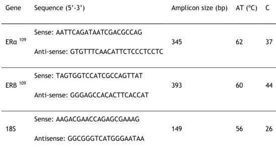
Acknowledgements ... i Publications in Peer-Reviewed International Journals ... iii Communications in Meetings ... iii Resumo ... v Resumo Alargado ... vii Abstract ... xi Table of Contents ... xiii List of Figures ... xvi List of Tables ... xvii List of Abreviations ... xix 1. General Introduction ... 1 Cellular and Molecular Overview of Mammalian Spermatogenesis ... 3 The somatic cells ... 4 The germ cells ... 5 Regulation of spermatogenesis ... 7 The Steroid Hormone Receptor Superfamily ... 8 Estrogens and Estrogen Receptors: α and β ... 11 ERα and ERβ genes and proteins ... 11 Mechanisms of estrogen action ... 12 Role of estrogens/estrogen receptors in male reproduction ... 14 Androgens and Androgen Receptor ... 16 The AR gene and protein ... 17 Mechanisms of androgen action ... 19 Role of androgens/androgen receptors in male reproduction ... 21 Aim and Outline of the Thesis ... 23 2. Estrogen Receptors α and β in Human Testis: Both Isoforms are Expressed ... 25 Abstract... 27 Introduction ... 27 Materials and Methods ... 28 Tissues ... 28 RT-PCR ... 28 Antibodies ... 29 Immunohistochemistry ... 29 Results ... 30 Both ER and ER mRNA are expressed in human testis ... 30 ER is expressed in somatic and germ cells of human testis... 30 ER is expressed in Leydig and germ cells of human testis but not in Sertoli cells .... 31 Discussion ... 33 3. Identification and characterization of androgen receptor variants: tissue and
vertebrate evolutive line expression ... 35 Abstract... 37 Introduction ... 37 Materials and Methods ... 38 Samples ... 38 RT-PCR ... 39 Results and Discussion ... 41 Several AR alternative transcripts are expressed in human testis ... 41 AR alternative transcripts identified in the testis are expressed in several other human tissues ... 44 AR alternative transcripts identified in the testis are expressed in vertebrate evolutive line ... 44 Conclusion ... 47 Acknowledgments ... 48
xiv
novel estrogen target gene in mammalian testis ... 49 Structured Abstract... 51 Introduction ... 51 Materials and Methods ... 52 Reagents and chemicals ... 52 Animals and tissues ... 52 Ex vivo culture of rat SeT ... 53 RNA isolation and cDNA synthesis ... 53 Reverse transcription polymerase chain reaction (RT-PCR) ... 53 Quantitative RT-PCR (qPCR) ... 54 Western blot (WB) ... 54 Immunohistochemistry (IHC) ... 55 Statistical analysis ... 55 Results ... 56 Aven expression and localization in human and rat testis ... 56 Aven expression in human testicular biopsies with different pathologies ... 56 Effect of 17β-estradiol on Aven expression in rat seminiferous tubules cultured ex-vivo ... 57 Discussion ... 58 Acknowledgments ... 60 5. Regucalcin, a calcium-binding protein, as an androgen target gene in rat testis ... 61 Abstract... 63 Introduction ... 63 Material and Methods ... 64 Animals and tissues ... 64 Reagents ... 64 Primary SC culture ... 64 STF collection... 65 Ex vivo culture of rat SeT ... 65 RNA isolation and cDNA synthesis ... 66 RT-PCR ... 66 In situ hybridization ... 67 Western blot ... 67 Immunohistochemistry ... 68 Immunocytochemistry ... 68 qPCR ... 69 Statistical analysis ... 69 Results ... 69 Regucalcin expression and localization in rat and human cell types of the testis ... 69 Regucalcin expression in rat reproductive tissues and STF ... 72 DHT regulation of regucalcin expression in rat SeT ... 74 Regucalcin expression is up-regulated in rat SeT cultured in presence of survival factors ... 74 Discussion ... 75 Acknowledgments ... 78 6. Regucalcin, a calcium-binding protein with a role in male reproduction? ... 79 Introduction ... 81 Regucalcin Expression in Male Reproductive Tract ... 81 Regucalcin Expression in Distinc Spermatogenic Phenotypes ... 82 Effects of Sex Steroids on Regucalcin Expression ... 83 Regucalcin Actions in Testis Physiology ... 84 Conclusion ... 85
xv integrative view in spermatogenesis ... 87 Introduction ... 89 Estrogen Receptors in Spermatogenesis: Monologue or Double Act? ... 89 Androgen Receptor in Spermatogenesis: the Many Faces of a Single Gene ... 91 Estrogen and Androgen Regulated Genes in Testis: a Matter of Life or Death? ... 91 Conclusion ... 93 8. Concluding remarks ... 95 9. References ... 99 Supplements ... 127 The ballad of Calcium and Regucalcin ... 129
xvi
List of Figures
Figure 1.1 Schematic view of the testicular histology ... 3 Figure 1.2 Steroidogenic pathway ... 5 Figure 1.3 Cell division and differentiation events occurring on spermatogenesis ... 6 Figure 1.4 Overview of the hypothalamic-pituitary-testicular axis ... 7 Figure 1.5 Schematic representation of a typical nuclear receptor ... 10 Figure 1.6 Schematic view of DNA binding domain ... 11 Figure 1.7 Amino acid identity between human estrogen receptors α and β ... 12 Figure 1.8 Mechanisms of estrogen control of gene expression ... 13 Figure 1.9 Structural organization of the AR gene and protein ... 18 Figure 1.10 Classical mechanism of androgen action ... 20 Figure 2.1 Expression of ERα and ERβ mRNAs in human testicular tissues ... 30 Figure 2.2 Immunohistochemical localization of ERα adult human testis ... 31 Figure 2.3 Immunohistochemical localization of ERβ in adult human testis ... 32 Figure 2.4 Alignment of human ERβ and ERβcx proteins (amino acids 1-175) ... 32 Figure 3.1 Electrophoresis of PCR (primers hAREx1Fwd and hAREx4Rv) products ... 42 Figure 3.2 Electrophoresis of PCR (primers hAREx1Fwd and hAREx5Rv) products ... 43 Figure 3.3 Schematic representation of the structure of PCR products ... 43 Figure 3.4 Schematic representation of the structure of PCR products ... 45 Figure 4.1 Aven expression and localization in human and rat testis ... 56 Figure 4.2 Expression levels of Aven in testicular biopsies ... 57 Figure 4.3 Effect of 17β-estradiol on Aven expression and Caspase-9 activation ... 58 Figure 5.1 Expression of regucalcin in reproductive tract and seminiferous tubule fluid .. 70 Figure 5.2 Immunochemical localization of regucalcin in testis and Sertoli cells ... 72 Figure 5.3 Immunohistochemical localization of regucalcin in rat prostate, epididymis
and seminal vesicles ... 73 Figure 5.4 Effect of DHT and survival factors on regucalcin expression ... 75 Figure 6.1 Expression levels of Regucalcin in testicular biopsies ... 83 Figure 6.2 Potential signalling pathways involved in the control of regucalcin expression
in testis, and the possible roles of RGN protein in testicular cells ... 86 Figure 7.1 Integrative view of the potential actions of Aven and Regucalcin in testicular
xvii
List of Tables
Table 1.1 Subfamily 3 of the NR superfamily ... 9 Table 1.2 Knockout mice for the study of estrogen action in spermatogenesis and male
fertility ... 16 Table 1.3 Published AR mRNA splice variants ... 19 Table 1.4 Main knockout mice for the study of AR in spermatogenesis and male fertility .... 22 Table 2.1 Oligonucleotides and cycling conditions for PCR amplification of ERα and ERβ .... 29 Table 3.1 Oligonucleotides for the detection of human AR alternative transcripts ... 40 Table 3.2 Primer pairs used for amplification of human AR alternative transcripts ... 40 Table 3.3 Primers designed for the detection of AR splice variants in different species... 41 Table 3.4 AR sequences used for analysis of the RT-PCR amplified transcripts ... 41 Table 3.5 AR splice variants identified in several human tissues ... 44 Table 3.6 Potential AR alternative transcripts detected in several species ... 47 Table 4.1 PCR primer sequences, amplicon size and conditions used ... 54 Table 5.1 PCR primers sequences, amplicon size and cycling conditions ... 67 Table 6.1 Localization of Regucalcin in male reproductive organs ... 82 Table 6.2 Hormonal factors regulating Regucalcin expression ... 84 Table 7.1 Expression of ERα and ERβ in rodent testicular cells ... 90 Table 7.2 Expression of ERα and ERβ in human testicular cells ... 90
xix
List of Abreviations
ABP – androgen binding protein AF1 – transactivation function 1 AF2 – transactivation function 2 AR – androgen receptor
ARE – androgen response elements ArKO – aromatase knockout mouse AT – annealing temperature Bp – base pair C – number of cycles Ca2+ - Calcium [Ca2+] i – intracellular calcium
cDNA – complementary DNA CREM - cAMP-responsive element
modulator
DBD – DNA binding domain DHT – 5α-dihydrotestosterone DNA – deoxyribonucleic acid
dNTP – deoxyribonucleotide triphosphate E2 – 17β-estradiol
ERα – estrogen receptor α ERβ – estrogen receptor β ERE – estrogen response element GnRH – gonadotropin releasing hormone H – heart
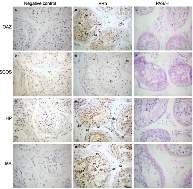
HP – hypospermatogenesis HSP – heat shock protein IHC – immunohistochemistry K – kidney
L – liver
LBD – ligand binding domain
LC – Leydig cell Lg – lung
LH – luteinizing hormone MA – meiotic arrest Met – methionine mRNA – messenger RNA OAZ – obstructive azoospermia NR – nuclear receptor
NTD – amino-terminal domain PCR – polymerase chain reaction qPCR – quantitative (real-time) PCR RGN – regucalcin
RNA – ribonucleic acid RNA P II – RNA polymerase II RT-PCR – reverse transcription PCR Sa – round spermatids
SC – Sertoli cell
SCOS – Sertoli cell-only syndrome Sd – elongated spermatids SeT – seminiferous tubules SG – spermatogonia SR - Sex steroid hormone ST – spermatocytes
STF – seminiferous tubule fluid Sz – spermatozoa T - testosterone TNFα - tumour-necrosis factor-α WB – western blot WT – wild type Zn – zinc
Ge ner al In tr od ucti on
3
Cellular and Molecular Overview of Mammalian Spermatogenesis
Spermatogenesis is a complex process occurring in testis involving cell division and maturation that culminates with the production of the male gametes, spermatozoa. Besides spermatogenesis, the testes are also the place for the synthesis of steroids. Testes are enclosed by a fibrous capsule, tunica albuginea, composed of an outer visceral peritoneum and an inner layer of fibroblasts, collagen fibers, and smooth muscles cells. Inside each testis there are several coiled hairpin-shaped seminiferous tubules (SeT) with both ends emptying into a structure called rete testis1. The SeT is the functional unit of spermatogenesis andcontains the somatic Sertoli cells (SC) and various types of germ cells (Figure 1.1). The tubules are surrounded by peritubular tissue composed of connective tissue and peritubular myoid cells, which move immature spermatozoa towards the rete testis2,3. The synthesis of
steroids occurs mainly in the interstitial space, which consists of the Leydig cells (LC), macrophages, blood and lymph vessels, and nerves. The SeT open into transitional zones in the rete testis lined by cells resembling Sertoli cells which appear to form a valve or plug.
Spermatogenesis can be defined as the sequence of cytological events that result in the formation of mature spermatozoa from precursor cells4. It is characterized by three
functional phases: proliferation, meiosis, and differentiation5. The process requires
spermatogonial stem cells renewal and amplification by mitosis and differentiation. Spermatogonial stem cells respond to differentiation signals and undergo mitosis without completing cytokinesis. Spermatogonia then differentiate into primary spermatocytes, which proceed to meiosis and originate secondary spermatocytes. These divide to become haploid spermatids and are transformed into spermatozoa (Sz) by spermiogenesis5. For
spermatogenesis to occur a unique environment within the SeT is necessary, which is achieved by the blood-testis barrier, formed by adjacent SC6.
Ge ner al In tr od ucti on
4
The somatic cells
There are two main types of somatic cells in the testis: SC and LC. While SC are an integrant part of the seminiferous epithelium and have nursing functions, LC are localized in the interstitial space between the tubules and their main function is testicular steroidogenesis4.
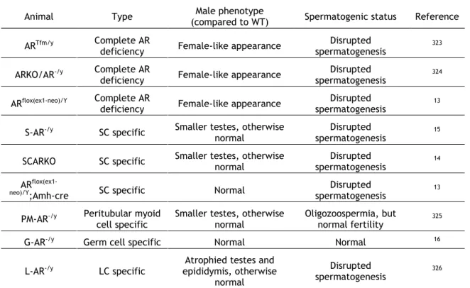
Sertoli cells play an essential role in spermatogenesis as they supply the developing germ cells with nutrients and growth factors, and the number of SC determines testicular size and sperm output8. The critical importance of SC to the survival of germ cells is also demonstrated by
the fact that although SC-only tubules exist, in cases of Sertoli cell-only syndrome (SCOS), there are no cases describing germ cell-only tubules9. Moreover, because germ cells do not
express androgen receptor (AR), the action of androgens on germ cells is achieved through SC10-12. In fact targeted deletion of AR in Sertoli cells results in complete disruption of
spermatogenesis13-15, while deletion in germ cells causes no effect on male fertility16 (see also
Androgens and Androgen Receptor, page 16). Sertoli cells were first described by Enrico Sertoli in 1865 as individual elements extending from the basement membrane to the lumen of the seminiferous tubule involving the many clusters of associated germ cells17. These cells
regulate the flow of nutrients and growth factors to the germ cells while protecting them from harmful agents and the host‟s immune system18. This is done by the so-called
blood-testis barrier, formed by tight junctions between neighbouring SC (Figure 1.1)18. The barrier
divides the basal compartment containing diploid cells from the adluminal compartment, containing haploid germ cells and bathed by the SeT fluid (STF) secreted by SC. Also, they are responsible for the phagocytosis of apoptotic germ cells and residual bodies, which result from the spermiation (release) of spermatids into the lumen of the tubules19,20.
Sertoli cells produce numerous molecules, including the androgen binding protein (ABP) which enables the achievement of high concentrations of androgens in the STF and the excurrent ductal system21.
The proliferation of SC is stimulated by follicle-stimulating hormone (FSH)22, which also
induces SC to produce factors such as ABP, inhibin, lactate and transferrin23-26 (see also
Regulation of spermatogenesis, page 7). Not only androgens but also estrogens act on SC and in addition, immature SC are known to express active P450 aromatase, which converts testosterone into 17β-estradiol (E2), making SC simultaneously a source and a target for
estrogens (see also Estrogens and Estrogen Receptors: α and β, page 11).
Leydig cells are localized in the interstitial spaces between SeT (Figure 1.1)4. These cells
contain lipid droplets containing cholesterol esters, which are used for the synthesis of testosterone. Leydig cells are, in fact, the main site of steroidogenesis in the testis and the main source of testicular androgens27. They have well developed smooth endoplasmic
reticulum (SER), which contains membrane-bound steroidogenic enzymes28,29. Only one step
Ge ner al In tr od ucti on
5
pregnenolone (Figure 1.2) happening in the membrane of the mitochondrial cristae29,30. The
SER is therefore the main site of steroidogenic enzymes in LC, and testosterone production is actually directly proportional to the volume of this organelle31. In LC, testosterone can be
further metabolized to a more potent androgen, 5α-dihydrotestosterone (DHT), by the action of 5α-reductase (Figure 1.2) within the microsomes of SER, although testosterone is the prevalent intratesticular androgen29,32,33. Alternatively, testosterone can also be metabolized
by P450 aromatase complex into the E2 (Figure 1.2), which locates to the mitochondria of the
LC34,35.
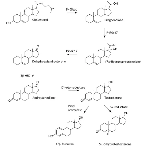
Figure 1.2 Steroidogenic pathway leading to the synthesis of testosterone from cholesterol.
Testosterone can be further metabolized to estrogen 17β-estradiol or to more potent androgen 5α-dihydrotestosterone. Legend: P450scc – cholesterol side-chain cleavage enzyme; P450c17 – 17α-hydroxylase/17,20-lyase; 3β-HSD – 3β-hydroxy steroid dehydrogenase.
The germ cells
The germinal epithelium is constituted by germ cells in different developmental stages nested in invaginations of SC4. The earliest cells to appear in the germinal epithelium are
spermatogonia (Figure 1.3). In order for spermatogenesis to be continuous spermatogonia must renew themselves by mitosis in addition to proceeding to meiosis, originating two types of cells: type A and type B spermatogonia4. As they undergo mitosis they remain connected by
intracellular cytoplasmic bridges, due to incomplete cytokinesis36. These bridges, which
persist until Sz are released into the SeT lumen, enable the movement of several macromolecules including mRNAs and proteins, allowing the transport of Y chromosome
Ge ner al In tr od ucti on
6
encoded gene products to X-bearing cells and vice versa37. While type A spermatogonia
continue proliferating and remain in the periphery of the tubules, type B spermatogonia progressively loose contact with the basal membrane and are able to enter the process of meiosis by differentiating into primary ST. At this point, primary spermatocytes pass through the blood-testis barrier into the adluminal compartment.
Figure 1.3 Cell division and differentiation events occurring on spermatogenesis (modified from 38)
Meiosis involves a round of DNA replication followed by two cell divisions (Figure 1.3). Meiosis I begins with primary spermatocytes and results in secondary spermatocytes, bearing a haploid number of chromosomes and diploid DNA content. Meiosis II is similar to a mitotic division and occurs in secondary ST resulting in haploid spermatids. Secondary spermatocytes have a very short life-span, and are therefore very rare in testis tissue sections. These cells are located close to the lumen and have a spherical shape. They undergo meiosis II and each one gives rise to two spermatids (Figure 1.3). Early spermatids are smaller than secondary spermatocytes and can be found in the lumen of SeT. The transformation of spermatids into Sz does not involve further cell division, it is a cellular restructure process called spermiogenesis4. This involves formation of the acrosome (Golgi originated lysosomal
structure that undergoes acrosome reaction at fertilization)39, nuclear changes (from central
to eccentric position, condensation of chromatin), development of the flagellum, reorganization of cytoplasm and organelles and spermiation (release of the spermatozoon by Sertoli cells into the SeT lumen)4.
Ge ner al In tr od ucti on
7
Regulation of spermatogenesis
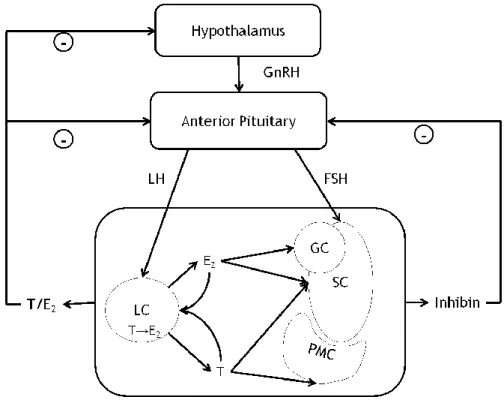
Spermatogenesis is a process that requires tight control for its maintenance. This is achieved by hormonal and non-hormonal regulators. The major mechanism for hormonal control of spermatogenesis is the hypothalamic-pituitary-gonadal axis40 (Figure 1.4). The hypothalamus
releases gonadotropin releasing hormone (GnRH) which acts on the pituitary inducing the release of luteinizing hormone (LH) and FSH. In the testis, LH will act on LC, stimulating the synthesis of testosterone (T), while FSH will act on SC stimulating spermatogenesis and the production of several signalling factors41.
Figure 1.4 Overview of the hypothalamic-pituitary-testicular axis. GnRH, gonadotropin releasing
hormone; LH, luteinizing hormone; FSH, follicle-stimulating hormone; LC, Leydig cell; SC, Sertoli cell; PMC, peritubular myoid cell; GC, germ cell; T, testosterone; E2, 17β-estradiol.
After being produced by LC, T diffuses into the SeT where it acts on SC, or enters circulation, acting in numerous target organs42,43 (more on this subject in section Androgens and Androgen
Receptor, page 16). Testosterone can be converted (Figure 1.2) into E2 (more details in the
section Estrogens and Estrogen Receptors: α and β, page 11) or into a more potent androgen DHT. Testosterone/DHT/E2 can act in a negative feedback mechanism to inhibit the
hypothalamic release of GnRH and pituitary release of LH (Figure 1.4). Testes also produce inhibins, mostly inhibin B, part of the transforming growth factor β (TGF-β) family which will exert a negative feedback effect on the production and release of FSH by the pituitary44.
Inhibin B is a dimeric glycoprotein which is formed by an α subunit, produced mainly by SC and to a lesser extend by LC25,45, and a β
B subunit, produced mainly by germ cells46,47.
Because of this inhibin B is a combined product of somatic and germ cells and therefore its levels reflect the quality of spermatogenesis48,49.
Ge ner al In tr od ucti on
8
Other factors from the TGF-β family besides inhibin B play a role in the regulation of spermatogenesis. For example, stem cell factor and glial cell line-derived neurotrophic factor, which seem to be important for the survival and differentiation of spermatogonia9,50,51. Moreover, anti-mullerian hormone (AMH) is secreted by SC until puberty
and induces the regression of the Mullerian duct in male embryos52. An interesting part of the
control of spermatogenesis is played by cytokines. For example, tumour-necrosis factor-α (TNFα) is produced in the testis by a variety of cells including SC53,54 and several germ
cells54,55, and is capable of inhibiting LC steroidogenesis56 and increasing AR expression in
SC57. The expression of TNFα receptors in SC is hormonally controlled by FSH58.
Sertoli cells produce other factors necessary for successful spermatogenesis. As previously said they produce ABP, which enables a high concentration of androgens on the adluminal compartment milieu. Other factors synthesized by SC are, for example, transferrin, ceruloplasmin, proteases, protease inhibitors and structural components of the basement membrane9,41. Transferrin in particular has been a marker for SC number and function, and its
concentration in seminal fluid has been correlated with sperm production59-61. This protein
transports iron to the germ cells in the adluminal compartment62,63. Although germ cells seem
to have high iron requirements for proliferation and differentiation64,65, its levels must be
tightly regulated as excessive iron is damaging to spermatogenesis66. Iron is not, however, the
only inorganic regulator of spermatogenesis. There has been some data indicating that calcium (Ca2+) has an important role to play in the regulation of spermatogenesis. This ion is
essential for the maintenance of SC tight junctions forming the blood-testis barrier67 and
modulates the activity of enzymes interfering in SC architecture68,69. A tight regulation of
intracellular Ca2+ concentration is essential for LC steroidogenesis, for example by controlling
the expression of steroidogenic acute regulatory protein70,71. Moreover, it has been shown
that administration of Ca2+ channel blockers has deleterious effects on mammalian
spermatogenesis, being associated with reversible infertility72-78.
The Steroid Hormone Receptor Superfamily
Sex steroid hormone (SR) receptors act mainly through nuclear receptors (NR), a superfamily comprising receptors which act as transcription factors79,80. However, ligands were only
identified for 24 members of the family, the rest being called orphan receptors79. The NR
superfamily can be divided into six subfamilies or classes based on evolutionary data79,81,82:
Subfamily 1 has 21 members and includes among others thyroid hormone receptors α and β, retinoic acid receptors α, β, and γ, and vitamin D receptor.
Subfamily 2 is slightly smaller, with 13 members, including retinoid X receptors α, β, and γ, and testis receptors α and β.
Subfamily 3 includes all SR in addition to estrogen-related receptors α, β, and γ (Table 1.1).
Ge ner al In tr od ucti on
9
Subfamily 4 is smaller, having only three members (nerve growth factor-induced clone B α, β, and γ).
Subfamily 5 has only two members, steroidogenic factor 1 and Fushi Tarazu factor 1. Subfamily 6 has only one member, germ cell nuclear factor.
Table 1.1 Subfamily 3 of the NR superfamily. Adapted from 79,82
Receptor Denomination Subtypes Nomenclature Ligand
ER Estrogen receptor α β NR3A1 NR3A2 E2, tamoxifen, raloxifen,
various synthetic compounds
ERR Estrogen-related receptor
α NR3B1 None β γ NR3B2 NR3B3 Diethylstilbestrol, 4-hydroxy-tamoxifen GR Glucocorticoid receptor NR3C1 Cortisol, dexamethasone, RU486 MR Mineralocorticoid receptor NR3C2 Aldosterone, apirolactone PR Progesterone receptor NR3C3
Progesterone,
medroxyprogesterone actate, RU486
AR Androgen receptor NR3C4 Testosterone, flutamide
The first SR to be identified, albeit by biochemical methods, was a receptor for estrogens in 1962 83. However, it took 26 years for estrogen receptor α (ERα) to be cloned84,85.
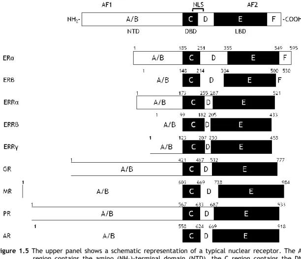
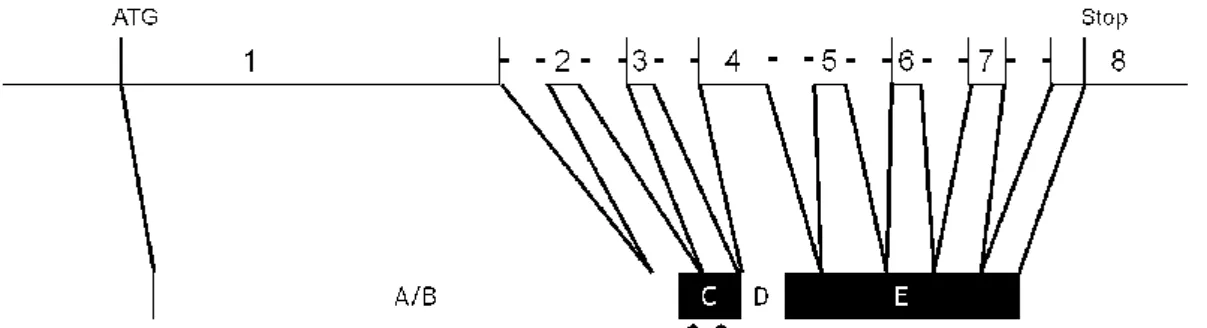
All SR share common structural features, consisting of basically the same functional domains (Figure 1.5): amino-terminal domain (NTD; A/B region), DNA-binding domain (DBD; C region), a hinge region (D region), and ligand binding domain (LBD; E region). Estrogen receptors also contain a region F of yet unknown function. The NTD, which contains the transcription activation function AF1, is the most variable region. In contrast, the DBD is the best conserved, as this is the domain by which SR bind to DNA at specific sequences called hormone response elements. The DBD contains two cystine-rich zinc (Zn) fingers (Figure 1.6) which fold to form a compact structure with two perpendicular α-helices that are important for DNA recognition and binding86-88. The first Zn-finger contains the P-box, a sequence of five
amino acids which is involved in DNA recognition, while the second contains the D-box, also formed by five amino acids which is involved in receptor dimerization89.
Ge ner al In tr od ucti on
10
Figure 1.5 The upper panel shows a schematic representation of a typical nuclear receptor. The A/B
region contains the amino (NH2)-terminal domain (NTD), the C region contains the
DNA-binding domain (DBD), the hinge is located in region D, while region E constains the ligand binding domain (LBD) in the carboxy(COOH)-terminal end of the protein. In some members a region F with unknown function may also be present. Two activation functions, AF1 and AF2, are localized on the NTD and LBD, respectively and a nuclear localization signal is localized between the DBD and the hinge region D. The schematic representation of the members of subfamily 3 of the nuclear receptor superfamily are also shown (ERα and ERβ, estrogen receptors α and β; ERRα, ERRβ, and ERRγ, estrogen-related receptors α, β, and γ; GR, glucocorticoid receptor; MR, mineralocorticoid receptor; PR, progesterone receptor; AR, androgen receptor). The amino acid position of each region is shown with arabic numerals and refers to the human proteins90-92
The DBD is linked to the LBD through the hinge region, a poorly conserved domain which often contains a nuclear localization signal. The LBD is less conserved than the DBD, however it is responsible for ligand binding, dimerization, interaction with heat-shock proteins, and contains ligand-dependent transactivation function AF293,94.
Ge ner al In tr od ucti on
11
Figure 1.6 Schematic view of DNA binding domain with 2 zinc fingers and corresponding cystine
residues. Aminoacids common between nuclear receptors are indicated by their corresponding letters, while non-conserved residues are indicated in black (based on 82.
Localization of P-box and D-box is pointed.
Hormone response elements are located on the regulatory regions of target genes and consist of a 6 bp consensus motif82. For SR the sequence of the motif is AGAACA separated by three
spacer nucleotides however, naturally occurring response elements can show variation from this sequence82,95. Most receptors will bind as dimers to these response elements, which are
usually made from two of these hexameric motifs (called half-sites), arranged into palindromes, inverted palindromes, or direct repeats82. The exception is the ERs, which bind
to palindromes of the half-site GGTCA96.
Is this thesis, attention will be focused on two groups of sex steroid hormones, androgens and estrogens, and their respective receptors.
Estrogens and Estrogen Receptors: α and β
Estrogens are steroid hormones synthesized from C19 androgens (e.g. T) by aromatase encoded by the CYP19 gene97. The most prevalent estrogen in humans is E
2, although smaller
amounts of several metabolites are present. Estrogens act through ERα and ERβ, sex steroid receptors, through various mechanisms in order to exert their effects98. Although classically
viewed as “female” hormones, estrogens have been shown to play pivotal roles in male physiology, including in reproduction99. Estrogens and ERs have also been suggested to play
important parts in the pathophysiology of numerous types of cancer100-103, including prostate
cancer104.
ERα and ERβ genes and proteins
The first evidence of a receptor for estrogens appeared in the 1960‟s83 based on the specific
binding of E2 in the uterus. In 1986 the cDNA for estrogen receptor (ER) was cloned by two
groups84,85. However, 10 years later a second ER was cloned and named ERβ, while the first ER
cloned was re-named ERα105,106. The two ER subtypes are encoded by genes in different
chromosomes. In humans, ERα is encoded by a gene in chromosome 6107,108 while ERβ gene is
Ge ner al In tr od ucti on
12
and 6111, respectively. Both the genes encoding human ERα and ERβ are organized into eight
exons109,112. Expression of the ERα/β gene is regulated by estrogens113-115, similar to what
happens with the AR and other SR116,117.
The human ERα gene encodes a 595 aa protein85 while ERβ protein is 485 aa long105. Both ERα
and ERβ proteins have the typical structure of a NR with functional domains from A/B to F (Figure 1.5). The NTD (domain A/B) is the least conserved between α and β subtypes and contains a transactivation domain while only 58% of the LBD is conserved between the two receptors98 (Figure 1.7). Alike other members of NR superfamily, ERs have two transactivation
functions, AF1 (located in NTD) and AF2 (located in LBD)118. Similarly to what happens with
the AR, binding of ligands to the LBD will cause a conformational change that will render the AF2 domain fully active93,119. Between the DBD and the LBD localizes the flexible hinge region,
which is poorly conserved between the two receptors106 and may account for differences in
receptor-induced changes in DNA structure necessary for gene transcription120. The hinge
region also harbours the nuclear localization signal121. Both receptors have a high degree of
homology concerning their DBD98 (Figure 1.7), which contain the two typical Zn-fingers
responsible for interaction with estrogen response elements (ERE) in the regulatory sequences of target genes, and the P-box (involved in DNA recognition) is identical in both receptor proteins (Figure 1.6)105.
Figure 1.7 Percentage of amino acid identity between human estrogen receptors α (ERα) and β
(ERβ)92,106
Several ERα and ERβ have been described, some of which originated by alternative splicing122-124, however whether all of them are translated into proteins or have important
biological functions remain to be determined125. However, some variants have been showed
to modulate the activity of the prototype ERs126,127.
Mechanisms of estrogen action
Estrogens can act through two main groups of mechanisms: genomic and non-genomic actions. In the classical mechanism, estrogens bind to its receptor triggering a series of conformational changes and events that ultimately lead to changes in the transcription rate of estrogen-regulated genes (Figure 1.8). Although this process is not entirely understood, it is very similar to what happens with other SR, namely AR, and it involves release of chaperone proteins, phosphorylation128, dimerization of the estrogen-bound receptor,
interaction with DNA, recruitment of transcription factors and coregulators, and assembly of the transcriptional complex129,130. Phosphorylation regulates ER activation131,132, and
Ge ner al In tr od ucti on
13
described134,135. Estrogen receptors bind to EREs in the regulatory regions of target genes,
containing the consensus motif GGTCA organized in palindromic repeats with a variable 3 bp spacer96,136. Both ERs bind these ERE with similar affinity, however the addition of a
nucleotide to the consensus, forming palindromes of AGGTCA with a 3 bp spacer, determines the affinity with which ERα binds to the ERE137. The activity of ERs can be modulated by
several factors, including alternatively spliced variants126,127. More importantly, ERβ acts as a
dominant negative regulator of ERα transcriptional activity138. Therefore, in cells and tissues
expressing both ERs, estrogenic actions are determined by the balance between ERα and ERβ. ERα and ERβ sometimes show opposite transcriptional regulatory activities. A classical experiment which involved binding of agonist raloxifene to ERβ induced an increase in reporter gene expression, however binding to ERα resulted in minimal activation139. Opposing
actions of ERα and ERβ also occur in the regulation of cyclin D1 expression140. Curiously ERs
bind DNA not only as ERα/ERα or ERβ/ERβ homodimers but also as ERα/ERβ heterodimers, adding new levels of complexity to estrogenic action141,142. It has been demonstrated that
these heterodimers bind to DNA with higher affinity than ERβ homodimers142 and are able to
bind not only to classical but also to specific EREs in the genome resulting in specific gene expression profiles different from those regulated by homodimers143.
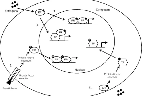
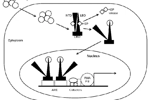
Figure 1.8 Mechanisms of estrogen control of gene expression. 1. Classical mechanisms – estrogens
enter the cell, bind to their cognate nuclear receptors (ER) which travel to the nucleous and bind, as dimmers, to estrogen responsive elements in the genome, controlling the rate of gene expression. 2. Estrogen-bound ER can bind and activate other transcription factors, that in turn bind to their responsive elements on the genome, controlling gene expression. 3. Estrogen-independent ER-mediated mechanism – growth factors can bind to their membrane receptor, triggering a protein-kinase cascade that phosphorilates and activate ER, which enters nucleus and binds to estrogen responsive elements, controlling gene expression. 4. Membrane ER action – estrogens can bind to ERs located at the cell membrane, triggering the activation of protein-kinase cascades, that will end with the phosphorilation of transcription factors that bind to their own response elements in genome, thereby controlling gene expression.
Ge ner al In tr od ucti on
14
Estrogen receptors can also regulate gene expression by non-classical mechanisms144
(Figure 1.8), the so-called nongenomic-to-genomic pathways, through interaction of the receptors with transcription factors which in turn bind to their own response elements in DNA144-146. Also, ERs can activate protein-kinases that activate transcription factors, thereby
influencing gene expression147. These pathways are sometimes initiated in the plasma
membrane, which has sparked the debate about the localization of classical ERs to the plasma membrane versus existence of novel ER forms which are specifically membrane-bound148-150.
Also, a transmembrane G-protein coupled receptor (GPR30) has been shown to be able to mediate estrogen signalling151, however the true contribution of this receptor to estrogenic
signalling has been questioned (reviewed by 152). There are also evidences indicating that ERs,
like AR, can have ligand-independent transcriptional activity153,154 (for an extensive review on
this subject read 155).
Non-genomic ER actions are faster since they do not involve the activation of RNA transcription or translation into protein, which is a common feature among steroid receptors149,156,157. They usually involve the activation of protein-kinase cascades158-160,
mobilization of intracellular Ca2+ 161,162, increase in cAMP concentration163, modulation of
nitric oxide release162, amongst other effects. Some of the non-genomic estrogenic actions
occur through membrane-bound ERs, as shown also by the use of membrane-impermeant forms of estrogens164,165. It is interesting to note that the opposite actions of ERα and ERβ are
also recognisable in these non-genomic effects, for example whereas ERβ activates c-Jun N-terminal kinase, ERα inhibits its activation149.
Role of estrogens/estrogen receptors in male reproduction
Although classically viewed as “female hormones”, the first evidence of the presence of estrogens in male gonad was known in 1934166. In recent times the importance of estrogens to
males as been stressed by many studies regarding male-specific physiological functions, such as spermatogenesis (for review see 167 and 168). In testes, E
2 is produced through the
aromatization of T mostly in LC, but also several different germ cells, and even spermatozoa have been shown to contain active aromatase169-172. Intratesticular E
2 reaches levels that can
be up to 100-400x higher than the serum levels173-175. Estrogens act in a negative feedback
fashion in the hypothalamus and pituitary, regulating the secretion of GnRH and FSH, respectively (Figure 1.4).
The localization of ERα and ERβ in testis has been matter of much debate. In rodents, ERα seems to be expressed by LC176-178 and peritubular myoid cells179, although expression in some
germ cells has also been described176. On the other hand, ERβ has been localized to LC179,180,
SC176,179,181,182, peritubular myoid cells179,180, and some germ cells179-181. In humans, the
controversy surrounding ERα and ERβ testicular localization is even greater. Some authors have localized ERα to LC183,184, SC184, and some germ cells185-189. Espression of ERβ was
Ge ner al In tr od ucti on
15
Some authors have not detected ERα expression in human testis190-192 which, together with
data from immunolocalization studies in rodent testis, led to the idea that ERβ is the main mediator of estrogenic action in the testis. However, data from ER mutations and KO animals does not corroborate this theory.
Contrarily to AR, whose defects cause dramatic consequences to male phenotype and fertility, very little is known of the effect of defective estrogenic action in humans. There is only one disruptive mutation of ERα known in humans193. This man had normal male
phenotype, however he presented with low sperm counts and decreased sperm viability193.
Until now, there have been no reports of ERβ disruptive mutations in humans. There are few reports of men with disruptive mutations of aromatase gene, and therefore with an absence ofendogenousestrogens,but not all of them had their reproductive parameters tested194-196.
Although the reproductive phenotype varies slightly with the type of mutation, all of the aromatase-deficient men studied had normal male phenotype however, reproductive parameters showed impairment of fertility with decreased sperm motility194-196,
oligozoospermia196, hypospermatogenesis and meiotic arrest195. The major contribution to the
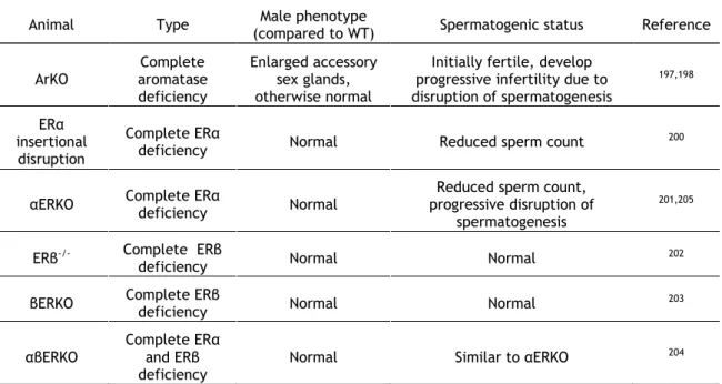
understanding of estrogenic action in male reproduction, and more importantly in spermatogenesis, came from the generation of KO mice for aromatase and both ERs. Table 1.2 shows the reproductive phenotypes of some estrogen related KO mice. Initially, male mice lacking aromatase (ArKO) are fertile and phenotypically normal, which indicates that estrogens are not necessary for development of male phenotype197. However, they
progressively develop disruption of spermatogenesis198. Also, inclusion of phytoestrogens in
the diet of these animals partially prevented disruption of spermatogenesis, which indicated a potentially important role for exogenous estrogens in the regulation of male fertility199. The
non-requirement of estrogen action for normal male phenotype is confirmed by the development of KO mice for ERα, ERβ, or both, which show normal male reproductive tract 200-204. The absence of ERβ does not cause changes in male fertility202,203, in contrary to ERα
lacking animals, in which a reduced sperm count can be observed200,201. However, these
animals present progressive disruption of spermatogenesis, which was explained as a result of defective fluid resorption in the efferent tubules, causing SeT atropy due to fluid back pressure205.
There has been increased attention given to the effects of estrogens in male reproduction in the past two decades because of the reports that exposure to environmental contaminants with estrogenic activity may have deleterious effects on male reproductive development and may be causing a decline in male fertility206-208. Although the issue is controversial, there is
evidence that excessive estrogenic exposure can lead to disturbances of spermatogenesis, mainly through disruption of the balance between germ cell survival and death209-212. There
are many studies relating estrogens to apoptosis in testis and identifying E2 as a germ cell
survival factor185,213, highlighting the importance of estrogens to male fertility. The disruption
Ge ner al In tr od ucti on
16
due to absence of E2198. Therefore estrogens are thought to play a role in the delicate balance
governing cell survival and death in the testis.
Table 1.2 Knockout mice for the study of estrogen action in spermatogenesis and male fertility
Animal Type (compared to WT) Male phenotype Spermatogenic status Reference
ArKO Complete aromatase deficiency Enlarged accessory sex glands, otherwise normal
Initially fertile, develop progressive infertility due to disruption of spermatogenesis 197,198 ERα insertional disruption Complete ERα
deficiency Normal Reduced sperm count 200
αERKO Complete ERα deficiency Normal
Reduced sperm count, progressive disruption of
spermatogenesis
201,205
ERβ-/- Complete ERβ
deficiency Normal Normal 202
βERKO Complete ERβ deficiency Normal Normal 203
αβERKO
Complete ERα and ERβ deficiency
Normal Similar to αERKO 204
Legend: ERα, estrogen receptor α; ERβ, estrogen receptorβ; KO, knockout; WT, wild type.
Although supportive and valuable evidences allow recognizing that estrogens have an important role in the regulation of spermatogenesis, there are gaps concerning the regulatory pathways involved in the testicular estrogenic functions. For instance, the cellular expression patterns of ERα and ERβ in human testis are not clearly defined, and the role of ERα in spermatogenesis remains to be clarified. On the other hand, the nature of estrogenic actions in testis, wether genomic or non-genomic, is slowly being uncovered. Regulation of kinase intracellular signalling pathways by estrogens has been described in testis188,214,215. Few
studies have aimed at identifying genes regulated by estrogens in testis216-219, and for most of
the identified targets their roles in male fertility and the expression patterns within the testis remain unstudied218,219. Also, the administration of endocrine disruptors acting through ERs,
which have been pointed as one of the reasons for the decline in human fertility, have been shown to alter testicular gene expression patterns207,220,221, highlighting the importance of
genomic estrogen action in the regulation of spermatogenesis. Therefore, it is important to know the targets of estrogenic action in order to better understand the regulation of spermatogenesis by this class of hormones.
Androgens and Androgen Receptor
Androgens have been classically viewed as predominantly “male hormones”. Androgens are responsible for initiation and maintenance of spermatogenesis, maintenance of male phenotype, among other physiological effects in the male222. Testosterone, the most