Vol.58, n.6: pp. 854-863, November-December 2015 http://dx.doi.org/10.1590/S1516-89132015060284
ISSN 1516-8913 Printed in Brazil
BRAZILIAN ARCHIVES OF BIOLOGY AND TECHNOLOGY
A N I N T E R N A T I O N A L J O U R N A L
Genes Regulating Maternal Recognition of Pregnancy in
Domestic Animals: an Update
Avantika Mor
1, Sukanta Mondal
1*Ippala Janardana Reddy
1and N.P Soumya
21
National Institute of Animal Nutrition and Physiology; Adugodi – India. 2National Dairy Research Institute; (SRS), Bangalore -India
ABSTRACT
Early embryonic mortality is one of the main sources of reproductive wastages and major constraints for full exploitation of the production potential of livestock. The survivality of embryo during early embryonic life is mostly dependent on the efficiency with which the maternal recognition of pregnancy (MRP) is established. Maternal recognition of pregnancy involves molecular dialogue between the trophoblast of conceptus and uterine endometrium. Embryonic development to the blastocyst stage and uterine differentiation to the receptive environment are crucial for successful establishment of the embryo-uterine cross-talk that leads to the initiation and progression of successful implantation. Unravelling the complex intricate molecular and cellular dialogues between the conceptus and uterine environment will facilitate development of strategies to augment early embryo survivality.
Key words: embryonic mortality, maternal recognition of pregnancy, embryo-uterine cross-talk
*Author for correspondence:[email protected]
INTRODUCTION
Successful establishment of pregnancy in the mammals involves concerted events where the uterus and terminally differentiated embryos
exchange signals culminating in maternal
recognition of pregnancy (MRP). Recent advances in molecular and genetic approaches have led to the discovery of few molecules involved in
embryo-uterine dialogue; however, a
comprehensive understanding of the MRP and implantation process is still not deciphered. Among various developmental events during early pregnancy, implantation of embryo is a critical step and its success mostly depends on the efficiency with which the MRP is established. The MRP involves the complex cellular and molecular cross-talking between the trophoblast of conceptus and uterine endometrium at embryo-uterine interface. In ruminant, the principal signal for
MRP is identified to be interferon- secreted by trophoectoderm of blastocyst for a limited period during early pregnancy. Interferon- exerts its antiluteolytic action by suppressing the normal pattern of pulsatile release of PGF2 in late estrous cycle, possibly by a mechanism that involves down regulation of estrogen receptor in uterine epithelium, which in turn prevents a rise in
oxytocin receptor. The survivability and
opportunity of successful development of an embryo are generally influenced directly and indirectly by various paracrine and autocrine factors (steroid hormones, growth factors, and cytokines), controlling uterine microenvironment.
Out of these factors, hormones such as PGF2α,
PGE2, integrin, COX-II, osteopontin and galectin
interactions between the conceptus and
endometrium (Spencer et al.1996).
The recent advent of state-of-the-art transcriptomic and proteomic technologies allows, for the first time, a holistic analysis of mechanisms involved in signaling between an embryo and its maternal environment before implantation. Proteome and transcriptome studies have revealed quantitative and qualitative changes in the genes at different stages of the estrous cycle and pregnancy (Bauersachs et al. 2005). These spatial and temporal changes were mainly due to the expression pattern of sets of genes in the uterine environment, where the endometrium is especially important for the embryo-maternal interaction and successful establishment of pregnancy. This review focuses primarily on the MRP and genes
controlling its events that facilitate the
development of optimal reproductive management strategies and paradigm to reduce early embryonic wastage in livestock.
MATERNAL
RECOGNITION
OF
PREGNANCY (MRP)
Higher rates of early embryonic mortality is a major cause of reproductive failure during implantation period in livestock. These pre-implantation losses are associated with inadequate luteal function during early pregnancy in ruminants (Mondal and Prakash 2002). The survivality of embryo during early embryonic life is mostly dependent on the efficiency with which the MRP is established. MRP was coined by Roger Short and defined as the physiological process whereby the conceptus signaled its presence to the maternal system and prolonged the lifespan of the corpus luteum (CL). Progesterone produced by the CL acts on the uterus to stimulate and maintain uterine functions that are conducive for early
embryonic development, implantation,
placentation and successful fetal and placental development to term (Mondal et al. 2007; 2010). The MRP occurs between days 16-19, 12-13 and around day 17 of pregnancy in cow, sheep and goat, respectively. During the MRP, the mononuclear cells of the conceptus trophectoderm
synthesize and secrete IFNτ between Days 10 and
21 to 25 with maximal production on Days 14 to
16 (Roberts et al. 1999). IFNτ appears to be the
sole factor produced by the conceptus that prevents the development of the endometrial
luteolytic mechanism. IFN-ι does not act to
stabalize the PR expression in the endometrial
epithelium during the pregnancy; however, IFN-ι
acts in a paracrine fashion on endometrial LE and GE to suppress the transcription of ERα and OTR
genes (Spencer and Bazer 1996; Fig. 1). In vitro
culture of sheep conceptus homogenates and analysis for radiolabelled proteins released into the culture medium indicates that oTP-1 is secreted by
the mononuclear cells of ovine
trophoectoectoderm. oTP-1 is secreted in two phases: one between days 10 and 21 of pregnancy, and second by the chorion between days 25 and 45 of pregnancy. oTP-1 is thought to exert a paracrine antiluteolytic effect on the endometrium as there is no evidence that it is transported from the uterus to directly affect CL. Secretion of oTP-1 (ng/uterine flushing) begins on about 10 and increases with the morphological changes: from spherical (312 ng) to tubular (1380 ng) to filamentous (4455 ng) forms on days 12-13 (Spencer et al. 1996). Goat conceptus secretes caprine IFN-tau between days 16-21 that is assumed to be antiluteolytic signal. Intra-uterine injections of recombinant o IFN-tau in cyclic goats extend CL lifespan in the goats. The endocrine-exocrine theory of maternal recognition of pregnancy in the pigs has been studied. It is assumed that uterine endometrium
secrets luteolysin PGF2 and that the conceptus
secretes estrogen that are antiluteolytic. The
present theory is that PGF2 is secreted in an
endocrine direction towards uterine vasculature in cyclic gilts and transported to CL to exert its luteolytic effect. However, in pregnant pigs, the direction of secretetion of PGF is exocrine into uterine lumen where it is sequestered to exert its biological effects in utero and / or to be metabolized to prevent luteolysis.
GENES
REGULATING
MATERNAL
RECOGNITION OF PREGNANCY
INTERFERRON-TAU (IFN-
-é
)
IFN-ι is a member of the Type I IFN family that
acts differentially on the endometrial luminal epithelium (LE), glandular epithelium (GE) and stroma to regulate expression of a number of IFN-stimulated genes (ISGs) that are hypothesized to play roles in the endometrial differentiation and conceptus implantation (Hansen et al. 1999). It is a subclass of the 172 amino acids type I omega (w) IFNs, which competes with IFNα and β for
binding of a common cell surface receptor. IFN-ιs
approximately 1 kb in length, which arise from multiple genes. Like other type I interferon genes,
IFN-ι genes are intronless and consist of 585 bp
ORF coding for a 195 amino acids preprotein containing a 23 signal sequence, which after cleavage yield a mature protein of 172 amino
acids. Bovine IFN-ι transcripts contain potential
N-glycosylation site at Asn78 and multiple glycosylation variants are present in the secreted
proteins. Bovine, ovine and caprine IFN-ι are more
similar in sequence to each other than bIFNw. In the coding region, the nucleotide sequences exhibit approximately 90% identity and their inferred amino acid sequence about 80% identity (Roberts et al. 1999). The predicted amino acid identity
between bIFN-ι and IFNα1 and IFNw is 50% and
72%, respectively.
Recently, Saugandhika et al. (2013) reported 13
distinct IFN-τ cDNA variants that encoded for
eight distinct buffalo IFN-τ isoforms. These
buffalo IFN-τ isoforms had a greater nucleotide
and amino acid homology with caprine IFN-τ (98
-100% and 96--100%) than ovine (94-97% and 90-95%) and bovine (89.6-90.6% and 82-86%),
respectively. The novel buffalo IFN-τ isoforms
showed pronounced nucleotide and amino acid sequence identity with one another (99.1-99.8% and 98-99%) but only moderate identity with
previously identified buffalo IFN-τ (90-92% and
82-86%). IFN-ι acts differently on the endometrial
LE, GE and stroma to regulate expression of various IFN stimulated genes (ISGs) that play crucial roles in endometrial differentiation and
implantation of conceptus. The actions of IFN-ι to
signal pregnancy recognition and induce or increase expression of ISGs including ISG15 are dependent on the effects of progesterone. Type 1 IFN receptor subunits - IFNAR1 and IFNAR2 are expressed in all endometrial cell types with highest expression in endometrial LE. The majority of ISGs are induced or increased in response to the conceptus or IFNγ only in endometrial stroma and middle to deep GE of the ovine uterus. In sheep,
IFN-ι does not induce ISG in endometrial LE and
sGE due to expression of potent repressor of gene transcription of IFN regulatory factor 2 (IRF 2). Bazer et al. (1997) proposed the mechanism of
action of IFN-ι on endometrial cells. oIFN-ι binds
to a dimeric IFN-R whose intracellular domain binds tyrosine kinases (Janus kinases, JAKs).
After binding of IFN-ι, JAKs viz., JAK1 and Tyk2
are activated and subsequently phosphorylate tyrosine residues of signal transducers and
activators of transcription (STATs: STAT1, STAT1A, STAT2). The STATs dimerize and bind DNA binding protein to form trimeric interferon simulated gene factor (ISGF) complex which then translocates to the nucleus where it binds an interferon stimulated regulatory element (ISRE) resulting in expression of the IRF-1 gene. The product of this gene in turn activates the expression of negative acting transcription factor IRF2, which interact with other regulatory elements to control the expression of IFN-responsive genes including the OTR and ERs.
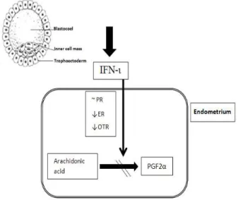
Figure 1 - Interferron tau (IFN-ι) production resulting from embryo-uterine crosstalking during early pregnancy in mammals. During maternal recognition of pregnancy, trophoectoderm of embryo produces IFN-ι which stabilizes progesterone receptor (PR) and reduces the number of estrogen receptor (ER) and oxytocin receptor (OTR) resulting in attenuation of uterine luteolysin, PGF2α. This pathway results
in maintenance of corpus luteum function.
CYCLOOXYGENASE 2 (COX 2)
Prostaglandins (PGs) are the major contributors to the regulation of reproductive processes and successful establishment of pregnancy (Mondal 2009). The production of endometrial PGs is mainly governed by the rate-limiting enzymes, cyclooxygenase (COX)-1 and COX-2, also known as prostaglandin endoperoxidase H synthetases 1 and 2 (PGHS-1 and PGHS-2). These enzymes are responsible for the conversion of arachidonic acid
into PGH2, the common precursor of the various
forms of PGs including PGE2 and PGF2α. The
down stream enzymes, PGE synthase and PGF
to PGE2 and PGF2α, respectively. PGE2 and PGF2
are the major secretory products of uterine
endometrium in ruminant. PGE2 is considered
important for blastocyst spacing, implantation and
decidualization, maternal recognition of
pregnancy, regulation of placental blood flow and function and corpus luteum life span and function
whereas PGF2 acts as the luteolytic agent to
control the estrous cycle (Arosh et al. 2004). Till-date, three isoforms of COXs have been identified. Cyclooxygenase-1 (COX-1) is a glycoprotein of 71 kDa, which is constitutively expressed in different tissues. COX-1 is encoded by a gene on chromosome 9 and plays a role in tissue homeostasis by modulating several cellular processes ranging from cell proliferation to angiogenesis or platelet aggregation due to
thromboxane production. Cyclooxygenase-2
(COX-2) is the inducible isoform, which is regulated by the growth factors and different cytokines such as IL1â, IL6, or TNFá and overexpressed during inflammation. The COX-2 gene is located on chromosome 1 and its promoter displays an NFêB response element as well as other cytokine-dependent (i.e., IL6) response elements. COX-3 has been identified as a splice variant of COX-1, and it is present mainly in brain and spinal cord. Currently, the role of COX-3 is not known. However, reports suggest a possible role in pain sensitivity, based on the studies focused on the mechanism of action of acetaminophen (paracetamol), evoked as a selective inhibitor of COX-3 (Blitek et al. 2006). Although COX-1 deficient female mice are fertile, they have specific defects in parturition, whereas COX-2 deficient female mice are infertile with
abnormalities in ovulation, fertilization,
implantation, and decidualization.
PROSTAGLANDIN
E
SYNTHASE
(PGES)
PGES is a member of the membrane associated protein in eicosanoid and glutathione metabolism (MAPEG) superfamily, which consists of six
human proteins with divergent functions.
Basically, PGES is a terminal prostanoid synthase that can enzymatically convert cyclooxygenase
product PGH2 to PGE2. The activity of PGES has
been detected both in cytosolic and microsomal fractions of various cells. Cytosolic PGES (cPGES) is constitutively expressed in a wide variety of cells and tissues and predominantly
linked with COX-1 to promote immediate response whereas microsomal PGES (mPGES) is a
membrane associated, inducible perinuclear
enzyme with glutathione dependent activity and is expressed in variety of tissues, including prostate, testis and small intestine. mPGES is preferentially coupled with inducible COX-2 to promote delayed
PGE2 generation. The PGES gene size is 14.8 kb
and has been mapped to chromosome 9. The genomic sequences of human PGES (Acc. No. AC 007936) indicates three exons (136, 83 and 1526 nt) separated by two introns (4.2 and 8.8 kb). The complete cDNA sequences of PGES has been characterized in the rat (Acc. No. NM 0211583), cattle (Acc. No. AY 032727), horse (Acc. No. AY 057096) and pig (Acc. No. AY 857634). Filion et al. (2001) reported the complete cDNA sequences of 462 base pairs of bovine PGES (Acc. No. AY 032727). The nucleotide sequence of buffalo PGES cDNA encoding the entire mature peptide, except the first three amino acids are also available (Acc. No. DQ167808). The nucleotide sequence of bubaline mPGES-1 cDNA exhibited 98, 90.5, 89.2, 87.2, 86.5, 80.4, 80.4% identity with that of cattle, pig, dog, human, horse, rat and mouse, respectively (Mondal et al. 2006). Expression of the PGES mRNA has been evaluated in a variety of tissues such as uterus, placenta, corpus luteum and ovary (Arosh et al. 2004; Parent and Fortier 2005; Waclawik et al. 2006; Mondal et al. 2007; 2008).
observed 3-fold increased GSH- dependent PGES
activity in vivo after lipopolysaccharide (LPS)
challenge in cytosol of rat brain but not of other tissues. Studies have reported that estradiol, oxytocin, LPS and TNF- increased the
production of PGE2 in buffalo endometrial
epithelial and stromal cells in vitro (Mondal et al.
2009a, 2009b; 2010). PGES mRNA expression is also increased in the presence of LPS, TNF- and IFN- in bovine epithelial cells (Parent et al. 2002). They also reported that phorbol -12 myristate 13 acetate increases PGES mRNA,
COX-2 mRNA and PGE2 production, suggesting
that the expression of PGES was correlated with
that of COX-2 for the production of PGE2.
Increasing the production would modulate
PGE2/PGF2 ratio and contribute to the
establishment of pregnancy.
PROSTAGLANDIN F SYNTHASE (PGFS)
PGFS is a member of aldo-keto reductase (AKR) superfamily based on the substrate specificity, molecular weight and amino acid sequence. AKR superfamily consists of monomeric NAD(P)H dependent oxido-reductase with broad specifity of substrates: endogenous substrates such as steroid hormones, prostaglandins and xenobiotics such as drugs and carcinogens. PGFS gene consists of nine exons and eight introns. The initiation codon is located at exon 1 and stop codon at exon 9 and encode a protein of 323 amino acid residues. Human PGFS gene is spanned around 11.597 kb and located at chromosome 10. The complete cDNA sequences of PGFS has been characterized in the cattle (Acc. No. J03570), buffalo (Acc. No. DQ884879), sheep (Acc. No. AY135401), horse (Acc. No. AY304536) and pig (Acc. No. AY863054). PGFS is known to exist in six isoforms viz., lung type PGFS (PGFS1), lung type PGFS found in liver (PGFS2), liver type PGFS (DDBX) and three other PGFS isolated from human, sheep and Trypanosoma brucei. All these isoforms belong to the AKR1C subfamily, except from Trypanosoma, which belongs to the AKR5A subfamily. In bovine, six isoforms of PGFS, viz., PGFS1, PGFS2, DDBX, PGFS like 1 (PGFSL1), PGFSL2 and 20 -HSD have been identified (Madore et al. 2003). A new isoform, AKR1B5 possessing aldoreductase activity has been implicated as the most probable enzymeresponsible for the production of PGF2 in bovines
(Madore et al. 2003). Kuchinke et al. (1992)
reported the complete cDNA sequence (CDS) of
972 base pairs of bovine lung PGFS (Acc. No.
M86544). The cDNA sequences of bovine PGFS like 1 protein (Acc. No AY135400) and PGFS like 2 protein (Acc. No. AY135401) are of 972 base pairs. The nucleotide sequence of buffalo PGFSL-2 cDNA exhibited 98, 87.6, 87.3, 77.PGFSL-2, 68.9, 68.7, 65, 77.7% identity with bovine PGFSL2, bovine lung PGFS, bovine liver PGFS, horse, pig, monkey, dog and human, respectively (Mondal et al. 2007). The expression of PGFS mRNA has been studied in the domestic ruminants, including cattle, buffalo, sheep, horse, pig and monkey and rat. Immuno-histochemical studies showed that PGFS proteins were localized in the luminal epithelium and stroma of endometrium and smooth muscle cells of bovine myometrium (Arosh et al. 2004). In corpus luteum, PGFS were localized in the large luteal cells and IFN- increased PGES expression (~2-fold) but not PGFS. Palliser et al. (2004) demonstrated that the expression of PGFS increased in the placentome
following dexamethasone induced and
spontaneous labour onset in the sheep. Mondal (2009) reported that the expression of PGFSL-2 mRNA could not be detected in buffalo uterine endometrium at any stage of estrous cycle. Xiao et al. (1999) reported that steroid hormones had no significant effect on PGFS expression in bovine endometrial cells. Oxytocin was reported to upregulate PGFS mRNA but rbIFN- significantly inhibited this induction at 3, 6, 12 and 24 h, respectively (Xiao et al. 1998). Similarly, Arosh et al. (2004) reported that IFN- decreased PGFS expression in endometrium by 1.5 fold and myometrium by 3.5 fold but not in corpus luteum.
It has been found that the production of PGF2α
increased in the presence of estradiol, LPS and TNF- in epithelial and stromal cells of buffalo (Mondal et al. 2009a; 2009b; 2010). The growth of buffalo uterine epithelial cells were significantly (P<0.05) higher in the presence of LPS, TNF- , linoleic acid and linolenic acid (Nandi et al. 2012) as compared to the control and lower doses. Progesterone, estradiol and oxytocin did not significantly increase the growth of buffalo uterine epithelial cells.
GALECTIN
cell adhesion and defence against invading microorganisms. Galectins do not possess a signal peptide or transmembrane spanning domain, and are secreted from the cells by a nonclassical pathway. Several forms of galectins: galectin-1 (LGALS1), galectin-3 (LGALS3), Galectin-5 (LGALS5), Galectin-9 (LGALS9) and Galectin-15 (LGALS15) have been discovered. Galectin-1
possess adhesive and anti-adhesive roles,
immunomodulator in maternofetal tolerance as well as embryo implantation. However, galectin-1-null mouse embryos develop normally and do not produce any overt phenotypic abnormalities
(Poirier and Robertson32). Galectin-3 (LGALS3) is
a galactose-specific lectin, expression of which increases significantly during the secretory phase of the menstrual cycle (von Wolff et al. 2005). Mice lack both galectins-1 and -3 implant normally but a further family member, galectin-5, is present and may compensate. Galectin-9 has been identified in mid- and late-secretory and decidual phases in human endometrium, with the expression in glandular and luminal epithelial but not stromal or immune cells (Smalley and Ley 2005). Recently, a new galectin family member,
galectin-15, has been discovered in the
endometrium of sheep, which has a prospective role in trophectoderm attachment (Farmer et al. 2008). Ovine endometrial Galectin-15 contains a
conserved carbohydrate recognition domain
(CRD) that binds β-galactosides, but the
carbohydrate-binding specificity for each galectin appears to be different. In addition to a conserved CRD, it also contains predicted cell attachment sequences (LDV and RGD) that could mediate binding to integrins in the ECM proteins (Kimber and Spanswick 2000). Galectin-15 mRNA was detected only in the endometrial LE and superficial ductal GE (Gray et al. 2004) of ovine uterus and was not detected before day 10, appeared and then increased 13-fold between days 10 and 14, and then noticeably decreased between days 14 and 16 in cyclic, but not pregnant ewes. It was present at low levels on days 10 and 12, but was abundant on days 14 and 16 of pregnancy in uterine flushing. The proposed extracellular role of galectin-15 in the uterine lumen is functionally to
bind and cross-link β-galactosides on
glycoproteins, such as mucins, integrins,
fibronectin, laminin and other glycoproteins and glycolipids, thereby allowing it to function as a heterophilic cell adhesion molecule bridging the blastocyst and the endometrial LE.
OSTEOPONTIN (OPN)
OPN belongs to the member of the small integrin-binding ligand, N-linked glycoprotein (SIBLING) family of genetically related ECM proteins recognized as key players in a number of diverse processes such as bone mineralization,
cell-mediated immune responses, inflammation,
angiogenesis, cell survival and implantation
(Butler et al. 1996). It is a negatively‐charged
acidic hydrophilic protein of approximately 300 amino acid residues, and is secreted into all body fluids. The OPN cDNA from various mammalian species exhibits a high degree of sequence homology. There is evidence of alternative splicing, although its functional significance is unclear. The molecule undergoes considerable
post‐translational modification, and is
phosphorylated and glycosylated. OPN has an
arginine‐glycine‐aspartic acid (RGD) cell binding
sequence, a calcium binding site and two heparin binding domains. In general, OPN is a monomer ranging in length from 264 to 301 amino acids that
undergoes extensive posttranslational
modification, including phosphorylation,
glycosylation, and cleavage, resulting in molecular mass variants ranging from 25 to 75 kDa. OPN
contains a hydrophobic leader sequence
characteristic of a secreted protein, a potential calcium phosphate aspatite binding region of consecutive asparagine residues, a cell attachment GRGDS sequence, a thrombin cleavage site, and two glutamines that are recognized substrates for transglutaminase-supported multimer formation. Genes encoding OPN have been cloned from eight species, including rat, mouse, human, cow, chicken, rabbit and sheep. The cDNA sequences from these species reveal only moderate
conservation except in the NH2-terminal region,
around the Arg-Gly-Asp (RGD) integrin-binding sequence, and the COOH-terminus.
Multiple forms of OPN have been identified as products of normal and transformed cells derived from rodent tissues. Osteopontin gene in buffalo encodes a protein of 280 amino acids and has insertion of two amino acids at positions 94 (Aspartic acid) and 227 (Asparagine), thus making a total of 280 amino acids as compared to 278 amino acids in bovines and ovines (Tantia et al.
2008). Multiple integrin receptors for OPN are
Evidence suggests that secreted OPN binds
integrin receptors expressed on conceptus
trophoblast and endometrial LE, where it can stimulate changes in proliferation, migration, survival, adhesion and remodeling of the conceptus as it elongates, apposes and adheres to the LE. OPN is hypothesized to serve as a bi-functional bridging ligand that mediates the adhesion between LE and trophoblast essential for implantation and placentation (Johnson et al.
2003). OPN binds to integrin heterodimers (αvβ1,
αvβ3, αvβ5, αvβ6, αvβ8, α4β1, α5β1 and α8β1) via
its Arg-Gly-Asp (RGD) sequence, and to α4β1 and
α9β1 by other sequences to promote cell adhesion, spreading and migration. In sheep, OPN is also a
component of histotroph secreted from
endometrial GE into the uterine lumen during pregnancy. During the peri-implantation period of pregnancy in sheep, OPN mRNA is expressed only by the endometrial glands, is first detected in some glands of some ewes by day 13, and is present in all glands by day 19 (Johnson et al. 2003).
INTEGRINS
Integrins belong to a family of heterodimeric intrinsic transmembrane glycoprotein receptors that play a major role in cellular differentiation, motility and adhesion. They mediate the interactions with ECM to transduce cellular signals in uterine epithelial cells and conceptus trophoblast (Johnson et al. 2003). The central role of integrins in the implantation adhesion cascade is to bind ECM ligand(s) to cause cytoskeletal reorganization, stabilize adhesion, and mediate cell migration, proliferation and differentiation through
numerous signaling intermediates. Altered
expression of integrins is correlated with several causes of infertility, null mutations of several integrins leads to peri-implantation lethality and functional blockade of selected integrins reduces the number of implantation sites (Illera et al.
2000). Integrin subunits α (v, 4, 5) and β (1, 3 and
5) are constitutively expressed on the apical surfaces of both conceptus trophoblast and endometrial LE during the peri-implantation period of pregnancy in ewes (Johnson et al. 2003). These integrin subunits are detected at the apical surfaces of the LE and GE and on conceptus trophoblast; expression of these integrins is constitutive and not influenced by pregnancy or presence of the conceptus. In the sheep, receptivity
to implantation does not appear to involve changes in either temporal or spatial patterns of integrin expression, but may depend on expression of other glycoproteins and ECM proteins, such as galectin-15, OPN and fibronectin, which are ligands for heterodimers of these integrins (Johnson et al. 2003). In species such as pig, mouse and humans, interactions between specific integrins and ECM
proteins frame the putative window of
implantation (Carson et al. 2000). A 2310 bp long buffalo (Bubalus bubalis) cDNA sequence of Integrin b2 (ITGB2) gene was characterized (Sharma et al. 2010). At nucleotide level, buffalo ITGB2 exhibited 96-98% homologies with other ruminants such as cattle, bison, sheep, goat and deer. The transmembrane and cytoplasmic regions are highly conserved among buffalo and other ruminants.
CONCLUSION
This review has highlighted maternal recognition of the pregnancy (MRP) and factors/genes controlling the pregnancy recognition signalling at embryo-uterine interface. Understanding of the
factors/genes regulating the conceptus
roles played by these genes in the MRP and implantation. A genome-wide screening approach coupled with functional assays might help elucidate these complex signaling pathways.
AUTHOR CONTRIBUTIONS
The authors Avantika Mor and Sukanta Mondal contributed equally to this work.
ACKNOWLEDGEMENTS
We are thankful to National Agricultural Science Fund (formerly National Fund for Basic, Strategic and Frontier Application Research in Agriculture), Ministry of Agriculture, ICAR, New Delhi for providing financial support to carry out the work. The authors express their gratitude to Dr. A. Bandyopadhyay, Ex. National Coordinator and Dr. P. K. Agarwal, ADG (NASF) for their help and suggestions. We thank the Director, NIANP, for providing the necessary facilities for conducting the research work. The help rendered by A. Jagannath is duly acknowledged.
REFERENCES
Arosh JA, Banu SK, Kimmins S, Chapdelaine P, Mac Laren LA, Fortier MA. Effect of interferon-tau on prostaglandin biosynthesis, transport and signaling at the time of maternal recognition of pregnancy in cattle: evidence of polycrine action of prostaglandin E2.Endocrinol. 2004; 145: 5280-5293.
Bauersachs S, Ulbrich SE, Gross K. Gene expression profiling of bovine endometrium during the oestrous cycle: detection of molecular pathways involved in functional changes. J MolEndocrinol. 2005; 34: 889-908.
Bazer FW, Spencer TE, Ott TL. Interferon tau: a novel pregnancy recognition signal. American J Reprod Immunol. 1997; 37: 412-420.
Blitek A, Waclawik A, Kaczmarek MM, Stadejek T, Pejsak Z, Ziecik AJ. Expression of cyclooxygenase-1 and -2 in the porcine endometrium during the oestrous cycle and early pregnancy. Reprod Domestic Anim. 2006; 41: 251-257.
Butler WT, Ridall AL, McKee MD. Osteopontin. In: Principals of Bone Biology, New York: Academic Press. pp.167-181 1996.
Carson DD, Bagchi I, Dey SK, Enders AC, Fazleabas AT, Lessey BA, et al. Embryo implantation. Dev Biol. 2000; 223: 217-237.
Farmer JL, Burghardt RC, Jousan FD, Hansen PJ, Bazer FW, Spencer TE. Galectin 15 (LGALS15) functions in trophectoderm migration and attachment. FASEB J. 2008; 22: 548-560.
Gray CA, Adelson DL, Bazer FW, Burghardt RC, Meeusen EN, Spencer TE. Discovery and characterization of an epithelial-specific galectin in the endometrium that forms crystals in the trophectoderm. Proc Natl Acad Sci USA. 2004; 101: 7982.
Hansen TR, Austin KJ, Perry DJ, Pru JK, Teixeira MG, Johnson GA. Mechanism of action of interferon-tau in the uterus during early pregnancy. J Reprod Fertil. 1999; 54: 329-339.
Illera MJ, Cullinan E, Gui Y, Yuan L, Beyler SA, Lessey BA. Blockade of the alpha (v) beta (3) integrin adversely affects implantation in the mouse. Biol Reprod. 2000; 62: 1285-1290.
Johnson GA, Bazer FW, Jaeger LA, Ka H, Garlow JE, Pfarrer C, et al. Muc-1, integrin, and osteopontin expression during the implantation cascade in sheep. Biol Reprod. 2001; 65: 820-828.
Johnson GA, Burghardt RC, Joyce MM, Spencer TE, Bazer FW, Gray CA, et al. Osteopontin is synthesized by uterine glands and a 45-kDa cleavage fragment is localized at the uterine–placental interface throughout ovine pregnancy. Biol Reprod. 2003; 69: 92-98.
Kimber SJ, Spanswick C. Blastocyst implantation: the adhesion cascade, Seminars Cell Dev Biol. 2000; 11: 77-92.
Kuchinke W, Barski O, Watanabe K, Hayaishi O. A lung type prostaglandin F synthase is expressed in bovine liver: cDNA sequence and expression in E.coli, Biochem Biophy Res Comm. 1992; 183: 1238-1244.
Martin RL, Whittle WL, Gyomarey S, Gribb W, Challis JRG. Ontogeny and regulation of ovine placental prostaglandin E synthase. Biol Reprod. 2002; 67: 868-873.
Madore E, Harvey N, Parent J, Chapdelaine P, Arosh JA, Fortier MA. An aldose reductase with 20 hydroxysteroid dehydrogenase activity is most likely the enzyme responsible for the production of prostaglandin F2 in the bovine endometrium. J Biol Chem. 2003; 278: 11205-11212.
Mondal S, Varshney VP, Kumar N, Shukla SN, Sukumar K, Agarwal SK, Joshi P, Sharma AK, Mitra A. Cloning and molecular characterization of prostaglandin E synthase gene in buffalo (Bubalus bubalis), In : Proc. of Symposium in Frontier in Reproduction : Concepts and Applications in Genomic Era in India, 2006, Karnal; p. 73.
Mondal S, Varshney VP, Sukumar K, Kumar N, Shukla SN, Agarwal SK, Joshi P, Mitra A. Molecular characterization of prostaglandin F synthase gene in buffalo (Bubalus bubalis), In : Proc. of Section of Medical Sciences (including Physiology), 94th Indian Science Congress Association in India, 2007, Chidambaram; p. 17-18.
Mondal S, Prakash BS, Palta P. Endocrine aspects of estrous cycle in buffalo: an overview. Asian Australasian J Anim Sci. 2007; 20: 124-131.
Mondal S, Varshney VP, Mitra A. Prostaglandin E and F synthases: genomic insight, In: Proceedings of National Symposium on Animal Biotechnology in India, 2007, Bangalore, p. 34-40.
Mondal S, Varshney VP, Mitra A. Early Embryonic Mortality : Physiogenomic approaches, Lead Paper presented at National symposium on Current Concepts in Productivity Management in Livestock and Poultry-Environment, Nutrition and Stress in India, 2008; p. 47-57.
Mondal S. Early embryonic loss: Genomic insight. Ind. J Physiol Allied Sci. 2009; 63: 44-51.
Mondal S, Nandi S, Reddy IJ, Suresh KP. Effect of steroids on in vitro prostaglandin production in buffalo endometrial epithelial cells, In: Proceedings of XVIII Annual SAPI conference and National Symposium on Physiogenomic prospects in augmenting Livestock Production in India 2009a, Bangalore, p. 170.
Mondal S, Nandi S, Reddy IJ, Suresh KP. Regulation of prostaglandin production by steroid hormones in buffalo endometrial stromal cells, In: Proceedings of XVIII Annual SAPI conference and National Symposium on Physiogenomic prospects in augmenting Livestock Production in India 2009b, Bangalore, p. 170.
Mondal S, Reddy IJ, Nandi S. Modulation of prostaglandin production by LPS and TNF in endometrial epithelial and stromal cells of buffalo, In: Proceedings of XXXVIII Dairy Industry Conference in India, 2010, NIMHANS, Bangalore; 2010. P. 7. Mondal S, Suresh KP, Nandi S. Endocrine profiles of
estrous cycle in buffalo - A meta-analysis. Asian Australasian J Anim Sci. 2010; 23: 169-172.
Nandi S, Mondal S, Reddy IJ. Effect of prostaglandin producing modulators on in vitro growth characteristics in buffalo endometrial epithelial cells. Theriogenol. 2012; 77: 1014-1020.
Palliser HK, Ooi GT, Hirst JJ, Rice G, Dellions NL, Escalon RM, et al. Changes in the expression of prostaglandin E and F synthases at induced and spontaneous labour onset in the sheep. J Endocrinol. 2004; 180: 469-477.
Parent J, Fortier MA. Expression and contribution of three different isoforms of prostaglandin E synthase in the bovine endometrium. Biol Reprod. 2005; 73: 36-44.
Parent J, Chapdelaine P, Sirois J, Fortier MA. Expression of microsomal prostaglandin E synthase in bovine endometrium: coexpression with cyclooxygenase type 2 and regulation by interferon-tau. Endocrinol. 2002; 143: 2936-2943.
Poirier F, Robertson EJ. Normal development of mice carrying a null mutation in the gene encoding the L14 S-type lectin. Dev. 1993; 119: 1229-1236.
Roberts RM, Ealy AD, Alexenko AP, Han CS, Ezashi T. Trophoblast interferons, Placenta. 1999; 20: 259-264.
Saugandhika S, Malik HN, Singhal DK, Singhal R, Dubey A, Boateng S, et al. Identification of the relatively predominant buffalo Interferon tau isoform and its expression in Escheria Coli. Reprod Fertil Dev. 2013; 26(1): 174-174.
Sharma D, Niranjana SK, Kumar S, Deb SM, Naskar S, Sharma A, et al. Molecular Characterization of
Bubaline Integrin β2 (ITGB2) cDNA. J Applied Anim Res. 2010; 37(2): 217-220.
Smalley DM, Ley K. L-selectin: mechanisms and physiological significance of ectodomain cleavage. J Cell Mol Med. 2005; 9: 255-266.
Spencer TE, Johnson GA, Bazer FW, Burghardt RC. Implantation mechanisms: insights from the sheep. Reprod. 2004; 128: 657-668.
Spencer TE, Bazer FW. Ovine interferon tau suppresses transcription of the estrogen receptor and oxytocin receptor genes in the ovine endometrium. Endocrinol. 1996; 137: 1144-1147.
Spencer TE, Ott TL, Bazer FW. Tau-interferon: Pregnancy recognition signal in ruminants. Proc Soc Exp Biol Med. 1996; 213: 215-229.
Tanioka T, Nakatani Y, Semmyo N, Murakami M, Kudo I. Molecular identification of cytosolic prostaglandin E synthase that is functionally coupled with cyclooxygenase-1 in immediate prostaglandin E biosynthesis. J Biol Chem. 2000; 275: 32775-32782. Tantia MS, Mishra B, Bharani KST, Mishra BP,
Kataria RS, Mukesh M,et al. Characterization of Osteopontin gene of Bubalus bubalis. Anim. 2008; 2(7): 987-990.
Waclawik A, Rivero-Muller A, Blitek A, Kaczmarek MM, Brokken LJS, Watanabe K, et al. Molecular cloning and spatiotemporal expression of prostaglandin F synthase and microsomal prostaglandin E synthase-1 in porcine endometrium. Endocrinol. 2006; 147: 210-221.
Xiao CW, Murphy BD, Sirois J, Groff AK. Downregulation of oxytocin induced cycloxygenase-2 and prostaglandin F synthase expression by interferon-tau in bovine endometrial cells. Biol Reprod. 1999; 60: 656-663.
Xiao CW, Liu JM, Sirois J, Goff AK. Regulation of cyclooxygenase-2 and prostaglandin F synthase gene expression by steroid hormone and interferon tau in bovine endometrial cells. Endocrinol. 1998; 139: 2293-2299.