• Nenhum resultado encontrado

Supressão da resposta do HIF-1a pelo NF-kB via competição pela ligação com o coativador transcricional p300

N/A
N/A
Protected

Academic year: 2017

Share "Supressão da resposta do HIF-1a pelo NF-kB via competição pela ligação com o coativador transcricional p300"

Copied!
138
0
0

Texto

(1)

DANIELA BACCELLI SILVEIRA MENDONÇA

SUPRESSÃO DA RESPOSTA DO HIF-1

PELO NF-

B VIA COMPETIÇÃO PELA

LIGAÇÃO COM O COATIVADOR TRANSCRICIONAL p300

Tese apresentada ao Programa de

Pós-graduação

Strictu

Sensu

em

Ciências

Genômicas e Biotecnologia da Universidade

Católica de Brasília como requisito para

obtenção do Título de Doutor em Ciências

Genômicas e Biotecnologia.

Orientador:

Prof. Dr. Francisco José Lima

Aragão.

Co-orientador:

Prof. Dr. Lyndon Frederick

Cooper.

(2)

À Deus pela vida e por todas as

oportunidades a mim oferecidas.

Ao querido Gustavo pelo carinho, amor,

compreensão, paciência, e por todo auxílio

que foram essenciais para que eu pudesse

terminar mais essa etapa.

Aos meus queridos pais Mac Tulio e Maria

Inez, exemplos de força e determinação,

agradeço pela educação, amor e apoio

incondicionais.

(3)

AGRADECIMENTO ESPECIAL

Aos meus Orientadores, Prof. Dr. Francisco José Lima Aragão e Prof. Dr. Lyndon

Frederick Cooper, agradeço pela confiança em mim depositada, pela disponibilidade

de tempo, pelos conhecimentos compartilhados e por terem disponibilizado os

recursos necessários para o término desse projeto

(4)

AGRADECIMENTOS

À Universidade Católica de Brasília.

À Embrapa Recursos Genéticos e Biotecnologia.

À Universidade da Carolina do Norte em Chapel Hill-EUA.

À Coordenação de Aperfeiçoamento de Pessoal de Nível Superior – CAPES – pela

bolsa de doutorado Sandwich que me permitiu expandir meus conhecimentos e na

Universidade da Carolina do Norte em Chapel Hill-EUA.

À todos os professores do Programa de Pós-graduação em Ciências Genômicas e

Biotecnologia da Universidade Católica de Brasília.

Aos funcionários do Programa de Pós-graduação em Ciências Genômicas e

Biotecnologia da Universidade Católica de Brasília, especiamente ao Francisco

Fábio Gomes da Costa.

Aos amigos de pós-graduação pelos momentos que passamos juntos e aprendemos

uns com os outros.

À Sara Valencia, reponsável pelo andamento de todas as atividades no Laboratório

de Biologia Óssea e Mineralização da Universidade da Carolina do Norte, e que

muito ajudou para que nossos trabalhos no laboratório funcionassem perfeitamente.

À Gidget Jenkins e Martha Taylor do departamento de Prótese da Faculdade de

Odontologia da Universidade da Carolina do Norte, por também nos ajudarem em

tudo que fosse necessário durante nossa estadia nos Estados Unidos.

Aos amigos da Universidade da Carolina do Norte, Gustavo e Greice, Ricardo e

Patrícia, Luiz e Silvana, Sodsi “Nid” Wirojchanasak e Ghadeer Thalji.

(5)

(6)

RESUMO

MENDONÇA, Daniela Baccelli Silveira. Supressão da resposta do HIF-1

pelo

B via competição pela ligação com o coativador transcricional p300. 2010.

138p. Tese. Ciências Genômicas e Biotecnologia – Universidade Católica de

Brasília, Brasília, 2010

(7)

dependente. Conclui-se que a sinalização inflamatória mediada pelo NF-

B é capaz

de bloquear a transativação do HIF-1

nos sítios HRE dos genes que respondem ao

HIF-1

através de uma competição direta pela ligação ao p300. A inflamação pode

influenciar o nicho de células-tronco e a regeneração tecidual por interferir com a

resposta das células à hipóxia.

(8)

ABSTRACT

Hypoxia has emerged as a key determinant of osteogenesis. HIF-1

is the

transcription factor mediating hypoxia responses that include induction of VEGF and

related bone induction. Inflammatory signals antagonize bone repair via the NF-

B

pathway. The present investigation explored the functional relationship of hypoxia

(HIF-1

function) and inflammatory signaling (NF-

B) in stem like and

osteoprogenitor cell lines. The potential interaction between HIF-1

and NF-

B

signaling was explored by co-transfection studies in hFOB with p65, HIF-1

and

9x-HRE-luc or HIF-1

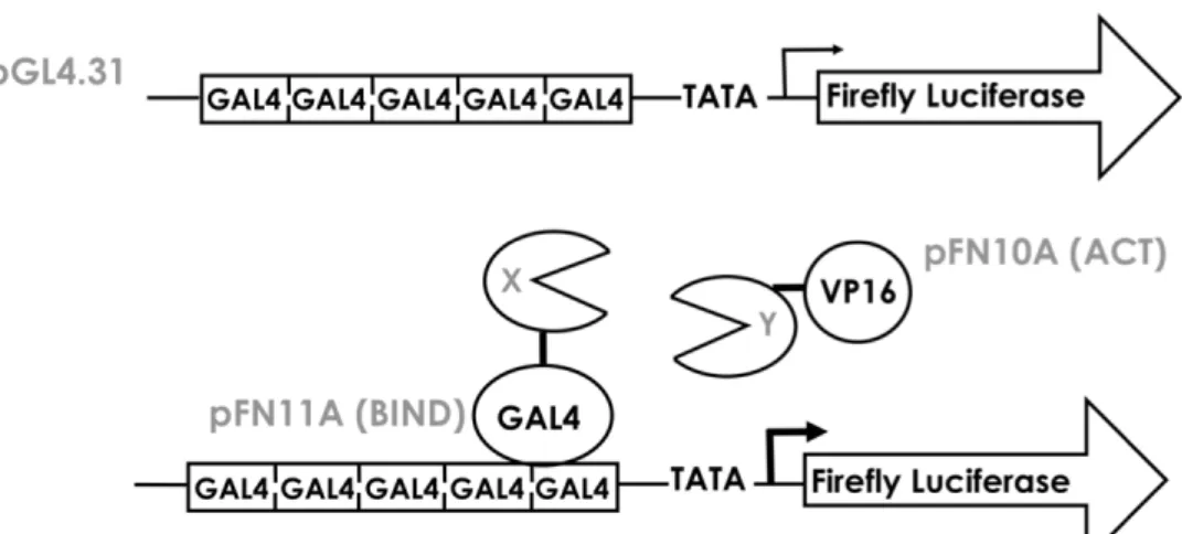
target genes reporter plasmids. Nuclear cross-talk was directly

tested using the mammalian Gal4/VP16 two-hybrid, and confirmed by

co-immunoprecipitation/western blotting assays. The results show that inflammatory

stimulation (TNF-

treatment) causes a marked inhibition of HIF-1

function at the

HRE in all cell lines studied. Also, co-transfection with p65 expression vector leads to

reduced hVEGFp transcription after DFO-induced hypoxia. However, TNF-

treatment or NF-

B expression from encoding virus or plasmid had little effect on

HIF-1

mRNA or protein levels nor did HIF- 1

nuclear translocation change with

treatment. The functional interaction of Gal4-HIF-1

and VP16-p300 fusion proteins

is effectively blocked by expression of p65 in a dose dependent manner. It was

concluded that NF-

B-mediated inflammatory signaling is able to block HIF-1

transactivation at HRE-encoding genes by direct competition for p300 binding at the

promoter. Inflammation may influence the stem cell niche and tissue regeneration by

influencing cellular responses to hypoxia.

(9)

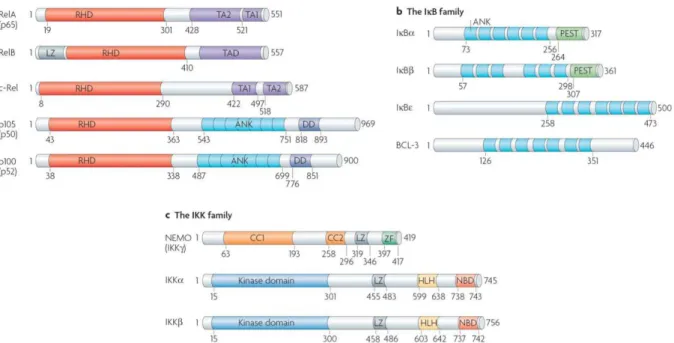
LISTA DE ABREVIATURAS

ACTB – Beta actin

ANG-1 – Angiopoietin 1

ANG-2 – Angiopoietin 2

ANK – Ankyrin-repeat motifs

ARNT – Aryl hydrocarbon receptor nuclear translocator

ATM – Ataxia telangiectasia mutated checkpoint kinase

BAFF – B-cell activating factor

bHLH – Basic helix-loop-helix domain

BMP – Bone morphogenetic protein

BSA – Bovine serum albumin

C-TAD – Carboxi-terminal Activation Domain

CAD – Carboxi-terminal Activation Domain

CD14 – Cluster of differentiation 14

CH1 – Cysteine-histidine-rich zinc finger motif

CK2 – Casein kinase-II

COX2 – Ciclooxigenase 2

CSC – Cancer stem cells

CX3CR1 – CX3C chemokine receptor 1

CXCR4 – CXC chemokine receptor 4

DFO – Desferrioxamine

EGF – Epidermal growth factor

eNOS – Endothelial nitric oxide synthase

ERK – Extracellular signal-regulated kinase

FAK – Focal adhesion kinase

FBS – Fetal bovine serum

FIH-1 – Factor inhibiting HIF-1

FITC – Fluorescein isothiocyanate

(10)

HA – Hemagglutinin

HAT – Histone acetyltransferases

HDAC – Histone deacetylases

HET – Heterozygous

hFOBs – Human fetal osteoblasts
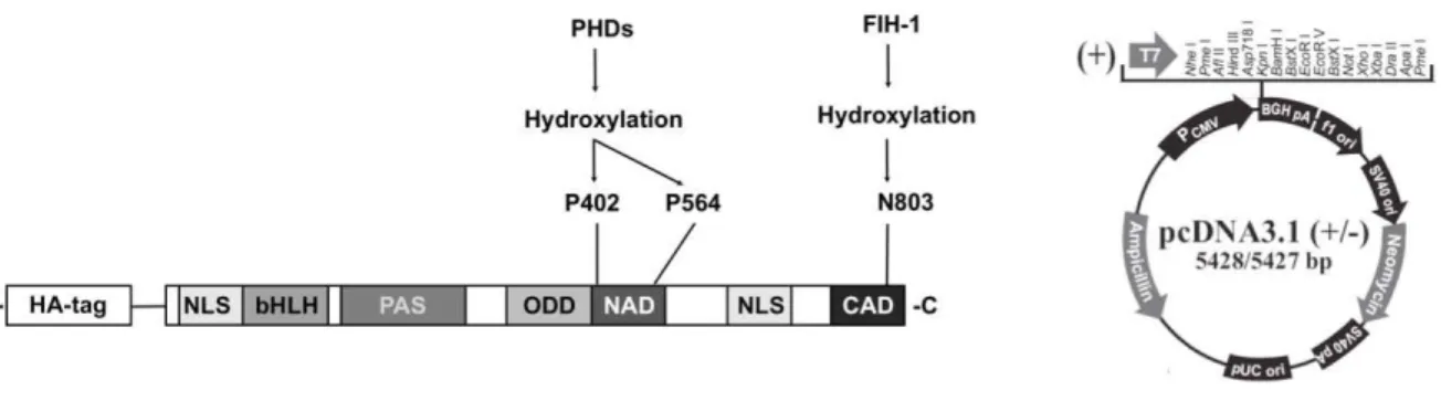
HIF-1

– Hypoxia inducible factor 1

hMSCs – Human mesenchymal stem cells

HRE – Hypoxia responsive element

HRP – Horseradish peroxidase

ID – Inhibitory domain

IGF-1 – Insulin-like growth factor 1

IGF-2 – Insulin-like growth factor 2

IKK – Inhibitor of

B kinase

IL-1 – Interleukin-1

IL-4 – Interleukin 4

IL-8 – Interleukin-8

iNOS – Inducible nitric oxide synthase

I

B

-SR – I

B

super repressor

LAR II – Luciferase Assay Reagent II

LMP1 – Latent membrane protein 1

LMP1 – Latent membrane protein-1

LOX – Lysyl oxidase

LPS – Lipopolysaccharide

LT

R – Lymphotoxin-

receptor

MAPK – Mitogen-activated protein kinase

ml – milliliter

MMP-2 – Matrix metalloproteinase 2

MMP-2 – Matrix metalloproteinase 9

MOI – Multiplicity of infection

mTOR – Mammalian target of rapamycin

N-TAD – Amino-terminal Activation Domain

N803 – Asparaginyl residue 803

(11)

NEMO – NF-

B essential modifier

NF-

B: Nuclear factor kappa B

ng – nanogram

NGF – Nerve growth factor

NIK – NF-

B inducing kinase

NO – Nitric oxide

ODD – Oxygen-dependent domain

PAI-1 – Plasminogen activator inhibitor 1

PAS – Per-ARNT-Sim domain

PBS – Phosphate buffered saline

PCR – Polymerase Chain Reaction

PDGF-

– Platelet-derived growth factor

PHD – Prolyl hydroxylase

PI-3k/AKT – Phosphatidylinositol-3 kinase / Serine-threonine protein kinase pathway

PKA – Protein kinase A

PKC – Protein kinase C

pRL-TK –

Renilla

luciferase reporter that contains the Herpes simplex virus

Thymidine Kinase

Pro 564 – Proline residue 564

Pro402 – Proline residue 402

pVhl – Von Hippel-Lindau protein

Rel homology domain – RHD

RIPA – Radio-immunoprecipitation assay

ROS – Reactive oxygen species

RT-PCR – Reverse transcriptase – Polymerase chain reaction

SDF-1

– Stromal-derived factor 1

SDS-PAGE – Sodium dodecyl sulfate – Polyacrylamide gel electrophoresis

SNIP1 – Smad nuclear-interacting protein 1

SV40 – Simian vacuolating virus

TBS – Tris-buffered saline

(12)

TNF-

– Tumor necrosis factor

VEGF – Vascular endothelial growth factor

µg – microgram

(13)

SUMÁRIO

Resumo

Abstract

Lista de Abreviaturas

Introdução

14

Revisão da literatura

25

Objetivos

47

Materiais e métodos

49

Resultados

59

Discussão

90

Conclusões

97

Perpectivas futuras

100

Referências Bibliográficas

101

(14)
(15)

O fator induzido por hipóxia 1 alfa (Hypoxia Inducible Factor, HIF-1

) foi

inicialmente identificado como um fator de transcrição responsável pela regulação do

gene da eritropoetina em resposta à uma diminuição de oxigênio nos tecidos renais

(WENGER et al., 2005). O complexo HIF consiste de uma de três sub-unidades alfa

(HIF-1

, HIF-2

ou HIF-3

) que se ligam ao Translocador Nuclear Receptor Aril

Hidrocarboneto (Aryl Hydrocarbon Receptor Nuclear Translocator, ARNT), também

conhecido como subunidade beta do fator 1 induzível por hipóxia HIF-1

(WANG et

al., 2007a). Geralmente, o HIF-1

é constitutivamente expresso e seus níveis não

variam de acordo com a disponibilidade de oxigênio, ao contrário das subunidades

alfa que são estritamente reguladas em resposta à hipóxia (HIROTA; SEMENZA,

2005). As variações na concentração de oxigênio não são diretamente reguladas

pelo HIF-1

e sim por uma classe de dioxigenases dependentes de ferro e de

2-oxoglutarato. Dois tipos de sensores do nível de oxigênio estão envolvidos na

regulação do HIF-1

: as prolil hidroxilases (PHDs) e a asparaginil hidroxilase

(SCHIPANI et al., 2009). Na presença de níveis normais de oxigênio (normóxia), as

PHDs 1, 2 e 3 hidroxilam dois resíduos de prolina (Pro402 e Pro564) na porção

amino-terminal (N-terminal transactivation

domain, N-TAD) do domínio de

degradação dependente de oxigênio (Oxygen-dependent Domain, ODD). Essa

hidroxilação promove a interação do HIF-1

com a proteína do gene supressor de

tumor von Hippel-Lindau (von Hippel-Lindau protein, pVHL), que é o componente de

reconhecimento da ligase de ubiquitina E3. O HIF-1

é então marcado com cadeias

de poliubiquitina e será degradado pelo proteassomo. O segundo sensor de oxigênio

é uma asparaginil hidroxilase conhecida como Fator Inibidor do HIF-1 (Factor

Inhibiting HIF-1, FIH-1). Essa enzima hidroxila um resíduo de asparagina (N803) na

porção carboxi-terminal (C-terminal transactivation domain, C-TAD) do domínio ODD

do HIF-1

. Essa modificação covalente bloqueia a interação do C-TAD com os

co-ativadores transcricionais, como CBP e p300 (SCHIPANI et al., 2009).

(16)

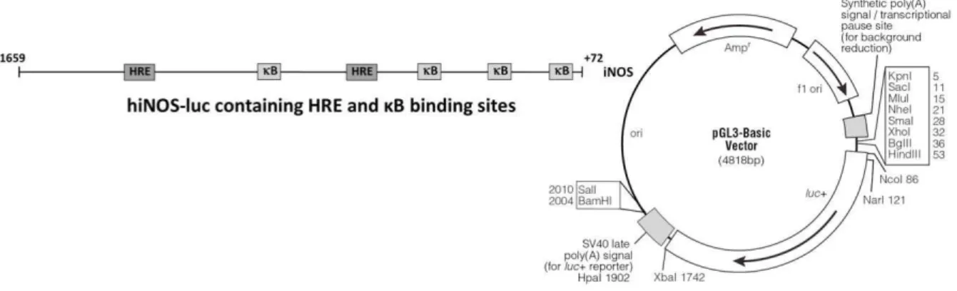
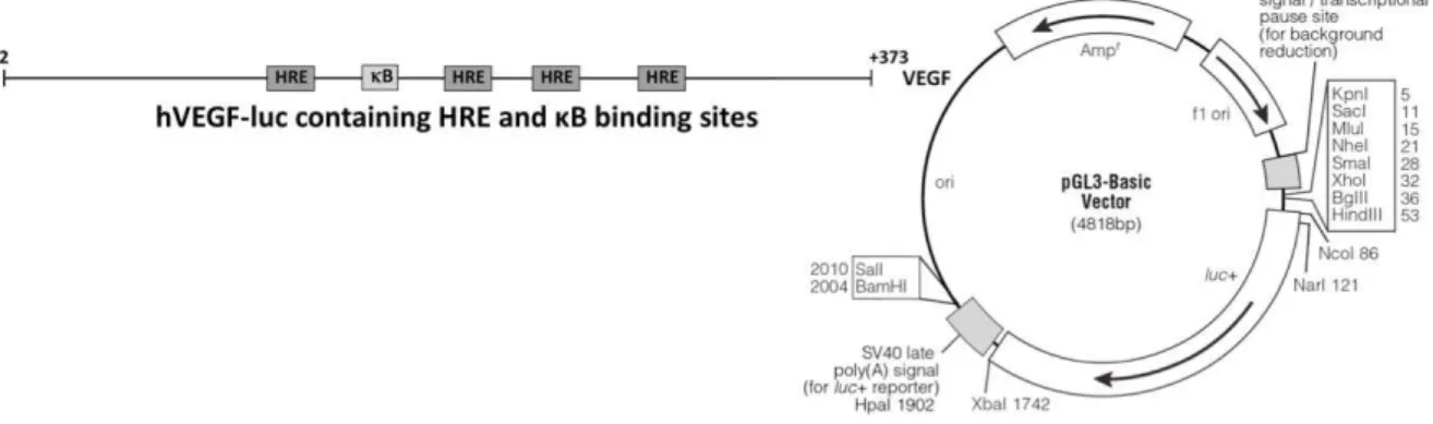
(RCGTG) na região promotora dos genes alvo do HIF-1

. Em células bem

oxigenadas, os dois sensores de oxigênio, PHD e FIH, que regulam a destruição e

atividade do HIF-1

, respectivamente, asseguram a repressão da via do HIF-1

em

células bem oxigenadas (SCHIPANI

et al.,

2009). A

figura 1 mostra o HIF-1

em

condições de normóxia e hipóxia (SEMENZA, 2004).

(17)

Acredita-se que a hipóxia seja o principal estímulo responsável pelo início da

cascata angiogênica durante o desenvolvimento e após o traumatismo ósseo. Os

osteoblastos estão idealmente localizados no osso de maneira que possam sentir a

tensão de oxigênio e responder à hipóxia ativando a via do HIF-1

(WANG et al.,

2007b). Utilizando uma abordagem genética para determinar os efeitos celulares e

moleculares de ganho ou perda da função do HIF-1

por mutagênese condicional

em osteoblastos de camundongo, esses autores demonstraram que a

superexpressão do HIF-1

em osteoblastos de camundongo por meio da truncagem

do Vhl resultou em aumento profundo na angiogênese e osteogênese. Ao contrário,

a truncagem do HIF-1

nos osteoblastos produziu o fenótipo reverso, ou seja, ossos

mais finos e menos vascularizados, o que demonstra a importância da via do HIF-1

na formação óssea guiada por osteoblastos (WANG et al., 2007a). O mesmo grupo

também demonstrou que a ativação farmacológica da via do HIF-1 por meio do

DFO induziu uma robusta resposta angiogênica acompanhada de uma subsequente

resposta osteogênica in vivo (WAN et al., 2008).

Atualmente, mais de 100 genes alvo do HIF-1

foram identificados, incluindo

genes relacionados à inflamação como a cicloxigenase-2 (COX-2), citocinas como o

fator de necrose tumoral (Tumor Necrosis Factor alpha, TNF-

) e a interleucina 8

(Interleukin-8, IL-8). Então, além de facilitar a adaptação, a hipóxia pode também

afetar os processos inflamatórios (OLIVER et al., 2009).

(18)

Gene product

1B-Adrenergic receptor

Adrenomedullin Aldolase A (ALDA) Atrial natriuretic peptide Carbonic anhydrase 9 CD18

Ceruloplasmin C-MET

Connective tissue growth factor CYP3A6

CXCR4 DEC1 DEC2

Ecto-5_-nucleotidase (CD73) Endocrine gland-derived VEGF Endoglin Endothelin-1 Enolase 1 ENOS Erythropoietin ETS-1

Glucose transporter 1 (GLUT1) Glyceraldehyde-3-phosphate dehydrogenase

Glucose regulated protein, 94-kDa (GRP94)

Heme oxygenase-1

HIF-1_ prolyl hydroxylase PHD3 (EGLN3)

HIF-1_ prolyl hydroxylase PHD2 (EGLN1)

HGTD-P Integrin 2

Intestinal trefoil factor

Lactate dehydrogenase A (LDHA) Lactase

Leptin

Membrane type-1 matrix metalloproteinase

Multi-drug resistance 1 (ABCB1) Myeloid cell factor 1 (MNL1) Nitric oxide synthase 2 NIP3

NUR77

p35srj (CITED2)

Phosphoglycerate kinase 1

6-Phosphofructo-2-kinase/fructose-2,6-bisphosphatase-3 (PFKFB3)

6-Phosphofructo-2-kinase/fructose-2,6-bisphosphatase-4 (PFKFB4)

Plasminogen activator inhibitor 1 Procollagen prolyl-4-hydroxylase (I) ROR

Stromal-derived factor 1 (SDF-1) Telomerase (TERT)

Transferrin

Transferrin receptor

Transforming growth factor 3 Vascular endothelial growth factor (VEGF)

(19)

O reparo ósseo é antagonizado pela inflamação. Vários pesquisadores

mostraram que estímulos inflamatórios diminuem ou impedem o reparo ou formação

óssea. Como exemplo, o tratamento de células osteoprogenitoras com TNF-

bloqueia

a diferenciação osteoblástica. Um possível mecanismo seria o antagonismo da via das

proteínas morfogenéticas ósseas (Bone Morphogenetic Protein, BMP) mediado pelo

TNF-

(GILBERT et al., 2000). Outro possível efeito da sinalização inflamatória em

células osteoblásticas é a inibição direta de fatores de transcrição chave para a

osteogênese como o Osterix (LACEY et al., 2009) (LU et al., 2006b).

O TNF-

é a citocina mais investigada entre as citocinas conhecidas por suas

ações inflamatórias (HADDAD; LAND, 2001). O efeito da hipóxia na biosíntese do

ainda não foi completamente investigado (LIU et al., 2008). Esses autores relataram

que o HIF-1

contribuiu predominantemente para a supra-regulação do TNF-

,

especialmente in vivo e foi dependente da sinalização via proteína quinase ativada por

mitógeno p38 (Mitogen-activated protein kinase, MAPK). De acordo com Albina

et al.

(ALBINA et al., 2001), o TNF-

aumentou o conteúdo protéico de HIF-1

em células

isoladas de um e cinco dias após o ferimento. Entretanto, o TNF-

não aumentou o

HIF-1

em fibroblastos de rato e camundongo. Blouin

et al. (BLOUIN et al., 2004)

demonstraram que células derivadas de macrófago apresentaram um aumento na

atividade do repórter HRE a um nível que ultrapassou o aumento causado pela hipóxia,

quando expostas a lipopolissacarídeo (LPS). Eles também afirmaram que quando

células derivadas de macrófago foram transfectadas com uma forma

dominante-negativa do HIF-1

, forte inibição do repórter HRE foi observada em células

estimuladas pelo LPS. Estas diferentes respostas apresentadas por diferentes tipos de

células estimulou o estudo com relação à células da linhagem osteoblástica.

(20)

(21)

Figura 2 –

Membros das famílias NF-

B, I

B e IKK. a)

Em células de mamíferos, a

família do NF-

B é composta de 5 membros: RelA (p65), RelB, c-Rel, p50/p105

B1) e p52/p100 (NF-

B2). p50 e p52 (não mostrados na figura) são derivados de

moléculas precursoras maiores p105 e p100, respectivamente. Todos os membros da

família do NF-

B contêm o domínio de homologia Rel, que contêm o domínio de

localização nuclear e é responsável pela ligação e dimerização do DNA. A sub-família

Rel, RelA, RelB e c-Rel possuem domínios independentes de ativação transcricional na

porção C- terminal. TA1 e TA2 são sub-domínios do domínio de transativação RelA.

b)

A família dos inibidores do NF-

B (I

B) é formada pelo membros: I

B

, I

B

, I

B

e

BCL-3. Assim como o p105 e o p100, as proteínas I

B possuem motivos de repetição

de anquirina (

Ankyrin-repeat motifs

– ANK) na porção C-terminal.

c)

As três principais

subunidades do complexo inibidor da proteína quinase kappa B (

Inhibitor of

B kinase

,

IKK) são mostradas: as subunidades catalíticas IKK

e IKK

e a subunidade regulatória

NEMO, também conhecida como IKK

. Os principais motivos da estrutura de cada

proteína são mostrados, assim como os números dos aminoácidos correspondentes às

proteínas humanas. CC,

coiled-coil

; DD,

region with homology to a death domain

; HLH,

helix–loop–helix

; LZ,

RelB-transactivation-domain containing a putative

leucine-zipper-like motif

; NBD,

NEMO-binding domain

; PEST,

domain rich in proline

(P),

glutamate

(E),

serine

(S)

and threonine

(T); ZF,

zinc-finger domain

(PERKINS, 2007).

Existem várias vias de ativação do NF-

B. A mais comumente observada é a via

canônica, também conhecida como via clássica. A via canônica é ativada através de

(22)

do proteassomo 26S. Em vários tipos celulares, I

B

e I

B

estão também sujeitos à

fosforilação e degradação, mas com uma cinética mais lenta (PERKINS, 2007). Na via

canônica, a fosforilação do I

B é causada pelo complexo IKK. Esse complexo consiste

de três subunidades principais: as subunidades catalíticas IKK

e IKK

(também

conhecidas como IKK1 e IKK2) e várias cópias de uma subunidade regulatória

conhecida como modificador essencial do NF-

B (NF-

B essential modifier – NEMO),

também conhecido como IKK

. Experimentos genéticos mostraram que o IKK

é a

quinase I

B predominante na via canônica (PERKINS, 2007). A via não canônica,

também conhecida como via alternativa, é ativada através de estímulos como o CD40,

receptor da linfotoxina beta (Lymphotoxin- receptor, LT

R), fator ativador de células B

(23)

(24)

ativação do IKK

por meio da quinase que induz o NF-

B NIK, seguida da fosforilação

da sub-unidade p100 do NF-

B pelo IKK

. Isso resulta na transformação dependente

do proteassomo da sub-unidade p100 para a p52, o que pode levar à ativação dos

heterodímeros p52–RelB, que ativam vários alvos

B. A fosforilação das sub-unidades

do NF-

B pelas quinases nucleares e modificação dessas subunidades por acetilases e

fosfatases pode resultar na ativação e repressão da transcrição, assim como causar

efeitos específicos nos promotores. Além disso, a interação com fatores de transcrição

heterólogos podem fazer com que o complexo do NF-

B seja alvo de promotores

específicos, o que resulta na ativação seletiva da expressão gênica após a exposição

celular à diferentes estímulos. Ac,

acetylation; bZIP,

leucine-zipper-containing

transcription factor; HMG-I,

high-mobility-group protein-I; I

B,

inhibitor of

B; IKK,

I

B

kinase; LMP1,

latent membrane protein-1; LPS,

lipopolysaccharide; NF-

B,

nuclear

factor-

B; RHD,

Rel-homology domain; TAD,

transcriptional activation domain; TF,

transcription factor; UV,

ultraviolet; Zn-finger TF,

zinc-finger-containing transcription

factor (PERKINS, 2007).

O HIF-1

e o NF-

B são fatores de transcrição que respondem à hipóxia e que

além de atuarem independentemente na regulação de genes adaptativos e

inflamatórios, demonstram um alto nível de interdependência e parecem compartilhar

as mesmas vias regulatórias (TAYLOR et al., 1998). Apesar do fato de que vários

estudos já demonstraram um

cross-talk entre as vias do NF-

B e HIF-1

, incluindo o

compartilhamento de alguns genes alvo, um

link direto ainda não foi elucidado (VAN

UDEN et al., 2008).

(25)

(26)

LITERATURE REVIEW

Biological functions of HIF-1 in angiogenesis and osteogenesis and its significance in

tissue engineering

Oxygen concentration is an important component of stem cell “niche,” where it

plays a fundamental role in maintaining the stem cells' proliferation and plasticity (MA

et

al.,

2009). Changes in oxygen concentrations affect many of the innate characteristics of

stem and progenitor cells (GRAYSON

et al.,

2007). Nevertheless, the effects of hypoxia

on stem cells are somewhat controverse.

Potier

et al.

(POTIER

et al.,

2007) investigated the effects of transient (48 h)

exposure to hypoxia (

1% O2) on primary human mesenchymal stem cells (hMSC)

survival, osteogenic potential and angiogenic factor expression. They discovered that

temporary hypoxia had no effect on hMSC survival, but it may inhibit the osteoblastic

differentiation of hMSCs since a long-term inhibition of

RUNX2

,

osteocalcin

and

type I

collagen

expressions were observed. It was also observed a 2-fold increase in Vascular

Endothelial Growth Factor (VEGF) expression at both the mRNA and protein levels.

They concluded that temporary exposure of hMSCs to hypoxia leads to limited

stimulation of angiogenic factor secretion but to persistent down-regulation of several

osteoblastic markers, which suggests that exposure of hMSCs transplanted in vivo to

hypoxia may affect their bone forming potential. Similarly, hypoxia reduced the

expressions of

RUNX2

,

type I collagen

,

osteocalcin

in human osteoblast-like MG63 cells

(PARK

et al.,

2002). In addition, hypoxia decreased the viability and affected osteoblast

survival, which may result in decreased bone formation. On the other hand, Salim

et al.

(SALIM

et al.,

2004) observed a down-regulation of

RUNX2

,

bone-associated collagens

and

osteocalcin

only after the complete absence of oxygen (anoxia) but not in the

presence of hypoxia on mouse primary osteoblasts and human bone marrow-derived

mesenchymal cells. Although a decrease or delay in osteogenic differentiation under low

oxygen conditions was noted for hMSCs, the preconditioning of these cells in hypoxia

restored their capacity to differentiate osteogenically under constant hypoxia. Also, the

prolonged exposure to 2% oxygen was neither cytotoxic nor did it negatively affect cell

(27)

stimulated a time-dependent increase in VEGF mRNA synthesis. This increase in VEGF

may subsequently promote angiogenesis required for successful osteogenesis after

fracture (STEINBRECH et al., 2000).

The relationship between hypoxia-related angiogenesis and osteogenesis have

been explored by different authors. Wang

et al. (WANG et al., 2007b) stated that

osteoblasts are ideally situated in bone to sense oxygen tension and respond to hypoxia

by activating the HIF-1

pathway. Using a genetic approach to determine the cellular

and molecular effects of gain and loss of HIF-1

function by conditional mutagenesis in

mouse osteoblasts, these authors have showed that mice overexpressing

HIF-1

in

osteoblasts through selective deletion of the von Hippel-Lindau gene (Vhl) expressed

high levels of

VEGF

and developed extremely dense and heavily vascularized long

bones. By contrast, mice lacking

HIF-1

in osteoblasts had the reverse skeletal

phenotype of that of the

Vhl

mutants: long bones were significantly thinner and less

vascularized than those of controls (WANG et al., 2007a). They also showed that

pharmacological activation of the HIF-1

pathway by the iron chelator desferrioxamine

(DFO) also induced a robust angiogenic response that was coupled to a subsequent

osteogenic response in vivo

(WAN et al., 2008). In a recent report, these authors

identified the osterix promoter, a major transcription factor related to osteoblast

differentiation, as having two HRE binding motifs and stated that HIF-1

influences

mesenchymal cells to differentiate along the osteoblast pathway, in part through its

ability to activate osterix gene expression (WAN et al., 2010).

More than that, tissue hypoxia may be a fundamental mechanism governing stem

and progenitor cell recruitment and retention. As such, transiently hypoxic

microenvironments (such as injured tissue) may represent a conditional stem and

progenitor cell niche, in which HIF-1

stabilization and activation of both the trafficking

stimulus stromal-derival factor 1 alpha (SDF-1

) and its CXC chemokine receptor 4

(CXCR4) facilitate progenitor cell recruitment and retention in ischemic tissue requiring

repair (CERADINI et al., 2004). The effects of hypoxia on hMSCs’ proliferation and

differentiation were studied by Grayson et al. (GRAYSON et al., 2007) and they showed

that hMSCs cultured in hypoxic conditions displayed significantly improved expansion

(28)

2007) also demonstrated that short-time hypoxia (1% O2) treatment of hMSCs

enhanced their engraftment in vivo, suggesting that short-term exposure to hypoxia

before transplantation might be a simple way to improve the transplant survival. Also,

both chemokine receptors CXCR4 and CX3C chemokine receptor 1 (CX3CR1) were

upregulated by exposure of hMSCs to hypoxia or DFO. This upregulation of CXCR4 and

CX3CR1 probably explained the enhanced migration of hypoxia-exposed MSCs in

response to SDF-1

, and their enhanced in vivo engraftment. In accordance to this,

Ceradini

et al.

(CERADINI

et al.,

2004) stated that recruitment of CXCR4-positive

progenitor cells to regenerating tissues is mediated by hypoxic gradients via HIF-1

-induced expression of SDF-1

. SDF-1

expression is transcriptionally activated by

oxygen-dependent stabilization of HIF-1

. Then a more thorough understanding of

HIF-1

as an upstream regulator of stem cell biology will enable us to harness the

physiologic response to hypoxia and augment endogenous pathways of tissue repair

and regeneration (CERADINI; GURTNER, 2005).

Understanding the mechanisms underlying the effects of oxygen concentration as

a potent regulator of in vitro cell physiology may provide new perspectives for

hMSC-based therapy in the field of tissue engineering and regenerative medicine (GRAYSON

et al.,

2007). Malda

et al.

(MALDA

et al.,

2007) stated that for the in vitro engineering of

tissues, a balance must be reached between the stimulatory and inhibitory effects of the

low oxygen supply. The ability to culture cells and enhance their rate of proliferation,

differentiation potential, and in vivo survival simply by lowering oxygen tension offers

exciting new potential for the field of tissue engineering. (DAS et al., 2010). Therefore it

may be of great interest to determine which in vitro hMSC culture conditions are most

appropriate for preserving their osteogenic potential after their in vivo implantation

(POTIER

et al.,

2007). In relation to this, it was stated that an important pretreatment of

hMSC prior to transplantation is to grow them under a low oxygen atmosphere in order

to preserve their stemness and their differentiation capacity (VOLKMER et al., 2010)

(29)

(SIMON; KEITH, 2008). Clearly, hypoxia plays a crucial role in many aspects of MSC

culture and therapy, but lack of consensus remains, especially when hypoxia is

(30)

Significance of HIF-1

in wound healing

Even though oxygen is a prerequisite for successful wound healing due to the

increased demand for reparative processes such as cell proliferation, angiogenesis and

collagen synthesis (SCHREML et al., 2010a), relative hypoxia is essential in wound

healing since it normally plays a pivotal role in regulating all the critical processes

involved in tissue repair (BOTUSAN et al., 2008). Acute hypoxia may stimulate fibroblast

proliferation, collagen synthesis and expression of transforming growth factor beta 1

(TGF-

1), whereas chronic hypoxia decreases these processes in human dermal

fibroblasts (SIDDIQUI et al., 1996).

Hypoxia and consequently

HIF-1

and its target gene

VEGF are important

stimulators of angiogenesis. VEGF stimulates endothelial cells to migrate, proliferate

and form new capillaries (SCHREML et al., 2010a). Some authors have showed the

importance of VEGF in wound healing. It was demonstrated by Hong

et al. (HONG et

al., 2004) that the overexpression of VEGF in the skin caused enhanced wound healing

in transgenic mice. However, the deletion of VEGF in murine epidermal keratinocytes

resulted in delayed wound healing due to impaired neoangiogenesis (ROSSITER et al.,

2004).

The importance of HIF-1

in wound healing has been suggested by studies in

which animals with conditions that impair excisional wound healing, such as diabetes

and/or aging, were found to have impaired induction of HIF-1 as a secondary effect

(31)

Loh

et al. (LOH et al., 2009) stated that aging impairs wound healing

predominantly as a result of decreased neovascularization. They showed that aged

wounds demonstrated significantly decreased levels of SDF-1

that was attributed to

reduced HIF-1

levels.

These authors have previously demonstrated that reduced

HIF-1

stabilization seen in aging can be attributed in part to increased prolyl hydroxylase

(PHD)-mediated HIF-1

degradation, which correlated with a resultant decrease in the

downstream effectors VEGF and SDF-1

, that are critical for neovascularization. This

effect could be reversed with the iron chelator DFO, which results in HIF-1

stabilization

and increased tissue survival (CHANG

et al.,

2007).

Zhang

et al.

(ZHANG

et al.,

2010) checked the effect of burn wound healing in

mice that were heterozygous (HET) for a null (knockout) allele at the locus encoding

HIF-1

. It was observed that HIF-1

heterozygous knockout mice displayed a significant

impairment in wound healing and angiogenesis. It was also observed a significant

impairment of burn wound angiogenesis in HET mice as documented physiologically by

reduced blood flow during the early hypervascular phase of wound healing and

histologically by reduced vascularity in the early phase that persisted even in the late

phase of wound healing (ZHANG

et al.,

2010). Impaired HIF-1

activity in response to

burn wounding may occur in patients as a result of genetic polymorphisms at the

locus-encoding HIF-1

(ZHANG

et al.,

2010).

Many of the pathophysiologic factors (hypoxia, pH changes, and bacterial

colonization) that contribute to delayed wound healing are well known. However, the

exact pathogenesis of chronic wounds remains unclear. Activity regulation of cells

involved in wound healing seems to be a hot topic in future wound therapy as many new

(32)

Significance of HIF-1

in cancer

Besides wound healing and tissue repair, hypoxia is also an important

mechanism in cancer and metastasis areas. Over the 10 years since the molecular

identification of HIF-1 there has been growing evidence for the importance of this

transcription factor in cancer (MAXWELL, 2005). The first line of evidence that

HIF-1

activation is clinically significant comes from clinical series in which higher levels of

HIF-1

activation correlate with poor clinical outcomes (MAXWELL, 2005). HIF-1 have

been found to promote key steps in tumorigenesis, including angiogenesis, metabolism,

proliferation, metastasis, and differentiation (RANKIN; GIACCIA, 2008). Tumors are

frequently hypoxic because of a combination of high oxygen consumption and

inadequate blood supply (ESTEBAN; MAXWELL, 2005). Tumorigenesis involves a

number of alterations in cell physiology that contribute to malignant growth (RANKIN;

GIACCIA, 2008). Hypoxia profoundly affects the biological behavior, response to

therapy and prognosis of human cancers (MELILLO, 2007). Rankin and Giaccia (2008)

(RANKIN; GIACCIA, 2008) stated that HIF-1

can be activated in tumors under

normoxic conditions through genetic alterations in its oxygen-signaling pathway.

The ability of tumor cells to induce angiogenesis occurs through a multistep

process, termed the ‘angiogenic switch,’ which ultimately tips the balance toward

pro-angiogenic factors. HIF-1 expression in tumor cells can directly activate the expression

(33)

was noted over 70 years ago that cancer cells shift glucose metabolism from oxidative

to glycolytic pathways. And it is well established that HIF-1

directly regulates the

expression of a number of genes involved in glycolytic metabolism, including glucose

transporters, glycolytic enzymes, lactate production, and pyruvate metabolism in both

hypoxic and normoxic (e.g. VHL deficient) cells (RANKIN; GIACCIA, 2008).

Metastasis is a critical step in tumor pathogenesis and is the primary cause of

human cancer deaths. HIF-1 activation correlates with metastasis in multiple tumors

and can promote metastasis through the regulation of key factors governing tumor cell

metastatic potential, including E-cadherin, lysyl oxidase (LOX), CXCR4, and SDF-1.

CXCR4 is the most common chemokine expressed in tumors and SDF-1

is highly

expressed at sites of metastasis, including the lung, bone marrow, and liver. Studies

have shown that HIF-1 is a potent inducer of both CXCR4 and SDF-1 expression in a

variety of cell types (RANKIN; GIACCIA, 2008).

Accumulating evidence suggests that cancer stem cells (CSC) are important

mediators of tumor growth (RANKIN; GIACCIA, 2008). One important characteristic of

these cells is their ability to restrict DNA damage and by doing this, the CSC population

survives injury and can continue to propagate the tumor. It has also been demonstrated

that one of the important roles the CSC population has in a tumor is in regulating tumor

angiogenesis by VEGF signaling, which is a HIF-1 target gene. The presence of

hypoxic areas within a tumor suggest possible niches for cancer stem cells

(GILBERTSON; RICH, 2007). Growing evidence supports the hypothesis that stem cells

residing in hypoxic niches rely on HIF-1 activity to maintain their undifferentiated

phenotype. The correlation of tumor hypoxia to poor patient outcome may be related to

an increase in the presence of cancer stem cells (HEDDLESTON et al., 2010).

Functional inhibition of HIF-1

target genes, such as

CXCR4,

MET,

LOX and

VEGF, all of which are “druggable” targets, has been or is being pursued. The inhibition

of signaling pathways, which are frequently dysregulated in human cancers, may also

lead to or be associated with down-regulation of HIF-1 functions. In addition, efforts to

identify small molecule inhibitors that directly target HIF-1 have been pursued

(34)

Since stem and progenitor cells and cancer cells share the CXCR4–SDF-1 axis

for selective tissue homing (STALLER et al., 2003; MULLER et al., 2001), the efforts to

decrease tumor vascularity (such as anti-angiogenesis approaches) may be

counterproductive because they increase tumor hypoxia, thereby potentially enhancing

the recruitment of circulating stem and progenitor cells and enlisting host mechanisms

for survival and growth (CERADINI et al., 2004). However, it has been shown that

overexpression of HIF-1

was implicated in tumor progression (SEMENZA, 2003)

and

also that the recruitment of progenitor cells contributed to the tumor vasculature (LYDEN

et al.,

2001). These contrasting roles of HIF-1

dysfunction in ischemic and neoplastic

states provide a strong foundation for the development of therapeutic agents that

modulate HIF-1 function to both augment and block the formation of new vessels

(35)

HIF-1

activation, expression, and function under normoxia/hypoxia and other signaling

pathways

The diverse nature of HIF-1

activation, expression and function under various

signaling pathways beyond hypoxia is a topic of intense study and this review will focus

on some of the aspects related to this.

Initially, activation of the HIF-1

pathway has been regarded to require hypoxia

which is normally associated with a reducing environment. Indeed, the HIF-1

subunits

are rapidly degraded by the proteasome upon reoxygenation after hypoxia, a process

which has been associated with increased Reactive Oxygen Species (ROS) levels

(BELAIBA

et al.,

2004). ROS are chemically-reactive molecules containing oxygen. The

identification of the signalling pathways linking the mitochondrial oxidant generation to

the regulation of PHD activity is important. One possibility is that the ROS act directly at

the Fe

2+

ion associated with PHDs, inactivating the enzyme by preventing its redox

cycling. Another possibility is that phosphorylation or other post-translational

modifications of PHDs mediate changes in its activity (GUZY; SCHUMACKER, 2006).

Interestingly, many of the non-hypoxic stimuli are able to increase ROS production

suggesting that elevated levels of ROS may be required to promote the HIF-1

response to non-hypoxic stimuli (BELAIBA

et al.,

2004). In accordance to this, it was

demonstrated that mitochondrial-derived ROS are essential intermediates leading to the

stabilization and activation of HIF-1

, and increased HIF-1

target genes expression in

nonhypoxic conditions (PATTEN

et al.,

2010). Simon (SIMON, 2006) showed that ROS

are essential for O

2

sensing and subsequent HIF-1

stabilization in hypoxia since in the

absence of this signal, HIF-1

subunits continue to be hydroxylated and degraded via

the proteasome. However, Chua

et al.

(CHUA

et al.,

2010) demonstrated that HIF-1

protein stabilization in hypoxia occurs independently of ROS production.

Besides ROS-induced HIF-1

activation and stabilization, it was recently reported

that Nitric Oxide (NO) can modulate oxygen sensing and HIF-1

target genes

expression (BERCHNER-PFANNSCHMIDT

et al.,

2010). NO is certainly one of the key

mediators in inflammation but has also been implicated in the interaction between tumor

(36)

been shown that NO, exogenously added or endogenously produced, stabilizes HIF-1

protein and causes transactivation of HIF-1

under normoxia. In contrast, when HIF-1

expression is analyzed under hypoxic conditions, NO reduces accumulation of HIF-1

protein (BRUNE; ZHOU, 2007). NO dependent HIF-1

accumulation under normoxia

was due to direct inhibition of PHDs and Factor inhibiting HIF-1 (FIH-1) most likely by

competitive binding of NO to the ferrous iron in the catalytically active center of the

enzymes. In contrast, reduced HIF-1

accumulation by NO under hypoxia was mainly

due to enhanced HIF-1

degradation by induction of PHD activity

(BERCHNER-PFANNSCHMIDT

et al.,

2010). NO attenuates HIF-1

ubiquitination in an in vitro-assay

and decreases PHD activity, implying that hypoxia and NO use overlapping signaling

pathways to stabilize HIF-1

(BRUNE; ZHOU, 2007). Although not formally proven for

PHDs, it is rational to argue that NO coordinates the catalytic iron of PHDs, thus

blocking enzyme activity (DEHNE; BRUNE, 2009). Surprinsingly, the hypoxia mimetic

agent DFO, which is known to abrogate pVHL–HIF-1

interactions under normoxia, was

antagonized by the presence of NO (CALLAPINA

et al.,

2005). These authors showed

that stabilization of HIF-1

by DFO was antagonized by the presence of an NO donor

under normoxia, was associated with regained PHD activity and proteasomal

destruction of the protein.

Cellular responses to hypoxia are not only dependent on oxygen sensor(s) and

effectors but also require integration of the hypoxia signaling pathway within the

regulatory cascades (MICHIELS

et al.,

2002). In addition to being stabilized, HIF-1

is

subject to regulation by different phosphorylation cascades, which are required to

regulate the transcriptional activity of HIF-1

(MINET

et al.,

2001; WENGER, 2002).

Generally, two types of kinases, mitogen-activated protein kinases (MAPKs) and PI3K

pathways, decide the phosphorylation level of HIFs (WEI; YU, 2007). Sodhi

et al.

(SODHI

et al.,

2001) showed that whereas the MAPK pathway appears to specifically

upregulate HIF-1

transactivation activity, the PI3K/Akt pathway appears to regulate

HIF-1

protein stability.

(37)

HIF-1

stabilization under hypoxia (MAZURE et al., 1997; ZUNDEL et al., 2000; ZHONG

et al., 2000), but the HIF-1

stabilization induced by growth factors does not seem to be

dependent on the Akt pathway (RICHARD et al., 2000).

Alvarez-Tejado

et al. (ALVAREZ-TEJADO et al., 2002) demonstrated that under

hypoxic conditions the PI3K/Akt pathway was activated in some (PC12 and HeLa) but

not all cell types (HepG2) tested, whereas the HIF-1

protein was induced by hypoxia in

all cases. They also stated that the activation of PI3K/Akt by hypoxia is cell type-specific

and, when observed, lies downstream of HIF-1

activation or in a parallel pathway. They

concluded that the activity of the PI3K/Akt is not sufficient for the activation of HIF-1

nor

is it essential for its induction by hypoxia. In line with this, Arsham et al. (ARSHAM et al.,

2002) reported that, whereas serum stimulation can induce a slight accumulation of

HIF-1

protein in a PI3K/Akt pathway-dependent fashion, hypoxia induces much higher

levels of HIF-1

protein and HIF-1

DNA binding activity independently of PI3K and

mammalian target of rapamycin (mTOR) activity. In addition, they found that the effects

of constitutively active Akt on HIF-1

activity are cell type specific. High levels of Akt

signaling can modestly increase HIF-1

protein, but this increase does not affect HIF-1

target gene expression. They concluded that the PI3K/ Akt pathway is not necessary for

hypoxic induction of HIF-1

subunits or activity, and constitutively active Akt is not itself

sufficient to induce HIF-1

activity.

(38)

Some other vias of HIF-1

activation have also been considered, but their roles

are somehow unclear. It was related that the extracellular signal-regulated kinase (ERK)

pathway is involved in the activation of the transcriptional activity of HIF-1

, under

normoxic and/or hypoxic conditions, depending on the cell type and on the stimuli

(RICHARD

et al.,

1999; MINET

et al.,

2000; SALCEDA

et al.,

1997). The p38-mediated

regulation of HIF-1

has recently been suggested by Sodhi

et al. (SODHI et al., 2000).

Page

et al. (PAGE et al., 2002) related that the non-hypoxic induction of the HIF-1

transcription factor via vasoactive hormones (Ang II and thrombin) is triggered by a dual

mechanism, i.e. a protein kinase C (PKC)-mediated transcriptional action and a

ROS-dependent increase in HIF-1

protein expression. Lee

et al.

(LEE

et al.,

2010) reported

that hypoxia increases migration of hMSCs via VEGF-mediated focal adhesion kinase

(FAK) phospholylation and involves the cooperative activity of the ROS, MAPK,

endothelial nitric oxide synthase (eNOS) and HIF-1

pathways. In conclusion, whereas

the PI3K/Akt pathway increases the availability of HIF-1

, the MAPK pathway leaves the

HIF-1

level unaffected but increases mainly the transactivation ability of HIF-1

in the

sense of an augmented interaction with transcriptional cofactors. The role of other

kinase pathways in the control of HIF-1

transcriptional activity requires further

exploration (HELLWIG-BURGEL

et al.,

2005).

A scheme of how these multiple signals affect transcription, translation, and

(39)

Figure 4:

O2-(in)dependent regulation of HIF-1

in the inflammatory

microenvironment. Multiple signals affect transcription, translation or posttranslational

modification of HIF-1

. These multiple signals, exemplified for macrophages, affect the

protein amount of HIF-1

, the activity of HIF-1 and concomitant target gene expression.

Pathways blocking PHD activity are marked in red. Signaling pathways leading to an

active HIF-1

/HIF-1

heterodimer formation are given in blue.

stimulation;

inhibition

(DEHNE; BRUNE, 2009).

(40)

Significance of the cross-talking between HIF-1

and NF-

B pathways

Although NF-

B has been considered to be activated by oxidative stress, in

contrast to HIF-1

being activated by low O2, some studies provided evidence that both

transcription factors are responsive to both conditions (KOONG et al., 1994; BELAIBA et

al., 2004; ZAMPETAKI et al., 2004; CUMMINS et al., 2006; BONELLO et al., 2007). At

inflammation sites, HIF-1

can be regulated independently of hypoxia by LPS or

cytokines, such as TNF-

(DEHNE; BRUNE, 2009). There is also evidence that hypoxia

activates NF-

B-dependent gene transcription and increases the sensitivity of this

pathway to activation by proinflammatory stimuli or cytokines (CUMMINS et al., 2006;

CUMMINS; TAYLOR, 2005). Although HIF-1

activation is desirable in inflammation,

HIF-1

suppression is potentially beneficial to prevent tumor angiogenesis and tumor

cell adaptation to hypoxia and energy deprivation in malignant diseases

(HELLWIG-BURGEL et al., 2005).

Whether LPS or TNF-

leads to a modification in either HIF-1

mRNA, protein

levels or promoter activity, there exists conflicting results which may be caused by the

different cell types and treatment conditions explored by several investigators. A variety

of studies in cultivated cells did not detect up-regulation of HIF-1

mRNA levels under

hypoxia (BELAIBA et al., 2007). In relation to this, Minet

et al. (MINET et al., 1999)

proposed that sequences within the HIF-1

promoter exist that may prevent transcription

of the HIF-1

gene in a cell type-specific way, which may provide an explanation for the

controversial data found in the literature. According to Belaiba

et al. (BELAIBA et al.,

2007), the predominant mode of HIF-1

regulation occurs at the level of protein stability.

However, Safranova

et al. (SAFRONOVA; MORITA, 2010) affirmed that unlike

post-translational regulation of HIF-1

in a hypoxic environment, inflammatory mediators

activate HIF-1

mainly via transcriptional activation of mRNA expression.

Blouin

et al. (BLOUIN et al., 2004) reported an LPS-induced increase in

HIF-1

mRNA in alveolar-derived rat macrophages. Frede

et al. (FREDE et al., 2006) stated

that hypoxia alone failed to influence HIF-1

mRNA levels in human monocytic cell lines

(41)

normoxic and hypoxic conditions. Also, analysis of the HIF-1

promoter demonstrated a

significant increase in promoter activity after LPS treatment (FREDE

et al.,

2006).

LPS-stimulation of NF-

B also induced HIF-1

promoter activity in mouse fibroblasts (RIUS

et

al.,

2008). Nishi

et al.

(NISHI

et al.,

2008) showed that LPS increases HIF-1

protein

expression in a dose- and time-dependent manner in macrophages that have

differentiated from the monocytic cell line THP-1 but not in undifferentiated THP-1 cells.

They affirmed that ROS generation, but not NF-

B activity, is required for HIF-1

activation induced by LPS. However, as demonstrated by Mi

et al.

(MI

et al.,

2008), LPS

did not significantly affect hypoxic induction of HIF-1

protein or HIF-1

DNA binding

activity in mouse macrophages, suggesting that LPS may affect signaling pathways that

regulate HIF-1

transcriptional activity. They affirmed that LPS and hypoxia act

synergistically to induce HIF-1

transcriptional activity and emphasize the existence of a

cross-talk between hypoxic and non-hypoxic signaling pathways in the regulation of

macrophages gene expression.

Belaiba

et al.

(BELAIBA

et al.,

2007) showed that hypoxia leads to activation of

NF-

B involving a PI3K-dependent pathway. NF-

B binds to the HIF-1

promoter,

resulting in a rapid and transient increase in HIF-1

mRNA and protein levels in

pulmonary artery smooth muscle cells. However, Flamant

et al.

(FLAMANT

et al.,

2009)

demonstrated that hypoxia does not lead to NF-

B activation in endothelial cells. The

expression of the HIF-1

during acute inflammation was also investigated in

experimental wounds. HIF-1

induction in primary inflammatory cells was reported to be

TNF

-dependent, the expression of which in early wounds may contribute to the

regulation of inducible nitric oxide synthase (

iNOS

) and

VEGF

, two HIF-1

responsive

genes intimately related to the process of repair (ALBINA

et al.,

2001).

It is already known that NF-

B and HIF-1

share common regulatory pathways.

NF-

B activation is controlled by I

B kinases (IKK), but components of IKK complex

have also been shown to be important contributors to the HIF response. In addition,

(42)

hypoxic inhibition of PHD1 leads to the activation of IKK

via decreased hydroxylation

and the subsequent liberation of NF-

B from I

B. Another oxygen-sensitive hydroxylase,

FIH, efficiently hydroxylates specific asparaginyl residues within proteins of the I

B

family, p105 (NF

B1) and I

B

(COCKMAN

et al.,

2006). HIF-1

itself has been shown

to mediate NF-

B activation through two independent pathways (SCORTEGAGNA

et

al., 2008). One is ERK1/2-dependent phosphorylation of S276 on p65, which is required

for both DNA binding and transcriptional activity of NF-

B. The other is an increase in

HIF-1

-dependent I

B

phosphorylation and subsequent enhancement of p65 nuclear

localization (WALMSLEY

et al.,

2005).

Both HIF-1

and NF-

B pathways have the p300 as a common coactivator

(ARANY

et al.,

1996) (GERRITSEN

et al.,

1997). CBP and p300 are paralogous,

multidomain proteins that serve as transcriptional coactivators by binding the

transactivation domains of a vast array of transcription factors and by binding

components of the general transcriptional apparatus (FREEDMAN

et al.,

2002).

Although CBP and p300 have a high degree of homology, some functional differences

between them are found that may be due to histone acetyltransferases (HAT)-specific

associating proteins (KALKHOVEN, 2004).

For transcription factors to activate gene expression, they must be recruited to

the promoters or enhancers of their target genes through direct binding to specific DNA

sequences. Appropriate gene expression therefore requires interplay with complexes

that when recruited by transcriptional activators, or repressors, can adjust the chromatin

structure to alter its accessibility (KENNETH; ROCHA, 2008). Chromatin structure and

gene transcription regulation are regulated by hystone acelylation. Nonhistone protein

acetylation has been shown to regulate protein function and stability (QIAN

et al.,

2006).

Two groups of enzymes, histone acetyltransferases (HATs) and histone deacetylases

(HDACs) modulate the acetylation status of histones and nonhistone proteins.

Generally, acetylation is associated with active transcription, whereas HDACs are found

in repressive complexes of transcription (FATH

et al.,

2006). HDACs are involved in

tumorigenesis and angiogenesis, being among the most promising targets for treating

(43)

CBP and p300 coactivators have HAT activity (FREEDMAN et al., 2002) and

have been shown to be essential for full activation of HIF-1

target genes (ARANY et al.,

1996). But there are few reports of investigators directly analyzing histone acetylation at

the promoters of HIF-1

target genes (JOHNSON; BARTON, 2007). Paradoxically,

different HDACs isoforms are also associated with HIF-1

transcriptional activity

(SAFRONOVA; MORITA, 2010). HDACs have been demonstrated to regulate diverse

aspects of HIF-1

function, such as protein stability, subcellular localization, and

transactivation function, which suggest that each HDAC subtype may have a distinct

mechanism for controlling HIF-1

(SEO et al., 2009). HDAC1 and HDAC3 enhanced

HIF-1

protein stability and transactivation function in hypoxic conditions through

interaction with the ODD of HIF-1

(KIM et al., 2007). In contrast, HDAC4 and HDAC6

induced HIF-1

protein stability via a pVHL-independent-, but proteasome-dependent

pathway. The specific inhibition of HDAC4 and HDAC6 compromised HIF-1

stability

and transcriptional activity (QIAN et al., 2006). Seo et al. (SEO et al., 2009) investigated

the roles of HDAC4 and HDAC5 in the regulation of HIF-1

function and its associated

mechanisms. They showed that HDAC4 and HDAC5 enhanced transactivation by

HIF-1

without stabilizing HIF-1

. HDAC4 and HDAC5 physically associated with HIF-1

through the inhibitory domain (ID) that is the binding site for FIH-1. In the presence of

these HDACs, binding of HIF-1

to FIH-1 decreased, whereas binding to p300

increased. They concluded that HDAC4 and HDAC5 increase the transactivation

function of HIF-1

by promoting dissociation of HIF-1

from FIH-1 and association with

p300. Kato

et al. (KATO et al., 2004) found that HDAC7 translocates to the nucleus

along with HIF-1

and enhances transcription of HIF-1

target genes VEGF and

GLUT-1 by forming a complex with HIF-GLUT-1

and p300 under hypoxic conditions. These results

suggest that HDAC7 is a new transcriptional regulatory partner of HIF-1

. The apparent

paradox between HDACs and HATs both being co-activators for HIF-1

activity may

reflect differing co-factor requirements for different target genes (KATO et al., 2004).

Imagem

Figura 1 – Regulação do HIF-1   em normóxia e hipóxia. A porção amino-terminal  do  HIF-1    é  composta  dos  domínios  de  homologia  basic  helix-loop-helix  (bHLH)  e  Per-ARNT-Sim (PAS)
Figura  2  – Membros  das  famílias  NF-  B,  I  B  e  IKK.  a)  Em  células  de  mamíferos,  a   família  do  NF-  B  é  composta  de  5  membros:  RelA  (p65),  RelB,  c-Rel,  p50/p105    B1)  e  p52/p100  (NF-  B2)
Figure 5: Mechanisms of HIF regulation in hypoxic inflammation. In inflamed tissue,  HIF-1    transcription  is  induced  via  an  NF-  B-dependent  mechanism
Figure  7  –  Scheme  of  the  HA-HIF-1    WT  expression  plasmid  and  pcDNA3.1  vector  backbone
+7

Referências

Documentos relacionados

To date, the hypoxia inducible factor-1 alpha (HIF-1 ␣ ) has increased its relevance in the assessment of cartilage since its participation is essential in the homeostasis of

Angiogenesis is mainly an adaptive response to tissue hypoxia and depends on the accumulation of the hypoxia- inducible factor (HIF-1 α ) in the ischemic myocardial area,

The anti-metastasis effect of K5 is likely to be mediated by suppressing the protein stabilization and nuclear accumulation of HIF-1 a , consequently inhibited the HIF- 1

The median percentage of HIF-1 α -positive cells showed significant difference in HIF-1 α immunoexpression observed among gingivitis, periodontitis and healthy gingival

In conclusion, the expression of HIF-1 α and HIF-2 α takes place during specific time frames in distinct subsets of mammary epithelial cells during separate phases of mammary gland

In the UUO group, HIF-1 a expression was increased in tubular epithelial cells and fibrotic areas at day 3 compared with the control and hAFSC groups; this increase in HIF-1

As shown in Figure 1A, HIF-1a protein levels increased in these cells when compared to those cultured at atmospheric oxygen concentrations (20%), while levels of HIF-2a did not

To further validate the role of nicotine in regulating VEGF/ PEDF ratio through HIF-1 a signaling, we blocked the expression of HIF-1 a proteins by transfecting NPC cells with an