Revista Portuguesa de
Cardiologia
Portuguese Journal of Cardiology
www.revportcardiol.orgPOSITION STATEMENT
Position Statement on bioresorbable vascular scaffolds
in Portugal
夽
Rui Campante Teles
a,∗, Hélder Pereira
b, Henrique Cyrne de Carvalho
c,
Lino Patrício
d,e, Ricardo Santos
f, José Baptista
g, João Pipa
h, Pedro Farto e Abreu
i,
Henrique Faria
j, Sousa Ramos
k, Vasco Gama Ribeiro
l, Dinis Martins
m, Manuel Almeida
aaHospital de Santa Cruz-CHLO, Carnaxide, Portugal bHospital Garcia de Orta, Almada, Portugal cHospital de Santo António-CHP, Porto, Portugal dHospital de Santa Marta-CHLC, Lisboa, Portugal eHospital Espírito Santo, Évora, Portugal fHospital de São Bernardo-CHS, Setúbal, Portugal gHospital Particular do Algarve, Alvor, Portugal hHospital de São Teotónio, Viseu, Portugal iHospital Fernando Fonseca, Amadora, Portugal
jHospital Universitário de Coimbra-CHUC, Coimbra, Portugal kHospital Cuf Infante Santo, Lisboa, Portugal
lCento Hospitalar de Vila Nova de Gaia, Gaia, Portugal mHospital do Divino Espírito Santo, Ponta Delgada, Portugal
Received 24 May 2013; accepted 29 May 2013 Available online 10 January 2014
KEYWORDS
Bioresorbable
vascular scaffolds;
Coronary angioplasty;
Percutaneous
coronary
intervention;
Diabetic;
Stent
AbstractBackground: Bioresorbable vascular scaffolds (BVS) were recently approved for percutaneous coronary intervention in Europe. The aim of this position statement is to review the information and studies on available BVS, to stimulate discussion on their use and to propose guidelines for this treatment option in Portugal.
Methods and Results: A working group was set up to reach a consensus based on current evi-dence, discussion of clinical case models and individual experience. The evidence suggests that currently available BVS can produce physiological and clinical improvements in selected patients. There are encouraging data on their durability and long-term safety. Indications were grouped into three categories: (a) consensual and appropriate --- young patients, diabetic patients, left anterior descending artery, long lesions and diffuse disease; (b) less consensual but possible --- small collateral branches, stabilized acute coronary syndromes; and (c) inappropriate --- left main disease, tortuosity, severe calcification.
夽 Please cite this article as: Campante Teles R, Pereira H, Cyrne de Carvalho H, et al. Posic¸ão sobre suportes vasculares restaurativos transitórios coronários em Portugal. Rev Port Cardiol. 2013;32:1013---1018.
∗Corresponding author.
E-mail address:[email protected](R. Campante Teles).
Conclusion: BVS are a viable treatment option based on the encouraging evidence of their appli-cability and physiological and clinical results. They should be used in appropriate indications and will require technical adaptations. Outcome monitoring and evaluation is essential to avoid inappropriate use. It is recommended that medical societies produce clinical guidelines based on high-quality registries as soon as possible.
© 2013 Sociedade Portuguesa de Cardiologia. Published by Elsevier España, S.L. All rights reserved.
PALAVRAS-CHAVE
Suportes vasculares
restaurativos
transitórios;
Angioplastia
coronária;
Intervenc
¸ão coronária
percutânea;
Diabetes;
Stent
Posic
¸ão sobre suportes vasculares restaurativos transitórios coronários em Portugal
Resumo
Introduc¸ão: Os suportes vasculares restaurativos transitórios (sVRT) foram recentemente aprovados para intervenc¸ão coronária percutânea (ICP) na Europa e possuem propriedades muito inovadoras. O objetivo desta declarac¸ão de posic¸ão é rever criticamente a informac¸ão e os estudos com os sVRT disponíveis e contribuir para uma reflexão científica que promova o seu uso racional com orientac¸ões estruturadas para a sua aplicac¸ão inicial em Portugal.
Métodos e resultados: Foi constituído um grupo de trabalho para alcanc¸ar um consenso com base na evidência científica conhecida, na discussão de casos clínicos modelo e na exper-iência individual. A evidência reunida sugere que os sVRT disponíveis podem produzir uma melhoria fisiológica e clínica em doentes selecionados. Os dados relativos à sua durabilidade e seguranc¸a a longo prazo são animadores. As indicac¸ões iniciais foram agrupadas em três cate-gorias: a) consensuais e apropriadas --- jovens, diabéticos, descendente anterior, lesões longase doenc¸a difusa, b) menos consensuais mas possíveis --- lesões com pequeno colateral, síndromas coronárias agudas estabilizadas; c) inapropriadas --- tronco comum, tortuosidade, calcificac¸ão grave.
Conclusão: Os suportes vasculares restaurativos transitórios constituem uma terapêutica válida pela evidência científica encorajadora da sua aplicabilidade, da melhoria fisiológica e clínica. Devemos privilegiar as indicac¸ões aconselhadas e adequar as técnicas de angioplastia coro-nária, bem como monitorizar e avaliar os resultados para evitar uma adoc¸ão inapropriada. É recomendável o desenvolvimento expedito de normas de orientac¸ão clínica pelas sociedades científicas apoiada em registos de elevada qualidade.
© 2013 Sociedade Portuguesa de Cardiologia. Publicado por Elsevier España, S.L. Todos os direitos reservados.
Preamble
Andreas Gruntzig performed the first coronary balloon
angio-plasty in 1977,
1and since then there have been continual
advances in the percutaneous treatment of coronary artery
disease by cardiac catheterization.
A major development occurred in 1986 with the
introduction of stents, which reduced the rate of
sub-acute coronary artery occlusion to 1.5%, considerably
decreasing the need for emergency coronary artery bypass
grafting.
2The next advance was in 2001 with the advent of
drug-eluting stents (DES), which, by lessening neointimal
hyperplasia, dramatically reduced the restenosis rate seen
with bare-metal stents by 39---61%, and hence the need for
secondary revascularization.
3---5The introduction of DES sparked a wealth of research
on coronary devices that included registries and
high-quality randomized trials, contributing to evidence-based
medicine in this area. This demonstrated that the
increas-ingly widespread use of DES had limitations, particularly in
terms of late thrombosis, which has now been thoroughly
studied and controlled.
5,6Despite the good clinical outcomes obtained with DES,
these stents have a fixed, rigid metal structure that cannot
be removed and that hinders the adaptive biological
pro-cess of remodeling. Furthermore, the polymers and drugs
involved cause local inflammation, which inhibits
physiolog-ical recovery of the artery and contributes to late thrombosis
and neoatherosclerosis.
After a decade of intense pre-clinical research, there
was a third revolutionary advance, that of bioresorbable
vascular scaffolds (BVS), which are designed to provide
temporary radial support to the vessel, to facilitate
admin-istration of antiproliferative drugs and to promote recovery
of the artery’s normal structure and physiological function
by gradual removal of the scaffolding through a process of
biodegradation.
BVS have several advantages, including physiological
recovery of the vessel, reduced stent thrombosis and need
for antiplatelet therapy, fewer constraints on future
inter-ventions in the vessel and its collaterals, and the possibility
of using noninvasive diagnostic exams, particularly
com-puted tomography angiography.
7---11These devices afford all
the benefits of a stent, plus the added advantage of being
absorbed by the body, ideally after they have fulfilled their
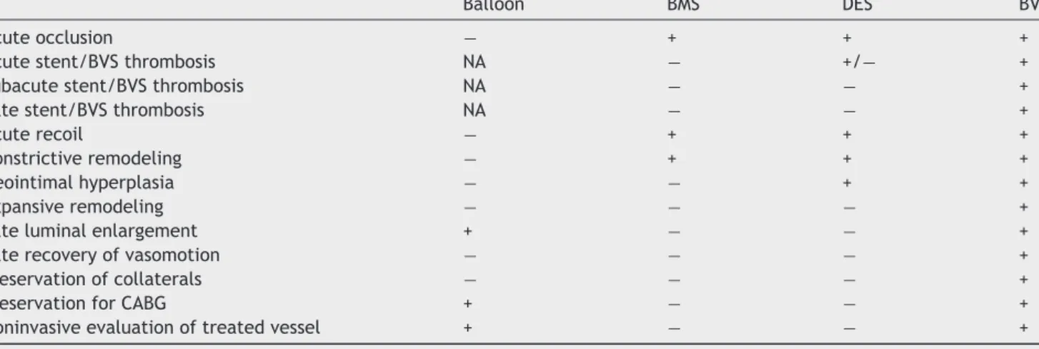
Table 1 Potential advantages of bioresorbable vascular scaffolds.
Balloon BMS DES BVS
Acute occlusion − + + +
Acute stent/BVS thrombosis NA − +/− +
Subacute stent/BVS thrombosis NA − − +
Late stent/BVS thrombosis NA − − +
Acute recoil − + + +
Constrictive remodeling − + + +
Neointimal hyperplasia − − + +
Expansive remodeling − − − +
Late luminal enlargement + − − +
Late recovery of vasomotion − − − +
Preservation of collaterals − − − +
Preservation for CABG + − − +
Noninvasive evaluation of treated vessel + − − +
Adapted from Onuma et al.9
BMS: bare-metal stent; CABG: coronary artery bypass grafting; DES: drug-eluting stent; NA: not applicable because of absence of stent; +: prevented or not restricted;−: not prevented or restricted.
function, imposing no constraints on future interventions
(
Table 1
).
Objective
BVS systems were recently approved for percutaneous
coronary intervention in Europe and possess highly
inno-vative properties. The aim of this position statement is
to review the information and studies on available BVS,
to stimulate discussion on their use and to propose
guide-lines for their application by interventional cardiologists in
Portugal.
Methods
A working group of experienced interventional cardiologists
was set up to evaluate current knowledge and to reach a
consensus based on available evidence, discussion of clinical
case models and individual experience.
Information and current evidence on bioresorbable
vascular scaffolds under development
BVS have been under development for more than a decade
(
Table 2
).
10The first BVS implanted in humans was the
Igaki-Tamai (Igaki Medical Planning Company, Kyoto, Japan)
in 2000, with poly-L-lactic acid (PLLA) scaffolds, using a
complex thermal delivery technique consisting of balloon
inflation with a heated dye at 80
◦C.
12The first metal BVS
used in humans, in 2007, was composed of 93% magnesium
(Biotronik, Berlin, Germany),
13while the first coated BVS
appeared in 2008 --- the everolimus-eluting ABSORB stent
(Abbott Vascular, Santa Clara, US) --- with a strut thickness
of 150 mm.
14Current clinical use of bioresorbable vascular
scaffolds
At present, the ABSORB everolimus-eluting system is the
only BVS available for clinical use.
In the ABSORB cohort A trial, with 30 patients, the major
event rate at four years was only 3.4%, demonstrating that
the concept is feasible and produces the expected results.
The data on durability and safety were extremely promising,
with no cardiac deaths, although the sample was small and
selected.
22---24The subsequent ABSORB cohort B trial used generation
1.1 devices incorporating changes in strut geometry that
provided more radial support than the initial version 1.0.
The study included 101 patients with stable or unstable
angina or silent ischemia, with de novo lesions in any native
artery with a maximum diameter of 3.0 mm, 50---99%
steno-sis, treatable with a 3.0 mm
× 18 mm BVS, and assessed by
different invasive imaging techniques. The study reported
a hierarchical major adverse cardiac event rate of 6.8%
and TLR of 5.9% at two years, and concluded that efficacy
and safety were satisfactory in arteries
≤2.5 mm in
diame-ter as well as in larger vessels, with no deaths or scaffold
thrombosis.
7,22,25---30The ABSORB cohort B trial included a predefined subgroup
of 56 patients (B2), who underwent intracoronary imaging
at 12 and 36 months. These patients showed encouraging
recovery of endothelium-dependent vasomotion, similar to
that observed in native coronary arteries. Endothelialization
parameters and negative remodeling, both constrictive and
elastic, were no worse than with metal stents, as reported
in various other studies. Strut absorption was confirmed,
although the devices could still be identified by
intravascu-lar ultrasound (IVUS) and by optical coherence tomography
(OCT).
25,31,32Significant expansive remodeling was observed
at 24 months, artery area increasing from 14.8 mm
2to 17.5
mm
2in large vessels, and from 12.7 mm
2to 13.1 mm
2in
small vessels, counteracting the negative effect of
neointi-mal hyperplasia (0.25 mm
2in all coronary arteries).
15,26,30Table 2 Bioresorbable vascular scaffolds.
Company Device Description/Study Drug Status
Abbott ABSORB PLLA, totally absorbed in 2 years 5.9% TLR at 2 years (ABSORB cohort B, 2009---2013)22---24
Everolimus EU approved
ART ART
Bioresorbable stent
PLA (2009)16 No FIM in progress
Biotronik DREAMS 93% magnesium alloy.
9.1% TLR at 6 months (BIOSOLVE-I, 2007---2013)13,17
Paclitaxel FIM complete
Elixir DESolve PLLA
BDES program (2009, unpublished)18
Novolimus FIM complete
Huaan Xinsorb PLLA (2012)19 Sirolimus FIM complete
Kyoto Medical Igaki-Tamai PLLA, absorbed in 2 years (2000)12 Yes FIM complete
REVA Medical REVA-ReZolve Tyrosine polycarbonate, absorbed in 18 months, ‘slide and lock’ design 66.7% TLR at 1 year (RESORB, 2007, unpublished)20
No FIM complete
Bioabsorbable Therapeutics Ideal BioStent Polysalicylate, absorbed in 12 months WHISPER (2009, unpublished)21
Sirolimus FIM complete
FIM: first-in-man; PLA: polylactic acid; PLLA: poly-L-lactic acid; TLR: target lesion revascularization.
We did not consider it relevant that in the ABSORB
trials, the devices were implanted in vessels visually
esti-mated to be 3.0 mm in diameter with lesions measuring
less than 14 mm, since only one platform was available at
the time (3.0 mm
× 18 mm) and there was a need to ensure
the safety of assessment by multiple intracoronary imaging
methods, including IVUS, OCT and palpography. A phase III
trial (ABSORB II) is currently underway, in which the
angi-ographic criteria are far more comprehensive (a maximal
luminal diameter between 2.25 mm and 3.8 mm as
esti-mated by online quantitative coronary angiography and a
lesion length of
≤48 mm).
38BVS implantation presents certain technical challenges,
particularly the importance of extremely accurate
measure-ment of minimum proximal and distal lumen diameters,
essential for effective anchoring of the device, the risk
of strut fracture resulting from balloon overdilation, and
the sometimes conflicting data from multiple intracoronary
imaging methods.
33---37Discussion of clinical case models and individual
assessment
The working group discussed clinical case models, leading
to individual reflections on the expectations and possible
limitations of BVS systems, based on the assumption that
the cost of the device would not be a deciding factor.
Results
Proposed indications and future review
The opinion of the working group is that BVS should
pref-erentially be introduced for recommended indications and
should be monitored.
This position statement reflects the analysis undertaken,
leading to a series of possible guidelines for the use of BVS
(
Table 3
).
These guidelines are necessarily limited by constant
developments in the state of the art and should be the
sub-ject of early review as technological advances and further
evidence become available, preferably by a medical society
specializing in the area.
Table 3 Indications for bioresorbable vascular scaffolds. Consensual and appropriate indications
1. Young patients (aged <50 years) 2. Diabetic patients
3. Lesions in segments that may undergo CABG, particularly the left anterior descending artery 4. Vessels with long lesions (>30 mm) and/or diffuse disease with a high probability of requiring secondary revascularization
Less consensual but possible indications 1. Small collateral branches (<1.5 mm)
2. Non-ST elevation acute coronary syndrome, stabilized, with intermediate and/or unstable plaques
3. ST-elevation acute coronary syndrome, stabilized, with intermediate and/or unstable plaques
Inappropriate indications 1. Left main disease
2. Moderate or severe tortuosity 3. Severe calcification
Monitoring, research and costs in Portugal
Medical societies in Portugal should organize and/or support
research that includes multicenter registries and
well-designed clinical trials, based on appropriate imaging and/or
functional studies. There is no published information on
the treatment’s economic aspects, so assessment of the
technological implications and incremental costs will be
par-ticularly relevant.
Conclusion
There is encouraging evidence that BVS are a viable
treat-ment option. They should be used in more consensual
indications and will require technical adaptations. Outcome
monitoring and evaluation is essential to avoid
inappropri-ate use. It is recommended that medical societies produce
clinical guidelines as soon as possible.
References
1. Gruntzig A, Schneider HJ. The percutaneous dilatation of chronic coronary stenoses --- experiments and morphology. Schweiz Med Wochenschr. 1977;107:1588.
2. Serruys PW, de Jaegere P, Kiemeneij F, et al., Benestent Study Group. A comparison of balloon-expandable-stent implantation with balloon angioplasty in patients with coronary artery dis-ease. N Engl J Med. 1994;331:489---95.
3. Sousa JE, Costa MA, Abizaid AC, et al. Sustained suppression of neointimal proliferation by sirolimus-eluting stents: one-year angiographic and intravascular ultrasound follow-up. Circula-tion. 2001;104:2007---11.
4. Mattos LA, Grines CL, Sousa JE, et al. One-year follow-up after primary coronary intervention for acute myocardial infarction in diabetic patients. A substudy of the STENT PAMI trial. Arq Bras Cardiol. 2001;77:549---61.
5. Bangalore S, Kumar S, Fusaro M, et al. Outcomes with var-ious drug eluting or bare metal stents in patients with diabetes mellitus: mixed treatment comparison analysis of 22 844 patient years of follow-up from randomised trials. BMJ. 2012;345:e5170.
6. Palmerini T, Biondi-Zoccai G, Della Riva D, et al. Stent thrombosis with drug-eluting and bare-metal stents: evi-dence from a comprehensive network meta-analysis. Lancet. 2012;379:1393---402.
7. Serruys PW, Garcia-Garcia HM, Onuma Y. From metallic cages to transient bioresorbable scaffolds: change in paradigm of coro-nary revascularization in the upcoming decade? Eur Heart J. 2012;33:16---25.
8. Brugaletta S, Garcia-Garcia HM, Garg S, et al. Temporal changes of coronary artery plaque located behind the struts of the everolimus eluting bioresorbable vascular scaffold. Int J Car-diovasc Imaging. 2011;27:859---66.
9. Onuma Y, Serruys PW. Bioresorbable scaffold: the advent of a new era in percutaneous coronary and peripheral revasculariza-tion? Circulation. 2011;123:779---97.
10. Ormiston JA, Serruys PW. Bioabsorbable coronary stents. Circ Cardiovasc Interv. 2009;2:255---60.
11. Okamura T, Serruys PW, Regar E. Cardiovascular flashlight. The fate of bioresorbable struts located at a side branch ostium: serial three-dimensional optical coherence tomography assess-ment. Eur Heart J. 2010;31:2179.
12. Tamai H, Igaki K, Kyo E, et al. Initial and 6-month results of biodegradable poly-L-lactic acid coronary stents in humans. Cir-culation. 2000;102:399---404.
13. Erbel R, di Mario C, Bartunek J, et al. Temporary scaffold-ing of coronary arteries with bioabsorbable magnesium stents: a prospective, non-randomised multicentre trial. Lancet. 2007;369:1869---75.
14. Ormiston JA, Serruys PW, Regar E, et al. A bioabsorbable everolimus-eluting coronary stent system for patients with sin-gle de-novo coronary artery lesions (ABSORB): a prospective open-label trial. Lancet. 2008;371:899---907.
15. Diletti R, Farooq V, Girasis C, et al. Clinical and intravascular imaging outcomes at 1 and 2 years after implantation of absorb everolimus eluting bioresorbable vascular scaffolds in small ves-sels. Late lumen enlargement: does bioresorption matter with small vessel size? Insight from the ABSORB cohort B trial. Heart. 2013;99:98---105.
16. Lafont A, Durand E. A.R.T.: concept of a bioresorbable stent without drug elution. EuroIntervention. 2009;5 Suppl. F:F83---7. 17. Haude M, Erbel R, Erne P, et al. Safety and performance of the drug-eluting absorbable metal scaffold (DREAMS) in patients with de-novo coronary lesions: 12 month results of the prospec-tive, multicentre, first-in-man BIOSOLVE-I trial. Lancet. 2013. 18. Yan J, Bhat VD. Elixir Medical’s bioresorbable drug eluting stent
(BDES) programme: an overview. EuroIntervention. 2009; Suppl. F:F80---2.
19. Wu Y, Shen L, Wang Q, et al. Comparison of acute recoil between bioabsorbable poly-L-lactic acid XINSORB stent and metallic stent in porcine model. J Biomed Biotechnol. 2012;2012:413956.
20. Pollman MJ. Engineering a bioresorbable stent: REVA pro-gramme update. EuroIntervention. 2009; Suppl. F:F54---7. 21. Jabara R, Pendyala L, Geva S, et al. Novel fully
bioab-sorbable salicylate-based sirolimus-eluting stent. EuroInterven-tion. 2009; Suppl. F:F58---64.
22. Serruys PW, Ormiston JA, Onuma Y, et al. A bioabsorbable everolimus-eluting coronary stent system (ABSORB): 2-year out-comes and results from multiple imaging methods. Lancet. 2009;373:897---910.
23. Onuma Y, Serruys PW, Ormiston JA, et al. Three-year results of clinical follow-up after a bioresorbable everolimus-eluting scaffold in patients with de novo coronary artery disease: the ABSORB trial. EuroIntervention. 2010;6:447---53.
24. Dudek D, Onuma Y, Ormiston JA, et al. Four-year clinical follow-up of the ABSORB everolimus-eluting bioresorbable vascular scaffold in patients with de novo coronary artery disease: the ABSORB trial. EuroIntervention. 2012;7:1060---1.
25. Gogas BD, Serruys PW, Diletti R, et al. Vascular response of the segments adjacent to the proximal and distal edges of the ABSORB everolimus-eluting bioresorbable vascular scaffold: 6-month and 1-year follow-up assessment: a virtual histology intravascular ultrasound study from the first-in-man ABSORB cohort B trial. JACC Cardiovasc Interv. 2012;5:656---65. 26. Brugaletta S, Heo JH, Garcia-Garcia HM, et al.
Endothelial-dependent vasomotion in a coronary segment treated by ABSORB everolimus-eluting bioresorbable vascular scaffold system is related to plaque composition at the time of biore-sorption of the polymer: indirect finding of vascular reparative therapy? Eur Heart J. 2012;33:1325---33.
27. Diletti R, Onuma Y, Farooq V, et al. 6-month clinical outcomes following implantation of the bioresorbable everolimus-eluting vascular scaffold in vessels smaller or larger than 2.5 mm. J Am Coll Cardiol. 2011;58:258---64.
28. Garcia-Garcia HM, Gonzalo N, Pawar R, et al. Assessment of the absorption process following bioabsorbable everolimus-eluting stent implantation: temporal changes in strain values and tis-sue composition using intravascular ultrasound radiofrequency data analysis. A substudy of the ABSORB clinical trial. EuroInt-ervention. 2009;4:443---8.
29. Gomez-Lara J, Brugaletta S, Farooq V, et al. Angiographic geo-metric changes of the lumen arterial wall after bioresorbable
vascular scaffolds and metallic platform stents at 1-year follow-up. JACC Cardiovasc Interv. 2011;4:789---99.
30. Ormiston JA, Serruys PW, Onuma Y, et al. First serial assess-ment at 6 months and 2 years of the second generation of absorb everolimus-eluting bioresorbable vascular scaffold: a multi-imaging modality study. Circ Cardiovasc Interv. 2012;5: 620---32.
31. Serruys PW, Onuma Y, Dudek D, et al. Evaluation of the sec-ond generation of a bioresorbable everolimus-eluting vascular scaffold for the treatment of de novo coronary artery steno-sis: 12-month clinical and imaging outcomes. J Am Coll Cardiol. 2011;58:1578---88.
32. Brugaletta S, Gogas BD, Garcia-Garcia HM, et al. Vascular compliance changes of the coronary vessel wall after biore-sorbable vascular scaffold implantation in the treated and adjacent segments. Circ J. 2012;76:1616---23.
33. Gomez-Lara J, Brugaletta S, Farooq V, et al. Head-to-head comparison of the neointimal response between metallic and bioresorbable everolimus-eluting scaffolds using opti-cal coherence tomography. JACC Cardiovasc Interv. 2011;4: 1271---80.
34. Okamura T, Garg S, Gutierrez-Chico JL, et al. In vivo evalua-tion of stent strut distribuevalua-tion patterns in the bioabsorbable
everolimus-eluting device: an OCT ad hoc analysis of the revi-sion 1.0 and revirevi-sion 1.1 stent design in the ABSORB clinical trial. EuroIntervention. 2010;5:932---8.
35. Bruining N, de Winter S, Roelandt JR, et al. Monitoring in vivo absorption of a drug-eluting bioabsorbable stent with intravas-cular ultrasound-derived parameters. A feasibility study. JACC Cardiovasc Interv. 2010;3:449---56.
36. Gomez-Lara J, Brugaletta S, Diletti R, et al. Agreement and reproducibility of gray-scale intravascular ultrasound and opti-cal coherence tomography for the analysis of the bioresorbable vascular scaffold. Catheter Cardiovasc Interv. 2012;79:890---902. 37. Gutierrez-Chico JL, Serruys PW, Girasis C, et al. Quantitative multi-modality imaging analysis of a fully bioresorbable stent: a head-to-head comparison between QCA IVUS and OCT. Int J Cardiovasc Imaging. 2012;28:467---78.
38. Diletti R, Serruys PW, Farooq V, et al. ABSORB II randomized controlled trial: a clinical evaluation to compare the safety, efficacy, and performance of the Absorb everolimus-eluting bioresorbable vascular scaffold system against the XIENCE everolimus-eluting coronary stent system in the treatment of subjects with ischemic heart disease caused by de novo native coronary artery lesions: rationale and study design. Am Heart J. 2012;164:654---63.