Clínica Universitária de Cardiologia
Iron Deficiency in patients with Heart
Failure with Mid-Range and Preserved
Ejection Fraction
Diogo Jácome Morgado
07’2019
Clínica Universitária de Cardiologia
Iron Deficiency in patients with Heart
Failure with Mid-Range and Preserved
Ejection Fraction
Diogo Jácome Morgado
Orientado por:
Dr. Pedro Moraes Sarmento
07’2019
2
Abstract
Introduction: Iron is essential in bioenergy production, immune system efficacity
and central nervous system development. In Chronic Heart Failure (CHF) patients, ID impairs functional capacity, worsens quality of life and increases mortality. There is no data on identification and correction of ID with intravenous ferric carboxymaltose (ivFCM) in CHF patients with midrange and preserved ejection fraction (HFmr/pEF).
Methods: Between 2015 and 2016 we identified and characterized symptomatic HFmr/pEF patients submitted to ivFCM treatment for ID correction with or without anemia and, compared them to the CHF patients with reduced ejection fraction (HFrEF) treated on the same period. Then, between 2015 and 2018, we investigated the evolution of HFmr/pEF patients’ NYHA class, NTproBNP and kidney function, at three and six months after treatment. Results: In the comparison study, 52 CHF patients with ID were evaluated: mean age 86 years, 69% were men, 34.6% had HFrEF and 65.4% HFmr/pEF. 90,4% had anemia. 13% and 23% of HFrEF and HFmr/pEF had respectively functional ID. HFmr/pEF patients had less ischemic heart disease (44% and 78%) and less diabetes mellitus (26% and 44%). No difference was seen in other comorbidities. In the evaluation study of patients submitted to ivFCM, 56 patients were included. 83% had absolute ID, 75% patients were anemic. 50% in NYHA class II and 46% in class III. NTproBNP was 6492pg/mL and eGFREPI was 47,8mL/min/m^2. At three and six months, 59% and 61% were in NYHA class II and 39% and 37% in class III. NTproBNP was 5331pg/mL and 4000pg/mL, the eGFREPI was 45,8mL/min/m^2 and 45,8mL/min/m^2. Conclusion: ID is per se poorly evaluated in routine practice. At three and six months after treatment with ivFCM, although no significant changes were seen in eGFREPI, a functional improvement, as assessed by the NYHA class, and a reduction of NTproBNP levels were observed.
3
Resumo
Introdução: O ferro é essencial para a produção de bioenergia, eficácia do sistema
imu-nitário e desenvolvimento do sistema nervoso central. Em pacientes com Insuficiência Cardíaca Crónica (ICC), o défice de ferro (DF) compromete a capacidade funcional, piora a qualidade de vida e aumenta a mortalidade. Não há dados sobre a identificação do DF e correção com carboximaltose férrica intravenosa (CMFiv) em pacientes com fração de ejeção intermédia e preservada (ICFEi/p). Métodos: Entre 2015 e 2016, identificámos e caracterizámos os pacientes sintomáticos com ICFEi/p que realizaram CMFiv para cor-reção do DF, com ou sem anemia, e comparámo-los com os pacientes com ICFEr tratados no mesmo período. Depois, entre 2015 e 2018, examinámos a evolução dos pacientes com ICFEi/p face à sua classe NYHA, porção N-terminal do péptido natriurético tipo B (NTproBNP), e função renal aos três e seis meses após o tratamento. Resultados: No estudo de comparação, 52 pacientes com ICC e DF foram avaliados: idade média 86 anos, 69% eram homens. 34,6% apresentavam ICFEr e 65,4% ICFEi/p. 90,4% tinha anemia. DF funcional estava presente em 13% e 23% de ICFEr e ICFEi/p, respetivamente. Os pacientes com ICFEi/p tinham menos doença arterial coronária (44% e 78%) e menos diabetes mellitus (26% e 44%). Não se observaram diferenças nas outras comorbidades. No estudo de seguimento dos pacientes submetidos a CMFiv, identificámos 56 pacientes. 83% tinha DF absoluto e 75% anemia. 50% na classe II da NYHA e 46% na classe III. NTproBNP foi 6492pg/mL e a eGFREPI foi de 47,8mL/min/m^2. Aos três e seis meses, 59% e 61% estavam na classe II da NYHA e 39% e 37% na classe III. O NTproBNP foi 5331pg/mL e 4000pg/mL, e a eGFREPI foi 45,8mL/min/m^2 e 45,8mL/min/m^2.
Con-clusão: O DF é per si subavaliado na prática clínica. Aos três e seis meses após o
trata-mento com CMFiv, não observámos alterações significativas na eGFREPI, e constatámos uma melhoria funcional, conforme avaliada pela classe NYHA, bem como uma redução dos níveis de NTproBNP.
Palavras-chave: insuficiência cardíaca crónica deficiência ferro
O Trabalho Final exprime a opinião do autor e não da FML
.4
Table of Contents
ABSTRACT ... 2
RESUMO ... 3
BACKGROUND ... 5
IRON: IN THE CENTER OF BIOENERGY PRODUCTION, CENTRAL NERVOUS SYSTEM PERFORMANCE AND IMMUNOLOGICAL PROCESSES ... 5
IRON METABOLISM ... 7
EVALUATION OF IRON DEFICIENCY ... 10
IRON DEFICIENCY,ANEMIA, AND CHRONIC HEART FAILURE ... 12
CORRECTION OF IRON DEFICIENCY ... 14
AIM ... 16
MATERIALS AND METHODS ... 17
STUDY A:A POPULATION CHARACTERIZATION ... 17
STUDY B:ID CORRECTION IN HFMREF&HFPEF, A FOLLOW UP AT 3 AND 6 MONTHS ... 17
RESULTS ... 18
STUDY A:A POPULATION CHARACTERIZATION ... 18
STUDY B:ID CORRECTION IN HFMREF&HFPEF, A FOLLOW UP AT 3 AND 6 MONTHS ... 19
DISCUSSION: ... 23
STUDY A:A POPULATION CHARACTERIZATION ... 23
STUDY B:ID CORRECTION IN HFMREF&HFPEF, A FOLLOW UP AT 3 AND 6 MONTHS ... 23
CONCLUSION ... 25
5
Background
Iron: in the center of bioenergy production, central nervous system performance and immunological processes
Iron ability to interconvert between ferric (Fe3+) and ferrous (Fe2+) forms makes it an essential cofactor for a wide variety of important cellular processes, such as oxygen transport, respiration, the tricarboxylic acid cycle, lipid metabolism, gene regulation and DNA synthesis.1,2 In order to execute such multiple tasks, iron is incorporated in the heme group of hemoglobin, myoglobin and cytochromes, or is associated to nonheme moieties or iron-sulfur (Fe-S) motifs. Not surprisingly, iron is considered an essential nutrient for virtually all organisms.4 The maintenance of normal iron metabolism is particularly important for cells that are characterized by high mitogenic potential (neoplastic cells, hematopoietic cells, including immune competent cells) and high energy demand (hepatocytes, adipocytes, renal cells, immune cells, skeletal myocytes, and cardiomyocytes).2–4
The elementary iron was firstly recognized as a critical component in systemic oxygen delivery and utilization. In fact, iron contributes to erythropoiesis and iron deficiency (ID) decreases oxygen-carrying capacity of the blood through reduced hemoglobin levels, the classic sign of ID.1,5 Furthermore, severe dietary ID, in addition to causing anemia, decreases the capacity of skeletal muscle to consume oxygen and produce ATP, i.e. tissue oxidative capacity.6 The tissue oxidative capacity, which determines endurance, energy efficiency, and submaximal exercise effort, is impaired proportionally across the whole spectrum of ID even when hemoglobin is normal.5 Likewise, it has been demonstrated that cellular ID results in a reduced activity of Fe-S cluster-based complexes in the mitochondria of human cardiomyocytes and is associated with impaired mitochondrial respiration and morphology, ATP production and, cardiomyocyte contractility and relaxation. Iron supplementation can revert these effects.7 In addition to reduced physical performance, fatigue is also closely associated with anemia and ID. Its correction improves mental quality of life, cognitive functions, self-rated alertness, contentment and calmness.8
6 Biologically, iron is required for proper myelination of the spinal cord and white matter of cerebellar folds and, it is a cofactor for several enzymes involved in neurotransmitter and DNA synthesis. As whole-brain iron content drops 15% below normal, biological and behavioral alterations occur due to changes in the dopaminergic system.2,9,10 There is compelling evidence that within critical periods, i.e. 6- to 24-month-old infants, ID have long-term effects on cognitive, motor, socioemotional and neurophysiologic development.11 Moreover, in women aged 18 to 35 years the severity of ID was linked with a reversible decreased cognitive performance and increased time to complete learning, memory, and attention tasks.12
Finally, being iron a critical component of peroxide-generating enzymes and nitrous oxide–generating enzymes that are critical for the proper enzymatic functioning of immune cells, it is likely involved in the regulation of cytokine production and second-messenger systems.2 Also, ID results in decreased T-lymphocyte numbers and in decreased T-T-lymphocyte blastogenesis and mitogenesis in response to mitogens.9 However, the role of iron in the immune system is ambiguous as the host must effectively withhold iron from pathogens and simultaneously provide a supply of iron that is not limiting to its immune system.2,13 An example of how iron overload might be disadvantageous was demonstrated by parenteral iron treatment association with life-threatening sepsis when given in the early neonatal period.14 Despite proven reversible functional immunological defects, a clinically important relationship between states of ID and susceptibility to infections remains controversial.14 Studies have shown us that the risk, if any, of intravenous iron causing infection and related morbidity and mortality is probably very small.15
7 Figure 1. ID impact in bioenergy production, erythropoiesis, immune system and central nervous system
Iron metabolism
From a nutritional viewpoint, both iron depletion and excess have dramatic consequences and great epidemiological significance. Indeed, in conformity with northern Europe, absolute ID is present in as much as 32% of the Portuguese population.16 There are no current global figures for ID, but using anemia as an indirect indicator it can be estimated that most preschool children and pregnant women in non-industrialized countries, and at least 30-40% in industrialized countries, are iron deficient. The estimated prevalence of ID worldwide is twice as high as that of iron-deficiency anemia.17 Not only it is the primary nutritional disorder in the world, but also hereditary hemochromatosis, which causes excess iron accumulation, is one of the most common genetic disorders - nearly 10% of the white population carries the most prevalent C282Y HFE mutation.3
↑ Erythropoiesis
↑ Tissue Oxidative Capacity ↑ Activity of Fe-S cluster-based Complexes
↑ Cognitive, Motor, Socioemotional and Neurophysiological Development
↑ T-lymphocyte blastogenesis and mitogenesis
↑ Contractility ↑ Relaxation
8 In order to maintain its homoeostasis, there are two major pathways which must be elucidated, the first being the mechanism of iron absorption, more specifically in the duodenum and proximal jejunum and, the second the regulation of iron bioavailability, which is mainly adjusted by the reticuloendothelial system.
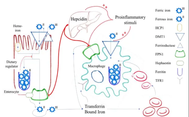
There are two different iron absorption pathways: one for heme-bound iron, mostly bound to porphyrins in meat-based foods, and another for non-heme iron, mostly found in vegetables. Heme-iron constitutes only 10% of dietary iron, but because of greater bioavailability it represents 30% of the total absorbed iron, being absorbed via a specific membrane transporter, the heme carrier protein-1.18,19 Non-heme iron is mostly found in the ferric form and is reduced to the ferrous form by a luminal ferrireductase, being thereafter transported into the enterocyte via a divalent metal ion transporter DMT1.3 Afterwards, ferrous iron may be either exported through the basolateral membrane of the enterocyte by ferroportin-1 (FPN1) or it is stored in the form of ferritin inside the enterocyte being lost once the cell is sloghed.1 When it follows the FPN1 way, an accompanying membrane hephaestin oxidizes ferrous into ferric iron, which is then released into the circulation and bound to transferrin. It may then be incorporated into cells by coupling with Transferrin Receptor 1 (TFR1), forming a complex that follows endocytosis.18 Alternatively, ferrous iron may be directly uptaken by DMT1, which is expressed in most tissues.20
The absorption of intestinal iron is regulated in several ways. Firstly, after a dietary iron bolus there is a direct resistance of absorptive enterocytes to iron, a mechanism referred as the dietary regulator.1 Secondly, hepcidin, an early inflammatory marker expressed predominantly in the liver, inactivates FPN1 promoting the sequestration of iron in the enterocyte and, subsequent lost.20
Although the major entry site for iron into the body is via dietary absorption of heme and non-heme bound iron in the duodenum, the major cellular system responsible for a sufficient supply of iron for erythropoiesis is the reticuloendothelial system.20 Approximately 95% of the daily needs of iron for erythropoiesis originates from this system, where activated macrophages phagocytose senescent erythrocytes for the recycling of iron. Once iron enters the macrophage cytoplasm it is either stored within ferritin, utilized upon incorporation into iron-containing enzymes or exported
9 and transferred to the circulation by FPN-1. Importantly, FPN-1, highly expressed in enterocytes, Kupffer cells and spleen macrophages., is so far the only known iron exporter, therefore the efficacy of iron recycling from erythrophagocytosis is linked to the expression of this protein. The earlier mentioned hepcidin plays a pivotal role by binding to this unique iron exporter leading to its internalization and degradation. Of note, the hepatic expression of hepcidin is stimulated by interleukin-6 and lipopolysaccharide. This results in a blockage of iron export and subsequent monocyte/ macrophage iron retention.20,21
Not only hepcidin but also proinflammatory stimuli, such as Interferon-γ, lipopolysaccharide and TNF-α, also induce the retention of iron in macrophages by up-regulating the expression of DMT1 expression and down-regulating the expression of FPN1, promoting simultaneously the uptake of ferrous iron while inhibiting its exportation.20,21
In summary, cytokines and hepcidin inhibit iron export from macrophages via downregulation of FPN1 expression, resulting in iron retention within cells of the reticuloendothelial system and an iron restricted erythropoiesis.2,18,22
Figure 2. Iron metabolic pathway and hepcidin regulation; FPN1, ferroportin-1; HCP1, heme carrier protein-1; TFR1 Transferrin Receptor 1. Adapted from Weiss G. Anemia of Chronic Disease, The New England Journal of Medicine, 2005:13.
10
Evaluation of Iron Deficiency
The most widely used screening measurement is Serum Ferritin (SF) which in the absence of inflammation, is the best noninvasive estimate of body iron stores. A ferritin level of less than 45 mg/L is 85% sensitive and 92% specific for ID.23 However, because it is an acute phase protein, SF is increased independent of iron status by acute or chronic inflammation. It is also unreliable in the setting of malignancy, hyperthyroidism, liver disease, and heavy alcohol intake.24 Another widely used screening tool is the degree of iron saturation of plasma transferrin (TSAT) calculated as the ratio of the plasma iron to total iron-binding capacity.25 Marked diurnal variation in plasma iron values and the numerous clinical disorders that affect the transferrin saturation limit its reliability.26 Other interesting markers are the serum transferrin receptor (sTfR), the sTfR/log ferritin ratio, the reticulocyte hemoglobin content (CHr), reticulocyte hemoglobin content equivalent (Ret-He), and the erythrocyte zinc protoporphyrin (ZPP).
The sTfR is a transmembrane glycoprotein that transfers circulating iron into developing red cells. Under iron-deficient conditions there is an increased concentration of surface sTfR. sTfR concentration appears to be a specific indicator of IDE that is not confounded by inflammation24. However, this assay was found to have a specificity of 84% and a positive predictive value of only 58% in a population of patients likely to be typical of the most difficult diagnostic environments for assessing iron status.27 When hemolysis or megaloblastosis can be excluded the sTfR measurement should be an attractive alternative to more conventional tests of iron status.28 Attempts to combine ferritin and sTfR results have led to the sTfR/log ferritin ratio, which was found to be highly sensitive and 93% specific for the diagnosis of ID.23 On the other hand, there was a significant number of patients in a “diagnostic grey zone” who could not be correctly classified.29
Flow cytometric analysis of reticulocytes allows determination of the mean cellular hemoglobin content of reticulocytes (CHr) or percentage of hypochromic reticulocytes (%HRC). The former is a premature phase marker (48 hours) while the latter reflects a longer period (20-120 days) of iron restricted erythropoiesis.28 The %HRC is the best-established variable for the identification of functional ID and thus
11 has the greatest level of evidence, while CHr is the next most established option. Falsely normal values of the CHr can be observed with an elevated MCV or thalassemia, but the main limitation to its wider use is that it can be measured with only one model of analyzer (Bayer ADVIA®).30 Among patients undergoing bone marrow examination for diagnostic purposes CHr had a better predictive value for iron depletion than MCV, serum ferritin or transferrin saturation values.30
An alternative measure of reticulocyte hemoglobin content, Ret-He (reticulocyte hemoglobin equivalent), is available on some analyzers manufactured by the Sysmex Corporation®. Ret-He alone was able to distinguish iron-deficient and iron-sufficient subjects using a cut-off of 25pg with reasonable sensitivity. Although less sensitive (80% agreement with sTfR and sTfR/log ferritin values), a Ret-He cut-off of 25 pg may help to distinguish absolute ID (values <25 pg) from functional ID (values >25 pg). With a Ret He cutoff level of 27.2 pg, ID could be diagnosed with a sensitivity of 93.3%, and a specificity of 83.2%. Ret-He is a reliable marker of cellular hemoglobin content and can be used to identify the presence of ID states.31
A simple and reliable measurement of IDE is the ZPP - the incorporation of iron into protoporphyrin IX is the final stage of heme synthesis and, an increase in ZPP is an indicator of defects at any point along this pathway. A major advantage of this well stablished assay is the ability to measure the ratio ZPP/heme directly on a drop of blood using a dedicated portable instrument called a hematofluorimeter. The ZPP is not widely used in large clinical laboratories, partly because of the difficulty in automating the assay. Due to lack of specificity, ZPP should not be used in isolation as a diagnostic test, but once a diagnosis is made it may be used to monitor response to therapy. The ZPP concentration reflects the entire circulating red cell population and is thus less sensitive than %HRC or CHr to acute changes in iron availability.30
In the setting of iron restricted erythropoiesis there are two different conditions to be distinguished: absolute ID and functional ID.28 Absolute ID reflects depleted iron stores, often with intact iron homoeostasis mechanisms and erythropoiesis. The commonest causes are: low-dietary iron, impaired gastrointestinal absorption, and blood loss.25 Currently, although the generally accepted SF cut-off level to diagnose
12 absolute ID is 30 mg/L, a higher value, 100 mg/L, may provide optimal sensitivity and specificity for the detection of depleted iron stores in inflammatory cohorts.26,28,32
A distinct situation is functional ID, which develops during increased erythropoiesis mediated either by endogenous erythropoietin responses to anemia, or by therapy with erythropoiesis-stimulating agents (ESAs): the iron supply, though adequate for baseline erythropoiesis, cannot meet the erythron requirements of supraphysiologic erythropoiesis. In these patients, the TSAT may be 20% as the hungry bone marrow strips iron off the circulating transferrin faster than the transferrin can replenish it with iron released from stores. The serum ferritin, which reflects iron stores, may be normal or elevated. This is a problem of supply and demand, not total body ID.25,26,28
An extreme case of functional ID is known as reticuloendothelial blockade and usually occurs in the setting of acute or chronic inflammation/infection, thus the designation anemia of chronic disease. This often correlates with a high C-reactive protein level and/or a high erythrocyte sedimentation rate. As explained above, the molecular mechanisms that underlie the redistribution of iron during inflammation center on the cytokine-stimulated overproduction of hepcidin, which “locks up” iron in the reticuloendothelial storage. As a result, transferrin-bound iron, which is reflected by the TSAT, is low despite a normal or elevated ferritin. When it is feasible to measure several indices, the initial laboratory evaluation, in addition to the SF and hemoglobin, should include a C-reactive protein, sTfR and/or ZPP. Due to the aforementioned issues it is widely accepted to define ACD as a SF≥100 mg/L and ≤300 mg/L when TSAT ≤20%.4,25
Iron Deficiency, Anemia, and Chronic Heart Failure
The complex pathophysiology of Chronic Heart Failure (CHF) involves dysfunc-tion of most body organs, including the cardiovascular, musculoskeletal, renal, neu-roendocrine, hemostatic, immune, and inflammatory systems.33 Despite optimal con-ventional therapy, many patients with CHF remain symptom limited, exercise
intol-13 erant, and subject to high rates of mortality.34 Clinical symptomatology of CHF re-sembles that of ID, therefore one can simply imply impaired iron homeostasis as one of the mechanisms underlying exercise intolerance in CHF. Interestingly, ID as an issue in the CHF syndrome only recently gained clinical interest, coinciding with the recognition of anemia as a frequent co-morbidity in CHF.35
At the beginning of this century, the first report of a decrease in blood hemoglobin content with increasing severity of CHF was published.36 Since then, several studies have documented how prevalent reduced hemoglobin is, being present in one-third of the CHF population, and how it is independently associated with increased risk of hospitalization and all-cause mortality, regardless of the ejection fraction.35,37,38 The etiology of anemia in CHF is not yet clearly understood due to the interaction of sev-eral elements such as: renal dysfunction, hemodilution, the angiotensconverting-enzyme inhibitors (ACEi) and angiotensreceptor blockers (ARAII), chronic in-flammation, bone marrow dysfunction, abnormal steroid metabolism, resistance to erythropoietin, and hematinic deficiencies, such as vitamin B12, folate and, in partic-ular, ID.22,37
The first efforts to address the correction of anemia in CHF fell short of the ex-pectations. Despite increasing the hemoglobin level, the ESA – darbepoetin alfa – was shown to have no positive impact on the primary outcome of death or hospitalization for worsening heart failure. Furthermore, there was an increase in fatal or nonfatal strokes, as well as all-cause mortality.36
Yet, when one door closes another one opens. Within the deranged milieu of CHF, iron is abnormally handled, being preferentially trafficked into storage sites and with-drawn from the circulation and erythroid marrow, triggering functional ID, with char-acteristic normal ferritin concentrations.4,34 In patients with CHF with reduced ejec-tion fracejec-tion (HFrEF), ID is common, ranging between 37% and 55%,4,37,39 being even as high as 73% when evaluated by bone marrow aspiration in those with ad-vanced CHF and anemia.40 Moreover, it constitutes a strong independent predictor of unfavorable outcome, irrespective of the presence of anemia and the severity of heart disease.4,22,34,40,41 Patients with ID are more frequently females, at higher New York
14 Heart Association (NYHA) class, with ischemic cardiomyopathy, a lower hemoglo-bin and a higher NT-pro-brain natriuretic peptide (NTproBNP).4 A decreased iron status is associated with exercise intolerance and breathlessness on exertion, as well as increased disease severity as assessed by NYHA functional class and NTproBNP level.39 Indeed, ID is per se prognostically more ominous than anemia in CHF.4,34
Although a greater number of studies have focused on HFrEF rather than CHF with midrange and preserved ejection fraction (HFmrEF & HFpEF), recent evidence has broadened the spectrum. Not only a high prevalence of ID has been confirmed in patients with HFmrEF (61%) and HFpEF (58.4-64%), but it has also been related to a reduced exercise capacity, VO2max, quality of life, worse clinical outcome, and all-cause mortality.42,43 Accordingly, ID prevalence increases with increased severity of diastolic dysfunction. Interestingly, in the HFpEF group 41.1% presented with iso-lated ID, i.e., without anemia, which reflects how it can be an unexpressed condition. ID is a frequent co-morbidity in patients with HFmrEF & HFpEF being associated with a more severe heart failure status.42,43
Correction of Iron Deficiency
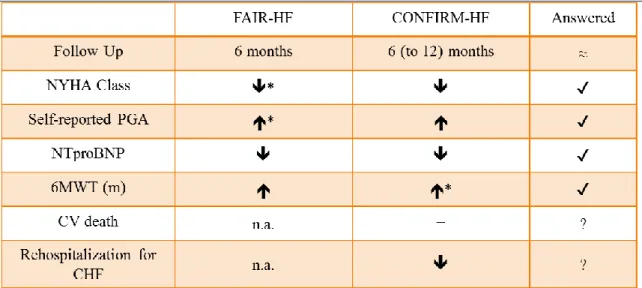
Facing all the evidence suggesting such an impact of ID in CHF corroborated by the first small trials,44 a pioneer trial was brought up. FAIR-HF demonstrates how treating ID with intravenous ferric carboxymaltose (IV FCM) for 24 weeks in patients with HFrEF and ID, with or without anemia, improves symptoms, functional capacity, and the quality of life, as assessed by the self-reported Patient Global Assessment and the NYHA class.45 It also suggests an improvement in distance on the 6-minute walking test distance, which was later stated by the CONFIRM trial. Besides proving that the treatment of ID, regardless the presence of anemia, in HFrEF patients with IV FCM results in a sustainable improvement in functional capacity as measured over a 1-year period using the 6-minute walking test, the CONFIRM trial notes a significant reduction of first hospitalization due to worsening CHF in the IV iron group.46
It is worth mentioning that IV FCM, contrary to other IV formulations - iron dextran, ferric gluconate and iron sucrose; is dextran-free and thus does not interact
15 with dextran antibodies limiting the hypersensitivity reactions that happen with those. Indeed, both studies had no side effect and adverse event issues.35,47
More recently, the EFFECT-HF study confirms that after 24 weeks of correction of ID with IV FCM in patients with HFrEF there is a beneficial effect on peak VO2, which has been linked with a more favorable outcome.48,49
Whether iron repletion of ID in HFrEF could be done orally was the main idea behind the IRONOUT trial, which contrary to the mentioned trials, found a lack of effect of oral iron therapy over 16 weeks on exercise capacity, including peak VO2 and 6-minute walking test distance and, quality of life scores, besides, it minimally augmented iron stores.50 Being this ID a functional ID related with high hepcidin levels, IV iron is advantageous over oral iron as it may bypass hepcidin actions by directly loading transferrin for further transport to the bone marrow.51,52 Interestingly, when comparing patients according to their hepcidin levels, after oral iron therapy those with higher hepcidin baseline levels demonstrated reduced TSAT and SF augmentation and an attenuated decline in soluble transferrin receptor levels, which corroborates that higher hepcidin levels may limit responsiveness to oral iron.50
Table 1. Outcome results of ID correction with IV FCM in HFrEF patients; *primary endpoint; 6MWT, 6-minute walking test; CHF, Chronic Heart Failure; CV, cardiovascular; n.a., not available; NTproBNP, NT-pro-brain natriuretic peptide; PGA, Patient Global Assessment.
16
Aim
Once perceived the impact of iron stores repletion with IV FCM in symptomatic patients with ID and HFrEF it is tempting to wonder whether it could be a therapeutic weapon in the HFmrEF & HFpEF population with ID. Until this moment there are no data about ID correction with IV FCM in HFmrEF & HFpEF patients. As we referred above, only data on ID prevalence on different populations of CHF patients is available, as well as its influence on their functional capacity.
Regarding this question, we collected data from symptomatic CHF patients who had been treated with IV FCM for correction of their anemia or ID. Within this population we analyzed and compared CHF patients according to their type of CHF: HFrEF and HFmrEF & HFpEF patients. Then we evaluated HFmrEF & HFpEF patients’ NYHA class, NTproBNP and glomerular filtration rate evolution at three and six months after IV FCM treatment.
17
Materials and Methods
The present work had two steps. In the first one, study A, we focused on the comparison of HFpEF and HFmrEF & HF&EF patients submitted to IV FCM. On the second one, study B, we evaluated the follow up of HFmrEF & HFpEF patients at three and six months after IV FCM treatment.
For both studies the type of CHF was determined according to the European Society of Cardiology recommendations, ID was designated as absolute (ferritin <100 mcg/L) or functional (ferritin 100-299 mcg/L and saturation of transferrin <20%), anemia was defined according to WHO definition, microcytosis was defined as a medium corpuscular volume <80 fL. Chronic kidney disease was defined as eGFR EPI <60 mL/min/m^2.
All statistical analyses were performed by using SPSS.
Study A: A population characterization
Between 2015 and 2016, we identified all symptomatic CHF patients referred to IV FCM treatment for their ID correction. We evaluated for each patient the type of CHF and NYHA class, demographic characteristics, comorbidities, ongoing therapy, the presence of anemia and type of ID. Afterwards we analyzed the difference between HFrEF and HFmrEF & HFpEF patients.
Study B: ID correction in HFmrEF & HFpEF, a follow up at 3 and 6 months
All the symptomatic HFmrEF & HFpEF patients submitted to IV FCM for their ID correction, between 2015 and 2018, were identified. We evaluated their demographic characteristics, comorbidities, presence of anemia, type of ID, NYHA Class, NTproBNP level and glomerular filtration rate (assessed by eGFR EPI) before and after IV FCM administration at three and six months.
18 Only patients submitted to 1000 mg of IV FCM administration, who were on NYHA Class II or higher and with available data on their medical records before, at three and six months after IV FCM administration were eligible.
For each patient we evaluated their diuretic therapy defining a daily furosemide-equivalent dose: 40 mg of furosemide were considered furosemide-equivalent to 10 mg of torasemide, 5 mg of metolazone, 2,5mg of indapamide and 25mg hydrochlorothiazide.
Results
Study A: A population characterization
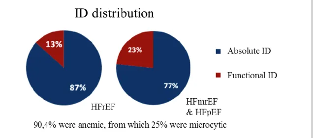
During the study period, 52 patients with CHF were submitted to IV FCM: 69% were men, and the mean age was 86±9 years. Regarding their type of CHF: 34.6% had HFrEF and 65.4% had HFmrEF & HFpEF. Patients were predominantly in NYHA class II 55% and class III 41%, with one patient being Class I and another Class IV. 90,4% had anemia: 25% were microcytic. HFrEF and HFmrEF & HFpEF patients had, respectively, 87% and 77% absolute ID, and 13% and 23% functional ID. Concerning comorbidities, HFrEF and HFmrEF & HFpEF had respectively: 78% and 44% ischemic heart disease, 72% and 76% hypertension, 50% and 62% atrial fibrillation (AF), 44% and 26% diabetes mellitus (DM), 67% and 76% chronic kidney disease (CKD), 28% and 29% chronic obstructive pulmonary disease (COPD), 11% and 15% peptic ulcer disease and 22% and 9% history of neoplasia of the digestive tract. As to their medication, 42.3% were being treated with antiplatelet drugs, 63.5% with anticoagulants, and 13.5% simultaneously on antiplatelet and anticoagulant therapy. 65.4% were on ACEi or ARAII, 26.9% on mineralocorticoid receptor antagonists, 86.5% on diuretics, 76.9% on beta blockers and 59.6% on proton pump inhibitors.
19 Table 2. ID characterization in HFrEF and HFmrEF & HFpEF patients.
Table 3. Comorbidity analysis of both populations – HFpEF and HFmrEF & HFpEF. AF, atrial fibrillation; CKD, chronic kidney disease; COPD, chronic obstructive pulmonary disease; DM, diabetes mellitus; GI, gastrointestinal; HTN, hypertension; IHD, ischemic heart disease; PUD, peptic ulcer disease.
Study B: ID correction in HFmrEF & HFpEF, a follow up at 3 and 6 months
56 patients with HFmrEF & HFpEF and ID were identified. From these 54% (30) were men, the mean age was 82,5±7 years. As to their iron status 83% (47) had absolute ID and 17% (9) had functional ID. 75% (42) patients were anemic, with a mean
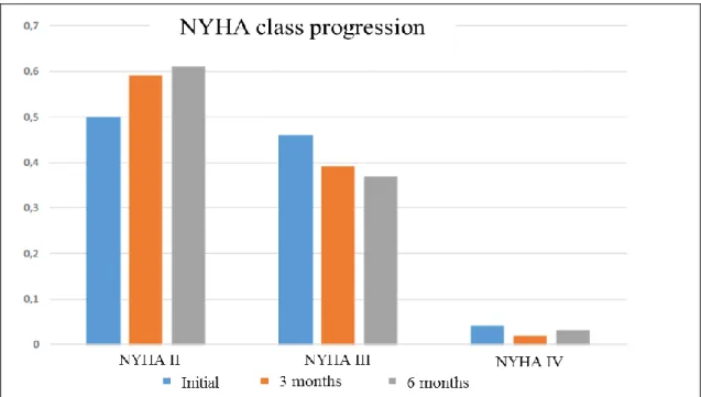
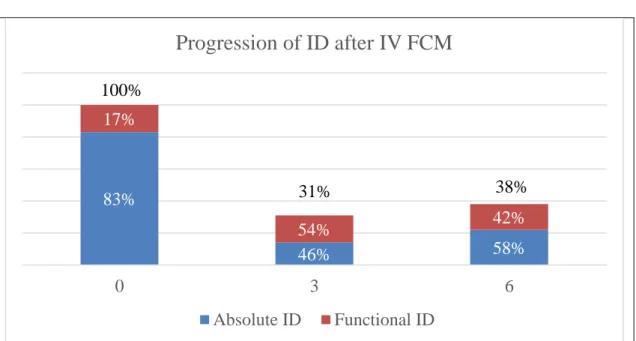
20 hemoglobin (Hb) value of 11±2,1 g/dL, of which 13% (7) were microcytic. Concerning their comorbidities, 25% (14) had ischemic heart disease, 70% (39) hypertension, 64% (36) AF, 32% (18) DM, 66% (67) CKD, 27% (15) COPD, 11% (6) peptic disease and 14% (8) history of neoplasia of the digestive tract. Regarding their NYHA class: 50% (28) patients were in class II, 46% (26) in class III and 4% (2) in class IV. The average NTproBNP was 6492±17459 pg/mL. The mean furosemide-equivalent dose was 40,7±26 mg per day, and the mean eGFR EPI: 47,8±22,6 mL/min/m^2. At three and six months after IV FCM administration: ID was absolute in 14% (6) and 22% (6) of the patients and functional in 17% (7) and 16% (5), with anemia being present in 57% (30) and 56% (22) of the patients, with a mean Hb value of 12±1,8 g/dL and 12±1,8 g/dL There was one patient (2%) with microcytosis. As to their NYHA Class: 59% (33) and 61% (23) patients were in Class II, 39% (22) and 37% (14) in Class III, 2% (1) and 3% (1) in Class IV. The average NTproBNP was 5331±9476 pg/mL and 4000±5262 pg/mL. The mean furosemide-equivalent dose was 45,8±23,4 mg and 45,6±30,0 mg per day, while the mean eGFR EPI: was 45,8±23,4 mL/min/m^2 and 45,8±24,8 mL/min/m^2.
21 0M 3M 6M Hemoglobin (g/dL) 11 ± 2.1 12 ± 1.8 12 ± 1.8 Anemia 75% 57% 56% Microcytosis 13% 2% 2% Iron Deficiency Absolute Functional 100% 31% 38% 83% 46% 58% 17% 54% 42%
Table 5. Analysis of hematinic parameters at 0, 3 and 6 months, after treatment with IV FCM
Table 6. Progression of absolute and functional ID after treatment with IV FCM
83% 46% 58% 17% 54% 42% 0 3 6
Progression of ID after IV FCM
Absolute ID Functional ID 100% 31% 38%22 Table 7. Evolution of NTproBNP, furosemide-equivalent dose and kidney function at zero, three and six months after treatment with IV FCM
6492 5331
4000
0M 3M 6M
Progression of NTproBNP, furosemide-equivalent dose
and glomerular filtration rate after IV FCM
NTproBNP (pg/mL)
40.7 46.9 45.6
0M 3M 6M
Progression of furosemide-equivalent dose with
treatment
Furosemide-equivalent dose (mg/day)
47.8 45.8 45.8
0M 3M 6M
23
Discussion:
Study A: A population characterization
Despite the lack of recommendations for IV FCM therapy in patients with HFmrEF & HFpEF this type of CHF prevailed over HFrEF patients (65.4% vs 34.6%). Nevertheless 90% of the patients were anemic, suggesting that ID per se continues to be poorly evaluated and referred to treatment in daily practice. In CONFIRM-HF only 1/3 of the population had anemia.46
CHF patients, despite of the ejection fraction, were very elderly with a mean age of 86 years, significantly older than in previous studies,45,46 and with several comorbidities. The distribution of these was overall similar throughout both group of patients, except for ischemic heart disease, DM and GI Neoplasia, which were more prevalent in HFrEF. We only found a statistically significant value for ischemic heart disease (78% vs 44%, p=0,013). This finding may reflect the difference on the etiology of the type of dysfunction.53 On the other hand, we found a trend on functional ID being more prevalent in HFmrEF & HFpEF than in HFrEF (13% vs 23%). These findings are in agree with the latest evidence which looks into HFrEF as a disease where left ventricular remodeling is driven by cardiomyocyte death mainly due to ischemia, infection or toxicity, while the new HFmrEF & HFpEF paradigm stands noncardiac comorbidities, namely HTN, obesity, DM, COPD, anemia and CKD, as triggers of coronary microvascular endothelial inflammation and, subsequentially, cardiomyocyte stiffening and hypertrophy.53
Study B: ID correction in HFmrEF & HFpEF, a follow up at 3 and 6 months
Between 2015 and 2018 there were 56 HFmrEF & HFpEF and ID patients treated with IV FCM. ID anemia was the main reason for IV FCM prescription in HFmrEF & HFpEF patients: only 25% of the patients didn´t have anemia. At three and six months after 1000 mg of IV FCM there was a discrete increase in Hb levels and a pronounced reduction in ID prevalence among these patients.
24 The modest increase of Hb levels raises the question, what is the etiology behind these patient’s anemia? Probably ID, while being a major culprit is not the only one.22 As we observed in study A, the prevalence of renal dysfunction, related to a lack of erythropoietin production, was considerably high (76%), as well as the usage of antiplatelet and anticoagulant drugs (42.3% and 63.5%) or the combination of both (13.5%), which may contribute to blood losses. Moreover 65.4% of the patients were on ACEi or ARAII which may inhibit the proliferation of bone marrow derived erythropoietic cells.54 Also, hemodilution might have influenced the degree of anemia.22
Above all, while there were no side effect and adverse event issues, we verified both a functional improvement, as assessed by the NYHA class and, an appealing reduction of NTproBNP levels.
Simultaneously, no changes were seen in eGFR EPI or furosemide-equivalent dose. The furosemide-equivalent dose was used as a surrogate of volume status. Diuretics, when increased, have the ability to decrease both the NYHA class and NTproBNP levels by decongesting the CHF patient and therefore reducing the wall stress of the left ventricle.55 Although it doesn’t counterbalance the lack of a control group, the absence of a significant change in the mean furosemide-equivalent dose allows us to expect that the improvement seen in the NYHA class and NTproBNP levels were not due to diuretic therapy adjustments, strengthening our hypothesis that it was rather due to ID correction with IV FCM.
25
Conclusion
Iron is a cornerstone micronutrient in bioenergy production, immune system efficacity and central nervous system development. Inflammation is characteristic of chronic diseases leading to a deranged milieu that disrupts iron metabolism contributing to a state of ID. In CHF patients, ID is responsible for the impairment of functional capacity, worse quality of life and increased mortality being per se prognostically more ominous than anemia. Although its identification and correction with IV FCM are currently recommended in HFrEF patients there is currently no data about the impact of IV FCM in HFmrEF & HFpEF patients.
Our study pretends to illustrate a daily practice scenario in which symptomatic patients with HFmrEF & HFpEF were recognized as being ID and thereafter submitted to its correction with IV FCM. Despite the lack of evidence for this practice, it was still conducted for correction of ID anemia. In the end, we realized it was a safe treatment, with no adverse event issues reported and without worsening the glomerular filtration rate. We also observed that it led to an improvement of the symptomatic variables NYHA class and NTproBNP, surrogate endpoints for CHF patients.56,57
Even though these results are noticeable, we highlight the lack of statistical significance of these numbers. This was a retrospective, non-randomized, nonblinded study with a small patient population and, therefore these results are merely suggestive of a potential improvement in the ID status, NYHA class and NTproBNP levels of HFmrEF & HFpEF patients with ID after IV FCM treatment. These findings support the need for prospective studies to investigate iron correction as a potential therapeutic target in patients with ID and HFmrEF & HFpEF.
The FAIR-HFpEF study is an undergoing clinical trial designed to access the impact of IV FCM in patients with ID and HFpEF focusing on the improvement of symptoms and exercise capacity as measured by the 6-minute walking test, as well as the impact on mortality and CHF related hospitalization rates, that may shed some light on the subject. [NCT03074591]
26
References
1. Andrews NC. Disorders of Iron Metabolism. The New England Journal of
Medicine. 1999:10.
2. Beard JL. Iron Biology in Immune Function, Muscle Metabolism and Neuronal Functioning. The Journal of Nutrition. 2001;131(2):568S-580S.
doi:10.1093/jn/131.2.568S
3. Cairo G, Bernuzzi F, Recalcati S. A precious metal: Iron, an essential nutrient for all cells. Genes & Nutrition. 2006;1(1):25-39. doi:10.1007/BF02829934
4. Jankowska EA, Rozentryt P, Witkowska A, et al. Iron deficiency: An ominous sign in patients with systolic chronic heart failure. European Heart Journal.
2010;31(15):1872-1880. doi:10.1093/eurheartj/ehq158
5. Haas JD, Brownlie T. Iron Deficiency and Reduced Work Capacity: A Critical Review of the Research to Determine a Causal Relationship. The Journal of
Nutrition. 2001;131(2):676S-690S. doi:10.1093/jn/131.2.676S
6. Davies KJ, Maguire JJ, Brooks GA, Dallman PR, Packer L. Muscle mitochondrial bioenergetics, oxygen supply, and work capacity during dietary iron deficiency and repletion. American Journal of Physiology-Endocrinology and Metabolism.
1982;242(6):E418-E427. doi:10.1152/ajpendo.1982.242.6.E418
7. Hoes MF, Grote Beverborg N, Kijlstra JD, et al. Iron deficiency impairs
contractility of human cardiomyocytes through decreased mitochondrial function: Impaired contractility in iron-deficient cardiomyocytes. European Journal of Heart
Failure. 2018;20(5):910-919. doi:10.1002/ejhf.1154
8. Favrat B, Balck K, Breymann C, et al. Evaluation of a Single Dose of Ferric Carboxymaltose in Fatigued, Iron-Deficient Women – PREFER a Randomized, Placebo-Controlled Study. Collins JF, ed. PLoS ONE. 2014;9(4):e94217. doi:10.1371/journal.pone.0094217
9. Arredondo M, Núñez MT. Iron and copper metabolism. Molecular Aspects of
Medicine. 2005;26(4-5):313-327. doi:10.1016/j.mam.2005.07.010
10. Ortiz E, Pasquini JM, Thompson K, et al. Effect of manipulation of iron storage, transport, or availability on myelin composition and brain iron content in three different animal models. Journal of Neuroscience Research. 2004;77(5):681-689. doi:10.1002/jnr.20207
11. Jáuregui-Lobera I. Iron deficiency and cognitive functions. Neuropsychiatric
Disease and Treatment. November 2014:2087. doi:10.2147/NDT.S72491
12. Murray-Kolb LE, Beard JL. Iron treatment normalizes cognitive functioning in young women. The American Journal of Clinical Nutrition. 2007;85(3):778-787. doi:10.1093/ajcn/85.3.778
27 13. Cassat JE, Skaar EP. Iron in Infection and Immunity. Cell Host & Microbe.
2013;13(5):509-519. doi:10.1016/j.chom.2013.04.010
14. Oppenheimer SJ. Iron and Its Relation to Immunity and Infectious Disease. The
Journal of Nutrition. 2001;131(2):616S-635S. doi:10.1093/jn/131.2.616S
15. Avni T, Bieber A, Grossman A, Green H, Leibovici L, Gafter-Gvili A. The Safety of Intravenous Iron Preparations. Mayo Clinic Proceedings. 2015;90(1):12-23. doi:10.1016/j.mayocp.2014.10.007
16. Fonseca C, Marques F, Robalo Nunes A, Belo A, Brilhante D, Cortez J. Prevalence of anaemia and iron deficiency in Portugal: The EMPIRE study: Anaemia and iron deficiency in Portugal. Internal Medicine Journal. 2016;46(4):470-478.
doi:10.1111/imj.13020
17. Camaschella C. Iron-Deficiency Anemia. Longo DL, ed. New England Journal of
Medicine. 2015;372(19):1832-1843. doi:10.1056/NEJMra1401038
18. Kell DB. Iron behaving badly: Inappropriate iron chelation as a major contributor to the aetiology of vascular and other progressive inflammatory and degenerative diseases. BMC Medical Genomics. 2009;2(1). doi:10.1186/1755-8794-2-2
19. González-Costello J, Comín-Colet J. Iron deficiency and anaemia in heart failure: Understanding the FAIR-HF trial. European Journal of Heart Failure.
2010;12(11):1159-1162. doi:10.1093/eurjhf/hfq165
20. Weiss G. Anemia of Chronic Disease. The New England Journal of Medicine. 2005:13.
21. Weiss G. Iron metabolism in the anemia of chronic disease. Biochimica et
Biophysica Acta (BBA)â”General Subjects. 2009;1790(7):682-693.
doi:10.1016/j.bbagen.2008.08.006
22. van Veldhuisen DJ, Anker SD, Ponikowski P, Macdougall IC. Anemia and iron deficiency in heart failure: Mechanisms and therapeutic approaches. Nature Reviews
Cardiology. 2011;8(9):485-493. doi:10.1038/nrcardio.2011.77
23. Hempel EV, Bollard ER. The Evidence-Based Evaluation of Iron Deficiency Anemia. Medical Clinics of North America. 2016;100(5):1065-1075.
doi:10.1016/j.mcna.2016.04.015
24. Zimmermann MB. Methods to assess iron and iodine status. British Journal of
Nutrition. 2008;99(S3). doi:10.1017/S000711450800679X
25. Wish JB. Assessing Iron Status: Beyond Serum Ferritin and Transferrin Saturation.
Clinical Journal of the American Society of Nephrology. 2006;1(Supplement
1):S4-S8. doi:10.2215/CJN.01490506
26. Cook J. Diagnosis and management of iron-deficiency anaemia. Best Practice &
Research Clinical Haematology. 2005;18(2):319-332.
28 27. Mast AE, Blinder MA, Gronowski AM, Chumley C, Scott MG. Clinical utility of
the soluble transferrin receptor and comparison with serum ferritin in several populations. Clinical Chemistry. 1998;(1):7.
28. Goodnough LT, Nemeth E, Ganz T. Detection, evaluation, and management of iron-restricted erythropoiesis. Blood. 2010;116(23):4754-4761. doi:10.1182/blood-2010-05-286260
29. Punnonen K, Irjala K. Serum Transferrin Receptor and Its Ratio to Serum Ferritin in the Diagnosis of Iron Deficiency. :7.
30. Thomas DW, Hinchliffe RF, Briggs C, et al. Guideline for the laboratory diagnosis of functional iron deficiency. British Journal of Haematology. 2013;161(5):639-648. doi:10.1111/bjh.12311
31. Brugnara C, Schiller B, Moran J. Reticulocyte hemoglobin equivalent (Ret He) and assessment of iron-deficient states. Clinical and Laboratory Haematology.
2006;28(5):303-308. doi:10.1111/j.1365-2257.2006.00812.x
32. Okonko DO, Grzeslo A, Witkowski T, et al. Effect of Intravenous Iron Sucrose on Exercise Tolerance in Anemic and Nonanemic Patients With Symptomatic Chronic Heart Failure and Iron Deficiency. Journal of the American College of Cardiology. 2008;51(2):103-112. doi:10.1016/j.jacc.2007.09.036
33. Jankowska EA, Ponikowski P. Heart Failure Classifications – Guidelines. In: Ronco C, Costanzo MR, Bellomo R, Maisel AS, eds. Contributions to Nephrology. Vol 164. Basel: KARGER; 2010:11-23. doi:10.1159/000313716
34. Okonko DO, Mandal AKJ, Missouris CG, Poole-Wilson PA. Disordered Iron Homeostasis in Chronic Heart Failure. Journal of the American College of
Cardiology. 2011;58(12):1241-1251. doi:10.1016/j.jacc.2011.04.040
35. Anker SD, Colet JC, Filippatos G, et al. Rationale and design of Ferinject®
Assessment in patients with IRon deficiency and chronic Heart Failure (FAIR-HF) study: A randomized, placebo-controlled study of intravenous iron supplementation in patients with and without anaemia. European Journal of Heart Failure.
2009;11(11):1084-1091. doi:10.1093/eurjhf/hfp140
36. Silverberg DS, Wexler D, Blum M, et al. The use of subcutaneous erythropoietin and intravenous iron for the treatment of the anemia of severe, resistant congestive heart failure improves cardiac and renal function and functional cardiac class, and markedly reduces hospitalizations. Journal of the American College of Cardiology. 2000;35(7):1737-1744. doi:10.1016/S0735-1097(00)00613-6
37. Tang Y-D, Katz SD. Anemia in Chronic Heart Failure: Prevalence, Etiology, Clinical Correlates, and Treatment Options. Circulation. 2006;113(20):2454-2461. doi:10.1161/CIRCULATIONAHA.105.583666
38. Groenveld HF, Januzzi JL, Damman K, et al. Anemia and Mortality in Heart Failure Patients. Journal of the American College of Cardiology. 2008;52(10):818-827. doi:10.1016/j.jacc.2008.04.061
29 39. Klip IjT, Comin-Colet J, Voors AA, et al. Iron deficiency in chronic heart failure:
An international pooled analysis. American Heart Journal. 2013;165(4):575-582.e3. doi:10.1016/j.ahj.2013.01.017
40. Nanas JN, Matsouka C, Karageorgopoulos D, et al. Etiology of Anemia in Patients With Advanced Heart Failure. Journal of the American College of Cardiology. 2006;48(12):2485-2489. doi:10.1016/j.jacc.2006.08.034
41. Ezekowitz JA, McAlister FA, Armstrong PW. Anemia Is Common in Heart Failure and Is Associated With Poor Outcomes: Insights From a Cohort of 12 065 Patients With New-Onset Heart Failure. Circulation. 2003;107(2):223-225.
doi:10.1161/01.CIR.0000052622.51963.FC
42. Bekfani T, Pellicori P, Morris D, et al. Iron deficiency in patients with heart failure with preserved ejection fraction and its association with reduced exercise capacity, muscle strength and quality of life. Clinical Research in Cardiology.
2019;108(2):203-211. doi:10.1007/s00392-018-1344-x
43. Martens P, Nijst P, Verbrugge FH, Smeets K, Dupont M, Mullens W. Impact of iron deficiency on exercise capacity and outcome in heart failure with reduced, mid-range and preserved ejection fraction. Acta Cardiologica. 2018;73(2):115-123. doi:10.1080/00015385.2017.1351239
44. Toblli JE, Lombraña A, Duarte P, Di Gennaro F. Intravenous Iron Reduces NT-Pro-Brain Natriuretic Peptide in Anemic Patients With Chronic Heart Failure and Renal Insufficiency. Journal of the American College of Cardiology. 2007;50(17):1657-1665. doi:10.1016/j.jacc.2007.07.029
45. Anker SD, Comin Colet J, Filippatos G, et al. Ferric Carboxymaltose in Patients with Heart Failure and Iron Deficiency. New England Journal of Medicine. 2009;361(25):2436-2448. doi:10.1056/NEJMoa0908355
46. Ponikowski P, van Veldhuisen DJ, Comin-Colet J, et al. Beneficial effects of long-term intravenous iron therapy with ferric carboxymaltose in patients with
symptomatic heart failure and iron deficiency. European Heart Journal. 2015;36(11):657-668. doi:10.1093/eurheartj/ehu385
47. Moore RA, Gaskell H, Rose P, Allan J. Meta-analysis of efficacy and safety of intravenous ferric carboxymaltose (Ferinject) from clinical trial reports and
published trial data. BMC Blood Disorders. 2011;11(1). doi:10.1186/1471-2326-11-4
48. van Veldhuisen DJ, Ponikowski P, van der Meer P, et al. Effect of Ferric
Carboxymaltose on Exercise Capacity in Patients With Chronic Heart Failure and Iron Deficiency. Circulation. 2017;136(15):1374-1383.
doi:10.1161/CIRCULATIONAHA.117.027497
49. Swank AM, Horton J, Fleg JL, et al. Modest Increase in Peak VO 2 Is Related to Better Clinical Outcomes in Chronic Heart Failure Patients: Results From Heart Failure and a Controlled Trial to Investigate Outcomes of Exercise Training.
Circulation: Heart Failure. 2012;5(5):579-585.
30 50. Lewis GD, Malhotra R, Hernandez AF, et al. Effect of Oral Iron Repletion on
Exercise Capacity in Patients With Heart Failure With Reduced Ejection Fraction and Iron Deficiency: The IRONOUT HF Randomized Clinical Trial. JAMA. 2017;317(19):1958. doi:10.1001/jama.2017.5427
51. Silverstein SB, Gilreath JA, Rodgers GM. Intravenous Iron Therapy: A Summary of Treatment Options and Review of Guidelines. Journal of Pharmacy Practice. 2008;21(6):431-443. doi:10.1177/0897190008318916
52. Van Wyck DB, Roppolo M, Martinez CO, Mazey RM, Mcmurray S. A randomized, controlled trial comparing IV iron sucrose to oral iron in anemic patients with nondialysis-dependent CKD. Kidney International. 2005;68(6):2846-2856. doi:10.1111/j.1523-1755.2005.00758.x
53. Paulus WJ, Tschöpe C. A Novel Paradigm for Heart Failure With Preserved Ejection Fraction. Journal of the American College of Cardiology. 2013;62(4):263-271. doi:10.1016/j.jacc.2013.02.092
54. van der Meer P, Lipsic E, Westenbrink BD, et al. Levels of Hematopoiesis Inhibitor
N -Acetyl-Seryl-Aspartyl-Lysyl-Proline Partially Explain the Occurrence of Anemia
in Heart Failure. Circulation. 2005;112(12):1743-1747. doi:10.1161/CIRCULATIONAHA.105.549121
55. Kalra P. Water and sodium regulation in chronic heart failure: The role of natriuretic peptides and vasopressin. Cardiovascular Research. 2001;51(3):495-509. doi:10.1016/S0008-6363(01)00297-8
56. Oremus M, Don-Wauchope A, McKelvie R, et al. BNP and NT-proBNP as prognostic markers in persons with chronic stable heart failure. Heart Failure
Reviews. 2014;19(4):471-505. doi:10.1007/s10741-014-9439-6
57. Aronson JK. Biomarkers and surrogate endpoints. British Journal of Clinical