UNIVERSIDADE DE LISBOA FACULDADE DE CIÊNCIAS DEPARTAMENTO DE FÍSICA
Enhancing reprogramming and transdifferentiation through long
non-coding RNAs
Miguel Torres Santana
Mestrado Integrado em Engenharia Biomédica e Biofísica Perfil de Engenharia Clínica e Instrumentação Médica
Dissertação orientada por:
Doutor Bruno Miguel Bernardes de Jesus Professor Doutor Pedro Miguel Dinis de Almeida
I UNIVERSIDADE DE LISBOA
FACULDADE DE CIÊNCIAS DEPARTAMENTO DE FÍSICA
Enhancing reprogramming and transdifferentiation through long
non-coding RNAs
Miguel Torres Santana
Mestrado Integrado em Engenharia Biomédica e Biofísica Perfil de Engenharia Clínica e Instrumentação Médica
Dissertação orientada por:
Doutor Bruno Miguel Bernardes de Jesus Professor Doutor Pedro Miguel Dinis de Almeida
II This page intentionally left blank
III
Agradecimentos
Quero expressar em primeiro lugar o meu agradecimento ao Doutor Bruno Jesus, pela orientação, ajuda e apoio constante ao longo deste último ano letivo e pela oportunidade que me proporcionou de participar neste projeto.
Agradeço ainda ao António Franco por me ter acompanhado, ajudado e ensinado no período de adaptação e ao Sérgio Marinho pela sua constante boa disposição e disponibilidade e boa vontade em me ajudar.
Agradeço ainda a todos os restantes colegas e amigos do laboratório e do IMM, pela companhia, apoio constante, boa disposição e amizade.
Agradeço também à minha família por todo o apoio, aconselhamento e por estarem sempre ao meu lado, mesmo estando distantes.
Por fim, não posso deixar de agradecer a todos os meus amigos que me acompanharam e ajudaram ao longo destes últimos anos.
IV
Resumo
Foi recentemente desenvolvido um novo método revolucionário capaz de reprogramar fibroblastos em células pluripotentes induzidas através da expressão de 4 fatores de transcrição (Oct4, Sox2, Klf4 e
c-Myc). A reprogramação de fibroblastos em células pluripotentes induzidas (iPSC) foi um grande
avanço científico com possíveis aplicações clínicas e fins terapêuticos, no entanto, à medida que as células diferenciadas (células somáticas) vão envelhecendo devido à acumulação de marcas genéticas e epigenéticas, estas tornam-se mais resistentes à sua conversão para um estado pluripotente.
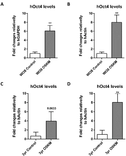
Posto isto, um dos principais objetivos deste projeto passava por perceber o impacto que o envelhecimento tem na reprogramação de células humanas em células estaminais pluripotentes induzidas humanas (hiPSCs). De facto, ao reprogramar fibroblastos adultos de ratinho em células pluripotentes induzidas (miPSCs – mouse induced pluripotent stem cells), observou-se que o envelhecimento celular estava a atuar como uma barreira, reduzindo a eficiência da reprogramação celular. Curiosamente, nenhuma correlação entre a eficiência da reprogramação celular e o envelhecimento foi observada na reprogramação de células humanas. Ao realizar a reprogramação celular de fibroblastos embrionários humanos (WI38) e fibroblastos humanos com 3 anos (3yr) com baixa passagem (passage 4), observou-se o mesmo número de células hiPSCs geradas. Sugerindo assim, e ao contrário do esperado e observado em células de ratinho, que a idade das células humanas utilizadas para formar hiPSCs não dificulta de forma significativa a reprogramação celular.
Contudo, foi observado que o número de passagens das células em cultura tinha uma contribuição importante na eficiência da reprogramação celular de fibroblastos humanos em hiPSCs. Ao tentar reprogramar fibroblastos humanos com uma baixa passagem (passagem 4) e uma alta passagem (passagem 7), e apesar de as células com passagem 7 expressarem níveis mais elevados de hOCt4, apenas as células humanas com uma baixa passagem reprogramaram. Estes resultados sugerem que o número de passagens celulares tem uma contribuição importante na eficiência da reprogramação celular.
De facto, estes resultados podem ser explicados pela simples razão de que cada passagem celular realizada em cultura, aumenta o risco de ocorrer dano no ADN, mutações e ainda alterações nas características celulares originando assim, alterações na morfologia, na resposta a estímulos, na taxa de crescimento, na expressão de proteínas e na eficiência da transfecção celular. De acordo com Leonard Hayflick e Paul Moorhead, as células humanas têm um número limitado de divisões celulares em cultura que poderá variar entre tipos de células. Cada divisão celular pode induzir um encurtamento dos telómeros que poderá resultar em senescência. Podendo esta ser ainda induzida através de dano molecular que ocorre de forma aleatória, pelo stress oxidativo e pela danificação do ADN. A senescência pode ainda ser acumulada em alguns tecidos contribuindo para a disfunção orgânica. Sugere-se portanto, que devido ao número elevado de passagens celulares, os fibroblastos embrionários humanos e os fibroblastos humanos com 3 anos sofreram o encurtamento dos seus telomeros, levando assim, a um fenótipo senescente, reduzindo a eficiência do processo de reprogramação celular em células humanas.
Um outro objetivo deste projeto passava por encontrar outras estratégias celulares que ajudem a ultrapassar a limitação do envelhecimento na reprogramação celular de células humanas, através da modulação de RNAs longos não codificantes (lncRNA). No entanto, como os resultados referentes ao envelhecimento demonstraram uma não influência na eficiência da reprogramação de células humanas em hiPSCs, decidiu-se desvendar e entender, qual a função do lncRNA Zeb2NAT na reprogramação celular e qualidade de células humanas. Embora, já reportado anteriormente pelo nosso laboratório,
V que a supressão do lncRNA Zeb2NAT em células de ratinho, utilizando olignucleótidos contra-senso (anti sense), denominados por LNAs aumentam significativamente a reprogramação celular de fibroblastos de ratinho envelhecidos ajudando assim contornar as barreiras mesenquimais, ainda nada se sabia sobre o impacto que a diminuição do Zeb2 e do Zeb2NAT poderia ter na reprogramação celular de células humanas.
Contudo, utilizando a mesma abordagem, acima descrita, em fibroblastos embrionários humanos e em fibroblastos humanos com 3 anos, verificou-se um atraso na formação de hiPSCs das células humanas que sofreram uma desregulação tanto do Zeb2 como do Zeb2NAT, comparativamente às células do controlo e do LNA-controlo (um LNA não específico a nenhuma sequência genómica humana, utilizado como um controlo negativo da transfeção de LNAs). De facto, 14 dias após a primeira transfeção com LNAs em ambas as duas linhas celulares, apenas começaram a formar-se
hiPSCs no controlo e no LNA-controlo, tendo o controlo um número maior de hiPSCs formadas em
ambas as células, comparativamente à condição com LNA-controlo. No entanto, 19 dias e 35 dias depois da primeira transfeção, as WI38 com a desregulação do Zeb2 e do Zeb2NAT, respetivamente, começaram a reprogramar. Nenhuma formação de hiPSCs nas células humanas de 3 anos com a desregulação do Zeb2 e do Zeb2NAT foi observada.
Tendo em consideração que as células de ratinho após diminuição dos níveis do Zeb2 e do Zeb2-NAT começaram a reprogramar de forma mais eficiente que o controlo e o LNA-controlo, estes resultados em células humanas sugerem que a desregulação do Zeb2 e do Zeb2NAT pode estar, de certa maneira, a atrasar e até mesmo a atuar como um bloqueador da reprogramação celular humana.
Este bloqueio/atraso que se observou na reprogramação de fibroblastos humanos em hiPSCs poderia ter como principal responsável a transfeção de LNAs, necessária para diminuir a expressão do Zeb2 e do Zeb2NAT. De facto, já foi demonstrado que o uso do reagente de transfeção Lipofectamine ativa algum stress nos genes afetando o ciclo da regulação e/ou a sinalização metabólica nas células. No entanto, as células com a condição do LNA-controlo reprogramaram com a mesma rapidez que o controlo (sem LNAs). E a diferença do número de hiPSCs geradas pelo LNA-controlo, comparativamente ao controlo, acaba por não ser significativamente diferente. Isto sugere, que embora a transfeção de LNAs tenha um impacto na reprogramação celular, diminuindo a eficiência desta, existe um outro fator que está a contribuir para que as células com a diminuição da expressão do Zeb2 e do Zeb2-NAT tenham uma redução na eficiência da reprogramação celular em células humanas.
Todavia, ainda é incerto a razão de a desregulação do Zeb2 e do Zeb2NAT terem atrasado e até mesmo bloqueado a reprogramação celular em células humanas. É possível que possa ser devido ao mecanismo que rege o Zeb2 que, de alguma forma, é diferente comparativamente ao mecanismo observado em ratinhos. Outra possibilidade a ter em conta é a necessidade de realizar uma otimização ao protocolo da transfeção de LNAs, de forma a adaptar esta ao protocolo da reprogramação celular.
Apesar disto, acreditamos que esta abordagem constitui uma nova estratégia para estudar o impacto dos lncRNAs na reprogramação celular e antecipamos ainda que os resultados produzidos irão gerar contribuições importantes na área de investigação do envelhecimento e da reprogramação celular.
Palavras-chave: Reprogramação celular, fibroblastos humanos, células estaminais pluripotentes induzidas humanas, envelhecimento, Zeb2NAT
VI
Abstract
Revolutionary progress has been achieved recently following the discovery of cellular reprogramming by the expression of a combination of 4 transcription factors (Oct4, Sox2, Klf4 and c-Myc). The reprogramming of fibroblasts to induced pluripotent stem cells (iPSC) was a major scientific advance however, as differentiated cells grow old and due to the accumulation of genetic and epigenetic marks, they become more resistant to be converted back to a pluripotent state.
In fact, when tried to reprogram mice cells into miPSCs, aging was acting as a barrier, reducing the efficiency of reprogramming aged cells. However, through our recent findings no correlation between cellular reprogramming efficiency and aging was found in human cells. However, we observed that the number of passages had an important contribution in the efficiency of reprogramming human fibroblasts into hiPSCs. Reprogramming experiments with human fibroblasts with lower passage (passage 4) and high passage (passage 7) show that only the human cells with a lower passage, reprogrammed. The higher the number of passages in vitro, the lower the efficiency of cellular reprogramming of human cells.
The other main interest of this project converged on cellular strategies to overcome this aging limitation by modulating long noncoding RNAs (lncRNAs). However, as the results showed a non-influence of aging on reprogramming human cells into hiPSCs it was decided to focus on understanding exactly the role of the lncRNA Zeb2NAT on cellular reprogramming of human cells. As previously reported by our lab, the suppression of Zeb2NAT in mice cells significantly increased the reprogramming of aged fibroblasts. However, when tested the same approach in human cells, a delay in generating hiPSCs was observed, suggesting that the knockdown of Zeb2 and Zeb2NAT can be acting as a blocker of cellular reprogramming in human cells, going against what was observed in mice cells.
Still, it’s uncertain why downregulation of Zeb2 and Zeb2NAT were delaying and blocking the cellular reprogramming in human cells. It is possible that the mechanisms of Zeb2 in humans are, somehow, different from those observed in mice. Another possibility to take in account can be due the fact the protocol isn´t fully optimized.
Despite this, we believe this approach constitutes a novel strategy to study the impact of lncRNAs in cellular reprogramming and we anticipate that the output of this proposal will generate important contributions to the aging and cellular reprogramming research field.
VII
Table of contents
Agradecimentos... III Resumo ... IV Abstract ... VI List of tables ... VIII List of figures ... IX List of abbreviations ... XII
1. Introduction and literature review ... 1
1.1. Stem cells ... 1
1.2. Cellular reprogramming ... 2
1.3. Cellular Aging ... 4
1.4. Non-coding RNAs ... 6
1.4.1. Long non-coding RNAs ... 6
1.5. Objectives ... 11
2. Materials and Methods ... 13
2.1. Cell lines ... 13
Human Fibroblasts ... 13
Human embryonic kidney 293T cell line (293T) ... 13
Human induced pluripotent stem cells (hiPSCs) ... 14
Mouse stem cells E14 ... 14
2.2. Cellular Reprogramming ... 15
2.3. Molecular Biological Techniques ... 16
3. Results and Discussion ... 21
3.1. Optimization of the Cellular reprogramming protocol ... 24
3.2. Aging as a barrier for cellular reprogramming... 28
3.3. Role of the lncRNA Zeb2NAT on cellular reprogramming of human cells ... 31
3.4. NORAD affects chromosomal stability of iPSCs after DNA damage ... 37
4. Conclusion ... 39
5. References ... 41
6. Annexes ... 45
Annex 1 ... 45
VIII
List of tables
Table 1.1 - Summary of the current literature on the impact of age on reprogramming19. ... 5 Table 3.1 - Number of hiPSCs clones reprogrammed from WI38 and 3yr human fibroblasts in 60 mm petri dish with a low passage (passage 4) following the optimized cellular reprogrammed protocol using OSKM plasmid. ... 30 Table 3.2 - Quantification of number of hiPSCs clones 28 days after firs cellular reprogramming infection and 25 days after 1º LNA transfection. ... 34 Table 3.3 –Reprogramming efficiency (%) of 3yr and Wi38 human cells with passage 4, 28 days after first cellular reprogramming infection and 25 days after 1º LNA transfection. ... 34
IX
List of figures
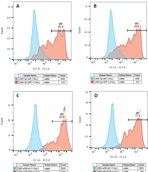
Figure 1.1 - Pluripotent stem cell differentiation into all three germ layers (Mesoderm, Endoderm and Ectoderm). Adapted from Juty N. et all, 2008. ... 2 Figure 1.2 - iPSC reprogramming protocol by introducing genes encoding four transcription factors13.3 Figure 1.3 - Illustration of the regulation of Zeb2 through lncRNA Zeb2NAT29. ... 8 Figure 1.4 - Representation of the lncRNA zeb2 regulation of the epithelial-mesenchymal transition of cells through the E-Cadherin inhibition. ... 9 Figure 1.5 - LncRNA NORAD regulates genomic stability by sequestering PUMILIO proteins34 ... 10 Figure 3.1 - Cellular reprogramming to test efficiency of OSKM and Pkp332 plasmids in reprogramming. (A) FUW-OSKM vector structure (B) pKP332 vector structure (C) WI38 Clone generated 25 days after transduction with OSKM plasmid. Amplification x10. Scale bar was set for 50μm. Image acquired through the Carl Zeiss PrimoVert microscope. Image was treated posteriorly with Fiji software. ... 22 Figure 3.2 - RT-qPCR results of hOCt4 expression levels of WI38 and 3yr human fibroblasts (after transduction using OSKM plasmid) presented as ΔΔCt normalized using non OSKM lentiviral transduced cells as control and hGAPDH and hActin as housekeeping gene. (A and B) WI38 human fibroblasts hOct4 levels using housekeeping hGAPDH (A) and hActin (B). (C and D) 3yr human fibroblasts hOct4 levels using housekeeping hGAPDH (C) and hActin (D). P-value ≤ 0.05*; p-value ≤ 0.01**; p-value ≤ 0.001***. ... 23 Figure 3.3 - GFP expression after a cellular reprogramming experiment with both 3yr (A) and WI38 (B) human fibroblasts performed using cellular reprogramming protocol. Scale bar was set for 50μm. Images acquired through the Zeiss Cell Observer fluorescence microscope, amplification x4. Images were treated posteriorly by using Fiji software. ... 23 Figure 3.4 - Flow cytometry histogram results of 3yr and WI38 human fibroblasts expressing green fluoresnce plotting in red. 3yr and WI38 control plotted in blue.. (A) and (B) 61.6% and 63.8% of viable 3yr human fibroblasts expressing GFP, correspondently; mean= 62.7%; (C) and (D) 82.9% and 77.4% of viable WI38 human fibroblasts expressing GFP, correspondently, mean= 80.2%. Analysis performed using BD Accuri C6 with a cell count of 3000. ... 24 Figure 3.5 - Illustration of cellular reprogramming protocol performed in 3yr and WI38 human fibroblasts with 293T transfection performed in day 1 and human fibroblasts infection in day 4 and 5. ... 25 Figure 3.6 - 11 days after first infection of cellular reprogramming with OSKM plasmid in 3yr and WI38 human fibroblasts (passage 4). (A) 3yr human fibroblasts control, amplification x4. (B) 3yr hiPSCs clones reprogrammed amplification x4. (C) 3yr hiPSCs clones reprogrammed, amplification x10. (D) WI38 human fibroblasts control, amplification x4. (E) WI38 hiPSCs clones reprogrammed, amplification x4. (F) WI83 hiPSCs clones reprogrammed, amplification x10. Scale bar was set for 50μm. Images acquired through the Carl Zeiss PrimoVert microscope. Images were treated posteriorly by using Fiji software. ... 26 Figure 3.7 - Reprogrammed hiPSCs isolated from feeders after using passing EDTA protocol. (A) Reprogrammed hiPSCs from 3yr human fibroblasts (passage 4º) (B) Reprogrammed hiPSCs from WI38 human fibroblasts (passage 4). Scale bar was set for 50μm. Images acquired through the Carl Zeiss PrimoVert microscope, amplification x4. Images were treated posteriorly by using Fiji software. ... 26 Figure 3.8 - hiPSCs after alkaline phosphatase treatment. A and B - WI38 hiPSCs with low confluence after AP treatment; C - WI38 hiPSCs with low confluence without AP treatment; D and E - 3yr
X hiPSCs with high confluence after AP treatment; F - 3yr hiPSCs with high confluence without AP treatment. ... 27 Figure 3.9 - Representative teratoma formed after injection of hiPSCs ... 27 Figure 3.10 - RT-qPCR results of hOCt4 expression levels of WI38 and 3yr human fibroblasts with passage 4 (p4) and passage 7 (p7) presented as ΔΔCt normalized using non OSKM lentiviral transduced cells as control and hGAPDH and hActin as housekeeping gene. (A and B) WI38 human fibroblasts (p4 and p7) hOct4 levels using housekeeping hGAPDH (A) and hActin (B). (C and D) 3yr human fibroblasts (p4 and p7) hOct4 levels using housekeeping hGAPDH (C) and hActin (D). P-value ≤ 0.05*; p-value ≤ 0.01**; p-value ≤ 0.001***. ... 28 Figure 3.11 –Cellular reprogramming protocol performed in WI38 and 3yr human fibroblasts with high passage (p7). (A and B) 6 days after first infection of 3yr and WI38 human cells lines with a high passage (passage 7) using the optimized cellular reprogramming protocol. (A1) 3yr human fibroblasts negative control. (A2) 3yr human fibroblasts transduced with OSKM. (B1) WI38 human fibroblasts negative control. (B2) WI38 human fibroblasts transduced with OSKM. (C and D) 31 days after first infection of 3yr and WI38 human cells lines with a high passage (passage 7 ) using the optimized cellular reprogramming protocol. (C1) 3yr human fibroblasts negative control. (C2) 3yr human fibroblasts transduced with OSKM. (D1) WI38 human fibroblasts negative control. (D2) WI38 human fibroblasts transduced with OSKM. Scale bar was set for 50μm. Images acquired through the Carl Zeiss PrimoVert microscope, amplification x4. Images were treated posteriorly by using Fiji software. ... 29 Figure 3.12 - Results of downregulation efficiency of hZeb2 and hNAT expression level in 3yr human fibroblasts presented as ΔΔCt normalized using non downregulation of lncRNAs Zeb2 and Zeb2NAT as control and HGAPDH and hActin as housekeeping gene. (A and B) ZEB2 expression levels in 3yr cells with knockdown of Zeb2 and (A) and hActin (B) as housekeeping (C and D) 3yr Zeb2, zeb2NAT and scramble levels compared to control values using hGAPDH (C) and hActin (D) as housekeeping. ... 32 Figure 3.13 – Cellular reprogramming protocol with LNAs GapmeRs transfection protocol in 3yr and WI38 human fibroblasts. Cellular reprogramming protocol started in day 1 with 293T transfection and ended in day 5 with second infection of human fibroblasts. LNAs GapmeRs transfection protocol was performed in day 8 and 9. ... 32 Figure 3.14 – HiPSCs generated after performing the cellular reprogramming protocol and the LNA transfection protocol in both human cell lines. 3yr (A) and WI38 (B) human fibroblasts with low passage (p4) after knockdown of Zeb2 and Zeb2NAT. (A1) 3yr human fibroblasts negative control for LNA transfection. Started to reprogram 14 days after first reprogramming infection, amplification x4. (A2) 3yr human fibroblasts transfected with scramble. Started to reprogram 14 days after first reprogramming infection, amplification x4. (B1) WI38yr human fibroblasts negative control for LNA transfection. Started to reprogram 14 days after first reprogramming infection, amplification x10 (B2) WI38yr human fibroblasts transfected with scramble. Started to reprogram 14 days after first reprogramming infection, amplification x4. (B3) WI38 human fibroblasts after knockdown of Zeb2. Started to reprogram 19 after first reprogramming infection, amplification x10. (B4) WI38 human fibroblasts after knockdown of Zeb2NAT. Started to reprogram 35 days after first reprogramming infection. Scale bar was set for 50μm. Images acquired through the Carl Zeiss PrimoVert microscope. Images were treated posteriorly by using Fiji software. ... 33 Figure 3.15 - Immunofluorescence performed in 3yr control (A and B) and WI38 control (C) hiPSCs. Cells stained with DAPI in blue and human antibodies against Nanog and Sox2 in red. (A1) 3yr hiPSCs stained with human antibody agains Sox2 in red. (A2) 3yr hiPSCs stained with DAPI in blue. (B1) 3yr hiPSCs stained with human antibody agains Nanog in red. (B2) 3yr hiPSCs stained with DAPI in blue. (C1) WI38 hiPSCs stained with human antibody agains Sox2 in red. (C2) Wi38 hiPSCs
XI stained with DAPI in blue. Scale bar was set for 50μm. Images acquired through the Carl Zeiss Axiovert 200M fluorescence microscope. Images were treated posteriorly by using Fiji software. ... 35 Figure 3.16 - Illustration of LNAs GapmeRs transfection protocol with cellular reprogramming protocol performed afterwards in 3yr and WI38 human fibroblasts. LNAs GapmeRs transfection protocol was performed in day 1 and 2. Cellular reprogramming protocol started in day 2 with 293T transfection and ended in day 6 with second infection of human fibroblasts. ... 36 Figure 3.17 - Representation of CRISPR/CAS9 technique used to delete NORAD gene. (A) Norad plasmids in red designed to cut in a specific location for NORAD deletion. (B) Two par of primers in yellow designed to detect if NORAD was successfully deleted through electrophoresis. (C) If NORAD was successfully deleted a band of 507bp in electrophoresis gel would be detected. (D) If NORAD deletion was not accomplished two bands of 434bp and 5703bp would appear in electrophoresis gel. 38 Figure 6.1 - RT-qPCR results of hKLF4 expression levels of WI38 and 3yr human fibroblasts presented as ΔΔCt normalized using non OSKM lentiviral transduced cells as control and hGAPDH and hActin as housekeeping gene. (A and B) WI38 human fibroblasts hKLF4 levels using housekeeping hGAPDH (A) and hActin (B). (C and D) 3yr human fibroblasts hKLF4 levels using housekeeping hGAPDH (C) and hActin (D). P-value ≤ 0.05*; p-value ≤ 0.01**; p-value ≤ 0.001***. ... 45 Figure 6.2 - RT-qPCR results of hOct4 expression levels of WI38 and 3yr human fibroblasts (after transduction using pKP332 plasmid) presented as ΔΔCt normalized using non pKP332 lentiviral transduced cells as control and hGAPDH and hActin as housekeeping gene. (A and B) WI38 human fibroblasts hOct4 levels using housekeeping hGAPDH (A) and hActin (B). (C and D) 3yr human fibroblasts hOct4 levels using housekeeping hGAPDH (C) and hActin (D). P-value ≤ 0.05*; p-value ≤ 0.01**; p-value ≤ 0.001***. ... 46 Figure 6.3 - Flow cytometry histogram results of 3yr and WI38 human fibroblasts expressing green fluoresnce plotting in red. 3yr and WI38 control plotted in blue.. (A) and (B) 72.3% and 72.5% of viable 3yr human fibroblasts expressing GFP, correspondently; mean= 72.4%; (C) and (D) 65.6% and 70.7% of viable WI38 human fibroblasts expressing GFP, correspondently, mean= 68.2%. Analysis performed using BD Accuri C6 with a cell count of 3000 for cells infected with GFP. Cell counting of control was set to 10000 cells. ... 46 Figure 6.4 - Flow cytometry histogram results of 3yr and WI38 human fibroblasts with high (p7) passage expressing green fluoresnce plotting in red. 3yr and WI38 control plotted in blue. (A) 67.8% of viable 3yr human fibroblasts with a high passage (p7) expressing GFP. (B) 65.6% of viable WI38 human fibroblasts with a high passage (p7) expressing GFP. Analysis performed using BD Accuri C6 with a cell count of 3000 for cells infected with GFP. Cell counting of control was set to 10000 cells. ... 47 Figure 6.5 - Flow cytometry histogram results of 3yr and WI38 human fibroblasts with low (p4) passage expressing green fluoresnce plotting in red. 3yr and WI38 control plotted in blue. (A) 67.8% of vialbe 3yr human fibroblasts with a low passage (p4) expressing GFP. (B) 61.0% of vialbe WI38 human fibroblasts with a low passage (p4) expressing GFP. Analysis performed using BD Accuri C6 with a cell count of 3000 for cells infected with GFP. Cell counting of control was set to 10000 cells. ... 47 Figure 6.6 - Results of downregulation efficiency of hNAT expression level in 3yr human fibroblasts presented as ΔΔCt normalized using non downregulation of lncRNAs Zeb2 and Zeb2NAT as control and HGAPDH and hActin as housekeeping gene. (A to C) ZEB2NAT expression levels in 3yr cells with knockdown of Zeb2 and hGAPDH as housekeeping. ... 48
XII
List of abbreviations
3yr – 3yr human fibroblasts
293T – Human embryonic kidney 293T cells AP – Alkaline phosphatase
cDNA – Complementary DNA c-Myc – MYC
CIN – Chromosomal instability
DMEM – Dulbecco’s Modified Eagle Medium E14 – Mouse stem cells E14
ECATs – ES cell-associated transcripts ESCs – Embryonic stem cells
EMT – Epithelial-mesenchymal transition FBS – Fetal Bovine Serum
HGPS – Hutchinson-Gilford Progeria syndrome hiPSCs – Human induced pluripotent stem cells GATA1 – Gata-binding protein 1
GFP – Green fluorescence protein hOct4 – Human Oct4
ICM – Inner cell mass
iPSCs – Induced Pluripotent Stem Cells IRES – Internal ribosome entry site KLF4 - Krüppel-like factor 4 KSR – KnockOutTM Serum LIF – Leukemia inhibitory factor LNA – Locked nucleic acid
lncRNA-RoR – Long non-coding RNAs regulator of reprogramming lncRNAs – Long non-coding RNAs
MEFs – Mouse embryonic fibroblasts mESCs – Mouse embryonic stem cells MET – Mesenchymal-epithelial transition
XIII miRNAs – Micro RNAs
miPSCs – mouse induced pluripotent stem cells mRNAs – Messenger RNAs
MYOD – Myoblast determination protein ncRNAs – Non-coding RNAs
NEAA – Non-essential amino acids solution SCNT – Somatic cell nuclear transfer
snoRNPs – Small nucleolar ribonucleoproteins snRNPs – Small nuclear ribonucleoproteins Oct4 - Octamer-binding transcription factor 4 PBS – Phosphate Buffered Saline
PCR – Polymerase chain reaction Pen/Strep – Penicillin Streptomycin PSCs – Pluripotent stem cells
PUF – Pumilio-Fem3-binding factor family PUM1 – PUMILIO 1
PUM2 – PUMILIO 2 RT – Room temperature
RT-qPCR – Real-time quantitative PCR Sox2 - SRY-box 2
XIV This page intentionally left blank
1
1. Introduction and literature review
1.1. Stem cells
The increase in life expectancy lead to an increase in the number of people affected by some kind of condition in particular associated to age-related diseases such as cancer or heart conditions. Due to this, different areas of science are being developed to find a faster and better response to age-related problems. One example is the stem cells field. Stem cells are one of the most promising fields for regenerative medicine and clinical application having an enormous therapeutic potential to replace damaged tissues and cells1.
One of the first studies identifying stem cells was performed in 1981 by Evans and Kaufman and Gail R. Martin describing independently how to successfully derive mouse embryonic stem cells (mESCs) from mouse blastocysts. These were seminal works on the stem cell field, demonstrating the capability to derive and expand stem cells in vitro2.
A stem cell has to satisfy three essential criteria. First, a stem cell is a cell not yet specialized for any particular function. The second, it must has the ability to differentiate into a specialized cell type under the right conditions, known as cell plasticity, and the third one argues that a stem cell must divide (self-renew) indefinitely. The term self-renewing refers to the ability to undergo multiple divisions while maintaining an undifferentiated state3.
Stem cells can be classified into two main categories based on their self-renewing capacity and plasticity, namely “embryonic stem cells” and adult or somatic stem cells (i.e. “non-embryonic”)2. Embryonic stem cells (ESCs) are a pluripotent cell type, meaning that they have the capacity to differentiate into the three germ layers (mesoderm, endoderm and ectoderm). Due to this capacity of pluripotency, ESCs are considered to have the greatest potential for regenerating new tissues for patient treatment2.
An adult stem cell is a type of cell that meets all the three of the above criteria but is found within already specialized human tissue. It’s also typically multipotent presenting the capacity to differentiate only into their corresponding tissues or organs to replace the injured cells. For instance, a hematopoietic stem cell can differentiate only into blood cells due to its lineage restriction4.
The presence of stem cells in adult tissues provides a chance of self-renewing after some trauma or natural cell death. This can be pertinent for many tissues such as the hair, skin, bone marrow, central nervous system, blood, liver, kidney or male germ cells3.
The three different primary germ layers that ESCs can differentiate into are: The mesoderm (middle layer), endoderm (internal layer) and the ectoderm (external layer) (Figure 1.1). Each layer has different types of cells corresponding to its specificity. For instance, alveolar cells, thyroid cells and pancreatic cells are from the endoderm layer while red blood cells and skin cells are from the mesoderm and ectoderm, correspondingly. The adult tissue-specific multipotent cells can only differentiate into one of this three embryonic germ layers5.
2
Figure 1.1 - Pluripotent stem cell differentiation into all three germ layers (Mesoderm, Endoderm and Ectoderm). Adapted from Juty N. et all, 2008.
1.2. Cellular reprogramming
Due to ethical limitations to acquire human embryonic stem cells for medical treatments (since it involves the manipulation of human blastocysts) new methodologies to obtain embryonic stem cells (ESC) were a request in the stem cell field2.
One of the first experiments to obtain in vitro ESC was reported in 1962 by Sir John Gurdon, who reported a method called somatic cell nuclear transfer (SCNT). During SCNT, the nucleus of a somatic cell is transferred to an enucleated and unfertilized egg of the same species. After some divisions of the egg a blastocyst is formed, an early stage embryo that is genetically identical to the donor of the somatic cell is generated. Through the isolation of inner cell mass (ICM) of the blastocyst by immunosurgery was possible to create a culture of embryonic stem cells6,7. This experiment demonstrated that the nuclei of a somatic cell maintain all genetic being possible to reprogram a somatic cell to an embryonic, pluripotent state by experimental manipulation8. Using this method was born the famous Dolly the sheep, the first mammal cloned from an adult somatic cell9.
Later, several studies revealed that the profile of gene expression in somatic cells can be changed through fusion with other cell types, thus causing reprogramming of these cells. In 1983, Helen M. Blau showed that silence of muscle-specific genes in human amniocytes is activated after cell fusion with mouse muscle cells (generating what is called of heterokaryons). It was also demonstrated, in the same year, that inactivated X chromosomes in female somatic cells, such as thymocytes or bone marrow cells, could be reactivated by fusion with teratocarcinoma-derived cells in which both X chromosomes were activated. Others reported that somatic cells could be reprogrammed to express genes that are predominantly expressed in pluripotent cells in vitro and/or in vivo, such as Oct4, by fusing them with pluripotent cells (for example, ES cells)10,11. This suggest that pluripotent stem cells (PSCs) have the potential to reprogram somatic cells toward pluripotency, suggesting the existence of one or more reprogramming factors that can “erase” the “memories” of somatic cells8,12.
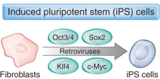
In 2006, Shinya Yamanaka lab, described a protocol to induce pluripotency in already differentiated cells by “reprogramming” them through the expression of four transcription factors, Octamer-binding transcription factor 4 (Oct4), SRY-box 2 (Sox2), Krüppel-like factor 4 (KLF4) and MYC (c-MYC), known has OSKM (Figure 1.2) 13,14.
3
Figure 1.2 - iPSC reprogramming protocol by introducing genes encoding four transcription factors15.
These cells were called induced pluripotent stem cells (iPSC) and have the same morphology, growth properties and markers of ESCs. The process to induce pluripotent stem cells from already differentiated cells, using transcription factors, is called cellular reprograming13,14.
The existence of reprogramming factors was also demonstrated for direct fate conversion of mammalian cells. A method that allow to convert directly somatic cells into different specialized cells without a pluripotent state transition16. For instance, myoblast determination protein (MYOD), alone was sufficient to transform mouse fibroblasts to myoblast. It was also demonstrated that the ectopic expression of erythroid transcription factor GATA-binding protein 1 (GATA1) could convert myeloblasts to megakaryocyte and erythrocyte precursors. The scientific term given to this process of converting somatic cells into a different somatic lineage is known as transdifferentiation8,17.
Yamanaka S. and Takahashi K. started to analyze mouse ES cells to identify genes underlying characteristics, such as pluripotency and proliferation. This investigation led to the identification of ES cell-specific genes, referred to as ES cell-associated transcripts (ECATs). After various experiments, including the generation of knockout ES cells and knockout mice, they discovered that NANOG is an ECAT being crucial for the maintenance of pluripotency in both ES cells and early embryos. It was also uncovered that the overexpression of NANOG allowed mouse ES cells to self-renew, even without the presence of leukemia inhibitory factor (LIF), which is an essential cytokine for the maintenance of mouse cell pluripotency in serum containing medium. Some others transcription factors of pluripotency were also discovered, such as KLF4, c-MYC, Sox2 and Oct4, as mentioned before8.
The transcription factors were tested in several murine and human cell lines. For this, it was initially used a retroviral transduction system, due to his high efficiency in gene delivery, developed by Toshio Kitamura at the University of Tokyo. The cells generated with the use of OSKM factors had ES cell-like properties but displayed somewhat incomplete pluripotency, that is, the characteristics of iPSCs were different to ES cells. This happened because in cellular reprogramming the expression of the four transcription factors (OSKM) should sustain for a sufficient period of time while the cell is being reprogrammed. When the cell is fully reprogrammed, the OSKM expression should be silenced in order to produce iPSCs with the same characteristics of ES cells8.
Aside of incomplete pluripotency, a crucial issue with iPSCs was the use of retroviral vectors to deliver the reprogramming factors. These vectors integrate into the genome of host cells, potentially causing disruption or aberrant activation of neighboring genes, and pose a risk of reactivation of the reprogramming factors. Indeed, the reactivation of c-MYC induces tumor formation in iPSC-derived chimeric mice. A way to solve this issue was the introduction of efficient, integration-free methods for
4 cell reprograming, such as adenoviruses, plasmids, transposons, Sendai viruses, synthetic mRNAs and recombinant proteins8.
Although the pluripotency of fully reprogrammed mouse iPSCs seems to be indistinguishable from that of mouse ES cells, it remains controversial for human iPSCS (hiPSCs), which seems to show differences in gene expression and DNA methylation patterns compared to human ES cells, demonstrating the urge for optimization of human cellular reprogramming8,18.
1.3. Cellular Aging
Cellular aging is one of most well characterized roadblock for cellular reprogramming. Aging is defined by the functional decline of cells, tissues and organs throughout life. It is also associated with a dramatic increase in a wide range of age-related diseases, including cardiovascular dysfunction, metabolic disorders, neurodegeneration and cancer19.
Cellular aging is influenced by exposure to extrinsic factors, such as inflammatory cytokines, and intrinsic factors. Old cells accumulate genomic damage, aggregated proteins and display telomere erosion and mitochondrial dysfunction. It’s also known that manipulation of some genetic pathways (e.g. insulin-FoxO, Tor, AMPK and Sirtuin) and environmental interventions can delay aging, even if initiated late in life19.
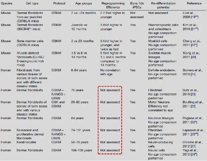
Through some initial studies that tested how age of the donor of cells affects reprogramming in mice, was suggested that cells from older donors tend to reprogram less efficiently than cells from young mice. For instance, dermal fibroblasts from old mice (>2 years) exhibited a reduction in their ability to generate colonies that stained positive for the stem cell marker alkaline phosphatase (AP) compared to fibroblasts from young adult mice (2 months old) upon expression of the four transcription factors (OSKM) by doxycycline induction. This was also observed in bone marrow cells from old mice (23 months old) that generated 5 times less AP+ colonies than cells from young adults (2 months old)19. These studies confirm an age-dependent decline in reprogramming efficiency in mice. Regarding human cells it is still unknown how the aging process affects cellular reprogramming, therefore further tests in human cells are needed to prove if the results observed in mice correlate with the human situation (Table 1.1)19.
5
Table 1.1 - Summary of the current literature on the impact of age on reprogramming19.
One of the mechanisms by which aging could be impairing the reprogramming efficiency in mice is cellular senescence and subsequent loss of division capacity characteristic of aged cells. Senescence is the gradual deterioration of the cellular functions leading to a virtually irreversible cell cycle arrest. Old mice donors contain more populations of cells with senescence or pre-senescence phenotypes thus decreasing reprogramming efficiency. Senescence is also common in human aged samples, although it’s not completely known how it affects the reprogramming efficiency of human cells19.
Reprogramming has the remarkable ability to reverse some cellular and molecular characteristics associated with aging, including cellular senescence, telomere erosion, mitochondrial dysfunction and global changes in gene expression, suggesting that many of the age-associated characteristics that were once thought to be permanent are, in fact, reversible. However the accumulation of nuclear and mitochondrial DNA damage that is associated with aging is unlikely an aspect of aging able to be reversed by reprogramming19.
6
1.4. Non-coding RNAs
The vast majority of genomic information is pervasively transcribed into not only a diverse range of protein-coding RNAs but also into long and short non-coding RNAs (ncRNAs). NcRNAs are molecules that are not translated into proteins but have a remarkable variety of biological functions, such as to regulate gene expression at the levels of transcription, RNA processing and translation, or even to protect genomes from foreign nucleic acids, guided DNA synthesis or genome rearrangement20.
Depending on their function, non-coding RNAs can be classified into the following: microRNAs, piwi-interacting RNAs, small interfering RNAs, long non-coding RNAs, enhancer RNAs and promoter-associated RNAs20.
Its function could be mediated through the formation of RNA-protein complexes21. This RNA-protein complexes occurs when a RNA-protein binds to a RNA playing a major role in RNA transcription, RNA processing or protein synthesis20.
1.4.1. Long non-coding RNAs
Long Non-coding RNAs (lncRNAs) are transcripts with more than 200 nucleotides that don’t encode proteins but have an important role in gene expression regulation and protein synthesis, such as, chromatin modification and structure, direct transcriptional regulation, regulation of RNA processing events (splicing, editing, localization, translation and turnover/degradation), facilitation of ribonucleoprotein (RNP) complex formation, gene silencing through production of endogenous small interfering RNAs and regulation of genomic imprinting. lncRNA can also be characterized as one or more of the following models of action: signal, decoy, guide, scaffold and enhancer. They are also usually transcribed by RNA polymerase II and frequently spliced and polyadenylated as other messenger RNAs (mRNAs). Unlike mRNAs, which is abundant and enriched in the cytoplasm, the lncRNAs tends to be expressed at lower levels and are predominantly localized in the nucleus22.
Several studies of lncRNAs suggested that they can operate through distinct manners, including as signals, scaffolds for protein-protein interactions, molecular decoys, antisense interference and guides to target elements in the genome or transcriptome. They are also involved in phenomena such as imprinting genomic loci, shaping chromosome conformation and allosterically regulating enzymatic activity, but the majority of lncRNAs functions are still unknown. Previous studies also suggested that lncRNAs expression is more cell-type-specific comparing to mRNA expression, meaning that lncRNAs may be involved in cell fate22.
The ability to regulate gene expression suggests that lncRNAs have influence in the reprogramming of the cells, becoming a viable and useful way to improve cellular reprogramming23.
An example of an lncRNA candidate with potential role in reprogramming has been described in 2015, by Mao Z. and his team who identified in human fibroblasts 986 down-regulated lncRNAs and 899 up-regulated lncRNAs in senescence cells compared with young cells. Among the lncRNAs, they characterized a senescence-associated lncRNAs, called SALNR that has low expression in senescent cells. When SALNR was overexpressed, cellular senescence was delayed. SALNR interacts with
7 NF90 (nuclear factor of activated T-cells) which suppress the biogenesis of senescence-associated miRNAs, such as miR-22 and miR181a. The NF90 inhibition results in premature senescence24.
When human fibroblasts were exposed to Ras-induced (oncogene Ras) stress to activate senescence, NF90 was translocated to nucleolus and couldn’t no longer suppress senescence-associated miRNA biogenesis. This was rescued by SALNR overexpression, which antagonized NF90 translocation into nucleolus and rescued its inhibitory activity on senescence-associated miRNA expression. These results suggests that lncRNA SALNR controls cellular senescence through a differential localization of NF9024.
To understand if lncRNAs could have a role in iPSC or cellular reprogramming, Rinn J. and his colleagues did, in 2010, a comparison between transcriptional profiles of human lncRNAs and protein-coding genes through iPSCs and hESCs derived from human fibroblasts. Gene expression profile analysis revealed that all iPSCs were similar to ESCs25.
Then, they designed a microarray probing ~900 lncRNAs in the human genome to analyze the expression of lncRNAs in the above cell lines. Through the results they identified 133 lncRNAs that were induced and 104 lncRNAs that were repressed across all iPSCs and ESCs compared with fibroblasts. They identified 28 lncRNAs that had a higher expression in fibroblasts iPSCs relative to ESCs (were referred to as “enriched” lncRNAs). With these results they hypothesized that iPSC-enriched lncRNAs have an important role in reprogramming. To test this hypothesis, they profile lncRNA expression in CD34+ hematopoietic stem and progenitor cells, two CD34+ iPSC lines and ESCs. The results correlate with their previous observations, showing that ten of the twenty-eight lncRNAs elevated in fibroblast iPSCs were also elevated in CD34+ iPSCs. Through RT-PCR, eight out of ten iPSC-enriched lncRNAs levels were independently validated and detected considerable variation in expression. All these results uphold the hypothesis that lncRNAs are tightly bound with the pluripotent state25.
The importance of iPSC-enriched lncRNAs for iPSC derivation suggests a link between lncRNAs and the pluripotency network. They observed that lncRNAs appears to be controlled by pluripotency transcription factors in ESCs and iPSCs. In particular, after the knockdown of a lncRNA candidate (lincRNA regulator of reprogramming - lncRNA-RoR) the iPSC colonies decreased relatively to the control whereas progenitor cells were unaffected, showing that lncRNA-RoR is required for iPSC derivation25.
Additionally, the authors investigated which cellular pathways were affected by lncRNA-RoR knockdown. The results suggested that the absence of lncRNA-RoR would led to a upregulation of genes involved in the p53 response, the response to oxidative stress and DNA damage-inducing agents, as well as cell death pathways. A knockdown of p53 partially rescued the apoptotic phenotype caused by ablation of lncRNA-RoR. These results led to the conclusion that lncRNA-RoR play an important role in promoting survival in iPSCs and ESCs, preventing the activation of cellular stress pathways including the p53 response25.
8 1.4.1.1. ZEB2 and his natural antisense transcript lncRNA ZEB2NAT
An lncRNA that is pursued in our current laboratory is Zeb2NAT, an antisense of the Zeb2 coding gene.
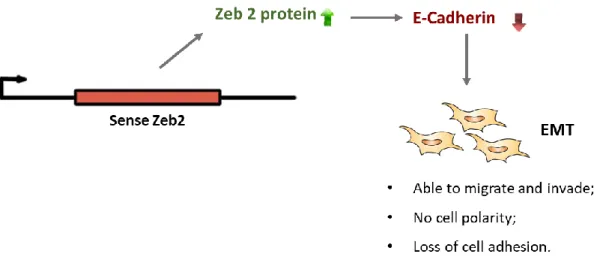
The change of cells morphology is one of the first signs to be observed during cellular reprogramming. Epithelial-mesenchymal transition (EMT) is a cellular process that occurs in early development and cancer through down-regulation of epithelial genes, such as E-cadherin, and up-regulation of mesenchymal genes, such as Snail1 (zinc finger transcription factors homologue of Snail) 26. During EMT epithelial cells lose their cell-cell adhesion and cell polarity and acquire invasive and migratory properties becoming mesenchymal cells. The mesenchymal-epithelial transition (MET), the reverse process of EMT, is also possible being crucial in reprogramming of fibroblasts to iPSCs. The reprogramming process is controlled by the balance between EMT and MET 27,28.
The expression of Zeb2 and Zeb1 transcription factor detected in mesenchymal cells is induced by Snail1 during EMT. The expression of Zeb2 assists the down-regulation of E-cadherin. Although Zeb2 mRNA levels remain stable, it is alternatively processed after Snail1 induction29. It’s also known that in MET the expression of Zeb2 mRNAs is repressed by miR-200 family members30.
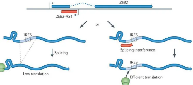
When Snail1 is induced in cells, an intron located in the 5’ -UTR of Zeb2 stop being processed. This intro contains an internal ribosome entry site (IRES) sequence crucial for the Zeb2 functional expression29.
The conservation of this intron is controlled by the expression of a noncoding transcript that occurs in an antisense direction (Zeb2NAT) overlapping the 5’ -UTR of Zeb2. Overexpression of Zeb2NAT in epithelial cells changes cell identity through increased Zeb2 protein levels (Figure 1.3)29.
Figure 1.3 - Illustration of the regulation of Zeb2 through lncRNA Zeb2NAT. LncRNA Zeb2NAT is the antisense of the sense Zeb2 and has an important role in incrising the Zeb2 proteins levels by binding to the pre-Zeb2 mRNA, preventing the splicing of the intro that contain the IRES (Internal Ribosome Entry Site), that is essential for Zeb2 mRNA translation. The increase of Zeb2levels promotes the inhibition of E-Cadherin leading cells to an EMT29.
9 Zeb2NAT increases the Zeb2 proteins levels by binding to the pre-Zeb2 mRNA, preventing the splicing of the intron that contains the IRES that is essential for Zeb2 mRNA translation. Zeb2 promotes the inhibition of E-Cadherin leading cells to an EMT (Figure 1.4). The knockdown of Zeb2NAT can lead the cells to a MET through the E-Cadherin expression that is increased with the decreasing of Zeb2 protein30.
Figure 1.4 - Representation of the lncRNA zeb2 regulation of the epithelial-mesenchymal transition of cells through the E-Cadherin inhibition.
The inhibition of protein translation, such as Zeb2, can be performed by using a locked nucleic acid (LNA), that prevent mRNA-protein interactions31. Locked nucleic acids (LNA) are a class of RNA analogs in which the ribose ring is “locked” into a rigid bicyclic formation by a methylene bridge between 2’ oxygen and the 4’ carbon, conferring an enhanced performance and an increased breadth of applications32.
LNAs can be used to overcome the difficulties of studying very short sequences and has greatly improved, and in many cases enabled, specific analysis and sensitive detection of DNA, micro RNA, ncRNA and other small RNA molecules. LNAs can be used to perform in situ hybridization, northern blotting, microarray analysis, real-time PCR, isolation, RNAi, gene repair/exon skipping, splice variant detection, antisense inhibition and more32.
Due to the high affinity of LNA to complementary nucleic acids, LNAs: RNA duplexes are much more stable that those formed between DNA and RNA, makes LNAs as an extremely potent antisense inhibitor, both for in vitro and in vivo use32. This means that LNAs are one of the best options to perform the downregulation of lncRNAs32.
1.4.1.2. LncRNA NORAD
Recently, Mendell and colleagues uncovered a novel lncRNA regulated by DNA damage, termed non-coding RNA activated by DNA damage (NORAD). NORAD plays a key role in maintaining genome integrity by modulating the activity of the RNA binding proteins PUM2 and PUM1 after DNA damage33,34. Mendell group was initially interested in identifying human lncRNAs that regulate the DNA damage response. To do so, they examined a set of previously identified mouse lncRNAs that are induced after doxorubicin treatment in a p53-dependent manner. Among all transcripts, they observed a poorly characterized 4.9kilobase (kb) unspliced murine lncRNA, annotated as
10 2900097C17Rik, that exhibits a high degree of evolutionary conservation in mammals and an unusually high abundance (500-1000 copies per cell). Human genome has a clear ortholog of this transcript, with 65% nucleotide identity to 2900097C17Rik, annotated in RefSeq as LINC00657 with 5.3kb. LINC00657 is a cytoplasmic lncRNA abundantly expressed in human cells and tissues34.
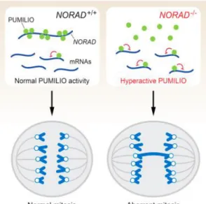
They observed that the presence of NORAD in cells is fundamental to maintain genomic stability by sequestering PUMILIO proteins. Otherwise, the absence of NORAD would trigger chromosomal instability by repressing mitotic, DNA repair and DNA replication factors through the release of PUMILIO proteins34.
NORAD affects genomic stability through its direct interaction with PUMILIO 2 (PUM2) and possibly PUMILIO 1 (PUM1), two human and mouse RNA-binding proteins belonging to the deeply conserved Pumilio-Fem3-binding factor (PUF) family. PUF proteins and more specifically PUM1 and PUM2 are capable of binding with high specificity to sequences in the 3’ UTRs of target mRNAs through an 8-nt specific sequence (UGUANAUA), referred to as the PUMILIO response element (PRE), causing genomic instability34(Figure 1
.5
).Figure 1.5 - LncRNA NORAD regulates genomic stability by sequestering PUMILIO proteins34
The presence of 15 PUMILIO-binding motifs, mostly located in five repeated 400-nt domains named NORAD Domains (ND1 through ND5), allows lncRNA NORAD to act as a potent molecular decoy for PUMILIO proteins (PUM1/PUM2), preventing the RNA-binding proteins from destabilizing their targets. In fact, NORAD is capable to bind approximately 7500-15000 PUMILIO molecules per cell, which represents 50%-100% of the total PUMILIO protein pool in the human colon cell line (HCT116)34.
Recent findings from Mendell group revealed a new pathway to marked chromosomal instability (CIN), a phenotype characterized by the frequent gain or loss of chromosomes during mitosis. Mendel and his lab findings support what they previously suggest earlier, that PUMILIO are an important regulator of genomic stability acting as repressors of a set of genes whose expression is necessary to maintain chromosomal stability, such as DNA replication and repair as well as key mitotic factors genes. They hypothesized that the deregulation of the targets under conditions of PUMILIO hype- or hypoactivity could induce a state of severe genomic instability as observed upon NORAD loss function, PUM1/2 overexpression or PUM1/2 inactivation34.
11 Another study, now from Pera group, identified an intriguing expression of PUM2 protein in embryonic stem cells. In fact, they observed that PUM2 can form a stable complex with DAZ protein and its homolog, DAZL (DAZ-Like) protein in diverse organism, including humans. DAZ/DAZL are proteins necessary for germ cell development in males and females (DAZ protein is only expressed in males)35.
They observed that the capacity of PUM2 protein to form a stable complex with DZA through RNA binding and protein-protein interaction makes PUM2 a requirement to maintain germ line stem cells in Drosophila and Caenorhabditis elegans. When the PUF repeat 8 of PUM2 was deleted, this interaction between PUM2 and DAZ decreased suggesting a link between both proteins, supporting the first observation of PUM2 importance in germ line maintenance. They also observed an interchange between DAZ and DAZL homologs when a human DAZ transgene was able to partially rescue a mouse DAZ mutation35.
This findings indicates that PUM2 function can pass by maintaining the human germ cell lineage giving rise to mature germ cells in men and women as PUM2 is expressed throughout the development of the female and male germ cell lines. They hypothesized that PUM2 together with DAZ and DAZL proteins can act as a translational regulator in the germ cell lineage35.
However, the PUM2 role in the germ cells remains unclear suggesting the need for a further investigation to uncover the PUMILIO proteins functions and how are regulated in stem cells34,35.
1.5. Objectives
The main objective of this project passed by understanding the cellular reprogramming of human aged cells. Previously results from our laboratory reported a decreased reprogramming efficiency when reprogramming adult somatic cells from mice. Here, we recently characterized an lncRNA capable of enhancing reprogramming of aged cells. When this lncRNA, ZEB2NAT, was down-regulated with LNAs we could achieve higher reprogramming efficiencies. Unfortunately, how aging influences the reprogramming of human cells and how Zeb2NAT works on human cell reprogramming is still unknown.
Actually, an objective of this project passed by testing if aging is a reprogramming barrier in human cells and if it’s possible to enhance reprogramming by downregulating Zeb2 and/or his antisense transcript ZEB2NAT.
Furthermore, was also intended to understand how a novel lncRNA, called NORAD, could affect stem cells properties, viability and also chromosomal stability since previous results have already shown that PUM2 have an important role in maintain germ cell lineage. Therefore, a last objective of this project was also to verify if there NORAD can regulate the stem cell stability and improve stem cells proprieties through PUM2 protein regulation.
13
2. Materials and Methods
2.1. Cell lines
Human Fibroblasts
The reprogramming of fibroblasts into hiPSCs was performed using two human fibroblasts cell lines, the human embryonic fibroblasts (WI38) and the 3yr human fibroblasts (3yr, supplied by ATCC), both with low passage (passage 2 and 4) and high passage (passage 7).
Human fibroblasts were maintained in Dulbecco’s Modified Eagle Medium (DMEM, GibcoTM) supplemented with 10% of Fetal Bovine Serum (FBS), 1% of L-Glutamina and 1% of Penicillin Streptomycin (Pen/Strep) under sterile conditions and incubated at 37°C and 5% CO2.
Fibroblasts were passed when reached a 90/95% of confluence by washing with Phosphate Buffered Saline (PBS) and detached with trypsin. Posteriorly, cells were resuspended in DMEM medium to inhibit trypsin and plated. When needed, fibroblasts were freeze by centrifuging the cells resuspended in DMEM medium at 1250 rpm during 5 minutes at room temperature (RT). The supernatant was removed and the pellet was resuspended in 1ml of 90% FBS + 10% DMSO (Sigma) and added to a cryogenic vial. Cells were stored at -80°C during 2 or more days and then stored at liquid nitrogen. The concentration of cells used per vial was between 1×10⁶ and 1×10⁷.
To perform a cellular reprogramming assay, human fibroblasts were counted during their passaging by mixing 20μl of a trypan blue solution with 20μl of cells resuspended in DMEM medium, before plating. The solution was homogenized and loaded into a Neubauer Chamber. As the ratio between cells and trypan blue used was 1:1, the formula to calculate cell concentration used was 𝑚𝑒𝑎𝑛 × 2 × 10⁴, were the mean is calculated by the sum of the farthest quadrants of the
extremities and divided by 4.
If needed to thaw fibroblasts to perform an assay, the frozen cryogenic vial would be withdrawn from the liquid nitrogen and thawed at 37°C for 3 minutes at maximum. All the volume of cryogenic vial was added to a falcon with PBS and resuspended. A pellet of cells was formed by centrifuging the falcon at 1000-1250 rpm for 5 minutes. Afterwards the supernatant was aspirated and the pellet was resuspended in DMEM medium and plated.
Human embryonic kidney 293T cell line (293T)
The 293T cell line (ATCC) was used to produce the viral medium to perform cellular reprogramming experiment. 293T cell line were maintained in DMEM supplemented with 10% of FBS, 1% of L-Glutamina and 1% of Penicillin Streptomycin under sterile conditions and incubated at 37°C and 5% CO2.
293T cells were maintained, passed, freeze and thawed by using the exact procedure as used for human fibroblasts. However, pre-coated culture petri dishes with gelatin were needed for 293T survival. Coating culture petri dishes was performed by adding gelatin 0.1% and incubated at 37°C during 10-15 minutes. After removing the gelatin, fresh DMEM cell medium was added along with 293T.
14
Human induced pluripotent stem cells (hiPSCs)
The human iPSCs obtained through cellular reprogramming of the human fibroblasts mentioned above were grown in mTeSRTM1 medium (GibcoTM) containing 90% of mTeSRTM1 Basal medium, 9,5% of mTeSRTM1 5X supplement and 0.5% of Pen/Strep, under sterile conditions and incubated at 37°C and 5% CO2. To maintain hiPSCs in culture, the medium was changed every day and hiPSCs were plated in pre-coated culture petri dishes with MatrigelTM (Corning).
Before hiPSCs clones started to touch each other, hiPSCs were passed by using one of the two protocols: hiPSCs passage with EDTA and hiPSCS passage with trypsin.
In the hiPSCs passage with EDTA protocol, cells were washed with PBS first. PBS-EDTA was added after removing PBS and incubated at RT for 3 minutes. Cells were rinsed and scraped from bottom, with care to avoid to getting single cell suspension. Cells were transferred to a 15ml falcon tube with additional medium according to the dilution wanted and resuspended carefully. Cells were plated in pre-coated culture petri dishes with MatrigelTM. If it were necessary to improve the recovery and growth of hiPSCs due to a single suspension a 1μl of ROCK inhibitor (Axon) per 1ml of mTeSRTM1 medium would be added. Rock inhibitor or selective Rho-associated kinase inhibitor increase the dissociated human stem cells survival by inhibiting apoptosis 36.
In the hiPSCs passage with Trypsin protocol, cells were washed with PBS. After removing PBS, cells were incubated with trypsin for 2-3 minutes at 37°C. Afterwards a few drops of FBS serum were added to inhibit trypsin effect. Cells were resuspended and centrifuged at 1250 rpm for 5 minutes. Supernatant was removed and the pellet resuspended in mTeSRTM1 medium. Afterwards hiPSCs were plated in a plate previously coated with MatrigelTM. To improve the recovery and growth of hiPSCs due to single cell suspension a 1μl of ROCK inhibitor per 1ml of mTeSRTM1 medium would be added. Culture petri dishes were previously pre-coated with MatrigelTM by thawing an aliquot of MatrigelTM on ice. In order to dilute MatrigelTM aliquot, DMEM-F12 medium (GibcoTM) was added. The diluted MatrigelTM was added to culture petri dishes and incubated at 37°C for 20-30 minutes. MatrigelTM was then aspirated and mTeSRTM1 was added.
When needed to freeze the hiPSCs resuspension from hiPSCs passage was centrifuged at 1250 rpm for 5 minutes. The supernatant was removed and the pellet resuspended in 90% KnockOutTM Serum (KSR) + 10% DMSO or 90% FBS (stem cell ready) + 10% DMSO solution. Cells were stored at -80°C during 2 or more days and then stored at liquid nitrogen.
The PBS-EDTA (0.5mM) used was made by diluting 200μl of 0.5 M EDTA with 200ml of PBS and the osmolarity was adjusted by adding 0.36g of NaCl, and sterilized using a filter-sterile. The freezing medium used contained KSR with 10% of DMSO.
Mouse stem cells E14
To study NORAD role in stem cells a mouse stem cell E14 cell line (a kind gift from Dr. Manuel Serrano, CNIO, Madrid) was used. E14 was maintained, under sterile conditions and incubated at 37°C and 5% CO2, in KnockOutTM DMEM medium with Leukemia inhibitor factor (KSR+LIF). KSR medium was made by supplementing basal DMEM medium with 15% of KSR (GibcoTM), 1% of L-Glutamina, 1% of Pen/Strep, 1.2% of non-essential amino acids solution (NEAA) and 0.18% of
Beta-15 mercaptoethanol. For an aliquot of 50ml of KnockOutTM DMEM medium was also added 0.012% of leukemia inhibitor factor supplement (LIF, Millipore) (KSR+LIF).
E14 were passed by washing with PBS. PBS was removed and the cells were trypsinized by adding a few drops of trypsin and incubated for 3-5 minutes at 37°C. Posteriorly, cells were resuspended in PBS or FBS (stem cell ready) and centrifuged at 1250 rpm during 5 minutes. Supernatant was removed and the pellet was resuspended in KSR+LIF medium and plated. Culture petri dishes were pre-coated with gelatin using the same coating gelatin protocol for 293T. When needed, the E14 were frozen by centrifuging at 1250 rpm during 5 minutes the E14 resuspension in PBS or FBS (stem cell ready). The supernatant was removed and the pellet resuspended in 90% KSR + 10% DMSO or 90% FBS (stem cell ready) + 10% DMSO solution. Cells were stored at -80°C during 2 or more days and then stored at liquid nitrogen.
To thaw E14, the frozen cryogenic vial would be withdrawn from the liquid nitrogen and thawed at RT. All the volume of cryogenic vial was added to a falcon with PBS and resuspended. Afterwards the supernatant was aspirated and the pellet was resuspended in KSR+LIF medium and plated in pre-coated plates with gelatin.
2.2. Cellular Reprogramming
293T transfection (viral medium production)
Cellular reprograming of human fibroblasts was performed based on the protocol of Li group 37. 293T cells were seeded with a confluence of 2×10⁶ on a 100 mm culture dish previously coated with gelatin and incubated 24 hours at 37°C. After the 24 hours of incubation two mixes were made:
- A containing lentiviral transfer vector and second generation packaging vectors; - B with 600μl of DMEM and 50μl of X-tremeGENETM 9 (Roche).
Both mixes were mixed together and incubated at RT for 20 minutes after a gently vortex. Transfection mixture was added dropwise to cells and incubated overnight at 37°C. 24 hours after, DMEM medium was replaced by new DMEM medium. Viral medium was collected 36h - 48 hours after transfection for first infection and new fresh medium was added to 293T. After 24 hours a second round of viral medium was collected to perform the second infection.
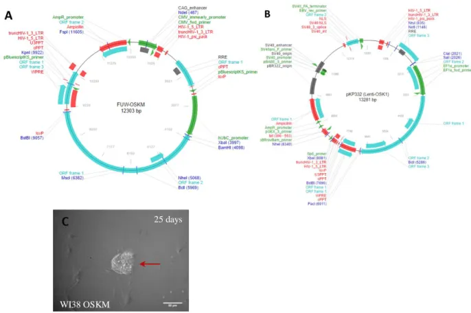
For viral medium production was used 10μg of transfer vector. FUW-OSKM plasmid (Addgene #20328), pkp332 plasmid (Addgene #21627) and a positive infection control, the green fluorescence protein plasmid (provided by Edgar Gomes lab of IMM). For the packaging vectors was used a second generation packaging system composed by 5μg of both pMD2.G (Addgene #12259) and psPAX2 (Addgene #12260) plasmids. All plasmids were produced using STBL3 competent bacteria (Thermo Fischer Scientific), following supplier protocol.
Fibroblasts Infection (transduction of transcription factors)
The target cells were plated at least 24 hours prior to viral infection with approximately 90% of confluence. Viral medium collected was filtrated by passing through a 0.45μM low protein-binding