Vol.50, n. 1 : pp.39-44, January 2007
ISSN 1516-8913 Printed in Brazil BRAZILIAN ARCHIVES OF
BIOLOGY AND TECHNOLOGY
A N I N T E R N A T I O N A L J O U R N A L
Development of an Enzyme Immunoassay for Poliovirus
Antigens
Newton Hashimoto, Fabrício José Benati, Flávio Lauretti, Rosa Elisa Carvalho Linhares and Carlos Mitihiko Nozawa*
Departamento de Microbiologia; CCB; Universidade Estadual de Londrina; C. P. 6001; 86051-970; Londrina - PR - Brasil
ABSTRACT
An indirect solid-phase enzyme immunoassay (EIA) was developed for the detection of poliovirus antigen. Virus antigen was obtained in LLC-MK2 cell cultures and used to prepare antibodies in rabbit and guinea pig. Antibodies were evaluated by double immunodiffusion and neutralization test. Optimal concentrations of guinea pig and rabbit immunoglobulins were determined by checkerboard titration. Microtitre plates were coated with 15.0 µg/ml guinea pig anti-polio immunoglobulin and rabbit anti-polio immunoglobulin at the concentration of 7.94 µg/ml was used as detecting antibody. The standard curve with eight different antigen concentrations in eight replicates resulted in a coefficient of variation (CV) between 2.1% to 7.8%. The dose-response relationship was determined by simple linear regression with a coefficient of correlation (R2) equal to 96.4%. The assay detected a minimum of 2.3 µg/ml poliovirus antigen.
Key words: Enzyme immunoassay, Poliovirus antigen
*
Author for correspondence
INTRODUCTION
The approaches for laboratory diagnosis of virus infection have been the isolation of the agent and/or detection of viral antigen, viral genome and serology (Lennette, 1995; Engleberg, 1999). The best methods should fulfill the five main prerequisites: speed, simplicity, sensitivity, specificity and low cost. Tests for viral antigen, which provide a diagnosis from a single specimen, are rather important for many purposes. Solid-phase enzyme immunoassay (EIA) is one of the methods of choice. Poliovirus, a member of the family Picornaviridae, genus Enterovirus, was responsible for the legacy of paralysis and deformity, since the fifties. The virus still poses a threat as the cause of poliomyelitis mainly in some underdeveloped countries (Pallansch and Roos,
2001). The identification of poliovirus relies mainly on the serum neutralization test (Nt) (Payment et al., 1982; Wahby, 2000). Because Nt test depends on living host system, it is costly, time consuming and the interpretation of neutralizing titers is influenced by subjective visual scoring of cytophatic effects (CPE). More rapid identification has been attempted using negative staining electron-microscopy, immunofluorescence or immunoelectrophoresis. These techniques are not always applicable to screening of large number of samples.
relatively short time with maximum of sensitive and minimum technical expertise and equipment (van Weemen, 1985; Pillot, 1996).
This work describes the development of an EIA for the detection of poliovirus antigen. The assay is useful for monitoring virus circulation in the environment, such as, in sewage treatment plant, water resources, poliomyelitis vaccine campaign and in the evaluation of anti-poliovirus compounds.
MATERIALS AND METHODS
Virus and cell culture
Poliovirus type 1 (ATCC, 192-VR) was propagated in LLC-MK2 (monkey kidney) cell
cultures cultivated in Dulbecco’s Modified Eagle’s Medium (DMEM) (Sigma Chem. Co., USA.) supplemented with 7% fetal bovine serum (Gibco BRL, USA), 100 µg/ml streptomycin, 100 UI/ml penicillin (Sigma Chem. Co., USA) and 2.5 µg/ml fungizon (Bristol-Myers Squibb, Brazil).
Antigen
The supernatant fluid of virus-infected cell cultures was harvested when cultures showed maximal CPE, 24-48 hours post-infection. Virus titer was determined as 0.5 x 10⁶ TCID50/ml.
Briefly, the supernatant fluid of infected cell cultures was clarified by centrifugation at 10,000 x g for 30 min. Partial concentration of virus was carried out by the addition of polyethylene glycol 6000 (8%, w/v), at 4oC overnight under agitation, and followed by centrifugation at 10,000 x g for 30 min at 4oC. The resulting precipitate was resuspended in phosphate-buffered saline (PBS), pH 7.4, and 1.0-2.0 ml layered on a cushion of 20% sucrose in PBS, and ultracentrifuged at 100,000 x g for 3 h. The pellet was resuspended, protein content was determined, and the material was stored at -20ºC.
Immunization
Rabbits and guinea pigs were inoculated five times with 0.2 ml purified viral antigen emulsified with Freund’s complete adjuvant, at a week interval. The animals were bled by cardiac puncture seven days after the final booster. Following inactivation at 56ºC/30 min, sera were adsorbed with 105 cells/ml (LLC-MK2), and immunoglobulin was
precipitated with ammonium sulfate. Protein contents were determined accordingly.
Antibody titers
Double immunodiffusion in agar gel was used for screening hyperimmune sera. Neutralization test was carried out as follows. Test sera were prepared in two fold dilution (from 1:2 on) in cell culture maintenance medium. Thereafter, 100 µl of each dilution were pooled with an equal volume of 100 TCID50/mlof poliovirus and incubated at 37oC for
1h. This was followed by inoculation onto cell monolayers grown in 96-well tissue culture plates and incubation at 37ºC for 5 days in 5% CO2
atmosphere. The antibody end point titer was determined as the highest dilution that inhibited CPE.
Checkerboard titration
Optimal concentrations of guinea pig and rabbit immunoglobulins were determined by checkerboard titration on microtiter plates. Briefly, polystyrene microtiter plates (Hemobag, Brazil) were coated with 100 µl per well of guinea pig antibodies containing 1.5 µg/ml (half plate) and 15.0 µg/ml (half plate) protein in 0.1 M carbonate-bicarbonate buffer, pH 9.6, overnight, at 4oC, in a humidified chamber. Plates were washed twice with PBS, pH 7.2, containing 0.05% Tween 20 (PBS-T). The coated plates were blocked with 200 µl/well of blocking solution (PBS, pH 7.2, containing 2% skimmed milk powder and 0.05% Tween 20) for 90 min at 37oC. Plates were washed with PBS-T throughout. Antigen at the concentration of 0.03, 3.0 and 30 µg/ml, in replica, was used as positive control at a volume of 100 µl/well. Blank was performed with PBS instead. Incubation was followed at 37oC for 60 min. Plates were washed with PBS-T. Aliquots of 100 µl of rabbit antibody, diluted in a serial dilution from 127.0 µg/ml to 2.0 µg/ml, in blocking solution were added and the plates were incubated at 37oC for 60 min. Incubation was followed by washing and the addition of 100 µl of goat anti-rabbit IgG peroxidase-conjugate (Sigma Chem. Co., USA) at 1:40,000 in blocking solution for 60 min at 37oC. Plates were washed and followed by the addition of 100 µl of substrate (0.03% H2O2 and 400 µg/ml
o-phenylenediamine dihydrochloride) (Sigma Chem. Co., USA) diluted in 0.05 M citrate-phosphate buffer, pH 5.0. After 15 min at 25oC in light tight box, the reaction was stopped by the addition of 50 µl 2 N H2SO4 and the optic density
Antigen standard curve for dose response
The standard curve was generated from decreasing antigen concentration (300.0 to 2.3 µg/ml) in eight replicas by previous protocol with immunoglobulin concentrations optimized by checkerboard titration (15.0 µg/ml for capture antibody and 7.94 µg/ml for detector antibody).
Statistical analysis
The standard curve was used to estimate the dose-response relationship and the variation of the analyte (antigen) concentration estimated by the coefficient of variation (n=8). The dose-response was determined by linear regression. The analysis was based on the number of data points and the correlation coefficient (R2). Normally, at least four data points were included and a R2 equal or larger than 95%.
Clinical specimens
Five fecal specimens from children between one to two years of age were collected in June and July 2003 after the poliomyelitis vaccination campaign, maintained in –20oC, were supplied by the Laboratório LAPAC (Ponta Grossa- PR). Four replicates of each sample diluted at 1:10 were tested by the previous optimized condition.
Cut-off
The cut-off value was established according to Porstmann and Kiessig (1992).
RESULTS
Optimal concentrations of capture and detector immunoglobulins were determined by checkerboard titration against the antigen used for immunization. The initial analysis indicated high background due to an excessive non-specific binding (NSB) hampering distinction of a positive signal derived from the analyte against the blank values. NSB was minimized by increasing blocking agent concentration from 1 to 2%. The concentration of guinea pig antibody (capture) of 15.0 µg/ml demonstrated a better result than 1.5 µg/ml. The slope of the curve indicated that
suitable results would be achieved by the use of 15.0 µg/ml of capture antibody and 7.94 µg/ml of rabbit antibody (detector) (Fig.1).
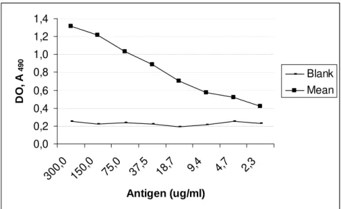
By the standard curve, the precision and the quantitative aspect of the assay could be evaluated (Fig.2).
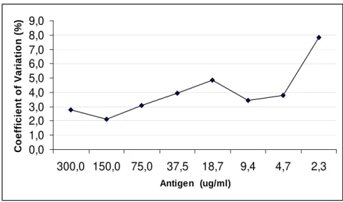
The precision, also referred to as reproducibility, a measure of variation between repeated determination of the same sample, was determined by the CV. Different concentrations of antigen were used to evaluate the performance of the method in the same day in eight replicas and the CV varied from 2.1 to 7.8 % (Fig.3). When evaluating the dose-response by standard curve for decreasing concentration of reference antigen, a sigmoid curve was obtained. Simple linear regression was used to determine if the assay showed a statistically significant dose-response relationship.
Antigen concentrations chosen empirically was analyzed to reach the best correlation (R2) between OD and antigen concentration.
The linear interval occurred between 2.3 and 37.5 µg/ml and resulted in a dose-response relationship represented by OD = 0.0124 x [Ag] + 0.4396, (Ag is the antigen concentration), with the coefficient of correlation, R2, equal to 96.4%. In optimized condition, assay detected cell antigen only at levels higher than 730 µg/ml indicating a low level of cross-reaction with cell antigen. The assay detected a minimum of 2.3 µg/ml polio antigen. The clinical samples resulted in OD of 0.146, 0.164, 0.201, 0.227, and 0.356, while the cut-off value resulted in an OD of 0.141.
DISCUSSION AND CONCLUSION
0,0 0,2 0,4 0,6 0,8 1,0 1,2
0,03 3 30
Antigen (ug/ml)
D
O
,
A
4
9
0
127,0 63,5
31,8 7,9 4,0
2,0
Figure 1 - Checkerboard titration for rabbit antibodies (Ab) tested with blank, 3.0, and 30.0 µg/ml of poliovirus antigen, on plates coated with 15.0 µg/ml of guinea pig detector antibodies.
0,0 0,2 0,4 0,6 0,8 1,0 1,2 1,4
300,
0
150,
0
75,0 37,5 18,7 9,4 4,7 2,3
Antigen (ug/ml)
D
O
,
A
4
9
0
Blank Mean
Figure 2 - Standard curve of poliovirus antigen. Arithmetic mean of eight replicates. Rabbit Ab
0,0 1,0 2,0 3,0 4,0 5,0 6,0 7,0 8,0 9,0
300,0 150,0 75,0 37,5 18,7 9,4 4,7 2,3
Antigen (ug/ml)
C
o
e
ff
ic
ie
n
t
o
f
V
a
ri
a
ti
o
n
(
%
)
Figure 3 - Coefficient of variation for different concentrations of poliovirus antigen (n=8).
Serological methods for poliovirus antigens detection evolved from complement fixation test to gel diffusion based on the detection of soluble and particulate (virus particles) antigens with low sensitivity.
The adaptation of EIA using poli- or monoclonal antibodies increased sensitivity and specificity for testing, particularly, the potency of inactivated poliovirus vaccine (Singer et al., 1989). Ukkonen et al. (1986) developed an EIA for the detection of poliovirus antigens in feces with a sensitivity between 2 to 5 ng/ml. However, due to small amount of poliovirus in feces the authors concluded that the assay was not suitable. In poliovirus natural infection it was determined that the titer of the virus shed in stool could vary from 102-107 TCID50/ml (Krugman et al., 1961). Most
of EIA detect antigen at level of 1 ng/ml (Deshpande, 1996). By deduction, this concentration was equivalent to 108 virus particles/ml (Ukkonen et al., 1986). Studies demonstrated that human feces rarely contain poliovirus at high titer (Payment et al., 1982; Ukkonen et al., 1986). This assay detected a minimum of 2.3 µg/ml polio antigen, considering that particulate and soluble antigens were used to obtain antisera. Both capture and detector antisera demonstrated neutralizing antibodies titers of approximately 1:5,120 (data not shown). The performance of present assay demonstrated results reasonable by the analysis of the CV, varying from 2.1 to 7.8 %. The estimated variation should be below 15% to be recommendable, whereas, 30%
would be the maximum acceptable value for repeatability of a diagnostic test (Porstmann and Kiessig, 1992; Deshpande, 1996; Gubbels et al., 2000). Using a better purified antigen, either particulate only or particulate and soluble antigens probably would increase the sensitivity of this test. The low sensitivity of the test associated with low titer of poliovirus in vaccinees clinical specimens could be the cause of OD values close to that of the cut-off for two specimens. Polioviruses grown in suitable cell cultures, even at low passage, render high titers, such as 1014 TCID50/ml in
HEp-2 cell cultures (human larynx carcinoma cells) (personal communication). Based on this property, the use of this test for monitoring poliovirus adapted in cell culture could be useful, such as, in antiviral studies, and water source contamination where virus concentration was required. In conclusion, the proposed test could have a potential use for laboratorial studies where cell culture adapted poliovirus was used or virus particles were accordingly concentrated.
ACKNOWLEDGEMENTS
RESUMO
O trabalho apresenta o desenvolvimento de um ensaio imunoenzimático indireto para a detecção de antígeno de poliovírus. O antígeno viral foi obtido em cultura de células LLC-MK2 e usado
para imunização de coelho e cobaia. Os soros hiperimunes foram avaliados por imunodifusão dupla e teste de neutralização. Após padronização, o soro de captura, produzido em cobaia, foi usado na concentração protéica de 15.0 µg/ml para sensibilizar microplacas de poliestireno e o soro de coelho (detector) foi usado na concentração de 7.94 µg/ml. A curva padrão resultante da utilização de oito diferentes concentrações do antígeno padrão definiu um coeficiente de variação de 2.1% a 7.8%. A relação dose-resposta foi determinada por regressão linear simples com o estabelecimento do coeficiente de correlação (R2) igual a 96.4%. O ensaio possibilitou a detecção mínima de 2.3 µg/ml de antígeno de poliovírus.
REFERENCES
Deshpande, S. S. (1996), Immunodiagnostics: a historical perspective. In: Enzyme Immunoassays: From Concept To Product Development. Chapman and Hall, New York, pp. 8-12.
Engleberg, N. C. (1999), Diagnostic principles. In:
Microbial Disease, eds. M. Schaechter; N.C. Engleberg; B.I. Eisenstein and G. Medoff, 3rd edition, Lippincott Williams and Wilkins Co., Philadelphia, pp. 503-512.
Gleaves, C. A.; Rice, D. H. and Lee, C. F. (1990), Evaluation of an enzyme immunoassay for the detection of herpes simplex virus (HSV) antigen from clinical specimens in viral transport media. J. Virol. Methods, 28,133-140.
Gubbels, M. J.; Oliveira, C. and Jongejan, F. (2000), Development of an indirect Tams1 enzyme-linked immunosorbent assay for diagnosis of Theileria annulata infection in cattle. Clin. Diagn. Lab. Immunol., 7, 404-411.
Krugman, S.; Warren, J.; Eiger, M.; Berman, P. H.; Michaeles, R. M. and Sabin, A. B. (1961), Immunization with live attenuated poliovirus vaccine.
Am. J. Dis. Child., 101, 23-29
Lennette, D. A. (1995), General principles for laboratory diagnosis of viral, rickettsial, and chlamydial infections. In: Diagnostic Procedures for Viral, Rickettsial and Chlamydial Infections, eds. E. H. Lennette; D. A. Lennette and E. T. Lennette, 7th
edition, American Public Health Association, Washington, pp. 3-11.
Middeldorp, J. M.; Hooymans, A. M.; Kocken, A. J. H. F.; Loon, A. M.; Emsbroek, J. A. and Coutinho, R. A. (1987), A sensitive enzyme-linked immunosorbent assay for the detection of herpes simplex virus antigens. J. Virol. Methods, 17, 159-174.
Pallansch, M. A. and Roos, R. P. (2001), Enteroviruses: Polioviruses, Coxsackieviruses, Echoviruses and newer Enteroviruses. In: Virology, eds. B. N. Fields; D. M. Knipe; P. M. Howley et al., 4th edition, Lippincott Williams and Wilkins Co., Philadelphia, pp. 723-775.
Payment, P.; Tremblay, C. and Trudel, M. (1982), Rapid identification and serotyping of poliovirus isolates by an immunoassay. J. Virol. Methods, 5, 301-308.
Pillot, J. (1996), The year of Pasteur: from the concept of antibody and antigen by Bordet (1895) to the ELISA. What future for immunological diagnosis?
Clin. Diagn. Virol., 5, 191-196.
Porstmann, T. and Kiessig, S. T. (1992), Enzyme immunoassay techniques - an overview. J. Immunol. Methods, 150, 5-21.
Singer, C.; Knauert, F.; Bushar, G.; Klutch, M.; Lundquist, R. and Quinnan Jr., G. V. (1989), Quantitation ofpoliovirus antigens in inactivated viral vaccines by enzyme-linked immunosorbent assay using animal sera and monoclonal antibodies. J. Biol. Stand., 17,137-150.
Ukkonen, P.; Huovilainnen, A. and Hovi, T. (1986), Detection of poliovirus antigen by enzyme immunoassay. J. Clin. Microbiol., 24, 6, 954-958. van Weemen, B. K. (1985), ELISA: Highlights of the
present state of the art. J. Virol. Methods, 10, 371-378. Wahby, A. F. (2000), Combined cell culture
enzyme-linked immunosorbent assay for quantification of poliovirus neutralization-relevant antibodies. Clin. Diagn. Lab. Immunol., 7, 915-919.