Identification and Characterization of a Novel Porin
Family Highlights a Major Difference in the Outer
Membrane of Chlamydial Symbionts and Pathogens
Karin Aistleitner1, Christian Heinz1, Alexandra Ho¨rmann1, Eva Heinz1, Jacqueline Montanaro1, Frederik Schulz1, Elke Maier2, Peter Pichler3, Roland Benz2,4, Matthias Horn1*
1Department of Microbial Ecology, University of Vienna, Vienna, Austria,2Rudolf-Virchow-Center, Deutsche Forschungsgemeinschaft - Research Center for Experimental Biomedicine, University of Wu¨rzburg, Wu¨rzburg, Germany,3Christian Doppler Laboratory for Mass Spectrometry, Vienna, Austria,4School of Engineering and Science, Jacobs University Bremen, Bremen, Germany
Abstract
TheChlamydiaeconstitute an evolutionary well separated group of intracellular bacteria comprising important pathogens of humans as well as symbionts of protozoa. The amoeba symbiontProtochlamydia amoebophilalacks a homologue of the
most abundant outer membrane protein of theChlamydiaceae, the major outer membrane protein MOMP, highlighting a
major difference between environmental chlamydiae and their pathogenic counterparts. We recently identified a novel family of putative porins encoded in the genome ofP. amoebophilabyin silicoanalysis. Two of theseProtochlamydiaouter membrane proteins, PomS (pc1489) and PomT (pc1077), are highly abundant in outer membrane preparations of this organism. Here we show that all four members of this putative porin family are toxic when expressed in the heterologous hostEscherichia coli. Immunofluorescence analysis using antibodies against heterologously expressed PomT and PomS purified directly from elementary bodies, respectively, demonstrated the location of both proteins in the outer membrane of P. amoebophila. The location of the most abundant protein PomS was further confirmed by immuno-transmission electron microscopy. We could show that pomS is transcribed, and the corresponding protein is present in the outer membrane throughout the complete developmental cycle, suggesting an essential role forP. amoebophila. Lipid bilayer measurements demonstrated that PomS functions as a porin with anion-selectivity and a pore size similar to the
ChlamydiaceaeMOMP. Taken together, our results suggest that PomS, possibly in concert with PomT and other members of this porin family, is the functional equivalent of MOMP inP. amoebophila. This work contributes to our understanding of the adaptations of symbiotic and pathogenic chlamydiae to their different eukaryotic hosts.
Citation:Aistleitner K, Heinz C, Ho¨rmann A, Heinz E, Montanaro J, et al. (2013) Identification and Characterization of a Novel Porin Family Highlights a Major Difference in the Outer Membrane of Chlamydial Symbionts and Pathogens. PLoS ONE 8(1): e55010. doi:10.1371/journal.pone.0055010
Editor:Gilbert Greub, University of Lausanne, Switzerland
ReceivedMay 30, 2012;AcceptedDecember 18, 2012;PublishedJanuary 31, 2013
Copyright:ß2013 Aistleitner et al. This is an open-access article distributed under the terms of the Creative Commons Attribution License, which permits unrestricted use, distribution, and reproduction in any medium, provided the original author and source are credited.
Funding:This work was supported by grants from the German Science Foundation to RB (BE 865/16) and the Austrian Science Fund (FWF) to MH (Y277-B03). The funders had no role in study design, data collection and analysis, decision to publish, or preparation of the manuscript.
Competing Interests:Matthias Horn is a PLOS ONE Editorial Board member. This does not alter the authors’ adherence to all the PLOS ONE policies on sharing data and materials. The other authors have declared that no competing interests exist.
* E-mail: [email protected]
Introduction
Chlamydiae are a group of obligate intracellular bacteria with an extraordinarily broad host spectrum. They include important human pathogens likeChlamydia(akaChlamydophila)pneumoniaeand Chlamydia trachomatis as well as many animal pathogens and symbionts of amoebae [1–4]. All chlamydiae share a biphasic developmental cycle in which they alternate between two developmental forms, the extremely stable and infectious elemen-tary body (EB) and the replicative reticulate body (RB) [5]. At the beginning of the developmental cycle, EBs attach to and are taken up by the host cell. Upon entry, chlamydiae reside within a host-derived vacuole [6] where the EBs differentiate into RBs. After several rounds of replication, RBs re-differentiate into EBs and leave the host cell by either lysis of the host or exocytosis in order to infect new host cells [2,7].
During all stages of the chlamydial developmental cycle, proteins in the bacterial outer membrane play an important role. They mediate the first contact to the host cell, and once inside the
host, they are involved in the uptake of nutrients and the removal of waste products. Being surface-exposed, outer membrane proteins represent promising candidates for vaccine development [8,9] and have therefore been thoroughly studied for the pathogenic chlamydiae, which have been grouped into the family Chlamydiaceae [10–16]. The major outer membrane protein (MOMP) and the two cysteine-rich proteins OmcA and OmcB are the most abundant proteins in the outer membrane of the Chlamydiaceaeand together form the chlamydial outer membrane complex (COMC) [17,18]. Chlamydiae lack detectable amounts of peptidoglycan [19]. Instead, the COMC and OmcA and OmcB in particular stabilize the outer membrane by forming extensive disulfide-bonds in the osmotically stable EBs whereas these bonds are reduced in the more fragile RBs [13,20–22].
form channels in the outer membrane of Gram-negative bacteria, facilitating passive diffusion of small molecules [24–26]. The porin function of MOMP was first suggested by Bavoil and coworkers based on liposome swelling assays [27] and later confirmed by lipid bilayer measurements using purified native and recombinant MOMP [28,29]. MOMP shows a beta-barrel structure with a pore size of 2 nm [30] and occurs as trimer in the outer membrane [31].
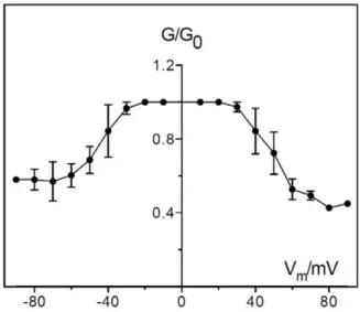
In contrast to the Chlamydiaceae, little is known about the composition of the outer membrane of other chlamydiae. Several key mechanisms for host cell interaction, such as a type three secretion system and its effector proteins, are conserved among all chlamydiae [32] [33,34]. Yet, the genome of the amoeba symbiont Protochlamydia amoebophila encodes no homologue of MOMP [33], and antibodies targetingChlamydiaceaeMOMP did not bind to the outer membrane of these bacteria [35]. A recent study identified 38 outer membrane proteins of P. amoebophilaby combining 1D and 2D gel electrophoresis of outer membrane fractions with mass spectrometry analysis [36]. The identified proteins included OmcA (pc0617) and OmcB (pc0616). Additionally, a novel protein family was identified consisting of four proteins that share an amino acid sequence identity of 22–28% and have no functionally characterized homologues in other organisms (Table 1). Two of these proteins were frequently detected in outer membrane fractions. Both were predicted to form beta-barrels by in silico analysis and to contain signal peptides. Their predicted structure and their high abundance in outer membrane fractions led to the hypothesis that they function as porins and together form the COMC ofP. amoebophilaby interactions with OmcA and OmcB. Because of the lack of significant sequence similarities with other characterized proteins we propose the names PomS, PomT, PomU, and PomV (Pom for ‘‘Protochlamydia outer membrane protein’’) for their designation.
In this study, we provide evidence that all members of the novel protein family found in P. amoebophila represent pore-forming proteins, and for two of them we confirmed their outer membrane location. The most abundant outer membrane protein of this family is expressed throughout the complete developmental cycle ofP. amoebophila, and lipid bilayer measurements further confirmed its function as porin. Our study provides a first detailed analysis of outer membrane proteins of an environmental counterpart of pathogenic chlamydiae and shows that a novel porin family represents the functional equivalent of theChlamydiaceaeMOMP in P. amoebophila.
Materials and Methods
Cultivation of Organisms and Infection Experiments Uninfected Acanthamoeba castellanii Neff or A. castellanii Neff infected withP. amoebophilawere grown axenically in 10 or 150 ml TSY medium (30 g/L trypticase soy broth, 10 g/L yeast extract, pH 7.3) at 20uC. Cultures were supplied with fresh medium every one to two weeks depending on amoebal growth. Escherichia coli strains were grown in standard LB medium at 37uC.
For infection experiments cultures of A. castellanii Neff were harvested and the number of amoebae was counted using a Neubauer counting chamber. Amoebae were seeded in the wells of a multiwell dish and were infected with purifiedP. amoebophilaEBs at an MOI of 5 or 10. Multiwell dishes were centrifuged at 6006g
for 15 min at 20uC, and the end of centrifugation was regarded as time point 0 hours post infection (p.i.). After centrifugation, the medium was exchanged and infected cultures were grown at 20uC. At selected time points cells were fixed for immunofluorescence analysis.
Table 1.Members of the putative porin family ofP. amoebophilaare predicted to be localized to the outer membrane byin silico
analysis.
PomS PomT PomU PomV
Locus tag pc1489 pc1077 pc0870 pc1860
Molecular mass in kDa 36.3 39.0 34.4 37.5
Signal peptide yes yes yes yes
Prediction of alpha-helix formation no no no no
Localization to outer membrane yes yes unknown yes
Prediction of beta-barrel formation yes yes no yes
Predicted lipoprotein no no no yes
Probability to be an OMP 97.72% 100% 97.68% 97.22%
pCOMP prediction integral outer membrane protein (cluster 081)
integral outer membrane protein (cluster 081)
ambiguous prediction lipoprotein (cluster 081)
Best blastp hit except for members of porin family
hypothetical protein of
Methylophagasp. JAM7 e-value 2e207 19.9% identity
hypothetical protein of
Methylomicrobium alcaliphilum
e-value 3e219 17.1% identity
hypothetical protein of
Methylophagasp. JAM7 e-value 8e204 19.1% identity
EGF-like domain-containing protein ofDictyostelium discoideume-value 0.15 8.9% identity
Presence in outer membrane protein fractions[36]
yes yes no yes
Experimental evidence for outer membrane location in this study
yes yes no no
Signal peptides were predicted using SignalP4 [80], alpha-helix formation was predicted with TMHMM, localization to the outer membrane with Cpsortdb [81], beta-barrel formation with MCMBB, BOMP and Pred-TMBB [82,83], lipoprotein signal peptides with LipoP [84], the probability of the localization to the outer membrane using HHOMP [85] and chlamydial outer membrane proteins with pCOMP [48].
Purification of Elementary Bodies of P. amoebophilaand Isolation of Pc1489
P. amoebophilaEBs for infection experiments were purified as previously described [36]. Highly enriched fractions of EBs were obtained using two additional centrifugation steps to further remove host cell debris [32]. Highly purified EBs were thawed and centrifuged at 10,6216g for 15 min at 4uC. The resulting pellet
was resuspended in 100ml POP05-buffer (0.087 g/L EDTA,
5.84 g/L NaCl, 300 mM NaxPO4, 0.5% n-octly-polyoxyethylen; pH 6.5) [37] with 100 mM freshly added dithiothreitol (DTT) per 3 mg EBs (wet weight) and incubated for 1 h at 37uC on a rocking platform. Cell debris was removed by centrifugation (10,6216g,
10 min, 4uC), an equal volume of ice-cold acetone was added to the supernatant and proteins were precipitated for 1 h at220uC. The suspension was centrifuged as before and the resulting pellet was resuspended in 400ml Buffer A (2.9 g/L HEPES, pH 7.5,
0.292 g/L NaCl, 0.5% n-octly-polyoxyethylen). Undissolved matter was removed by centrifugation at 10,6216g for 10 min
at 20uC. After equilibration of a Vivapure Q-Mini-spin column (Sartorius) with 400ml Buffer A, the supernatant was applied onto the column and centrifuged at 2,0006g for 5 min at 20uC. The
column was washed twice with Buffer A. Elution of proteins was achieved by applying a gradient with increasing NaCl concentra-tions ranging from 0.1 to 1 M NaCl. The flow-through of all steps was collected. 46Laemmli buffer without DTT was added to the samples and samples were run on a 12.5% SDS-PAGE gel, followed by staining with colloidal Coomassie (100 g/L
(NH4)2SO4, 20 g/L orthophosphoric acid, 25% methanol, 0.625 g/L Coomassie Brilliant Blue G-250). Fractions that showed only a single band for Pc1489 at the correct size were collected and pooled. To concentrate samples, proteins were precipitated with ice-cold acetone and resuspended in Buffer A without n-octyl-polyoxyethylen (n-octyl-POE). Protein concentrations were deter-mined using the BCATMProtein Assay Kit (Pierce Biotechnology). The identity of the purified protein was confirmed by SDS-PAGE analysis in combination with mass spectrometry.
Mass Spectrometry Analysis
Sample processing and high-performance liquid chromatogra-phy mass spectrometry (HPLC-MS) analysis were perfomed as described previously [36]. Briefly, bands were excised from the gel, reduced with DTT, alkylated with iodoacetamide and digested overnight with trypsin. Samples were separated on an Ultimate plus HPLC system (Dionex) coupled online to an LTQ mass spectrometer (Thermo Scientific). Raw files were searched with ProteomeDiscoverer 1.3.0.339 and Mascot 2.2 using the following settings: trypsin/P, maximum 2 missed cleavage sites, 1.5 Da precursor ion tolerance, 0.8 Da fragment ion tolerance, cabami-domethyl-cysteine as fixed modification, oxidation of methionine and deamidation of asparagine or glutamine as variable modifi-cations. The fasta database comprised sequences from P. amoebophilaas well as proteins of the amoeba host and contam-inants. Proteins were quantified by calculating spectral counts and normalized spectral abundance factor (NSAF) for allP. amoebophila proteins identified with p,0.05 [38]. As the precision of spectral
Figure 1. Toxic effect of the heterologous expression of PomS, PomT, PomU, and PomV inE. coli.(A) Survival ofE. coliBL21 (DE3) carrying different pet16b plasmid constructs after induction of protein expression (labeled with *) and without induction. As controls, the vector pet16b without insert and pet16b containing a gene fragment coding for the inclusion membrane protein IncA (pc0399) ofP. amoebophilawere used [32]. Induction of expression of proteins of the putative porin family lead to a rapid decrease in the number of colony forming units (cfus) compared to the non-toxic protein IncA. Ten minutes after induction, the numbers of cfus ofE. coliexpressing the putative porins were significantly lower than those of all controls (p,0.05, one-way ANOVA test and Dunnett’s post test); this difference was not significant anymore at sixty minutes after induction. The mean number of cfus for three independent replicates is shown+/2the standard error of the mean (SEM). (B) Visualization of the toxic effect of the putative porins onE. coli10 min after induction of protein expression. Colonies formed by 10ml droplets of the same dilutions for
the non-toxic IncA (left) and the putative porin PomU (right) are shown after incubation overnight at 37uC. Similar results were obtained for all four putative porins tested. Dilutions range from 1:10 to 1:10,000. (C) Detection of protein expression by SDS-PAGE (left) and Western blot analysis (right) for PomT and IncA. Time in min after induction of protein expression by addition of IPTG is indicated above the lanes. Expression of IncA can be detected by SDS-PAGE and Western blot analysis whereas expression of PomT can be detected only by the more sensitive Western blot analysis. doi:10.1371/journal.pone.0055010.g001
counting was shown to be optimal for proteins with five or more spectra, calculation of NSAF was restricted to proteins with a minimum of five spectra with a Mascot score of 20 or above [39]. NSAF values were converted to percentages as a measure of relative abundance.
Transcriptional Analysis
For the isolation of RNA, 3 ml ofA. castellanii infected withP. amoebophila were harvested by centrifugation (7,3236g, 10 min). The pellet was resuspended in 750ml Trizol and cells were disrupted using a bead beater at an intensity of 4.5 for 30 sec. The cell suspension was then centrifuged at 12,0006g for 5 min. The supernatant was incubated at room temperature for 5 min followed by the addition of 200ml chloroform. The solution was shaken vigorously for 15 sec, further incubated for 5 min and finally centrifuged at 12,0006g for 15 min at 4uC. The supernatant was taken off carefully, mixed with 500ml isopropyl alcohol per 750ml volume and incubated at room temperature for
10 min. After another centrifugation step (12,0006g, 10 min,
4uC), the resulting RNA pellet was washed with 1 ml 75% ethanol and centrifuged for 5 min at 7,0006g and 4uC. The pellet was air-dried for 10 min and dissolved in 30ml distilled water treated with
diethylpyrocarbonate (DEPC). Isolated RNA was quantified using a NanoDropHND-1000 spectral photometer and either stored at 280uC or treated directly with DNaseI (Sigma-Aldrich). DNA was digested by adding 1 U DNaseI/2mg RNA and incubation at room temperature for 1 h. RNA was precipitated with pure ethanol at 220uC overnight and resolved in DEPC-treated distilled water. RNA quality was assessed by agarose gel electrophoresis. To test for DNA still present in the RNA preparations, PCR without reverse transcription was performed and no product was obtained (data not shown). Three independent biological replicates of RNA isolated at 0 h p.i., 24 h p.i., 48 h p.i. and 96 h p.i were analyzed.
Reverse transcription was performed using the RevertAidTM First Strand cDNA Kit (Fermentas). 1mg RNA was applied per reaction and random hexamer primers were used for reverse transcription. A negative control without reverse transcriptase was performed in parallel. The RT conditions were 5 min at 25uC, 60 min at 37uC and 5 min at 70uC. The obtained cDNA was stored at220uC until further use. Primers targetingpc1489and the 16S rRNA gene of P. amoebophila were designed using the online tool Primer3plus [40] (Table S1). Primer concentrations and annealing temperatures were optimized using gradient PCR. Genes were cloned into the pCR-XL-TOPO vector (Invitrogen) and these constructs were used as standards for calibration of the PCR assay. The PlatinumH SYBRH Green qPCR SuperMix-UDG Kit (Invitrogen) was used for amplification following the manufacturer’s instructions by applying 1ml cDNA to the reaction mixture. All standards and samples were analyzed on one plate during a single run using an iCycler (Bio-Rad). Each plate included three different negative controls: a no template control (where all the reaction reagents except for cDNA were used), an RNA only control (to test for residual chlamydial DNA) and cDNA obtained from uninfected amoebae. The qPCR program was: denaturation at 95uC for 3 min, followed by 35 cycles of 40 sec at 95uC, 30 sec at 60uC and 30 sec at 72uC. Final elongation was allowed for 1 min at 72uC, followed by 30 sec at 95uC and a melting curve from 55–95uC. For interpretation and further analysis of the qPCR results the iCycler software was used. Copy numbers of pc1489 were adjusted to the number of organisms present at the different stages of the developmental cycle by normalization with copy numbers of the 16S rRNA gene at the respective time point [41]. Analysis of the melting curves of the PCR products forpc1489and the 16S rRNA showed a single-peak for the amplicons, indicating that a single PCR product was formed. This was confirmed by analysis of the products on a 2% agarose gel. The negative controls did not show any amplification product (data not shown).
Cloning and Heterologous Expression of Proteins in
E. coli
Genes encoding the predicted porins were cloned as full length copies or without the predicted signal peptide using two different cloning vectors. Full length pc0870, pc1077, and pc1860 were cloned into the BamHI and NdeI restriction sites of the vector pet16b (Novagen). Full length pc1489was cloned into the XhoI site of pet16b.Pc0870, pc1077and pc1489were cloned into the KpnI and PstI sites of pQE-30 (Qiagen) excluding the first 21 bp encoding the predicted signal peptide. DnaK (pc1499), which served as control in immunofluorescence analysis, was cloned into the SmaI and PstI restriction sites of pQE-30. Genes were
amplified using the High Fidelity PCR Enzyme Mix (Fermentas) or the Phusion High-Fidelity DNA Polymerase (New England BioLabs). Forward and reverse primers contained sequences to introduce recognition sites for the respective restriction enzymes. For cloning into the vector pet16b, an additional C-terminal 66
His-tag was introduced via the reverse primer to compensate for possible removal of the N-terminal His-tag by cleavage of the signal peptide. Amplified fragments were first cloned into the vector pCR-XL-TOPO (Invitrogen) and subsequently cloned into the expression vectors pet16b and pQE-30. All constructs were sequenced before transformation into E. coli BL21 (DE3) (Invitrogen) or E. coli M15 (Qiagen). Heterologous expression was induced by addition of 1 mM isopropyl b -D-1-thiogalacto-pyranoside (IPTG) to cultures at an OD600of 0.6. Heterologously expressed Pc1077 without leader sequence and Pc1499 were purified using HisTrap purification columns (GE Healthcare Biosciences) according to the instructions of the manufacturer. The identity of the purified proteins was verified by 1D gel electrophoresis combined with mass spectrometry (data not shown).
Toxicity Assay
To analyse the toxicity of the heterologously expressed proteins, LB medium was supplemented with 0.4% glucose to ensure repression of the T7 lac promoter of the vector pet16b. pet16b containing pc0870, pc1077, pc1489 or pc1860 was transformed into E. coli BL21 (DE3), and cells were grown on LB plates overnight at 37uC. Single colonies were inoculated in LB, and protein expression was induced by addition of 1 mM IPTG at an OD600 of 0.6. As controls, the vector pet16b without insert and pet16b containing a gene fragment coding for the inclusion membrane protein IncA (pc0399) of P. amoebophila were used [32]. Samples were taken at 0, 10 and 60 min after induction, they were diluted in phosphate buffered saline (PBS, 130 mM NaCl, 10 mM NaxPO4; pH 7.2–7.4) and plated on LB plates. After incubation overnight, the number of colony forming units (cfu) was
counted. All experiments were performed in three biological replicates.
Generation of Polyclonal Antibodies,
Immunofluorescence, and Immunoelectron Microscopy All antibodies used in this study were produced by Eurogentec (Belgium). To generate polyclonal antibodies against Pc1489, 400mg of Pc1489 purified from P. amoebophila EBs in Buffer A without n-octyl-POE were used for immunization of one chicken. To generate antibodies against Pc1077, heterologously expressed protein without leader sequence purified fromE. coliwas used for immunization of two rabbits. To generate antibodies against DnaK, one chicken and two guinea pigs were immunized with heterologously expressed DnaK. IgY from pre-immune sera and egg yolk collections was purified using a HiTrapTM IgY Purification HP column (GE Healthcare) according to the manufacturer’s instructions. Pre-immune sera were obtained for all immunizations and tested in Western Blot and immunofluo-rescence analyses. To remove antibodies targeting amoeba proteins, sera were adsorbed with amoeba lysate prior to experiments [36]. Pre-immune serum and antibodies targeting Pc1489 were affinity purified on a polyvinylidene fluoride (PVDF) membrane as previously described to further reduce unspecific background signals [42]. Immunofluorescence analysis of metha-nol and paraformaldehyde fixed cells and immunogold labelling of ultrathin cryosections was performed as described previously [32].
Western Blot Analysis
Cells were harvested by centrifugation (18,0006g for 2 min for E. coli or 7,3236g for 5 min for amoebae). Pellets were
resuspended in 46 Laemmli buffer with 400 mM DTT and heated to 95uC for 5 min. Nucleic acids present in the samples were removed by digestion with the nuclease Benzonase (Novagen) for 1 h. Outer membrane fractions ofP. amoebophilawere obtained either by treatment with N-laurylsarcosine (sarkosyl) as described previously [36] or by incubation in POP05 buffer for 1 h as
Figure 3. Detection of PomS and PomT inP. amoebophilawithin its natural amoeba host.Left panel: Localization of PomS and PomT by immunofluorescence in an asynchronous culture ofA. castellaniicontainingP. amoebophila. Halo-shaped fluorescence signals were observed around intracellularP. amoebophila. In contrast, signals for the heat-shock protein DnaK were confined to the cytoplasm. No differences were observed for methanol- and PFA- fixed samples. Identical microscopic fields are shown. Bar, 5mm. Right panel: Magnification of intracellularP. amoebophila; overlay
images of fluorescence signals for PomS and PomT (red), respectively, with DnaK (green) are shown. Bars, 2mm.
doi:10.1371/journal.pone.0055010.g003
described above. Proteins were separated by 1D gel electropho-resis, transferred to a PVDF membrane (GE Healthcare) by semi dry blotting in transfer buffer (5.8 g/L Tris, 2.9 g/L glycine, 20% methanol) using a Trans-BlotH SD Semi-Dry Electrophoretic Transfer Cell (Bio-Rad) and proteins were detected using specific antibodies [36]. Detection of heterologously expressed Pc1077 in E. colilysates by Western blot analysis proved to be difficult. The protein was not detectable in Western blot analysis after transfer with standard transfer buffer and staining of SDS-PAGE gels after blotting showed that Pc1077 did not elute from the gel (data not shown). Addition of 0.05% SDS to the buffer improved transfer,
but still a substantial amount of protein could be detected in the gel after blotting and blots showed high background (data not shown). The best results were obtained with a transfer buffer containing urea (12 mM Tris, 48 mMol glycine, 5 mM NaH2PO4, 0.55% SDS, 3 M Urea) which was developed for the transfer of membrane proteins [43] and this buffer was used for the detection of Pc1077 in subsequent experiments. To test for the presence of multimers of Pc1489 in the outer membrane, outer membrane protein fractions from highly enriched EBs were cross-linked as described previously using bis(sulfosuccinimidyl)suberate [30]. Cross-linked samples were analyzed by SDS-PAGE and Western blot analysis.
Infection Inhibition Assay
P. amoebophilaEBs were thawed at 37uC and 1 volume of PBS was added. Heat inactivation was achieved by incubation at 56uC for 30 min [44]. Anti-Pc1489 antibodies, targeting the putative porin, and anti-Pam antibodies, targeting the immunodominant components of the outer membrane ofP. amoebophila[32], were diluted in FA Block solution (2% bovine serum albumin in PBS) to reach final dilutions of 1:15 and 1:150. EBs and heat-inactivated EBs were incubated with the antibody solutions for 30 min at
Figure 4. Localization of PomS in the outer membrane ofP. amoebophila by immuno-transmission electron microscopy. Immunogold labeling with pre-immune serum (left) and polyclonal anti-PomS antibodies (right). Gold particles indicating PomS were confined to the outer membrane ofP. amoebophila. White arrow head, gold particle in the outer membrane; grey arrowhead, cytoplasmic membrane; black arrowhead, inclusion membrane. Bars, 200 nm. doi:10.1371/journal.pone.0055010.g004
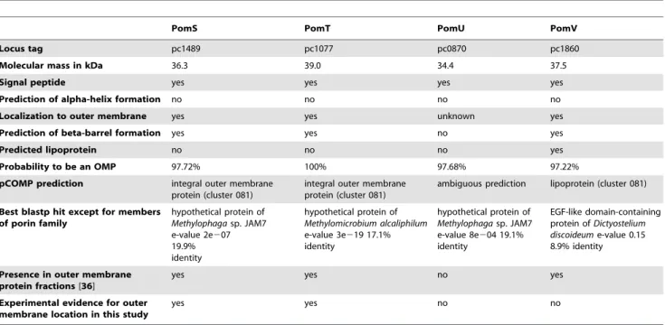
Figure 5. Expression of PomS during the developmental cycle ofP. amoebophilain its amoeba host.Upper panel: Relative levels of pomS transcripts measured by real-time quantitative PCR. pomS transcripts were normalized to the 16S rRNA to account for an increase in copy numbers due to multiplication ofP. amoebophila. Data are shown as the mean of five replicates+/2SEM from a total of three independent infection experiments. Lower panel: Expression of PomS at the same time points as in the upper panel detected by anti-PomS antibody in methanol-fixed cells. Outlines of the amoebae are drawn in white. Bars 5mm.
37uC with mild shaking. As controls, EBs and heat inactivated EBs were incubated with FA Block solution only. The pre-incubated EBs were used to infect amoebae at an MOI of 5 and samples were fixed at 0, 6, 12, 24, 48, 72 and 96 h p. i. and stored at 4uC for later analysis. Immunofluorescence analysis was performed as described above, and for the time point 48 h p.i. the ratio of infected amoebae to all amoebae was determined by counting at least 100 amoebae. All experiments were performed in three biological replicates.
Planar Lipid Bilayer Assays
The methods used for black lipid bilayer experiments have been described previously [45]. The black lipid bilayer instrumentation
consisted of a Teflon chamber with two compartments of 5 ml volume, which were separated by a thin wall and connected by a small circular hole with an area of about 0.4 mm2. A 1% (w/v) solution of diphytanoyl-phosphatidylcholine (PC) (Avanti Polar Lipids) inn-decane was used to form the membranes across the hole. The Pc1489 (PomS)-containing protein fractions were diluted 1:100 in 1% Genapol (Roth) and added at a concentration of about 10 ng/ml to the aqueous phase after the membrane had turned black. The membrane current was measured with a pair of Ag/AgCl electrodes with salt bridges connected in series to a voltage source and a highly sensitive current amplifier (Keithley 427). The temperature was throughout kept at 20uC. The amplified signal was recorded on a strip chart recorder. Zero-current membrane potential measurements were performed in the presence of a salt gradient as described earlier [46,47]. After the insertion of more than 100 channels into the PC membranes, the KCl concentration (300 mM KCl) was raised 2.5-fold by addition of 3 M KCl to one side of the membrane. The zero-current membrane potentials were measured with a high impedance electrometer (Keithley 617) and analyzed using the Goldman-Hodgkin-Katz equation [46,47].
Results
Toxicity of PomS, PomT, PomU, and PomV forE. coli
To characterize the putative novel outer membrane protein family of P. amoebophila [36,48], we initially tried to clone and express PomS (pc1489), PomT (pc1077), PomU (pc0870), and PomV (pc1860) in the heterologous hostE. coli. Our first attempts to express the full length proteins using different expression vectors inE. colifailed. When protein expression was induced, the optical density (OD600) of the cultures decreased and no overexpression of the proteins was observed by SDS-PAGE, which indicates host cell lysis due to detrimental effects of the heterologous proteins.
The overexpression ofChlamydiaceaeMOMP inE. coliresulted in a strong decrease in the number of colony forming units (cfus) after induction of protein expression [49]. To investigate whether the predicted porins of P. amoebophila show a similar effect, we
Figure 6. Infection-inhibition assays using anti-Pam and anti-PomS antibodies.Left panel: Incubation of host-freeP. amoebophilaEBs with anti-PomS and anti-Pam antibodies prior to fixation demonstrated that these antibodies can bind unfixed cells. Fluorescence signals derived from specific antibodies (left) and 49, 6-Diamidino-2-phenylindol (DAPI; right) are shown for identical microscopic fields. Bars, 2mm. The absence of DAPI
signals for some cells indicates cells that lysed during the purification procedure. Right panel: Infection-inhibition assay using preincubations of EBs with anti-Pam and anti-PomS antibodies in different dilutions. The proportion of infected amoebae compared to all counted amoebae of three replicates at 48 h p.i. is shown+/2SEM. Heat-inactivated EBs, used as negative controls, were taken up by the amoebae but did not multiply.
doi:10.1371/journal.pone.0055010.g006
Figure 7. Purification of PomS fromP. amoebophilaEBs.A gel stained with colloidal coomassie is shown; 1, outer membrane fraction after incubation of EBs with n-octyl-POE; 2, outer membrane fraction after precipitation with acetone; 3, column flow through; 4–7, fractions after elution with 0.1, 0.25, 0.3, 0.35 and 0.4 M NaCl. Molecular mass of marker bands (M) in kDa; the expected size of PomS (33.9 kDa) is indicated by an arrow head.
doi:10.1371/journal.pone.0055010.g007
performed a time-course experiment and compared induced and uninduced E. coli cultures with a control protein (IncA) and an empty vector. Consistent with our initial observations, expression of the proteins PomS, PomT, PomU, and PomV had an immediate lethal effect onE. colias indicated by a strong decrease in the number of cfus already 10 min after induction of protein expression. No decrease was observed at this time point when the empty vector alone or the expression of a non-toxicP. amoebophila protein was induced (Fig. 1A and 1B). Sixty minutes after induction, the number of cfus decreased even further for cells expressing the putative porins. At this time point also induced cells containing the control vector without an insert or encoding the non-toxic protein showed a decrease in the number of viable cells (Fig. 1A). This decrease was not as strong as for the putative porins and could result from toxicity of the expression vector for the host cells [49,50] or from interference of the strong production of heterologous proteins with cellular processes because of the high transcription rate of the T7 RNA polymerase [51,52].
To link the observed toxicity to protein expression, we tried to detect the heterologous proteins by SDS-PAGE. With this technique, a band for the non-toxic control protein was observed 10 and 60 min after induction, but no bands were visible for the putative porins (Fig. 1C). We therefore used the more sensitive Western blot analysis. This resulted in the detection of PomT at 10 and 60 min after induction of protein expression as well as in the detection of IncA (Fig. 1C). However, we could not detect expression of PomS, PomU, and PomV, probably due to the low amounts in which these proteins were expressed. The observed strong toxic effects onE. coliwere thus caused by minute amounts of these proteins.
Previous studies have shown that removal of the signal peptide, which prevents secretion, can help in the overexpression of porins [29]. Indeed, when we tested PomS, PomT, and PomV without the predicted leader sequence overexpression was successful for all three proteins as indicated by bands at the correct size on SDS-PAGE gels. We chose to analyse PomS and PomT in more detail,
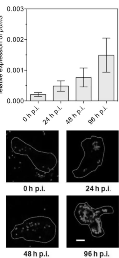
Figure 8. Porin function of purified PomS.Single channel experiments using a PC/n-decane membrane in the presence of purified PomS. The aqueous phase contained 1 M KCl and 10 ng ml21PomS dissolved in 1% Genapol. The applied membrane potential was 20 mV; T = 20
uC. Left panel: Single-channel recordings show a uniform stepwise increase as expected for a highly enriched purified porin. Right panel: Frequency of observed conductance increments. P(G) was calculated by dividing the number of fluctuations with a given conductance increment by the total number of conductance fluctuations. Data from both panels suggest that the purified protein fraction contains mainly PomS (about 82% of the total number of pores) and that there is only a very minor contribution of other pores in the histogram (about 18% of the total number of pores) caused either by contaminant porins or by degradation of PomS. The average single-channel conductance was 3.25 nS for 230 single-channel events.
doi:10.1371/journal.pone.0055010.g008
Table 2.Average single-channel conductance of PomS in different salt solutions.
Salt Salt concentration (M) Single-channel conductance G (nS)
LiCl 0.3 1.0
1.0 2.25
KCl 0.01 0.15
0.03 0.25
0.10 0.60
0.30 1.20
1.0 3.25
3.0 11
KCH3COO (pH 7) 0.30 0.60
1.0 1.50
The membranes were formed from diphytanoyl phosphatidylcholine (PC) dissolved to 1% in n-decane. The aqueous solutions were used unbuffered and had a pH of 6 unless otherwise indicated. The applied voltage was 20 mV, and the temperature was 20uC. The average single-channel conductance, G, was calculated from at least 80 single events.
which were most abundant in P. amoebophila outer membrane fractions [36]. Polyclonal anti-PomS and anti-PomT antibodies recognized PomS and PomT expressed in E. coli resulting in a strong band at the correct molecular mass (Fig. 2, upper panel).
Location of PomS and PomT in the Outer Membrane ofP. amoebophila
The transport function of porins is inherently linked to their presence in the outer membrane. We therefore investigated the location of the putative porins PomS and PomT with Western blot, immunofluorescence analysis, and immuno-transmission electron microscopy (immuno-TEM). First, soluble and insoluble protein fractions of highly purified EBs after treatment with the detergent sarkosyl were analyzed by Western blot. In contrast to the cytoplasmic protein DnaK, which was absent in the sarkosyl-insoluble fraction containing proteins of the outer membrane complex, strong bands were observed for PomS and PomT (Fig. 2, lower panel). In addition, a band for PomS was also detected in the sarkosyl-soluble fraction at a higher molecular mass, possibly representing the full length protein before removal of the signal peptide. For PomT, the band detected in the sarkosyl-soluble fraction was much weaker than the band detected in the outer membrane fraction. For this protein, additional bands with lower molecular mass were observed. This is consistent with a previous study, in which PomT was detected in lower molecular bands of outer membrane protein fractions of P. amoebophila by mass spectrometry [36].
Most known porins function as trimers [53], including MOMP of theChlamydiaceae [54]. We therefore tested for the presence of multimers of PomS by crosslinking outer membrane preparations of EBs ofP. amoebophila, but under the conditions used we could not observe any evidence for the presence of multimers (data not shown).
Immunofluorescence analysis with antibodies targeting PomS and PomT resulted in a halo-shaped fluorescence signal surrounding singleP. amoebophilacells, demonstrating the presence of these proteins at the periphery ofP. amoebophilacells (Fig. 3). As P. amoebophila is located in single-cell inclusions, it is difficult to distinguish between signals from the outer membrane, the inner membrane and the inclusion membrane by immunofluorescence. However, halo-shaped fluorescence signals were also observed when immunofluorescence analysis was performed with purified EBs. This is a strong evidence for a location of PomS and PomT in the bacterial outer membrane and excludes a location of these proteins in the host-derived inclusion membrane. To further demonstrate the location of the most abundant putative porin
PomS in the outer membrane we exploited the higher resolution of immuno-TEM. With this technique, signals for PomS were observed only in the outer membrane ofP. amoebophila, but not in the cytoplasmic membrane or the inclusion membrane (Fig. 4).
Transcription and Expression of PomS throughout the Developmental Cycle
To get first insights into the role of PomS during infection and intracellular replication ofP. amoebophila, we analyzed expression of the gene coding for PomS by real-time quantitative reverse transcription PCR (RT-qPCR) throughout a complete develop-mental cycle. Expression ofpomS was detected at all time points during the developmental cycle, with a significant increase from 0 to 48 h p.i. (Mann-Whitney U-test, p#0.05) and was highest at 96 h p.i. (Fig. 5).
In addition, the presence of PomS in P. amoebophila was monitored by immunofluorescence analysis during the develop-mental cycle. Consistent with our RT-qPCR data, PomS protein was detected at all investigated time points. Fluorescence signal intensities increased, and the halo-shaped signals were better defined at later time points, confirming an elevated expression of PomS at later time points and suggesting an increase of the amount of PomS during the developmental cycle (Fig. 5 and Fig. S1).
Preincubation with PomS Antibodies does not Alter Infection of Amoebae
Proteins in the bacterial outer membrane can be important for attachment to and uptake by host cells. As our and previous results identified PomS as a major component of the outer membrane of P. amoebophila[36], we tested whether this protein is required for attachment to amoeba host cells and whether infection can be blocked by preincubation of EBs with PomS-specific antibodies. To ensure that the antibodies used in this experiment bind to the
Table 3.Zero-current membrane potentials of PC/n-decane membranes in the presence of PomS measured for a 2.5-fold gradient of different salts (300 mM versus 750 mM).
Salt Vm/mV Pcation/Panion
KCl 27.8 0.48
LiCl 29.5 0.40
KCH3COO, pH 7 22.6 0.79
The zero-current membrane potential Vmis defined as the difference between
the potential at the dilute side and the potential at the concentrated side. The aqueous salt solutions were used unbuffered and had a pH of 6, if not indicated otherwise; T = 20uC. The permeability ratio Pcation/Panionwas calculated using
the Goldman-Hodgkin-Katz equation [46] from at least 3 individual experiments.
doi:10.1371/journal.pone.0055010.t003
Figure 9. Voltage dependence of PomS.PomS was added in a concentration of 500 ng ml21
to the trans-side side of a PC/n-decane membrane in multi-channel experiments. The aqueous phase contained 1 M KCl, pH 6.0. After 30 min the conductance had increased considerably. At this point different potentials were applied to the membrane. The ratio of the conductance G at a given membrane potential (Vm) divided by the conductance Goat 10 mV was calculated as a function of the membrane potential Vm [79]. The membrane potential refers to the cis-side of the membrane. T = 20uC. Means (6SD) of three membranes are shown.
doi:10.1371/journal.pone.0055010.g009
outer membrane of unfixed cells, EBs were incubated with the specific antibodies prior to fixation, subsequently fixed and incubated with a secondary antibody. All antibodies were found to bind to the outer membrane of unfixed EBs (Fig. 6). Nevertheless, preincubation with the PomS antibody had no significant effect on bacterial uptake and entry, or on the progress of infection (Fig. 6). Interestingly, also no effect was observed when EBs were preincubated with polyclonal antibodies targeting the immunodominant components ofP. amoebophilaEBs (Fig. 6). High concentrations of this antibody lead to agglutination of EBs at the start of the infection experiment, but even this did not influence the outcome of the infection.
Porin Function of PomS
To investigate the putative porin function of PomS we purified this protein directly from P. amoebophila EBs. Outer membrane proteins were solubilized with the detergent n-octyl-POE, and DTT was added to reduce disulfide bridges, which are responsible for extensive crosslinking of proteins in the EB cell envelope. A single band at the expected size of PomS in fractions eluted with 250 mM NaCl from an anion exchange column was visible on protein gels, and no other bands were present in this fraction (Fig. 7). This indicates the absence of larger amounts of contaminating protein in this fraction and demonstrates the successful enrichment of PomS. To further analyze this protein fraction, quantitative mass spectrometry analysis was performed. This highly sensitive method identified several P. amoebophila proteins. In total, 767 peptide-spectrum matches were assigned to PomS, while only 121 peptide-spectrum matches were assigned to the other P. amoebophila proteins. This analysis confirmed that PomS was highly enriched and represented the by far most abundant protein in this fraction (86% based on NSAF quantification). Further proteins included other putative porins and outer membrane proteins with different molecular masses, and at least 18-fold lower abundance (5% and below, based on NSAF) (Table S2).
To perform planar lipid bilayer assays purified PomS was added to a lipid membrane consisting of phosphatidylcholine. Single-channel conductance increased in a stepwise fashion indicating that the protein formed defined channels (Fig. 8). The average single-channel conductance was about 3.25 nS in 1 M potassium chloride (KCl). Only a minor fraction of channels (about 18% of the total number of fluctuations) with other conductance was observed suggesting that the protein preparation was essentially free of pore-forming contaminants (Fig. 8). The channels formed by PomS had a long lifetime, similar to porins of other Gram-negative and Gram-positive bacteria [25,55–57]. Analysis of the average single-channel conductance at different KCl concentra-tions in the aqueous phase showed that the conductance was a nearly linear function of the KCl concentration (Table 2). This is characteristic of many porins of Gram-negative bacteria [25,26,47,57] and suggests that PomS forms a wide and water-filled channel, similar to MOMP of theChlamydiaceae. Lipid bilayer assays were also performed with salts other than KCl to obtain information on the size and selectivity of the channels formed by PomS. Conductance was highest for KCl, followed by lithium chloride (LiCl), and lowest values were observed for potassium acetate (KCH3COO, Table 2). Replacement of the Cl
--ion by the less mobile acetate-ion resulted in a stronger decrease in conductance than replacement of the K+-ion by the less mobile Li+-ion. This means that the influence of anions of different size and mobility on the conductance was more pronounced than that of cations, suggesting anion-selectivity of the PomS channel.
Anion-selectivity and Voltage-dependence of PomS Additional information about the structure of the channel formed by PomS was obtained from zero-current membrane potential measurements in the presence of salt gradients. A 2.5-fold KCl gradient (300 mM versus 750 mM), across a lipid bilayer membrane in which about 100 to 1000 PomS channels were reconstituted, resulted in an asymmetry potential of27.8 mV at the more dilute side. This result indicated preferential movement of chloride over potassium ions through the pore at neutral pH. The ratio of the chloride permeability, PCl, divided by the potassium permeability, PK, was around 2.0, indicating indeed low anion selectivity of the PomS channel. This was further confirmed by measurements with LiCl and potassium acetate; we observed under the same conditions as for KCl, diffusion potentials around 29.5 and22.6 mV at the more dilute side, respectively (Table 3). The observed selectivity changes dependent on the mobility of the cations and anions indicated that PomS forms a general diffusion pore similar to OmpF and OmpC ofEscherichia coli [25,47] and MOMP ofC. psittaci[28].
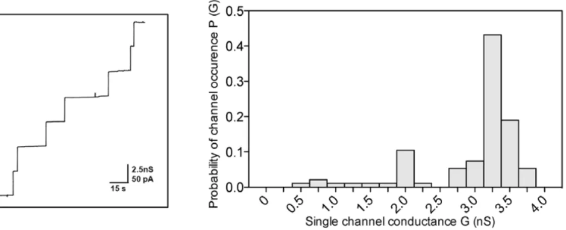
Some porins of Gram-negative bacteria show voltage-depen-dent closure in reconstitution bilayer experiments [58] although the physiological role of this channel gating is obscure [59]. This effect was also observed for PomS when voltages of positive and negative polarity higher than 30 mV or lower than230 mV were applied (Fig. 9). The voltage dependence of PomS was essentially unchanged if the protein was added to either the trans- or to the cis-chamber of the membranes. These results indicated a symmetric response of the pore to the applied voltage.
Discussion
In the Chlamydiaceae, the most abundant protein and a major structural component in the outer membrane is the porin MOMP [11,18]. While two copies of MOMP are encoded in the genome ofSimkania negevensis[34], no homologue of this protein was found in the two sequenced genomes of members of theParachlamydiaceae [33,34] – a major difference between this family and its pathogenic relatives from theChlamydiaceae. In a previous study, the presence of a putative novel porin family inP. amoebophilahas been proposed [36]. Here we show that two members of this porin family, PomS and PomT, are indeed localized in the outer membrane of P. amoebophila (Figures 2, 3, 4). PomS, the most abundant outer membrane protein [36], is expressed throughout the developmen-tal cycle with an increase in expression in the later phase of the developmental cycle similar to the expression profile of MOMP in different serovars of C. trachomatis (Fig. 5) [60–62]. This is consistent with a key function of PomS in the outer membrane of both RBs and EBs.
MOMP is probably not the only factor with respect to attachment to host cells or tissue-specificity inChlamydiaceae[63]. However, preincubations of EBs with antibodies targeting MOMP inhibited infection by either blocking attachment to host cells [64,65] or at steps after internalization [44,66]. Preincubation with antibodies targeting the outer membrane proteins OmcB, PorB and members of the Pmp family also reduced infectivity [67–70]. In contrast, the infection of amoebae by P. amoebophila is not impaired by pre-incubation with anti-PomS or anti-Pam antibod-ies (Fig. 6). This suggests that neither PomS nor other immunodominant components of the outer membrane of P. amoebophilaplay an important role in attachment to and uptake into amoebae, a process which might be fundamentally different from the uptake into non-phagocytic mammalian cells.
outer membrane. Similar to theChlamydiaceaeMOMP it is also a major porin, facilitating transport of small molecules across the outer membrane [24]. The pore formed by PomS is wide, water-filled and anion-selective, presumably because of an excess of positively charged amino acids in or near the pore. The single channel conductance was with 3.25 nS in 1 M KCl relatively high (Fig. 8, Table 3), about 3-times higher than that reported for MOMP which has a single channel conductance of 1 nS in C. trachomatisand 1.3 nS inC. psittaci[30,71]. The channels formed by PomS are voltage-dependent in a more or less symmetrical way starting at about 630 mV (Fig. 9). The voltage dependency of PomS is thus similar to that of the mitochondrial porin VDAC [72], and the channel conducts at high voltages about 50% of its open configuration. Different ion-selectivities have been reported forChlamydiaceae MOMPs. Native MOMP ofC. psittaciis weakly anion-selective [28], but cation-selective when expressed inE. coli [73]. Full length recombinant MOMP of C. trachomatis was reported to be either anion- [71] or cation-selective [29], suggesting that tags of the cloning-vector added during the cloning procedure may influence the functional characteristics of these proteins. In this study, ion selectivity was determined using native PomS. Therefore, modifications introduced by recombinant expression can be excluded.
MOMP is highly crosslinked and mostly present as a trimer in Chlamydiaceae EBs [30,54], whereas it is found mostly in its monomeric form in RBs [17]. For PomS we found no evidence for the formation of multimers, which might suggest that this protein is present in the outer membrane of EBs as monomer. Alternative explanations for our observation would be a highly instable PomS multimer that breaks down during purification, or an untypical migration behavior in SDS-PAGE. Trimers of MOMP are stabilized by disulfide-bridges between the monomeric subunits [29], and it has been suggested that opening of the pore is regulated by the reduction of these disulfide bonds [27]. PomS contains only two cysteine residues in contrast to 7–10 cysteine residues in MOMP [74] possibly hindering the formation of stable multimers by PomS and suggesting a different opening mechanism for this pore.
Whether the other members of the PomS protein family also function as porins has to be determined. The predicted beta-barrel structure of PomT and its location in the outer membrane strongly suggests a porin function. PomU is not predicted to form a beta-barrel, and PomV represents a lipoprotein according to in silico analysis [48]. However, all proteins of this family were predicted to encode a signal peptide and showed a pronounced toxic effect on E. coliwhen expression of the full length constructs was induced (Fig. 1). This is consistent with the notion that outer membrane proteins, and porins in particular, are generally difficult to overexpress inE. coli, because if properly folded, they might insert into the outer membrane, and their pore-forming activity can be toxic for the host [49,75].
The different members of the PomS protein family could play a role in the adaption to different environmental conditions as they
show an only low degree of amino acid sequence identity (22– 28%) and hence likely differ in pore size, uptake rates or ion specificity. Well-studied examples of homologous yet differentially sized porins are OmpF and OmpC of E. coli whose levels of expression depends on the osmolarity of the extracellular milieu [76,77]. The smaller pore formed by OmpC is dominant under conditions of high osmolarity whereas OmpF is upregulated under iso- or hypoosmotic conditions (Benz et al., 1985). Expansions of genes encoding porins are apparent in several chlamydial genomes. In theChlamydiaceae, the protein PorB was identified as putative porin based on its weak sequence similarity to MOMP by genome analysis; its pore-forming function was later confirmed [70]. The much larger genomes ofW. chondrophilaandS. negevensis encode 11 and 35 MOMP-like genes, respectively, which are more diverged from knownChlamydiaceaeMOMPs [34,78]. Within the chlamydiae, the PomS protein family analyzed here seems to be restricted toP. amoebophilaandParachlamydia acanthamoebaeand lacks close homologues in other bacteria. The P. amoebophila PomS corresponds to the MOMP of the Chlamydiaceae with respect to abundance in the outer membrane and its function as major porin, and it thus represents a specific adaptation of this amoeba symbiont.
Supporting Information
Figure S1 Fluorescence intensity derived from anti-PomS antibodies increases during infection. Quantifica-tion of fluorescence intensity was performed using the image analysis software daime [86]. The mean fluorescence intensity (6 SD) is shown for each time point.
(TIF)
Table S1 Primers used for qPCR targeting genes ofP. amoebophila.
(DOCX)
Table S2 Quantification by mass spectrometry shows that PomS is highly enriched in purified porin fractions.
The percent abundance of proteins with five or more assigned spectra was calculated based on the normalized spectral abundance factor (NSAF) [38,39].
(DOCX)
Acknowledgments
We thank Gustav Ammerer, Karl Mechtler and Sonja Kolar for access to and help with mass spectrometry analysis, Holger Daims for help with image analysis, and Barbara Sixt and Ilias Lagkouvardos for helpful discussions. Technical assistance by Christian Baranyi is greatly acknowl-edged.
Author Contributions
Conceived and designed the experiments: KA EH RB MH. Performed the experiments: KA CH AH EH JM FS EM PP. Analyzed the data: KA AH EH JM EM RB MH. Wrote the paper: KA RB MH.
References
1. Longbottom D, Coulter LJ (2003) Animal chlamydioses and zoonotic implications. J Comp Pathol 128: 217–244.
2. Horn M (2008) Chlamydiae as symbionts in eukaryotes. Ann Rev Microbiol 62: in press.
3. Blasi F, Tarsia P, Aliberti S (2009) Chlamydophila pneumoniae. Clinical Microbiology and Infection 15: 29–35.
4. Be´be´ar C, De Barbeyrac B (2009) Genital Chlamydia trachomatis infections. Clinical Microbiology and Infection 15: 4–10.
5. Abdelrahman YM, Belland RJ (2005) The chlamydial developmental cycle. FEMS Microbiol Rev 29: 949–959.
6. Hackstadt T, Fischer ER, Scidmore MA, Rockey DD, Heinzen RA (1997) Origins and functions of the chlamydial inclusion. Trends in Microbiology 5: 288–293.
7. Hybiske K, Stephens RS (2007) Mechanisms of host cell exit by the intracellular bacteriumChlamydia. Proc Natl Acad Sci U S A 104: 11430–11435. 8. Pal S, Theodor I, Peterson EM, de la Maza LM (1997) Immunization with an
acellular vaccine consisting of the outer membrane complex of Chlamydia trachomatisinduces protection against a genital challenge. Infect Immun 65: 3361–3369.
9. Tan TW, Herring AJ, Anderson IE, Jones GE (1990) Protection of sheep against
Chlamydia psittaciinfection with a subcellular vaccine containing the major outer membrane protein. Infect Immun 58: 3101–3108.
10. Everett KD, Hatch TP (1995) Architecture of the cell envelope ofChlamydia psittaci6BC. J Bacteriol 177: 877–882.
11. Hatch TP, Vance DW Jr, Al-Hossainy E (1981) Identification of a major envelope protein inChlamydiaspp. J Bacteriol 146: 426–429.
12. Raulston JE (1995) Chlamydial envelope components and pathogen-host cell interactions. Molecular Microbiology 15: 607–616.
13. Hatch T (1996) Disulfide cross-linked envelope proteins: the functional equivalent of peptidoglycan in chlamydiae? J Bacteriol 178: 1–5.
14. Salari SH, Ward ME (1981) Polypeptide composition ofChlamydia trachomatis. J Gen Microbiol 123: 197–207.
15. Tanzer RJ, Hatch TP (2001) Characterization of outer membrane proteins in
Chlamydia trachomatisLGV serovar L2. J Bacteriol 183: 2686–2690.
16. Liu X, Afrane M, Clemmer DE, Zhong G, Nelson DE (2010) Identification of
Chlamydia trachomatis Outer Membrane Complex Proteins by Differential Proteomics. Journal of Bacteriology 192: 2852–2860.
17. Hatch TP, Allan I, Pearce JH (1984) Structural and polypeptide differences between envelopes of infective and reproductive life cycle forms ofChlamydiaspp. J Bacteriol 157: 13–20.
18. Caldwell HD, Kromhout J, Schachter J (1981) Purification and partial characterization of the major outer membrane protein ofChlamydia trachomatis. Infect Immun 31: 1161–1176.
19. McCoy AJ, Maurelli AT (2006) Building the invisible wall: updating the chlamydial peptidoglycan anomaly. Trends Microbiol 14: 70–77.
20. Hatch TP, Miceli M, Sublett JE (1986) Synthesis of disulfide-bonded outer membrane proteins during the developmental cycle ofChlamydia psittaciand
Chlamydia trachomatis. J Bacteriol 165: 379–385.
21. Hackstadt T, Todd WJ, Caldwell HD (1985) Disulfide-mediated interactions of the chlamydial major outer membrane protein: role in the differentiation of chlamydiae? J Bacteriol 161: 25–31.
22. Newhall WJ (1987) Biosynthesis and disulfide cross-linking of outer membrane components during the growth cycle ofChlamydia trachomatis. Infect Immun 55: 162–168.
23. Stephens RS, Tam MR, Kuo CC, Nowinski RC (1982) Monoclonal antibodies to Chlamydia trachomatis: antibody specificities and antigen characterization. J Immunol 128: 1083–1089.
24. Nikaido H (2003) Molecular basis of bacterial outer membrane permeability revisited. Microbiol Mol Biol Rev 67: 593–656.
25. Benz R (1994) Solute uptake through bacterial outer membrane. In Bacterial cell wall.; Ghuysen JM, Hakenbeck, R, editor. Amsterdam: Elsevier Science B. V. 26. Benz R, Bauer K (1988) Permeation of hydrophilic molecules through the outer
membrane of gram-negative bacteria. Eur J Biochem 176 1–19.
27. Bavoil P, Ohlin A, Schachter J (1984) Role of disulfide bonding in outer membrane structure and permeability inChlamydia trachomatis. Infect Immun 44: 479–485.
28. Wyllie S, Ashley RH, Longbottom D, Herring AJ (1998) The major outer membrane protein ofChlamydia psittacifunctions as a porin-like ion channel. Infect Immun 66: 5202–5207.
29. Findlay H, McClafferty H, Ashley R (2005) Surface expression, single-channel analysis and membrane topology of recombinantChlamydia trachomatisMajor Outer Membrane Protein. BMC Microbiology 5: 5.
30. Sun G, Pal S, Sarcon AK, Kim S, Sugawara E, et al. (2007) Structural and Functional Analyses of the Major Outer Membrane Protein of Chlamydia trachomatis. Journal of Bacteriology 189: 6222–6235.
31. Newhall V, Wilbert JJ, Robert B (1983) Disulfide-linked oligomers of the Major Outer Membrane Protein ofChlamydiae. J Bacteriol 154: 998–1001. 32. Heinz E, Rockey DD, Montanaro J, Aistleitner K, Wagner M, et al. (2010)
Inclusion Membrane Proteins ofProtochlamydia amoebophilaUWE25 Reveal a Conserved Mechanism for Host Cell Interaction among the Chlamydiae. J Bacteriol 192: 5093–5102.
33. Horn M, Collingro A, Schmitz-Esser S, Beier CL, Purkhold U, et al. (2004) Illuminating the evolutionary history of chlamydiae. Science 304: 728–730. 34. Collingro A, Tischler P, Weinmaier T, Penz T, Heinz E, et al. (2011) Unity in
Variety–The Pan-Genome of theChlamydiae. Molecular Biology and Evolution 28: 3253–3270.
35. Collingro A, Toenshoff ER, Taylor MW, Fritsche TR, Wagner M, et al. (2005) ‘CandidatusProtochlamydia amoebophila’, an endosymbiont ofAcanthamoebaspp. Int J Syst Evol Microbiol 55: 1863–1866.
36. Heinz E, Pichler P, Heinz C, op den Camp HJM, Toenshoff ER, et al. (2010) Proteomic analysis of the outer membrane of Protochlamydia amoebophila
elementary bodies. PROTEOMICS 10: 4363–4376.
37. Heinz C, Roth E, Niederweis M (2003) Purification of Porins fromMycobacterium smegmatis. 139–150.
38. Zybailov B, Mosley AL, Sardiu ME, Coleman MK, Florens L, et al. (2006) Statistical Analysis of Membrane Proteome Expression Changes inSaccharomyces cerevisiae. Journal of Proteome Research 5: 2339–2347.
39. Collier TS, Sarkar P, Franck WL, Rao BM, Dean RA, et al. (2010) Direct Comparison of Stable Isotope Labeling by Amino Acids in Cell Culture and Spectral Counting for Quantitative Proteomics. Analytical Chemistry 82: 8696– 8702.
40. Untergasser A, Nijveen H, Rao X, Bisseling T, Geurts R, et al. (2007) Primer3Plus, an enhanced web interface to Primer3. Nucl Acids Res 35: W71– 74.
41. Borges V, Ferreira R, Nunes A, Nogueira P, Borrego MJ, et al. (2010) Normalization strategies for real-time expression data inChlamydia trachomatis. Journal of Microbiological Methods 82: 256–264.
42. Ritter K (1991) Affinity purification of antibodies from sera using polyvinyli-denedifluoride (PVDF) membranes as coupling matrices for antigens presented by autoantibodies to triosephosphate isomerase. Journal of Immunological Methods 137: 209–215.
43. Abeyrathne PD, Lam JS (2007) Conditions that allow for effective transfer of membrane proteins onto nitrocellulose membrane in Western blots. Can J Microbiol 53: 526–532.
44. Caldwell HD, Perry LJ (1982) Neutralization ofChlamydia trachomatisinfectivity with antibodies to the major outer membrane protein. Infect Immun 38: 745– 754.
45. Benz R, Janko K, Boos W, La¨uger P (1978) Formation of large, ion-permeable membrane channels by the matrix protein (porin) ofEscherichia coli. Biochim Biophys Acta 511: 305–319.
46. Benz R, Janko K, La¨uger P (1979) Ionic selectivity of pores formed by the matrix protein (porin) ofEscherichia coli. Biochim Biophys Acta 551: 238–247. 47. Benz R, Schmid A, Hancock RE (1985) Ion selectivity of gram-negative bacterial
porins. Journal of Bacteriology 162: 722–727.
48. Heinz E, Tischler P, Rattei T, Myers G, Wagner M, et al. (2009) Comprehensive in silico prediction and analysis of chlamydial outer membrane proteins reflects evolution and life style of theChlamydiae. BMC Genomics 10: 634.
49. Koehler JE, Birkelund S, Stephens RS (1992) Overexpression and surface localization of the Chlamydia trachomatis major outer membrane protein in
Escherichia coli. Mol Microbiol 6: 1087–1094.
50. Miroux B, Walker JE (1996) Over-production of Proteins inEscherichia coli: Mutant Hosts that Allow Synthesis of some Membrane Proteins and Globular Proteins at High Levels. Journal of Molecular Biology 260: 289–298. 51. Hoffmann F, Rinas U (2004) Stress Induced by Recombinant Protein
Production inEscherichia coli. Physiological Stress Responses in Bioprocesses: Springer Berlin/Heidelberg. 73–92.
52. Iost I, Guillerez J, Dreyfus M (1992) Bacteriophage T7 RNA polymerase travels far ahead of ribosomes in vivo. J Bacteriol 174: 619–622.
53. Welte W, Nestel U, Wacker T, Diederichs K (1995) Structure and function of the porin channel. Kidney Int 48: 930–940.
54. McCafferty MC, Herring AJ, Andersen AA, Jones GE (1995) Electrophoretic analysis of the major outer membrane protein of Chlamydia psittaci reveals multimers which are recognized by protective monoclonal antibodies. Infect Immun 63: 2387–2389.
55. Trias J, Benz R (1994) Permeability of the cell wall ofMycobacterium smegmatis. Molecular Microbiology 14: 283–290.
56. Trias J, Jarlier V, Benz R (1992) Porins in the cell wall of mycobacteria. Science 258: 1479–1481.
57. Benz R (2001) Porins - structure and function; Winkelmann G, editor. Weinheim: Wiley-VCH.
58. Schindler H, Rosenbusch JP (1978) Matrix protein fromEscherichia coliouter membranes forms voltage-controlled channels in lipid bilayers. Proceedings of the National Academy of Sciences 75: 3751–3755.
59. Sen K, Hellman J, Nikaido H (1988) Porin channels in intact cells ofEscherichia coliare not affected by Donnan potentials across the outer membrane. Journal of Biological Chemistry 263: 1182–1187.
60. Gomes JP, Hsia RC, Mead S, Borrego MJ, Dean D (2005) Immunoreactivity and differential developmental expression of known and putative Chlamydia trachomatis membrane proteins for biologically variant serovars representing distinct disease groups. Microbes Infect 7: 410–420.
61. Belland RJ, Zhong G, Crane DD, Hogan D, Sturdevant D, et al. (2003) Genomic transcriptional profiling of the developmental cycle of Chlamydia trachomatis. Proc Natl Acad Sci U S A 100: 8478–8483.
62. Albrecht M, Sharma CM, Reinhardt R, Vogel J, Rudel T (2010) Deep sequencing-based discovery of theChlamydia trachomatistranscriptome. Nucleic Acids Research 38: 868–877.
63. Stothard DR, Boguslawski G, Jones RB (1998) Phylogenetic Analysis of the
Chlamydia trachomatis Major Outer Membrane Protein and Examination of Potential Pathogenic Determinants. Infect Immun 66: 3618–3625.
64. Su H, Caldwell HD (1991) In vitro neutralization ofChlamydia trachomatisby monovalent Fab antibody specific to the major outer membrane protein. Infect Immun 59: 2843–2845.
65. Ward ME, Murray A (1984) Control mechanisms governing the infectivity of
Chlamydia trachomatisfor HeLa cells: mechanisms of endocytosis. J Gen Microbiol 130: 1765–1780.
66. Peeling R, Maclean IW, Brunham RC (1984) In vitro neutralization ofChlamydia trachomatis with monoclonal antibody to an epitope on the major outer membrane protein. Infect Immun 46: 484–488.
67. Moelleken K, Hegemann JH (2008) TheChlamydia outer membrane protein OmcB is required for adhesion and exhibits biovar-specific differences in glycosaminoglycan binding. Mol Microbiol 67: 403–419.
bacterial adhesion and activation of human host cells. Mol Microbiol 51: 319– 334.
69. Mo¨lleken K, Schmidt E, Hegemann JH (2010) Members of the Pmp protein family of Chlamydia pneumoniae mediate adhesion to human cells via short repetitive peptide motifs. Molecular Microbiology 78: 1004–1017.
70. Kubo A, Stephens RS (2000) Characterization and functional analysis of PorB, a
Chlamydiaporin and neutralizing target. Mol Microbiol 38: 772–780. 71. Hughes ES, Shaw KM, Ashley RH (2001) Mutagenesis and functional
reconstitution of chlamydial Major Outer Membrane Proteins: VS4 domains are not tequired for pore formation but modify channel function. Infect Immun 69: 1671–1678.
72. Benz R (1994) Permeation of hydrophilic solutes through mitochondrial outer membranes: review on mitochondrial porins. Biochimica et Biophysica Acta (BBA) - Reviews on Biomembranes 1197: 167–196.
73. Wyllie S, Longbottom D, Herring AJ, Ashley RH (1999) Single channel analysis of recombinant major outer membrane protein porins fromChlamydia psittaciand
Chlamydia pneumoniae. FEBS Lett 445: 192–196.
74. Stephens RS (1999) Chlamydia. Washington DC: ASM Press.
75. Wagner S, Bader ML, Drew D, de Gier J-W (2006) Rationalizing membrane protein overexpression. Trends in Biotechnology 24: 364–371.
76. Alphen WV, Lugtenberg B (1977) Influence of osmolarity of the growth medium on the outer membrane protein pattern ofEscherichia coli. J Bacteriol 131: 623– 630.
77. Pratt LA, Hsing W, Gibson KE, Silhavy TJ (1996) From acids to osmZ: multiple factors influence synthesis of the OmpF and OmpC porins inEscherichia coli. Molecular Microbiology 20: 911–917.
78. Bertelli C, Collyn F, Croxatto A, Ru¨ckert C, Polkinghorne A, et al. (2010) The
WaddliaGenome: A Window into Chlamydial Biology. PLoS ONE 5: e10890. 79. Ludwig O, De Pinto V, Palmieri F, Benz R (1986) Pore formation by the mitochondrial porin of rat brain in lipid bilayer membranes. Biochim Biophys Acta 860: 268–276.
80. Petersen TN, Brunak S, von Heijne G, Nielsen H (2011) SignalP 4.0: discriminating signal peptides from transmembrane regions. Nat Meth 8: 785– 786.
81. Rey S, Acab M, Gardy JL, Laird MR, deFays K, et al. (2005) PSORTdb: a protein subcellular localization database for bacteria. Nucleic Acids Res 33: D164–168.
82. Bagos PG, Liakopoulos TD, Hamodrakas SJ (2004) Finding beta-barrel outer membrane proteins with a markov chain model. WSEAS Transactions on Biology and Biomedicine 2: 186–189.
83. Bagos PG, Liakopoulos TD, Spyropoulos IC, Hamodrakas SJ (2004) PRED-TMBB: a web server for predicting the topology of beta-barrel outer membrane proteins. Nucleic Acids Res 32: W400–404.
84. Juncker AS, Willenbrock H, Von Heijne G, Brunak S, Nielsen H, et al. (2003) Prediction of lipoprotein signal peptides in Gram-negative bacteria. Protein Sci 12: 1652–1662.
85. Remmert M, Linke D, Lupas AN, So¨ding J (2009) HHomp–prediction and classification of outer membrane proteins. Nucleic Acids Research 37: W446– W451.
86. Daims H, Lu¨cker S, Wagner M (2006) daime, a novel image analysis program for microbial ecology and biofilm research. Environmental Microbiology 8: 200– 213.