Printed version ISSN 0001-3765 / Online version ISSN 1678-2690 http://dx.doi.org/10.1590/0001-3765201620150197
www.scielo.br/aabc
Academy Section:
/
Extraction of bioactive compounds and free radical scavenging activity of purple
basil (
Ocimum basilicum
L.) leaf extracts as affected by temperature and time
ALESSANDRA C. PEDRO1
, FERNANDA MOREIRA2
, DANIEL GRANATO1
and NEIVA D. ROSSO1,2
1
Programade Pós-Graduação em Ciência e Tecnologia de Alimentos, Universidade Estadual de Ponta Grossa, Av. Carlos Cavalcanti, 4748, 84030-900 Ponta Grossa, PR, Brasil 2
Departamento de Química, Universidade Estadual de Ponta Grossa, Av. Carlos Cavalcanti, 4748, 84030-900 Ponta Grossa, PR, Brasil
Manuscript received on March 19, 2015; accepted for publication on July 27, 2015
ABSTRACT
In the current study, response surface methodology (RSM) was used to assess the effects of extraction time and temperature on the content of bioactive compounds and antioxidant activity of purple basil leaf (Ocimum basilicum L.) extracts. The stability of anthocyanins in relation to temperature, light and copigmentation was also studied. The highest anthocyanin content was 67.40 mg/100 g extracted at 30 °C and 60 min. The degradation of anthocyanins with varying temperatures and in the presence of light followed a first-order kinetics and the activation energy was 44.95 kJ/mol. All the extracts exposed to light showed similar half-lives. The extracts protected from light, in the presence of copigments, showed an increase in half-life from 152.67 h for the control to 856.49 and 923.17 h for extract in the presence of gallic acid and phytic acid, respectively. These results clearly indicate that purple basil is a potential source of stable bioactive compounds.
Key words:anthocyanins, extraction, pigments, response surface methodology.
Correspondence to: Neiva Deliberali Rosso E-mail: [email protected]
INTRODUCTION
Purple basil (Ocimum basilicum L.) is one of the species that belongs to a group of medicinal and aromatic plants of economic value worldwide, especially in Brazil where it is vastly used in traditional dishes, in the
folk medicine, and various pharmaceutical applications (Rosas et al. 2004, Lee et al. 2005). The purple basil
and belongs to the Lamiaceae family and its chemical composition is composed of an appreciable amount
of phenolic compounds, especially flavonoids (Capecka et al. 2005, Carvalho-Filho et al. 2006). Nguyen et al. (2010) studied leaves of three cultivars of purple basil, namely Dark Opal, Sweet Thai, and Genovese,
and observed that the basil treated with a higher potassium rate (5.0 mM) resulted in an increase in the
Flavonoids are the major class of phenolic compounds and include the pigments called anthocyanins
which are responsible for the color of many plants (Holton and Cornish 1995, Wu and Prior 2005, Escribano-Bailón et al. 2004). Phippen and Simon (1998) studied different varieties of basil and found that the yield
of anthocyanins ranged from 6.49 to 18.78 mg/100 g wet basis. These authors identified a total of fourteen
acylated and glycosylated anthocyanins. Anthocyanins found in high concentrations in purple basil are: cyanidin based p-coumaryl and malonyl acids; cyanidin based p-coumaryl acid; cyanidin based p-coumaryl acid; peonidin 3-(p-coumaryl glucoside)-5-glucoside; cyanidin based p-coumaryl and malonyl acids.
Studies have shown that anthocyanins extracted from different botanical sources present beneficial health effects on humans, such as the reduction of inflammatory, circulatory, and coronary diseases (Bettini et al. 1985, Vincieri et al. 1992, Waterhouse 1995), in vitro anti-cancer properties (Lee et al. 2013), protection of neuronal cells (Pascual-Teresa 2014), in vitro and in vivo antioxidant properties (Kim et al.
2014, Szymanowska et al. 2015), and lipid-lowering effects (Zawistowski et al. 2009), anti-inflammatory
and antimicrobial (Usman et al. 2013). Anthocyanin-rich extracts from purple potatoes, Solanum tuberosum L. cv Vitelotte noire (Solanaceae) had been shown to present anti-proliferative activity against the colon cancer cells Caco-2, SW48 and MCF7, MDA-MB-231 breast cancer cells, antimicrobial activity against Bacillus cereus, Escherichia coli and Pseudomonas aeruginosa and antioxidant activity after in vitro simulated gastrointestinal digestion (Ombra et al. 2015). As anthocyanins can be used as colorants in the
food industry in replacement to synthetic dyes (Bridle and Timberlake 1997, Patil et al. 2009, Nontasan et
al. 2012), there is a need to study new sources of such compounds and to assess their stability toward pH, light, temperature, oxygen, enzymes, metalic ions, and copigments (Heredia et al. 1998).
The solvents most commonly used in anthocyanin extraction are methanol, ethanol, water acidified
with acetic acid and ethanol added with hydrochloric acid (Shao et al. 2014, Phetpornpaisan et al. 2014,
Pereira-Caro et al. 2013, Francis 1989). However, extraction procedures using ethanol and organic acids
are desirable from a public health and industrial perspectives because they are not as toxic as methanol and
hydrochloric acid (Escribano-Bailón et al. 2004, Pedro et al. 2016). Factors such as the type and volume of solvent, temperature and extraction time have a significant influence on the extraction of bioactive
compounds from different matrices, so the use of mathematical models that describe accurately the isolated and combined effects of different factors (i.e., time, temperature, solvent volume) seems to be a most promising approach to obtain a functional extract from purple basil (Shao et al. 2014). In this sense, response surface methodology (RSM) can be used to assess the effects of different experimental factors (independent variables, i.e.,time and temperature of extraction) on the content of chemical markers and bioactivity of plant extracts as they are the main contributors to the extraction and stability of bioactive compounds (Saha
et al. 2011, Zhang et al. 2013, Granato et al. 2014a, Uddin et al. 2015, O’Shea et al. 2015).
Still there is a clear lack of studies regarding the evaluation of the effects of the main factors that influence the extraction of bioactive compounds from purple basil, and the evaluation of the stability of anthocyanins toward temperature, light and copigmentation. Based on this fact and taking into account the
importance of new sources of natural colorants that can be applied in pharmaceutical, medical and food
products, the objective of this study were to evaluate the effect of time and temperature on the extraction of bioactive compounds (total flavonoids, total phenolic content, and anthocyanins) and antioxidant activity
MATERIALS AND METHODS
ChemiCals
Gallic acid, 2,2-diphenyl-1-picrylhydrazyl (DPPH), Folin-Ciocalteau’s phenol reagent, phytic acid (dipotassium salt) were acquired from Sigma Aldrich (St. Louis, USA). Ethyl alcohol (UV/HPLC grade, Vetec, São Paulo, Brazil), monohydrate citric acid (Reagen, Curitiba, Brazil), HCl, Na2CO3, KOH and KCl
(Merck, Germany) were of analytical grade. The solutions were prepared using ultrapure water (Millipore).
PreParationof samPles
Purple basil (Ocimum basilicum L.) samples were acquired in Ponta Grossa, PR, Brazil. The leaves were washed with distilled water, crushed and stored in low-density polyethylene sealed containers and protected from light, at 8 °C.
exPerimental DesignanD extraCtionof BioaCtive ComPounDs
A 2² factorial design (two factors and two levels coded as +1 and -1) added with a central point was employed assess the effects of extraction time and temperature on the content of bioactive compounds and antioxidant activity of the purple basil extracts. The factors (independent variables) were extraction time (20, 40 and 60 min) and temperature (10, 20 and 30 °C), assuming -1, 0, and +1 as coded values (Table I).
TABLE I
Total phenolic content, flavonoids, anthocyanins and in vitro antioxidant capacity of Ocimum basilicum L. leaf extracts obtained with different temperature and extraction times.
Temperature (o
C) Time (min) Total anthocyanins (mg/100 g)
Total flavonoids
(mg/100 g)
Total phenolic content (mg/100 g)
DPPH (% inhibition)
10 (-1) 20 (-1) 21.81e
± 0.28 40.49d
± 0.20 272.54d
± 0.68 43.00d ± 0.28
10 (-1) 60 (+1) 36.85d
± 0.04 58.66c
± 2.88 404.71c
± 0.69 54.11c ± 0.64
30 (+1) 20 (-1) 47.10c
± 0.50 74.85b
± 5.42 464.30bc
± 33.44 66.99b ± 1.79
30 (+1) 60 (+1) 64.70a
± 1.04 106.23a
± 10.38 688.22a
± 53.85 77.21a ± 0.47
20 (0) 40 (0) 52.48b
± 1.68 79.81b
± 2.83 508.43b
± 16.98 69.43b ± 3.41
p-value (Bartlett test)a 0.15 0.17 0.03 0.21
p-value (ANOVA/Welch)b
<0.0001 0.0001 <0.0001 <0.0001
Values are expressed as means ± standard deviation (n = 3); a
Probability values obtained by the Bartlett test for homogeneity of variances; bProbability values obtained by one-way analysis of variances (homoscedastic data) or Welch-ANOVA (heteroscedastic
data). Different letters in the same column represent statistically different results according to the Fisher LSD test (p ≤ 0.05).
The response variables were: total anthocyanins, flavonoids, total phenolic content and in vitro antioxidant activity toward the DPPH radical. As the temperature is one of the most important factors affecting the
stability of anthocyanins in natural extracts, in the current work we used only a range between 10 and 30
ºC. All experiments were performed in triplicate, including the extraction procedure.
The bioactive compounds were extracted from the purple basil according to the procedures described
by Lees and Francis (1972), with some modifications. For that purpose, 1 g of leaf was macerated and added to a mixture of ethanol and citric acid 1.0 mol/L (v=10 mL; 80:20 v/v) and the extraction was carried out
according to the experimental design in a thermostated cell, protected from light, under constant stirring.
washed until a volume of 25 mL was achieved. The extracts were stored at -20 °C, for fifteen days, until
analysis.
total anthoCyanins, flavonoiDsanD total PhenoliC Content
Total content of anthocyanins (λ = 535 nm) and flavonoids (λ = 374 nm) from the purple basil were quantified by UV-Vis spectrophotometry (Shimadzu UV-1800) using Equations 1 and 2, respectively and
were expressed as mg cyanidin-3-glucoside equivalents (CGE) per 100 g of fresh tissue and mg quercetin equivalents (CE) per 100 g of fresh tissue.
Total anthocyanins (TA) = A535nm x dilution factor
98.2 (1)
Total flavonoids (TF) =
A374nm x dilution factor
76.6
(2)The Folin-Ciocalteu assay (Singleton et al. 1999) was used to assess the total hydrophilic phenolic con
-tent of purple basil leaf extracts. Briefly, 3.0 mL of ultrapure water, 250 µL of Folin-Ciocalteu reagent 0.2 N
and 250 µL of diluted purple basil leaf extract were added to a test tube and the solution was agitated for 5 min. Then, an aliquot of 250 µL of a 10% Na2CO3 solution (w/v) was added, and the volume was completed to 5.0 mL with ultrapure water. After 60 min reaction, the absorbance was registered at λ= 761 nm using a spectrophotometer (Shimadzu 1800, Japan). For quantification purposes, an analytical curve of gallic acid (10 to 70 µmol/L, y = 0.01816x – 0.01015; R2 = 0.9982) as constructed and the total phenolic content was estimated. Data were expressed as mg of gallic acid equivalents per 100 g of fresh tissue (mg GAE/100 g).
Determinationof antioxiDant aCtivity
The free radical scavenging activity toward DPPH radical was quantified according to a previously described method (Brand-Williams et al. 1995), with modifications. The reaction time was based on a kinetic assay of DPPH with different concentrations of the extracts. Briefly, an aliquot (1.5 mL) of an ethanolic solution
of DPPH (2.2316 x 10⁻⁴ mol/L), 200 µL of sample, and 1.8 mL of ethyl alcohol were added to a test tube to
a final volume of 3.5 mL. After 60 min reaction at 25 ± 1 °C, the absorbance was registered at λ = 517 nm
and the DPPH inhibition of purple basil extracts was calculated using Equation 3.
DPPH inhibition (A517nm of control – A517nm of sample) x 100
A517nmof control (3)
staBility stuDiesofthe anthoCyanin extraCt effeCtof temPerature, lightanD CoPigmentation
The anthocyanins extract was diluted with ultrapure water according to the Lambert-Beer’ law (maximum
absorbance of 1.0 at λ = 516 nm) and the changes in pH was recorded using a previously calibrated pH meter (MICRONAL B-474). For that purpose, 8 mL of this solution were transferred into a capped test
tube protected from light and then tubed were immersed in a water bath (Microquímica MQBTC 99-20)
at different temperatures (60, 70, 80 and 90 °C). For each temperature, the absorbance was recorded at a
and activation energy (EA) were calculated using Equations 4, 5, 6 and 7, respectively (Reyes and
Cisneros-Zevallos 2007).
% R = (At/A0) x 100 (4)
ln (At/A0) = –k × t (5)
t1/2 = ln 2 / k (6)
ln k = – EA/RT + lnA (7)
whereby: At = final absorbance; A0 = initial absorbance; R = 8,314462 J K-1 mol-1; T = absolute tempera ture (K). The effect of light and copigmentation on the stability of anthocyanins was conducted according to the
procedures described by Amr and Al-Tamimi (2007) and Awika (2008), with modifications. Three solutions
of extract were prepared using a sodium citrate/citric acid buffer solution at 0.1 mol/L and pH 3.6. One of the solutions was considered the control (absence of copigments) and in the other phytic acid and gallic acid were added at a concentration of 7.5 x 10⁻⁴ mol/L. Each solution was divided into three flasks with
15 mL each, covered with plastic wrap and exposed continuously to the direct light of a white fluorescent lamp (20 W) in a closed wooden chamber (T = 32 ± 2 °C). The absorbance was recorded every 60 min at λ = 535 nm until a 50% decrease in the initial absorbance was achieved. The rate constant for degradation of anthocyanins (k) and its half-life (t1/2) (Reyes and Cisneros-Zevallos 2007) were calculated using Equations
5 and 6, respectively.
statistiCal analysis
Results are expressed as means followed by the standard deviation (n = 3). Differences among the five treatments were highlighted by one-way analysis of variances and means were compared using the Fisher’s least significant difference test (LSD) after checking the homoscedasticity by the Bartlett’s test. When
data were heteroscedastic, Welch analysis of variances was applied (Nunes et al. 2015) Linear regression analysis was carried out to assess, quantitatively, the correlation between responses. Probability values
below 5% were considered significant for inferential analysis (Granato et al. 2014a).
The effects of extraction time and temperature on the content of total phenolic content, flavonoids,
anthocyanins, and antioxidant activity (DPPH assay) were assessed by means of the response surface
methodology (Alvarez et al. 2015). For that purpose, the responses [E(y)] were described as a function of
the extraction time (x1), temperature (x2), and their linear interaction (x1x2) using Equation: E (y) = βo + β1x1 + β2x2 + β12x1x2, whereby βo is a constant, and β1,β2 and β12 are regression coefficients. When the regression
coefficient was not significant (p > 0.10), it was removed from the initial model and experimental data were
refitted to provide the final multiple regression model and to generate 2-dimensional contour plots for the
responses (Rebouças et al. 2014). All analysis were performed using Action v. 2.8 (Statcamp, Brazil) and Statistica v. 7 (Statsoft, USA) software.
RESULTS AND DISCUSSION
BioaCtive ComPounDsanD rsm moDeling
temperature of extraction impacted significantly in all response variables (p < 0.0001). The Ocimum basilicum L. leaf extracts presented a high content of total phenolics (272 – 688 mg/100 g) in which about 14.49 to 16.12% are made of total flavonoids (40.49 – 106.23 mg/100 g). It is important to note that the
content of anthocyanins ranged from 21.81 and 64.70 mg/100 g, representing from 8.0 to 10.3% of the total phenolic content (w/w fresh tissue).
Higher contents (p < 0.05) of total anthocyanins, flavonoids, total phenolic content and antioxidant activity were obtained when the extraction temperature and time were higher (t = 60min and T = 30 oC)
and this was corroborated by means of inferential statistical analysis (ANOVA/Welch-ANOVA followed by Fisher’s least significance difference test). When purple basis was extracted using t = 40 minand T =
20 oC, high contents of bioactive compounds and antioxidant activity were also obtained, however, lower (p < 0.05) than those obtained with t = 60 minand T = 30 oC. On the other hand, when purple basil was
extracted with using T = 10 oC and t = 20 min, the lowest values of all bioactive compounds and free radical scavenging activity were observed, indicating a poor extraction of antioxidant compounds.
After checking statistical differences among treatments, the main purpose of the current research work was to assess the quantitative effects of the response variables (extraction time and temperature) on the antioxidant compounds from purple basil leaves. For this purpose, multiple linear regression analysis
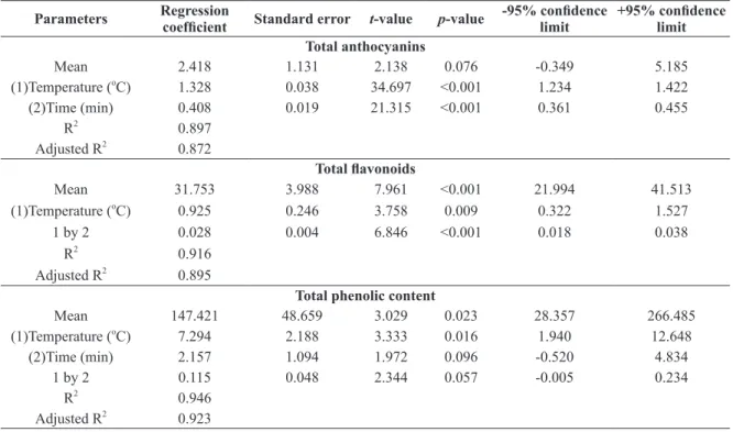
based on the response surface methodology was used and results are shown in Table II and the contour plots
generated using the experimental data are shown in Figure 1. Both extraction time and temperature were highly significant (t-value = 21.32 and 34.70, p < 0.001, respectively) in the extraction of total anthocyanins
and the model was deemed significant (R2 = 0.897 and adjusted R2 = 0.872).
TABLE II
Regression coefficients obtained by multiple regression analysis (response surface methodology) to assess the effects of time and temperature of extraction of total anthocyanins, flavonoids, total phenolic content, and antioxidant
activity of Ocimum basilicum L. leaf extracts.
Parameters Regression coefficient Standard error t-value p-value -95% confidence limit
+95% confidence limit Total anthocyanins
Mean 2.418 1.131 2.138 0.076 -0.349 5.185
(1)Temperature (o
C) 1.328 0.038 34.697 <0.001 1.234 1.422
(2)Time (min) 0.408 0.019 21.315 <0.001 0.361 0.455
R2
0.897
Adjusted R2
0.872
Total flavonoids
Mean 31.753 3.988 7.961 <0.001 21.994 41.513
(1)Temperature (o
C) 0.925 0.246 3.758 0.009 0.322 1.527
1 by 2 0.028 0.004 6.846 <0.001 0.018 0.038
R2
0.916
Adjusted R2
0.895
Total phenolic content
Mean 147.421 48.659 3.029 0.023 28.357 266.485
(1)Temperature (o
C) 7.294 2.188 3.333 0.016 1.940 12.648
(2)Time (min) 2.157 1.094 1.972 0.096 -0.520 4.834
1 by 2 0.115 0.048 2.344 0.057 -0.005 0.234
R2
0.946
Adjusted R2
For the total flavonoids content, the effect of temperature (t-value = 3.758, p = 0.009) and the linear
interaction between time and temperature (t-value = 6.846, p < 0.001) were the parameters that influenced the response, while the effect of time (t-value = 1.409, p = 0.208) was not important. This model also presented a high coefficient of determination (R2 = 0.916 and adjusted R2 = 0.895), implying that more than 90% of data variability was explained by the multiple linear regression model (Bassani et al. 2014).
Parameters Regression coefficient Standard error t-value p-value -95% confidence limit
+95% confidence limit Free radical scavenging activity toward DPPH
Mean 28.598 2.220 12.879 <0.001 23.165 34.031
(1)Temperature (o
C) 1.177 0.075 15.663 <0.001 0.993 1.361
(2)Time (min) 0.266 0.037 7.094 <0.001 0.175 0.358
R2
0.865
Adjusted R2
0.831
TABLE II (continuation)
Figure 1 - Contour plots to project the experimental data obtained for the extraction of total anthocyanins (a), total flavonoids (b), total phenolic content (c), and free radical scavenging activity (d) of Ocimum basilicum L. extracts. All data are expressed as
The higher the extraction time (t-value = 1.972, p = 0.096) and, more importantly the temperature
(t-value = 3.333, p = 0.016), the higher the content of total phenolic compounds. The interaction between these two factors also influenced positively the extraction of phenolic compounds from purple basil (t-value
= 2.344, p = 0.057). The regression model explained up to 95% of data variability, indicating the suitability
of RSM in modeling the extraction of total phenolic content in the range studied.
The free radical scavenging activity toward DPPH radical was also modeled by means of RSM and
results clearly indicated the statistical significance of extraction time (t-value = 7.094, p < 0.001) and temperature (t-value = 15.663, p < 0.001) on the response. Interestingly, the interaction of both independent
variables did not affect significantly (t-value = -0.293, p = 0.780) the antioxidant activity of purple basil extracts under the conditions adopted in the current work. The proposed multiple regression model presented
R2 = 0.865 and adjusted R2 = 0.831, indicating it was possible to model the effects of time and temperature on such a chemical response.
One of the most important advantages of using RSM is to provide useful regression equations that can be applied to predict certain responses based on pre-selected factors. In other words, if the equations
generated are statistically significant (p-value of regression coefficients are below the stipulated α-value) and the equations can explain more than 70% of data variability (R2 > 0.70), it is possible to assume
the model can be used to predict the response on the basis of specific values of the factors (independent
variables). In our case, all regression models generated presented p< 0.10 and R2 > 0.80. So, if one considers
the extraction of total anthocyanins conducted at T = 27 oC and t = 47 min, the mean theoretical value would
be 57.46 mg/100 g (confidence interval at ± 95% = 56.37 – 58.54 mg/100 g). The same is applied to the
other variables: if one wishes to have an idea of the experimental value to be obtained when the extraction
of total flavonoids is conducted at T = 18.8 oC and t = 36 min, the mean concentration of flavonoids should
be 68.15 mg/100 g and the confidence interval at ± 95% would be 64.21 and 72.08 mg/100 g. Clearly, it would be more valid if the analysis goes back to the laboratory and perform the experiment under the
simulated conditions (T and t) and compare the experimental and theoretical values. If both results are in
agreement (relative standard error below 10%), then the regression model can be regarded as significant and
predictive (Granato et al. 2014b).
Similarly to what was observed in the current study, Bassani et al. (2014) used a 32 design of experiment aimed to model the effects of extraction time (5, 7.5, and 10 min) and temperature (60, 75, 90 oC) on the
total content of phenolic compounds and total flavonoids content as well as on the antioxidant capacity
(DPPH assay) of water-soluble extracts from roasted leaves of Ilex paraguariensis and authors obtained high R2 values: 0.989 (antioxidant activity), 0.997 (total phenolic content), and 0.999 (total flavonoids).
Pedro et al. (2016) used a Box–Behnken design to assess the effects of temperature (10 – 50 ºC), time (20-80 min), and solid-solvent ratio (1:15 – 1:45 w/v) on the extraction of total phenolic content, total flavonoids
content, total anthocyanins, and in vitro antioxidant activity of extracts from Oryza sativa L. (black rice). Experimental data were subjected to RSM modeling and authors were able to identify the factors (alone
and in combination) that affected the extraction of bioactive compounds and presented regression equations based on RSM to explain the results.
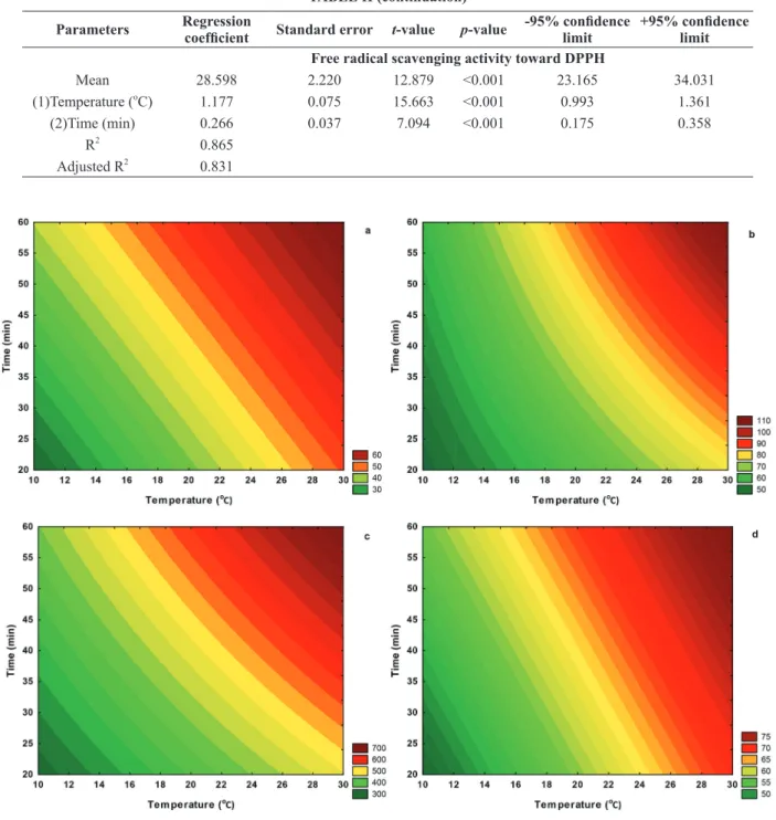
Using linear regression analysis, it was possible to analyze the correlation between the antioxidant
reaction medium. The proposed equations can be used experimentally (inside the range of the study) to have
an idea of the antioxidant activity toward DPPH radical in terms of the content of bioactive compounds. For
example, if one wishes to estimate the antioxidant activity of a purple basil extract containing 350, 87.50,
or 53.90 mg/100 g of total phenolic content, total flavonoids, and total anthocyanins, respectively using the exact extraction conditions employed in the current work, one would obtain, theoretically, 52.17, 70.50, and
69.85% of DPPH inhibition, respectively.
Figure 2 - Regression analysis based on the ordinary least-squares method between the free radical scavenging activity toward DPPH and the total content of phenolic compounds (a), flavonoids (b), and total anthocyanins (c). Note that TPC = total phenolic
content, TFC = total flavonoids content, and TAC = total anthocyanins content.
The antioxidant activity of phenolic compounds in a food extract depends on the number and type of functional groups present in the aromatic ring, such as the hydroxyl groups and the position they occupy, either ortho or para, as well as the presence of other groups. The combination of the non-binding electron
pair of the hydroxyl group with the PI system (л) of the aromatic ring strongly activates the ortho and para positions. Similarly, other functional groups, such as alkyl and methoxyl weakly activate the ortho and para positions by hiperconjugative effects. These groups favor electron delocalization and stabilize the phenoxyl
staBility stuDyof PurPle Basil anthoCyanins effeCtof temPerature
From the study of the effect of temperature on the stability of anthocyanins extracted from purple basil, a linear behavior was observed, indicating that the thermal degradation followed first-order kinetics. Table
III shows the degradation rate constant (k), the half-life time (t1/2) and color retention percentage (% R) of the anthocyanins as the temperature increased. These parameters show that the rate of degradation reaction increased when the temperature was raised as a function of exposure time. It is observed that the half-life
decreased markedly according to the increase in temperature. Patras et al. (2010) suggested that foods rich
in anthocyanins should be manufactured at low temperatures and be stored for reduced periods of time. This provides greater retention of color and stability to the pigment.
The activation energy for the reaction of anthocyanins degradation was 44.95 kJ/mol. Mourtzinos et al. (2008) found that the activation energy was 54 kJ/mol for anthocyanins extracted from Hibiscus sabdariffa
L. According to Silva et al. (2010) excessive heating may promote the loss of anthocyanin color and
cause the opening of the aromatic ring chalcones forming colorless compounds (Katsaboxakis et al. 1998,
Heredia et al. 1998). Reduced values of activation energy suggest that the anthocyanin extract is susceptible to degradation when temperature is increased (Hou et al. 2013).
effeCtof lightanD CoPigmentation
Table IV shows the rate constants and half-lives for reaction of anthocyanins degradation toward light
in the purple basil with or without copigments. It was found that after 24 hours, all extracts exposed to
light showed no significant difference in the half-life (control - 17.08 h, phytic acid -15.61 h and gallic acid - 17.20 h). However, all extracts protected from light showed a remarkably higher half-life than the
extracts exposed to light. Lima et al. (2005) observed that the anthocyanin extracts from purple cherry
showed greater stability when protected from light. Bąkowska et al. (2003) found that the heating and UV
irradiation affect negatively the stability of anthocyanins.
The half-life for the extracts protected from light in the presence of phytic and gallic acids was 923.17 and 856.49 h, after 24 hours, respectively, whereas half-life of the control was 152.67 h. The considerable increase in half-life of the extracts in the presence of phytic and gallic acids compared to the control shows the effect of cogpigmentation on the chemical stabilization of anthocyanins. Copigments have pi electron and may link with the flavylium ion. This combination protects the anthocyanin molecule against nucleophilic attack by water and consequently increases its chemical stability (Mazza and Brouillard 1990).
The interaction of copigments with the anthocyanins gives the bathochromic effect (increase of the
maximum absorbance) and hyperchromatic effect (shift of the peak wavelength) (Bąkowska et al. 2003). TABLE III
Effects of temperature on the degradation of anthocyanins as noted by the first-order rate constant (k), half-life time (t1/2), and retention of color (R).
Temperature (°C) k (h-1) a R² t1/2 (h) R (%) b p-value
60 0.01288 0.9974 53.82 0.963 <0.0001
70 0.02261 0.9990 30.66 0.935 0.0226
80 0.02515 0.9977 27.56 0.915 <0.0001
90 0.05466 0.9935 12.68 0.887 0.0006
a
R²= coefficient of determination; b
The increase in half-life of anthocyanin extracts (protected from light) in the presence of copigments
yielded a significant increase in the chemical stability of the extracts. Gauche et al. (2010) observed an
increased half-life of anthocyanins from Cabernet Sauvignon grapes added with tannic and gallic acids as compared to the control. Gris et al. (2007) found that a solution containing anthocyanins extracted from Caberbet Sauvignon grapes in the presence of caffeic acid and absence of light presented an increased half-life compared to the control.
Some experimental parameters, such as solvent polarity, temperature, extraction time, and solvent: food matrix ratio are important to extract bioactive compounds from a certain source. The most commonly
extraction solvents used are methanol and ethanol in acidic medium. Under these conditions the flavylium
cation of anthocyanins (red color) is more stable. Strong acids can hydrolyze the structure of the molecule,
while weak acids such as citric and tartaric acids are preferred (Strack and Wray 1989). Methanol is an efficient solvent for extraction of many bioactive compounds, however, its degree of toxicity should be
considered, especially by food companies (Escribano-Bailón et al. 2004). Thus, considering the stability of anthocyanins in acidic medium and the toxicity of the solvent, ethanol in the presence of citric acid was
used in the current work to extract antioxidant compounds from purple basil. The use of this solvent can
facilitate the application of the extract in food products due to its low toxicity.
As a conclusion, RSM was shown to be a suitable statistical approach to model the effects of time and temperature on the extraction of bioactive compounds as well as antioxidant activity from purple basil leaf. As the anthocyanins extract was stable at moderate temperature, the application of such extract in
food matrix may be advantageous over synthetic colorants. In this sense, further works should focus on
the application of purple basil extracts in beverages and other foods and study their sensory properties and chemical stability during shelf life.
ACKNOWLEDGMENTS
Authors are grateful for the financial support provided by Coordenação de Aperfeiçoamento de Pessoal
de Nível Superior/Programa de Apoio à Pós-Graduação (CAPES/PROAP) and Programa Nacional de Pós-Doutorado (CAPES/PNPD) (D. Granato) and to the support provided by Central de Laboratórios Multiusuários/Universidade Estadual de Ponta Grossa (CLABMU/UEPG).
RESUMO
No presente estudo, a metodologia de superfície de resposta (MSR) foi utilizada para avaliar o efeito de tempo e temperatura de extração no conteúdo de compostos bioativos e atividade antioxidante de extratos de folhas de manjericão
TABLE IV
Effects of light and copigments on the first-order rate constant (k) and half-life time (t1/2) on the degradation of anthocyanins extracted from Ocimum basilicum L. leaf.
Extract k (h-1) a R² t1/2 (h) b p-value
Control (protected) 0.00454 0.9998 152.67 <0.0001
Control (exposed) 0.04058 0.9963 17.08 <0.0001
Phytic acid (protected) 0.00075 0.9969 923.17 <0.0001
Phytic acid (exposed) 0.0444 0.9977 15.61 <0.0001
Gallic acid (protected) 0.00081 0.9985 856.49 <0.0001
Gallic acid (exposed) 0.04029 0.9987 17.20 <0.0001
a
R²= coefficient of determination; b
roxo (Ocimum basilicum L.). A estabilidade das antocianinas relacionada à temperatura, luz e copigmentação também foi estudada. O maior conteúdo de antocianinas foi 64,70 mg/100 g a 30 °C e 60 min. A degradação das antocianinas com diferentes temperaturas e em presença de luz seguiu uma cinética de primeira ordem e a energia de ativação foi 44,95 kJ/mol. Todos os extratos expostos à luz apresentaram tempos de meia vida semelhantes. Enquanto que os extratos protegidos da luz em presença dos copigmentos apresentaram um aumento do tempo de meia vida: 856,49 h para o ácido gálico e 923,17 h o ácido fítico enquanto que para o controle foi de 152,67 h. Estes resultados indicam claramente que o manjericão roxo é uma potencial fonte de compostos bioativos estáveis.
Palavras-chave: antocianinas, extração, pigmentos, metodologia de superfície de resposta.
REFERENCES
alvarez Pa, emonD C, gomaa a, remonDetto ge anD suBiraDe m. 2015. Predictive response surface model for
heat-induced rheological changes and aggregation of whey protein concentrate. J Food Sci 80: 326-333.
amr a anD al-tamimi e. 2007. Stability of the crude extracts of Ranunculus asiaticus anthocyanins and their use as food
colourants. Int J Food Sci Tech42: 985-991.
awika JM. 2008. Behavior of 3-deoxyanthocyanidins in the presence of phenolic copigments. Food Res Int 41: 532-538. Bąkowska a, kucharska aZ and osZmiański J. 2003. The effects of heating, UV irradiation, and storage on stability of the
anthocyanin–polyphenol copigment complex. Food Chem 81: 349-355.
Bassani DC, nunes Ds anD granato D. 2014. Optimization of phenolics and flavonoids extraction conditions and antioxidant activity of roasted yerba-mate leaves (Ilex paraguariensis A. St.-Hil., Aquifoliaceae) using response surface methodology. An Acad Bras Cienc 86: 923-934.
Bettini v, fiori a, martino r, mayellaro r anD ton P. 1985. Study of the mechanism whereby anthocyanosides potentiate
the effect of catecholamines on coronary vessels. Fitoterapia 54: 67-72.
BranD-williams w, Cuvelier me anD Berset C. 1995. Use of a free radical method to evaluate antioxidant activity. Lebensm Wiss-Technol 28: 25-30.
BriDle P anD timBerlake Cf. 1997. Anthocyanins as natural food colours–selected aspects. Food Chem 58: 103-109. capecka e, marecZek a and LeJa m. 2005. Antioxidant activity of fresh and dry herbs of some Lamiaceae species. Food
Chem 93: 223-226.
Carvalho-filhoJLS, Blank af, alves PB, ehlertPBA, melo as, CavalCantiSCH, arrigoni-Blank mf anD silva -mann r. 2006. Influence of the harvesting time, temperature and drying period on basil (Ocimum basilicum L.) essential oil.
Rev Bras Farmacogn 16: 24-30.
escriBano-BaiLón mT, sanTos-BueLga c and rivas-gonZaLo Jc. 2004. Review–Anthocyanins in cereals. J Chromatogr
A 1054: 129-141.
Francis FJ. 1989. Food Colorants: Anthocyanins. Crit Rev Food Sci 28: 273-314.
gauChe C, malagoli es anD luizMTB. 2010. Effect of pH on the copigmentation of anthocyanins from Cabernet Sauvignon grape extracts with organic acids. Sci Agr67: 41-46.
granato D, CalaDoVMAand Jarvis B. 2014a. Observations on the use of statistical methods in food science and technology.
Food Res Int 55: 137-149.
granato D, grevink r, zielinskiAAF, nunes Ds anD van ruth sm. 2014b. Analytical strategy coupled with response surface methodology to maximize the extraction of antioxidants from green, yellow, and red teas (Camellia sinensis var sinensis). J Agric Food Chem 62: 10283-10296.
gris ef, ferreira ea, falCão lD anD BorDignon-luiz mt. 2007. Caffeic acid copigmentation of anthocyanins from
Cabernet Sauvignon grape extracts in model systems. Food Chem 100: 1289-1296.
heredia FJ, Francia-aricha em, rivas-gonZaLo Jc, vicario im and sanTos-BueLga c. 1998. Chromatic chaterization
of anthocyanins from red grapes–I. pH effect. Food Chem 63: 491-498.
holton ta anD Cornish eC. 1995. Genetics and biochemistry of anthocyanin biosynthesis. Plant Cell7: 1071-1083.
hou z, Qin P, zhang y, Cui s anD ren g. 2013. Identification of anthocyanins isolated from black rice (Oryza sativa L.) and
their degradation kinetics. Food Res Inter 50: 691-697.
katsaBoxakis k, PaPaniColaou D anD melanitou m. 1998. Stability of pigmented orange anthocyanins in model and real
food systems. Ital J Food Sci 10: 17-25.
kim sJ, kim Jm, shim sh and chang hi. 2014. Anthocyanins accelerate the healing of naproxen-induced gastric ulcer in rats
Lee Jh, Lim Jd and choung mg. 2013. Studies on the anthocyanin profile and biological properties from the fruits of Acanthopanax senticosus (Siberian Ginseng). J Funct Foods 5: 380-388.
Lee sJ, umano k, shiBamoTo T and Lee kg. 2005. Identification of volatile components in basil (Ocimum baslicum L.) and
thyme leaves (Thymus vulgaris L.) and their antioxidant properties. Food Chem 91: 131-137.
Lees dh and Francis FJ. 1972. Standardization of pigment analyses in cranberries. Hortscience 7: 83-84.
limaVLAG, mélo ea anD limaDES. 2005. Efeito da luz e da temperatura de congelamento sobre a estabilidade das antocianinas da pitanga roxa.Ciênc Tecnol Aliment25: 92-94.
mazza g anD BrouillarD r. 1990. The mechanism of co-pigmentation of anthocyanins in aqueous solutions. Phytochem 29: 1097-1102.
mourtzinos i, makris DP, yannakoPoulou k, kalogeroPoulos n, miChali i anD karathanos vt. 2008. Thermal Stability of Anthocyanin Extract of Hibiscus sabdariffa L. in the Presence of β-Cyclodextrin. J Agric Food Chem 56: 10303-10310.
nguyen Pm, kwee em anD niemeyer eD. 2010. Potassium rate alters the antioxidante capacity and phenolic concentration of
basil (Ocimum basilicum L.) leaves. Food Chem 123: 1235-1241.
nontasan s, moongngarm a anD Deeseenthum s. 2012. Application of functional colourant prepared from black rice bran in yogurt. APCBEE Procedia 2: 62-67.
nunes ca, aLvarenga vo, sanT’ana as, sanTos JsanDgranato D. 2015. The use of statistical software in food science
and technology: Advantages, limitations and misuses. Food Res Int 75: 270-280.
omBra mn, fratianni f, granese t, CarDinale f, Cozzolino a anD nazzaro f. 2015. In vitro antioxidant, antimicrobial and anti-proliferative activities of purple potato extracts (Solanum tuberosum cv Vitelotte noire) following simulated gastro-intestinal digestion. Nat Prod Res 29: 1087-1091.
o’shea n, röBle C, arenDt eanD gallagher e. 2015. Modelling the effects of orange pomace using response surface
design for gluten-free bread baking. Food Chem 166: 223-230.
PasCual-teresa s. 2014. Molecular mechanisms involved in the cardiovascular and neuroprotective effects of anthocyanins. Arch Biochem Biophys 559: 68-74.
Patil g, maDhusuDhan mC, BaDu Br anD raghavaraoKSMS. 2009. Extraction, dealcoholization and concentration of anthocyanin from red radish. Chem Eng Process 48: 364-369.
Patras a, Brunton nP, o’Donnell C anD tiwari Bk. 2010. Effect of thermal processing on anthocyanin stability in foods;
mechanisms and kinetics of degradation. Food Sci Technol 21: 3-11.
PeDro aC, granato D anD rosso nD. 2016. Extraction of anthocyanins and polyphenols from black rice (Oryza sativa L.) by
modeling and assessing their reversibility and stability. Food Chem 191: 12-20.
pereira-caro g, waTanaBe s, FuJimura T, YokoTa T and ashihara h. 2013. Phytochemical profile of a Japanese black-purple rice. Food Chem141: 2821-2827.
pheTpornpaisan p, TippaYawaT p, JaY m and suTThanuT k. 2014. A local Thai cultivar glutinous black rice bran: A source
of functional compounds in immunomodulation, cell viability and collagen synthesis, and matrix metalloproteinase-2 and -9
inhibition. J Funct Foods 7: 650-661.
phippen wB and simon Je. 1998. Anthocyanins in Basil (Ocimum basilicum L.). J Agric Food Chem 46: 1734-1738.
reBouças mC, roDriguesMCPanD afonsoMRA. 2014. Optimization of the acceptance of prebiotic beverage made from
cashew nut kernels and passion fruit juice. J Food Sci 7: 1393-1398.
reyes lf anD Cisneros-zevallos l. 2007. Degradation kinetics and colour of anthocyanins in aqueous extracts of purple and red-flesh potatoes (Solanum tuberosum L.). Food Chem 100: 885-894.
rice-evans ca, miLLer nJ and paganga g. 1996. Structure antioxidant activity relationships and flavonoids and phenolic
acids. Free Radical Bio Med 20: 993-956.
rosas JF, siLvaACM, zoghBiMGBanD anDraDeEHA. 2004. Comparação dos voláteis das folhas de Ocimum micranthum Willd. obtidos por hidrodestilação e destilação-extração simultânea. Rev Bras Plantas Med 7: 26-29.
saha J, deBnaTh m, saha a, shosh T and sarkar pk. 2011. Response surface optimization of extraction of antioxidants
from strawberry fruit, and lipid peroxidation inhibitory potential of the fruit extract in cooked chicken patties. J Sci Food Agric
9: 1759-1765.
shao Q, huang y, zhou a, guo h, zhang a anD wang y. 2014. Application of response surface methodology to optimise supercritical carbon dioxide extraction of volatile compounds from Crocus sativus. J Sci Food Agric 94: 1430-1436.
shao Y, Xu F, sun X, Bao J and BeTa T. 2014. Identification and quantification of phenolic acids and anthocyanins as
antioxidants in bran, embryo and endosperm of white, red and black rice kernels (Oryza sativa L.).J Cereal Sci 59: 211-218. silvaGJF, ConstantPBL, figueireDo rw anD moura sm. 2010. Formulação e estabilidade de corantes de antocianinas
singleton vl, orthofer r anD lamuela-raventos rm. 1999. Analysis of total phenols and other oxidation substrates and
antioxidants by means of Folin-Ciocalteu reagent. Method Enzymol 299: 152-178.
straCk D anD wray v. 1989. In: Dey PM and Harborne JB (Eds), Anthocyanins, in Methods in Plant Biochemistry, Academic
Press, New York, p. 325.
sZYmanowska u, ZłoTek u, karaś m and Baraniak B. 2015. Anti-inflammatory and antioxidative activity of anthocyanins
from purple basil leaves induced by selected abiotic elicitors. Food Chem 172: 71-77.
uDDinMDS, sarkerMDZI, ferDosh s, akanDaMDJH, easminMSTS, BtshamsuDin sh anD yunus kB. 2015.
Phytosterols and their extraction from various plant matrices using supercritical carbon dioxide: a review. J Sci Food Agric 95:
1385-1393.
usman la, ismaeel ro, zuBair mf, saliu Bk, olawore no anD elelu n. 2013. Comparative studies of constituents and antibacterial activities of leaf and fruit essential oils of Ocimium basilicum grown in north central Nigeria. Int J Chem Biochem Sci 3: 47-52.
vinCieri ff, romani a, BalDi a, mulinaCCi n anD alBerti mB. 1992. Analysis HPLC of anthocyanins present in fluid extracts from Malva sylvestrisL. flowers and leaves. Bull. Liaison-Groupe Polyphenols16: 339-342.
waterhouse al. 1995. Wine and heart disease. Chem Ind-London9: 338-341.
wu xl anD Prior rl. 2005. Identification and characterization of anthocyanins by high-performance liquid
chromatograpy-electrospray ionization-tandem mass spectrometry in common foods in the United States: Vegetables, nuts, and grains. J Agric Food Chem 53: 3101-3113.
ZawisTowski J, kopec a and kiTTs dd. 2009. Effects of a black rice extract (Oryza sativa L. indica) on cholesterol levels and
plasma lipid parameters in Wistar Kyoto rats. J Funct Foods 1: 50-56.
Zhang Y, kong L, Yin c, Jiang d, Jiang J, he J and Xiao w. 2013. Extraction optimization by response surface methodology, purification and principal antioxidant metabolites of red pigments extracted from bayberry (Myrica rubra) pomace. LWT –