ACTA RADIOLÓGICA PORTUGUESA May-August 2018 Vol 30 nº 2 25-29
IgG4 Associated Cholangitis and Hepatic Pseudoinflammatory
Tumour: IgG4 Related Disease Mistaken by Neoplastic Disease
Colangite e Pseudotumour Inflamatório Hepático Associados a IgG4: Doença Associada
a IgG4 Simulando Doença Neoplásica
Ana Isabel S. Ferreira*, Natália Ferreira*, Raquel Gaio*, Afonso Gonçalves*, Isabel Távora**, José Fonseca Santos
*Departament of Radiology, Centro Hospitalar Lisboa Norte, Lisboa, Portugal
**Departament of Radiology, Hospital CUF Descobertas, Lisboa, Portugal
Address
Ana Isabel Ferreria Departamento de Radiologia Centro Hospitalar Lisboa Norte Av. Prof Egas Moniz 1649-035 Lisboa, Portugal email: anaisabelvnsf@gmail.com
Resumo
Apresentamos o caso de um homem de 71 anos com icterícia colestática inaugural, com estenose e espessamento parietal circunferencial do colédoco distal, assim como uma massa infiltrativa hepática condicionando “amputação” das vias biliares dilatadas à periferia. O doente foi submetido a pancreatoduodenectomia e hemi-hepatectomia por suspeita de colangiocarcinoma. A histologia revelou tratar-se de colangite e pseudotumor inflamatório hepático (PIH) associados a IgG4.
A doença associada a IgG4 (DA-IgG4) foi recentemente estabelecida como uma condição sistémica, em que ocorrem infiltrados fibro-inflamatórios e tumefação de um ou mais órgãos. As manifestações hepatobiliares incluem a colangite e o PIH associados a IgG4 – que fazem diagnóstico diferencial com colangiocarcinoma. Os médicos devem estar familiarizados com esta patologia, normalmente responsiva à corticoterapia, para evitar procedimentos invasivos desnecessários.
Os autores apresentam um caso de colangite e PIH associados a IgG4 e fazem uma revisão da literatura desta rara apresentação de DA-IgG4. Palavras-chave
Doença associada a IgG4; Colangite autoimune; Pseudotumor inflamatório hepático;
Colangiocarcinoma; Hemi-hepatectomia; Pancreateduodenectomia cefálica. Abstract
We report the case of a 71-year-old man presenting inaugural cholestatic jaundice, with a stricture and circumferential wall thickening of the distal common bile duct, as well as an ill-defined hepatic infiltrative mass with “amputation” of peripheral distended bile ducts. The patient underwent a pancreaticoduodenectomy and hemihepatectomy for suspected cholangiocarcinoma. Histology revealed IgG4 associated cholangitis (IgG4-AC) and hepatic pseudoinflammatory tumour (HPT). IgG4-related disease (IgG4-RD) is a recently established systemic disease, characterized by fibro-inflammatory tissue infiltrates and tumefaction of one or more organs. The hepatobiliary manifestations comprise IgG4-AC and associated HTP - which can be mistaken for cholangiocarcinoma. Clinicians must be familiar with this disorder, usually responsive to corticosteroid therapy, in order to avoid unnecessary invasive procedures.
We present a case of IgG4-AC and associated HTP and review the literature of this rare presentation of IgG4-RD.
Keywords
IgG4-related disease; Autoimmune cholangitis; Hepatic pseudoinflammatory tumour; Cholangiocarcinoma; Hemihepatectomy; Cephalic pancreaticoduodenectomy. Radiological Case Report / Caso Clínico
Introduction
Immunoglobulin G4-related disease (IgG4-RD) is a systemic condition affecting virtually every organ system that comprises common pathologic, serologic and clinical features. It has been
increasingly recognized since 2003, when Kamisawa et al1
proposed that a group of disorders, previously thought to be unrelated, might be part of a broader spectrum of IgG4-RD; nevertheless, pathogenesis is still poorly understood. Common
of involved organs, detecting multiorgan involvement and, eventually, for assessing response to therapy.
Clinical Case
We report the case of a 71-year-old man who presented himself at the emergency department of our hospital in August 2015 complaining of abdominal pain for the previous week, jaundice, choluria and diarrhea with acholic faeces. At physical examination, the abdomen was tender at
26
vildagliptin, enalapril and hydrochlorothiazide, diosmin and esomeprazole.
Blood tests at admission showed high levels of total bilirubin 9.90mg/dL (<1.2mg/dL) with a predominant increase of the direct bilirubin (conjugated component), as well as high levels of alanine aminotransferase 116mg/ dL (12-78mg/dL), aspartate aminotransferase 122mg/ dL (<34mg/dL), gamma-glutamyl transferase 1330mg/ dL (<73mg/dL), alkaline phosphatase 880mg/dL (40-130mg/dL); slightly elevated C-reactive protein 0.9mg/dL (<0.5mg/dL); abnormal presence of moderate quantity of bilirubin in urine analysis; low levels of amylase (3 U/L) and haemoglobin of 11.8 mg/dL (13-17.5mg/dL). Further workup, during the hospital stay, revealed normal levels of the tumour markers CEA of 1.9mg/dL (<5mg/dL) and CA 19.9 of 23.4mg/dL (0-37mg/dL), as well as further progressive increase of total bilirubin level.
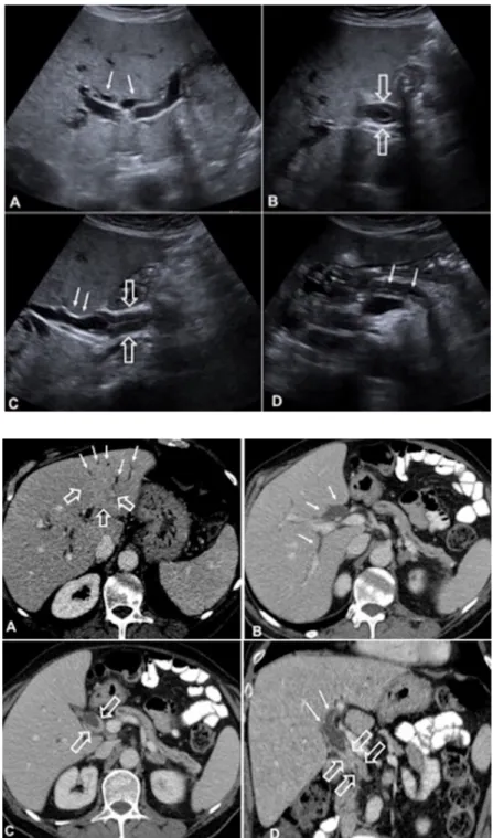
An abdominal ultrasound was performed at admission (Fig. 1), which revealed bilateral dilatation of the intrahepatic biliary ducts, as well as the proximal half of the common bile duct (CBD), with stenosis and a significant hipoechogenic circumferential wall thickening of the remaining CBD; non distended gallbladder, without signs of cholelithiasis; diffuse atrophy of the pancreatic parenchyma and mild dilatation of the main pancreatic duct.
A computed tomography (CT) was performed subsequently (Fig. 2), confirming the previous findings of biliary ducts dilatation (CBD calibre of 19mm), with stenosis and a significant circumferential and regular wall thickening of the distal common bile duct, with homogeneous contrast enhancement; also, diffuse atrophy of the pancreatic parenchyma, with some calcific foci, mild diffuse dilatation (4-5mm) and slight irregularity of the main pancreatic duct (suggestive of chronic pancreatitis). These findings were
Figure 1 – Ultrasound. (A) Axial plane, showing
bilateral dilatation of the intra-hepatic biliary ducts (arrows). (B) Axial plane and (C) sagittal plane, demonstrating dilatation of the proximal CBD (arrow), with stenosis and hipoechogenic circum-ferential wall thickening of the remaining CBD (open arrows). (D) Axial plane, showing diffuse atrophy of the pancreatic parenchyma and mild dilatation of the main pancreatic duct (arrows). It was also noticed moderate hepatomegaly with signs of steatosis and minor splenomegaly.
Figure 2 – Enhanced computed tomography
(CT). (A) Axial plane in a liver dedicated window, depicting a poorly circumscribed area of hetero-geneity on segment II of the left liver lobe, with mild delayed enhancement (open arrows), causing “amputation” of peripheral dilated intra-hepatic bile ducts (arrows). Axial plane at a higher level (B) and a lower level (C), as well as coronal plane (D), demonstrating dilatation of the bile ducts (arrows), with stenosis and a significant circumferential and regular wall thickening of the distal CBD, with ho-mogeneous contrast enhancement (open arrows). Also, notice the diffuse atrophy of the pancreatic parenchyma, with some calcific foci, and mild diffuse dilatation and slight irregularity of the main pancreatic duct.
not present on a previous CT (dated from September 2014). The magnetic resonance cholangiopancreatography, also confirmed the previous findings and further revealed an area of left hepatic parenchyma heterogeneity with ill-defined margins at segment II, presenting mild hiperintensity at T2-weighted images (WI) and hipointensity at T1-WI, associated with “amputation” of peripheral dilated intrahepatic bile ducts and an altered luminal signal intensity (hipointensity at T2-WI and hiperintensity at T1-WI) in the proximal dilated bile ducts. Retrospectively, a corresponding ill-defined infiltrative mass, with a mild delayed enhancement, was also depicted on CT.
Based on the clinical and imaging elements, a presumptive diagnosis of multifocal cholangiocarcinoma was made. Since no alternative diagnosis was proposed, biopsy was not performed and the patient underwent surgery. A cephalic pancreaticoduodenectomy and a left hemihepatectomy were performed. The pathological findings were highly suggestive of IgG4-related disease, with IgG4 associated cholangitis (IgG4-AC) and a hepatic pseudoinflammatory tumour (with approximately 4cm), probably related to a focal IgG4-AC exacerbation; no neoplastic tissue was found. Ensuing blood tests revealed IgG values of 2160 md/dL, but the IgG4 serum levels were not subsequently assessed.
The post-operative course was complicated by a surgical wound infection, a fluid collection adjacent to the jejunopancreatic anastomosis with fistulisation to the skin in the right upper abdomen, another infected haematic collection in the left upper abdomen, as well as a left empyema, that required intravenous broad spectrum antibiotics and percutaneous drainage. Except for the surgical wound infection, these complications were, eventually, managed after a long course of conservative treatment and the patient was discharged home 39 days after surgery. The surgical wound infection went on for several months, postponing the beginning of treatment with systemic corticosteroids.
Discussion
The hepatobiliary manifestations of IgG4-related disease (IgG4-RD) comprise cholangitis, pseudoinflammatory
tumour and IgG4 hepatopathy.3
After the pancreas, the bile ducts are the second most commonly involved organ, seen in up to 88% of patients
with IgG4-RD.3 IgG4-associated cholangitis (IgG4-AC)
occurs more frequently in patients with type 1 autoimmune pancreatitis (60-70%), but it might also occur without pancreatic involvement, making the diagnosis more
Figure 3 – Magnetic resonance (MR). T2-weighted image
(WI) (A), T2-WI with fat suppression (B) and T1-WI (C) showing at the level of hepatic segment II an area of parenchyma heterogeneity, with mild hiperintensity on T2-WI and hipointensity on T1-WI, and peripheral dilated intra-hepatic bile ducts with an altered luminal signal intensity. T2-WI (D), (E) and (F) showing dilatation of the proximal half of the CBD with stenosis and thickening of the distal CBD (open arrows). (G) MR cholangiography, showing the dilated bile ducts until the point of stenosis in the CBD (open arrow); and a defect in the intra-hepatic bile ducts at hepatic segment II (arrows), corresponding to the area of parenchyma heterogeneity.
28
difficult.4,5 Involvement by other extrapancreatic lesions
may be additionally seen.2,6 Generally, IgG4-AC affects
older patients and can induce obstructive jaundice.5
Both intra- and extrahepatic segments can be affected by dense bile duct infiltration of IgG4-positive plasma cells and extensive fibrosis, but the most commonly involved segment is the intrapancreatic portion of the common bile duct (CBD). The typical imaging appearance, at MRCP, is focal or diffuse bile duct wall thickening - usually long and continuous strictures are depicted - mostly associated with stenosis and upstream dilatation.7 Isolated strictures of the
distal CBD may also occur.8 CT and MR imaging usually
depict a circular and symmetric ring of tissue encasing the bile duct wall, with relatively smooth margins and homogeneous enhancement in the delayed phase.
The main differential diagnoses are cholangiocarcinoma and primary sclerosing cholangitis (PSC). The latter would not be expected in this case due to the acute onset of symptoms, older age and no previous history of inflammatory bowel disease; also, the typical findings for PSC are multifocal and short intrahepatic biliary strictures with beaded or “pruned-tree” appearance.5,8 Differentiation
from cholangiocarcinoma may be difficult, especially in the presence of a soft-tissue mass that produces stenosis of the hilar hepatic bile duct, presented as inaugural jaundice. In the case of cholangiocarcinoma, tumour infiltration is usually confined to either extrahepatic or intrahepatic bile ducts, whereas in IgG4-AC luminal irregularities and stenosis generally involve both the biliary and pancreatic
ducts.2,9 Differentiating hilar cholangiocarcinoma from
other causes of hilar obstruction remains an issue.6 There
are a few reports in the literature addressing cases of IgG4-cholangitis of the CBD, with no apparent pancreatic involvement, that underwent surgery for suspected cholangiocarcinoma.6,10-12
IgG4 related cholangitis and hepatic inflammatory pseudotumour (HIP) is an extremely rare association, in which the cholangitis is considered either as the condition preceding or associated with the inflammatory
pseudotumour.13 Inflammatory pseudotumours occur
most commonly in the lung and only occasionally in extrapulmonary organs, including the liver in about 8%
of cases.14 Inflammatory pseudotumours are characterised
histologically by the proliferation of fibroblasts or myofibroblasts and inflammatory cell infiltrate, independently of the organs where they occur. Infections and autoimmune reactions have been suggested as possible triggers, although pathogenesis of hepatic inflammatory pseudotumours is still incompletely understood and
might differ among cases.14 On CT, homogeneous delayed
enhancement was considered an important diagnostic imaging characteristic of IgG4-related disease. On MRI, these lesions are typically hypointense on T1-WI and hyperintense on T2-WI, but contrast enhancement patterns are variable.15 The differential diagnosis is generally with a
primary liver neoplasm or metastasis, if the bile ducts or pancreas are also involved by the inflammatory disease. The IgG4-RD usually demonstrates a favourable response to corticosteroid therapy. However, it is strongly recommended to confirm the diagnosis of IgG4-RD by a biopsy specimen instead of a steroid trial, since some patients are refractory to such therapy and some malignant lesions may improve after steroid treatment. Relapses may sometimes occur, even under maintenance of corticosteroid therapy;7,12 azathioprine is frequently used in these cases.10
In the case we present, there was a lymphoplasmacytic infiltrate enriched in IgG4-positive plasma cells at histology, involving both the intra- and extrahepatic bile ducts. However, at imaging, no luminal irregularities or stenosis of the intrahepatic bile ducts were seen, but only a dominant stricture of the distal CBD causing upstream dilatation. This showed typical circumferential regular thickening of the wall and homogeneous enhancement. There were also pancreatic findings of chronic pancreatitis (confirmed at histology). Additionally, a probable exacerbation of the cholangitis at the left hepatic lobe caused a “mass-like” appearance, related to an inflammatory pseudotumour. The finding of a hepatic “mass” coupled with CBD stenosis and thickening with upstream dilatation may mimic multifocal cholangiocarcinoma, which represents about 5%
of cholangiocarcinomas.16 The associated presentation of
IgG4-AC and HPT is extremely rare and was misleading. The older age of the patient and the inaugural presentation with obstructive jaundice did not help in establishing the differential diagnosis. Furthermore, tumour markers CA 19.9 and CEA are not elevated in 15% and 70% of cholangiocarcinomas, respectively.16
In conclusion, differentiating IgG4-related hepatobiliary disease from cholangiocarcinoma remains a challenge. Altogether, we believe that this was an extremely difficult case to differentiate from cholangiocarcinoma, based on clinical presentation and imaging alone. Therefore, we expect to emphasize the need for a biopsy specimen before potentially harmful and unnecessary invasive procedures are undertaken. Moreover, we suggest regular testing on IgG4 blood levels when there are overlapping hepatobiliary imaging findings, bearing in mind that it is neither specific nor uniformly increased in all patients, but it might guide clinicians into the right track for IgG4-RD diagnosis.
Received / Recebido 28/10/2017 Acceptance / Aceite 02/04/2018 Ethical disclosures / Divulgações Éticas
Conflicts of interest: The authors have no conflicts of interest to declare. Conflitos de interesse: Os autores declaram não possuir conflitos de interesse. Financing Support: This work has not received any contribution, grant or scholarship.
Suporte financeiro: O presente trabalho não foi suportado por nenhum subsídio ou bolsa.
Confidentiality of data: The authors declare that they have followed the protocols of their work center on the publication of data from patients. Confidencialidade dos dados: Os autores declaram ter seguido os protocolos do seu centro de trabalho acerca da publicação dos dados de doentes. Protection of human and animal subjects: The authors declare that the
procedures followed were in accordance with the regulations of the relevant clinical research ethics committee and with those of the Code of Ethics of the World Medical Association (Declaration of Helsinki). Protecção de pessoas e animais: Os autores declaram que os procedimentos seguidos estavam de acordo com os regulamentos estabelecidos pelos responsáveis da Comissão de Investigação Clínica e Ética e de acordo com a Declaração de Helsínquia da Associação Médica Mundial. References
1. Kamisawa T, Funata N, Hayashi Y, et al. A new clinico-pathological entity of IgG4-related autoimmune disease. J Gastroenterol. 2003;38:982-4.
2. Martínez-de-Alegría A, Baleato-González S, García-Figueiras R, et al. IgG4-related disease from head to toe. RadioGraphics. 2015;35:2007-25.
3. Hedgire SS, McDermott s, Borczuk D, Elmi A, Saini S, Harisinghani M G. The Spectrum of IgG4-related disease in the abdomen and pelvis. AJR. 2013;201:14-22.
4. Okazaki K, Yanagawa M, Mitsuyama T, Uchida K. Recent advances in the concept and pathogenesis of IgG4-related disease in the hepato-bilio-pancreatic system. Gut Liver. 2014;8:462-70.
5. Kawa S, Hamano H, Umemura T, Kiyosawa K, Uehara T. Sclerosing cholangitis associated with autoimmune pancreatitis. Hepatol Res. 2007;37:S487-S95.
6. Kawa S, Okazaki K, Kamisawa T, Shimosegawa T, Tanaka M. Working members of research committee for intractable pancreatic disease and japan pancreas society. Japanese consensus guidelines for management of autoimmune pancreatitis. II. Extrapancreatic lesions, differential diagnosis. J Gastroenterol. 2010;45:355-69.
7. Nakazawa T, Ohara H, Sano H, et al. Cholangiography can discriminate sclerosing cholangitis with autoimmune pancreatitis from primary sclerosing cholangitis. Gastrointest Endosc. 2004;60:937-44.
8. Itoh S, Nagasaka T, Suzuki K, Satake H, Ota T, Naganawa S. Lymphoplasmacytic sclerosing cholangitis: assessment of clinical, CT, and pathological findings. Clin Radiol. 2009;64:1104-14.
9. Nowatari T, Kobayashi A, Fukunaga K, Oda T, Sasaki R, Ohkohchi N. Recognition of other organ involvement might assist in the differential diagnosis of IgG4-associated sclerosing cholangitis without apparent pancreatic involvement: report of two cases. Surg Today. 2012 Nov;42:1111-5.
10. De Both A, Van Vlierberghe H, Geerts A, Libbrecht L, Verhelst X. IgG4-related cholangitis: Case report and literature review. Acta Gastroenterol Belg. 2015 Jan-Mar;78:62-4.
11. Lin HP, Lin KT, Ho1 WC, Chen CB, C Kuo CY, Lin YC. IgG4-associated cholangitis mimicking cholangiocarcinoma – Report of a case. 2013;24:137-41.
12. Ghazale A, Chari ST, Zhang L, Smyrk TC, Takahashi N, Levy MJ, et al. Immunoglobulin G4-associated cholangitis: clinical profile and response to therapy. Gatroenterology. 2008;134:706-15.
13. Zen Y, Harada K, Sasaki M, et al. IgG4-related sclerosing cholangitis with and without hepatic inflammatory pseudotumor, and sclerosing pancreatitis-associated sclerosing cholangitis do they belong to a spectrum of sclerosing pancreatitis? Am J Surg Pathol. 2004;28:1193-203. 14. Zen Y, Fujii T, Sato Y, Masuda S and Nakanuma Y. Pathological classification of hepatic inflammatory pseudotumor with respect to IgG4-related disease. Modern Pathology. 2007;20:884-94.
15. Naitoh I, Nakazawa T, Ohara H, et al. IgG4-related hepatic inflammatory pseudotumor with sclerosing cholangitis: a case report and review of the literature. Cases J. 2009;2:7029.
16. Khan SA, Davidson BR, Goldin R, et al. Guidelines for the diagnosis and treatment of cholangiocarcinoma: consensus document. Gut 2002;51:vi1-vi9.