of osteitis after BCG vaccination have been reported in the international literature, and most such studies were carried out in Finland, Sweden or countries in northern Europe in the 1980s.(6,7)
Bone involvement occurs due to lymphohe-matogenous dissemination, and the lesion site is not necessarily associated with the injection site.(8) The sites most often affected are the tibia, femur, vertebrae, sternum and ribs.
Since bone lesion after BCG vaccination is a poorly understood disease, as well as presenting slow progression and mild symptoms, the diag-nosis is difficult. Although clinical manifestations usually occur 18 months after vaccination, this interval can range from a few months to 5 years. The initial symptoms are sensitivity, pain and limited movement of the affected region. When
Introduction
Mycobacterium tuberculosis reaches the bones during lymphohematogenous dissemina-tion, clinical manifestations appearing in only 1-6% of the cases.(1) Osteomyelitis and arthritis can occur in weight-bearing joints. Knees, ankles, hips and vertebrae are the structures most frequently affected.
The anti-TB vaccine, which is typically applied in children, was obtained by the attenuation of M. bovis and was later designated the bacillus Calmette-Guérin (BCG) vaccine.(2,3) Recent studies using PCR have shown that M. tuberculosis and M. bovis differ by only one nucleotide (guanine or cytosine).(4)
Despite the safety of the vaccine, local and systemic complications can occur.(5) Osteitis after BCG vaccination is a rare condition, with an incidence of approximately 0.39/1,000,000, depending on the bacillus used.(6,7) Various cases
Osteitis after BCG vaccination*
Osteíte por BCG
André Fukunishi Yamada, Juliana Barbosa Pellegrini, Luciana Menezes Cunha, Artur da Rocha Corrêa Fernandes
Abstract
The authors report the case of a 21-month-old boy with an osteolytic lesion in the proximal region of the right humerus. Based on the clinical history and histological findings, the authors suspected osteitis following BCG vaccination. Symptoms remitted after antituberculosis therapy was initiated, and the patient presented radiological improvement. The authors describe this uncommon entity in pediatric practice and call attention to possible complications of BCG vaccination.
Keywords: Infant; Osteitis; BCG vaccine; Tuberculosis.
Resumo
Os autores relatam o caso de um menino de 1 ano e 9 meses que apresentou lesão osteolítica na região proximal do úmero direito. Com base na história clínica e em achados histológicos, os autores suspeitaram de osteíte pós-vacina BCG. Após o início do tratamento antituberculose, os sintomas desapareceram e o paciente apresentou melhora radiológica. Os autores descrevem esta entidade incomum na prática pediátrica e alertam para possíveis complicações da vacina BCG.
Descritores: Lactente; Osteíte; Vacina BCG; Tuberculose.
* Study carried out in the Department of Diagnostic Imaging, Universidade Federal de São Paulo/Escola Paulista de Medicina – UNIFESP/EPM, Federal University of São Paulo/Paulista School of Medicinve – São Paulo, Brazil.
Correspondence to: André Fukunishi Yamada. Rua Napoleão de Barros, 800, Vila Clementino, CEP 04024-002, São Paulo, SP, Brasil. Tel 55 11 5014-6813. E-mail: [email protected]
Financial support: None.
as well as pain upon palpation and upon move-ment, of the proximal third of the right upper limb, without signs of inflammation.
Complementary examinations revealed the following: leukocytes, 8,200 (9% rods and 66% neutrophils); erythrocyte sedimentation rate, 32 mm/h; C-reactive protein, < 6.0 mg/L; normal urine sediment; normal chest X-ray; and strongly positive PPD result, 18 mm. A CT scan of the humerus revealed multiple lytic lesions in the proximal region of the humerus, involving the epiphysis, the metaphysis and the proximal diaphysis, as well as cortical discontinuity and adjacent soft tissue edema (Figure 3). Bone biopsy and drainage of the lesion revealed a chronic granulomatous inflammatory process with tuberculous caseous necrosis and nega-tive histological culture for acid-fast bacilli. The PCR for M. tuberculosis in the biopsy material was negative. Bone scintigraphy revealed only increased uptake in the proximal humerus. Based on the histological findings, we decided to initiate antituberculosis therapy: rifampin (10 mg/kg/day), isoniazid (20 mg/kg/day) and pyrazinamide (35 mg/kg/day).
present, fever is low and does not affect the general status of the individual.(4,7)
On X-rays, lytic lesions with a sclerotic halo can be seen, as can periosteal reaction and peri-articular osteoporosis.(4,9-11) Histopathological studies show granulomatous inflammation with epithelioid cells, with or without caseous necrosis. Acid-fast bacilli are detected in approximately half of all cases, and most present strongly posi-tive PPD reactions.(9,12)
Some authors suggest that the risks involving BCG vaccination can be a reason for the vaccine not to be administered in developed countries. However, in Brazil, due to the high incidence of TB, the rare vaccine-related complications should not contraindicate its use.(4)
The objective of the present study was to report a case of osteitis after BCG vaccination and call attention to the possibility that this rare disease will occur.
Case report
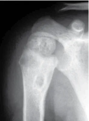
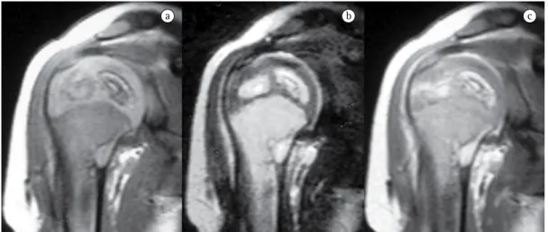
A 21-month-old boy, born in São Paulo, presented with fever and mild functional impair-ment of the right upper limb. The child had daily episodes of fever (> 38°C), predominantly at night. One week prior, the child had started presenting pain upon palpation and upon move-ment, as well as limited movement of the right arm. The mother stated that the boy had no history of local trauma. An X-ray of the right upper limb revealed a lytic lesion of the proximal epiphysis/metaphysis, with ill-defined borders, surrounded by a halo of reactive bone scle-rosis, periosteal reaction and soft tissue edema (Figure 1). Magnetic resonance imaging revealed areas of abnormal marrow signal and soft tissue edema with highlighting of the infected marrow by the contrast medium—signs consistent with osteomyelitis of the proximal humerus (Figure 2). The patient received cephalosporin for 10 days in an outpatient setting. On day 8 of the anti-biotic therapy, the child came to our facility due to fever and persistent pain.
Further anamnesis data revealed that there had been no contact with people with chronic cough or pulmonary TB. The chest X-rays of the parents were normal. The child had received BCG vaccination in the neonatal period.
Upon admission, the child was pale, had fever (38°C) and presented tachycardia (100 bpm). The child also presented functional impairment,
Discussion
This case report describes an uncommon disease that should be considered in small chil-dren with bone lesion of unknown etiology.(10)
Osteitis after BCG vaccination is rarely recog-nized in pediatric practice, since the disease is poorly understood and the diagnosis is difficult to make.(13) It is estimated that the only 25% of cases are diagnosed.(4)
In the case described, the patient presented symptoms such as low fever and pain upon palpation and upon movement of the right arm. In the literature, clinical symptoms are usually few.(4,7)
The patient was discharged on post-admis-sion day 19. He was in good clinical condition and had no fever or pain. Subsequently, monthly X-rays and another CT, performed 6 months later, revealed a decrease in the bone lesion, with reestablishment of the cortical contour, marked bone sclerosis, residual ill-defined lytic areas in the epiphysis and involution of the soft tissue edema.
Immunologic testing revealed CD4 cells, CD8 cells, normal Ig levels, normal IgG subclass levels and negative HIV serology.
Two months after treatment initiation, pyrazi-namide was discontinued, whereas rifampin and isoniazid were maintained for another 8 months.
a b c
Figure 2 - Magnetic resonance imaging of the humerus, in a T1-weighted sequence (a) and a T2-weighted sequence (b), as well as a T1-weighted sequence after the injection of a gadolinium contrast agent (c). Areas of bone marrow change in the humeral epiphysis/metaphysis accompanied by edema and highlighting of the adjacent soft tissues after contrast injection.
The cellular response to PPD also contradicted this hypothesis. This test can be positive in M. tuberculosis infections and also in M. bovis infections. Humoral immunity, determined based on Ig levels and on IgG subclass levels, was normal. Another cause of immunodeficiency that could not be investigated in our patient was IFN-α receptor deficiency.
The most important differential diagnosis is osteomyelitis caused by nonspecific bacteria against which the patient initially received anti-biotic therapy. The lack of response to antianti-biotic therapy, as well as the mildness of the clinical and laboratory profile, led to the suspicion of osteitis after BCG vaccination.
Bone TB is another important differential diagnosis. Lack of contact with TB and BCG vaccination within the last 4 years are criteria that suggest osteitis after BCG vaccination.
Many different treatment regimens are employed in patients with osteitis after BCG vaccination. One includes pyrazinamide, in combination with isoniazid and rifampin, for 2 months. The lesion following BCG vaccina-tion can be resistant to pyrazinamide. However, this resistance did not occur in our patient. In cases of treatment failure, alternative treatments should be recommended. Although the litera-ture recommends treatment with isoniazid and rifampin for 12 months, we opted for discon-tinuing both drugs after 8 months, due to the rapid clinical and radiological improvement of the patient.(4,8,10)
Similarly to our case, the long-term evolution of most patients is favorable.(7) The prognosis of this disease is good, and bone sequelae or growth deficit are described in only 3% of the cases.(7)
In conclusion, osteitis after BCG vaccination is a rare, underestimated and difficult-to-di-agnose complication. It should be considered in small children who have previously received BCG vaccination, have had no contact with TB and present clinical findings consistent with osteomyelitis but do not respond to antibiotic therapy.
In most cases, long-term antituberculosis therapy and surgical drainage are necessary for remission. Fortunately, the prognosis is good, with a low frequency of complications. Therefore, the use of BCG vaccine should be maintained in countries with a high incidence of TB.
In the literature, the lower limbs are described as the site most commonly affected, indicating that the osteitis site does not always correspond to the vaccination site.(7,8) However, in the case reported here, there was a clear relationship between the vaccination site and the apparent location of the soft tissue involvement, which allow us to conclude that it was a contiguous lesion.
The diagnosis of osteitis after BCG vaccina-tion was established according to the criteria proposed by Foucard & Hjelmsted in 1971: 1) BCG vaccination in the neonatal period; 2) a period of less than 4 years between vaccination and symptom onset; 3) no contact between the child and any adults with TB; 4) a consistent clinical profile; and 5) histopathology suggestive of TB.(14-16)
Lytic and sclerotic bone lesions with perio-steal reaction characterize the radiographic lesions. In the case described here, the CT find-ings in the epiphysis, the metaphysis and the diaphysis were consistent with previous descrip-tions in young patients. Older children usually present only metaphyseal changes.(4)
Although typical epithelioid cell granu-lomas, with or without caseous necrosis, are more frequently associated with TB, they are also found in lesions after BCG vaccination. The criteria for the diagnosis of osteitis after BCG vaccination were proposed several years ago by authors from countries where the incidence of TB was minimal at the time. In Brazil, the inci-dence of TB remains high, making it important to retain BCG vaccination. However, the fact that we did not isolate M. bovis in culture does not rule out the diagnosis, since this bacterium has been found in approximately half of all cases in the literature.(4,7,8) This can be due to the fact that M. bovis loses its viability after the initiation of antibiotic therapy.(4,7,8) The PCR was performed to test for M. tuberculosis and might have produced positive results if it had been performed to test specifically for M. bovis.
In patients with osteitis after BCG vaccina-tion, PCR has been used to identify the etiologic agent. The difference in a single nucleotide can distinguish M. tuberculosis from M. bovis.(4)
9. Kröger L, Brander E, Korppi M, Wasz-Höckert O, Backman A, Kröger H, et al. Osteitis after newborn vaccination with three different Bacillus Calmette-Guérin vaccines: twenty-nine years of experience. Pediatr Infect Dis J. 1994;13(2):113-6.
10. Moreno L, Gottrand F, Herbaux B, Savage C, Farriaux JP. Vertebral osteitis following BCG vaccination in a previously healthy child. Eur J Pediatr. 1990;149(9):668.
11. Web L, Torklus DV. Osteomyelitis nach BCG-Impfung. Z Orthop Ihre Grenzgeb. 1981;119:297-300
12. Peltola H, Salmi I, Vahvanen V, Ahlqvist J. BCG vaccination as a cause of osteomyelitis and subcutaneous abscess. Arch Dis Child. 1984;59(2):157-61.
13. Böttiger M, Romanus V, de Verdier C, Boman G. Osteitis and other complications caused by generalized BCG-itis. Experiences in Sweden. Acta Paediatr Scand. 1982;71(3):471-8.
14. Foucard T, Hjelmstedt A. BCG-osteomyelitis and -osteoarthritis as a complication following BCG-vaccination. Acta Orthop Scand. 1971;42(2):142-51.
15. Hanimann B, Morger R, Baerlocher K, Brunner C, Giger T, Schopfer K. BCG osteitis in Switzerland. A report of 6 cases [Article in German]. Schweiz Med Wochenschr. 1987;117(6):193-8.
16. Kolandaivelu G, Manohar K, Bose JC, Rajagopal P. Osteitis of humerus following BCG vaccination. J Indian Med Assoc. 1986;84(6):184-5.
References
1. Farhat CK, Carvalho ES, Carvalho LH, Succi RC, editors. Infectologia Pediátrica. São Paulo: Atheneu; 1999. p. 343-351.
2. Santana CC. A revacinação BCG em escolares: um estudo pioneiro. J Pediatr (Rio J). 2002;78(4):257-8.
3. Ferreira AA, Ferreira MF, Macedo EA, Cunha I, Santos SL, Reis AR, et al. Revacinação BCG em escolares: evolução da lesão vacinal entre 48 horas e 10 semanas. J Pediatr (Rio J). 2002;78(4):289-94.
4. Lin CJ, Yang WS, Yan JJ, Liu CC. Mycobacterium bovis osteomyelitis as a complication of Bacille Calmette-Guérin (BCG) vaccination: rapid diagnosis with use of DNA sequencing analysis: a case report. J Bone Joint Surg Am. 1999;81(9):1305-11.
5. Bricks LF. Vacina BCG: via percutânea ou intradérmica? J. Pediatr (Rio J). 2004;80(2):93-8.
6. Hoppe JE, Orlikowsky T, Klingebiel T, Niethammer D. Costal BCG osteomyelitis presenting as a tumor. Infection. 1992;20(2):94-6.
7. Kröger L, Korppi M, Brander E, Kröger H, Wasz-Höckert O, Backman A, et al. Osteitis caused by bacille Calmette-Guérin vaccination: a retrospective analysis of 222 cases. J Infect Dis. 1995;172(2):574-6.
8. Aftimos S, Nicol R. BCG osteitis: a case report. N Z Med J. 1986;99(800):271-3.
About the authors
André Fukunishi Yamada
Physician. Department of Diagnostic Imaging, Universidade Federal de São Paulo/Escola Paulista de Medicina – UNIFESP/EPM, Federal University of São Paulo/Paulista School of Medicine – São Paulo, Brazil.
Juliana Barbosa Pellegrini
Pediatric Resident. Professor Edmundo Vasconcelos Hospital, São Paulo, Brazil.
Luciana Menezes Cunha
Pediatric Resident. Professor Edmundo Vasconcelos Hospital, São Paulo, Brazil.
Artur da Rocha Corrêa Fernandes