Clara Maria Pinheiro
Efeito da estimulação elétrica sob a reinervação de
músculos desnervados em ratos
SÃO CARLOS Fevereiro de 2016
Universidade
Federal de São Carlos
Clara Maria Pinheiro
Efeito da estimulação elétrica sob a reinervação de
músculos desnervados em ratos
Orientador: Prof. Dr. Thiago Luiz de Russo
SÃO CARLOS
Fevereiro de 2016
Universidade
Federal de São Carlos
Centro de Ciências Biológicas e da Saúde (CCBS)
Departamento de Fisioterapia
Tese apresentada ao Programa de
Pós-Graduação
em
Fisioterapia
da
com os dados fornecidos pelo(a) autor(a)
P654e
Pinheiro, Clara Maria
Efeito da estimulação elétrica sob a reinervação de músculos desnervados em ratos / Clara Maria
Pinheiro. -- São Carlos : UFSCar, 2016. 90 p.
Tese (Doutorado) -- Universidade Federal de São Carlos, 2016.
AGRADECIMENTOS
Sinto-me uma pessoa privilegiada e honrada pelo grande número de pessoas que gostaria de agradecer nesse momento. Hoje agradeço especialmente...
À Deus por sempre guiar meus passos, mesmo que por linhas tortas. E também por colocar em meu caminho pessoas especiais, por me permitir alcançar e realizar mais esse sonho.
Ao Prof. Thiago Luiz de Russo, meu orientador, com quem aprendi muito, que me acompanha desde a iniciação científica. Obrigada pela paciência, pelos grandes ensinamentos de laboratório e de vida, pela sua sabedoria, cuidado e otimismo! Muito obrigada por me acolher quando eu precisei de orientação e por não me deixar desistir desse sonho. Sempre admirei suas conquistas e tenho certeza que muitas outras estão por vir. Obrigada por tudo!
À Profa. Tania de Fatima Salvini, que assim como o Prof. Thiago me acompanhada desde a iniciação científica, e que, me fez conhecer e gostar do mundo científico. Obrigada pelos ensinamentos, paciência, conversas e lição de vida. Muito obrigada por me tranquilizar em momentos difíceis. Vou levar sempre comigo seu exemplo e dedicação.
À Profa. Davilene Gigo-Benato, que me auxiliou e permitiu o desenvolvimento do meu projeto, transmitindo conhecimentos específicos e muito importantes para a realização do mesmo. Muito obrigada pela paciência, sabedoria, carinho e delicadeza que me foram dedicados. Sempre admirei seu jeito de levar e enfrentar a vida. Obrigada pelo apoio e ajuda na realização desse sonho.
À Bruna Erbereli, aluna de iniciação científica e uma amiga muito especial. Sem sua ajuda seria muito difícil conseguir realizar esse projeto. Obrigada pelas conversas, risadas e por sua companhia agradável.
À nossa querida e animada técnica de laboratório, Teresa Fatori Piassi. Obrigada pelos ensinamentos de laboratório e de vida. Pelas receitas, dicas e conselhos. Você tornou meu trabalho mais leve! Continue sempre assim! Parabéns pelo carinho e respeito que guarda em seu coração! Te admiro muito.
científica. Mesmo que eu te agradeça muito, seria pouco. Você está sempre em minhas orações.
Às meninas do LaFiN, Fernanda, Carol, Marcela, Gabriela e ao único menino
Luccas, que me acompanharam durante essa jornada e sempre estiveram dispostos a me ajudar no que fosse necessário. Obrigada por tudo!
As pessoas importantes e especiais, como Andriete, Ivana, Ari, Jean, Flávio, Paula, Maíra que me ouviram, ajudaram e deixaram minha jornada mais leve e divertida.
À CAPES e à FAPESP que viabilizaram a execução desse projeto através do apoio financeiro.
À todos os professores do Departamento de Fisioterapia que tanto contribuíram para minha formação, e que me auxiliam até hoje.
À minha família, meu pai Noel D. Pinheiro, minha mãe Maria de Fátima E. Néo Pinheiro (Beth), meu irmão Guilherme H. D. Pinheiro, minha cunhada Karina C.T. Pinheiro e meus sobrinhos Henrique T. Pinheiro e Ícaro T. Pinheiro, pessoas essenciais em minha vida! Obrigada pela força e compreensão do dia-a-dia, e me desculpe pela ausência em reuniões e comemorações, quando eu precisava ir para o laboratório. Muito obrigada pelo apoio, incentivo e coragem para chegar até aqui, nem tenho como agradecer. Aos meus pais, obrigada pela oportunidade e pelo exemplo, foi por vocês que cheguei até aqui. Amos todos vocês incondicionalmente.
Ao meu namorado Luiz Fernando Maffei Dardis, amigo e companheiro. Obrigada
pelo apoio incondicional durante a realização desse sonho, pela paciência e compreensão quando não podíamos nos ver por causa dos experimentos. Mesmo distante (São Paulo) através de suas palavras você conseguia nos deixar próximos! Sou mais feliz com você ao meu lado! Amo você!
As minhas amigas Maria Luiza Zeraik, Kamilla Ortega, Natália Troya e Ariani
Chiari, por sempre me incentivarem a continuar e me ensinaram a ser uma pessoa melhor. Obrigada pelo carinho e atenção! Vocês são muito especiais na minha vida.
Enfim, agradeço imensamente a todos que de forma direta ou indiretamente fizeram parte do meu crescimento intelectual, espiritual e pessoal durante esses 4 anos.
LISTA DE FIGURAS
MANUSCRITO 1
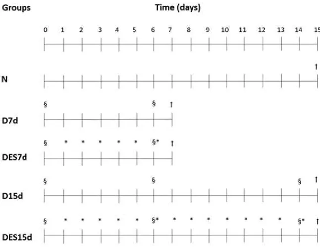
Figura 1. Grupos experimentais. Na parte superior da figura é apresentada uma linha do tempo; nas porções média e inferior, estão os grupos experimentais e os procedimentos realizados. À direita, a legenda. No final, a representação esquemática do protocolo de estimulação eléctrica do músculo tibial anterior incluindo avaliação eléctrica (EE). A EE foi realizada no momento inicial, seguido por 4 séries de estimulação elétrica com duração de 7,5 min cada, com um intervalo de 10 minutos entre as séries. Legenda: Ϯ: sacrificados; §: avaliações excitabilidade elétrica; *: estimulação elétrica.
Figura 2. Variáveis elétricas do músculo tibial anterior obtidas das avaliações elétricas durante 15 dias de desnervação. Os valores normais de reobase, cronaxia e acomodação foram considerados como o período pré-desnervação. Resultados são média ± erro padrão da media (A) Reobase, (B) Cronaxia, (C) Acomodação. a: representa diferença estatística (p < 0.05) quando dia 6 e dia 14 foi comparado ao período pré-desnervação; b: representa diferença estatística (p < 0.001) quando D15d foi comparado ao DES15d no dia 6 e no dia 14;c: representa diferença estatística (p < 0.05) do grupo D15d no dia 6 comparado ao dia 14; d: representa diferença estatística (p < 0.01) do grupo DES15d no dia 6 comparado ao dia 14. A cronaxia aumentou significativamente nos músculos desnervados submetidos ou não à ES, e o grupo DES15d apresentou maiores valores de cronaxia comparados com D15d nos dias 6 e 14. Note que ambas, reobase e acomodação diminuíram depois da desnervação.
Figura 3. Área de secção transversa e morfologia do músculo tibial anterior (TA). A AST diminuiu em todos os grupos desnervados, com uma importante diferença estatística entre os grupos D15d e DES15d (p < 0.05). Morfologia: a atrofia da fibra muscular foi observada em todos os grupos desnervados quando comparados ao grupo normal. A distribuição do tamanho das fibras musculares nos grupos desnervados foi significantemente menor do que no grupo normal. Barra: 100 µm.
Figura 4. Níveis de RNA mensageiro no músculo tibial anterior dos genes atrogina-1 (A), MuRF1 (B) and myoD (C). Resultados são apresentados em média ± desvio padrão. a: representa diferença estatística comparado ao N; b: representa diferença estatística comparado ao D7d; c: representa diferença estatística comparado ao DES7d; d: representa diferença estatística comparado ao D15d. Note que os músculos desnervados submetidos à ES (DES7d e DES15d) mostraram um declínio da expressão gênica de atrogina-1, myoD e MuRF1 quando comparado ao D7d e D15d. O grupo DES15d apresentou valores menores comparado ao grupo D15d (p < 0.05).
Figura 6. Níveis de expressão do MuSK (A), Dok-7 (b) e receptores de acetilcolina (C,D,E,F) no músculo tibial anterior. Resultados são mostrados em média ± desvio padrão. a: representa diferença estatística comparado ao N; b: representa diferença estatística comprado ao D7d; c: representa diferença estatística comparado ao DES7d; d: representa diferença estatística comparado ao D15d. Note que a expressão gênica do MuSK não foi afetada pela ES, entretanto os receptores de acetilcolina e Dok-7 foram modulados diferentemente.
MANUSCRITO 2
Figura 1. Cálculo do índice funcional do ciático (SFI), com déficit funcional dos grupos D15d e DES15d. O período pré-desnervação (PD) foi considerado normal nos dois grupos. Resultados são média ± erro padrão da média. a: representa diferença estatística (p < 0.05) quando dia 6 e dia 14 foram comparados ao PD; b: representa diferença estatística (p = 0.032) quando dia 6 foi comparado ao dia 14 do grupo D15d; c: representa diferença estatística (p = 0.001) quando os grupos D15d e DES15d forma comparados entre eles. Note que DES15d não recuperou sua função após 15 dias de lesão nervosa.
Figura 2. Níveis de RNA mensageiro de TWEAK, Fn14 e miostatina no músculo tibial anterior. Resultados são média ± desvio padrão. a: representa diferença estatística comparado a N; b: representa diferença estatística comparado a D7d; c: representa diferença estatística comparado a DES7d; d: representa diferença estatística comparado a D15d. Note que os músculos desnervados submetidos à ES (DES7d and DES15d) mostraram um declínio na expressão gênica do TWEAK e miostatina quando comparado a D7d e D15d. O grupo DES15d apresentou valores menores comparado ao D15d (p < 0.05). Fn14 e TGF-
apresentaram um pico depois de 7 dias no grupo DES7d.
LISTA DE TABELA
MANUSCRITO 1
Tabela 1: Oligonucleotídeos primers utilizados para amplificação da transcrição reversa de RNA em real-time PCR
Tabela 2. Deficit functional nos grupos D15d e DES15d através do cálculo do índice funcional do ciático.
MANUSCRITO 2
Tabela 1: Oligonucleotídeos primers utilizados para amplificação da transcrição reversa de RNA em real-time PCR
Tabela 2. Massa corporal final, massa muscular, área de secção transversa e tecido conjuntivo do músculo tibial anterior (TA)
PRINCIPAIS ABREVIATURAS
CSA = cross-sectional area
Dok-7 = cytoplasmic protein Downstream of kinase-7
ECM / MEC = extracelular matrix/ matriz extracelular
EE = avaliações elétricas
ES / EE = electrical stimulation / estimulação elétrica
Fn14 = receptor fibroblast growth factor inducible 14
MAFBx (atrogin-1) = muscle-specific E3-ubiquitin ligases
MuRF1 = muscle-specific E3-ubiquitin ligases MuSK = muscle-specific receptor tyrosine kinase
MMP = metaloproteinase
MMP-2 = metaloproteinase 2 MMP- 9 = metaloproteinase 9
NMJ = junção neuromuscular
N-CAM = neural cell adhesion molecule
PD = pré-desnervação
PNI / LNP = Peripheral nerve injury /lesão nervosa periférica
SFI / IFC = sciatic functional index / índice functional do ciático TA = tibial anterior
TGF- = cytokine transforming growth factor beta TNF- = tumor necrosis factor-alpha
RESUMO
A lesão nervosa periférica interrompe as funções normais dos neurônios e leva a alterações rápidas e progressivas no músculo esquelético, tais como a atrofia muscular e a fibrose, causando perdas funcionais. Para o tratamento dos músculos desnervados a estimulação elétrica (EE) tem sido utilizada. A escolha dos melhores parâmetros de EE para minimizar a atrofia muscular devido a desnervação, é controversa. Além disso, não está claro se a EE pode afetar, de fato, a reinervação de músculos desnervados. Assim, esta tese tem dois objetivos principais: 1) verificar se a EE, aplicada aos músculos desnervados com eletrodos de superfície, pode afetar a recuperação neuromuscular após axonotmese do nervo ciático em ratos; e 2) avaliar o impacto da EE no estabelecimento da fibrose do músculo desnervado. Dois manuscritos foram produzidos e contavam com os mesmos grupos experimentais. Trinta e cinco ratos Wistar foram divididos em 5 grupos: (1) Normal (N); (2) Desnervado 7 dias (D7d); (3) Desnervado e ES 7 (DES7d); (4) Desnervado 15 dias (D15d); e (5) Desnervado e ES 15 dias (DES15d). Foi realizada a axonotmese no nervo isquiático. O protocolo de EE do músculo tibial anterior consistiu em: 200 contrações por dia, divididas em 4 séries consecutivas de 50 contrações, com 10 minutos de descanso entre cada série. Foram utilizados os seguintes parâmetros: pulso monofásico exponencial; tempo: 2 vezes cronaxia; amplitude: nível motor; TON: 3s e TOFF: 6s. O índice funcional do ciático foi calculado. A excitabilidade muscular foi avaliada considerando a reobase, cronaxia e acomodação. Análises morfométricas, tais como área de secção transversa da fibra muscular e a porcentagem de proliferação do tecido conjuntivo foram utilizados para caracterizar a morfologia. Marcadores moleculares relacionados à reinervação como a N-CAM (molécula de adesão celular neural), organização e manutenção da junção neuromuscular (JNM) (MuSK, Dok-7 e receptores de acetilcolina), controle de massa muscular (atrogin-1, MuRF1, myoD e miostatina), fibrose (TGF-β e miostatina), remodelação da matriz extracelular (metaloproteinases) e, inflamação (TWEAK / Fn14) foram investigados por técnicas de biologia molecular como western-blot, qPCR ou zimografia. Os principais resultados mostraram que a EE provocou perda da recuperação natural dos músculos desnervados acentuando a perda funcional, a atrofia muscular e a fibrose, assim como, reduzindo a excitabilidade muscular. Estas alterações morfofuncionais e eletrofisiológicas foram relacionadas à diferentes modulações de todos os marcadores moleculares, no decorrer do tempo estudado. De modo geral, a presente tese forneceu provas de que a EE pode atrasar o processo de reinervação por fatores relacionados com a estabilidade e a organização da JNM, bem como a induzir à incapacidade e à atrofia muscular, com diminuição da excitabilidade muscular. Além disso, a EE aplicada aos músculos desnervados induziu fibrose através da modulação da via inflamatória e também pela produção e remodelamento da matriz extracelular. Cuidados devem ser tomadas pelas equipes de reabilitação ao utilizar a EE no tratamento de músculos desnervados.
ABSTRACT
Peripheral nerve injury disrupts the normal functions of neurons and leads to rapid and progressive alterations in structural skeletal muscle, such as muscle atrophy and fibrosis, causing functional deficits. Electrical stimulation (ES) has been recommended to treat denervated muscles. The best parameters of ES to minimize muscle atrophy due to denervation is controversial. Furthermore, it is not clear if ES can, in fact, affect reinnervation of denervated muscles. Thus, this thesis has two main objectives: 1) to verify if ES, applied to denervated muscles by surface electrodes, can affect neuromuscular recovery after nerve crush injury in rats; and 2) to assess the impact of ES on fibrosis establishment in denervated muscles. Two manuscripts were produced and used the same experimental groups. Thirty-five Wistar rats were divided into 5 groups: (1) Normal (N); (2) 7-day denervation (D7d); (3) 7-day denervation and ES (DES7d); (4) 15-day denervation (D15d); and (5) 15-day denervation and ES (DES15d). Tibialis anterior (TA) muscle denervation was induced by
crushing the sciatic nerve. The ES protocol to stimulate TA muscles consisted of: 200 contractions per day divided into 4 consecutive series of 50 contractions, with 10-minute rests between each set. The following parameters were used: exponential monophasic pulse; width time: 2x chronaxie; amplitude: motor level; time On: 3s and Off: 6s. The sciatic functional index was calculated. Muscle excitability was assessed considering the rheobasis, chonaxie and accommodation. Morphometric analyses, such as the muscle fiber cross-sectional area and percentage of connective tissue proliferation were used to characterize muscle morphology. Molecular markers related to reinnervation (neural cell adhesion molecule, N-CAM), neuromuscular junction organization and maintenance (MuSK, Dok-7 and nicotinic Acetylcholine Receptors (nAChR) subunits), muscle mass control (atrogin-1, MuRF1, myoD
and myostatin), fibrosis (TGF-β and myostatin), extracellular matrix remodeling
(metaloproteinases, MMPs) and inflammation (TWEAK/Fn14) were investigated by molecular biology techniques such as western-blot, qPCR or zymography. The main results showed ES impaired natural recovery of denervated muscles accentuating disability, muscle atrophy and fibrosis, as well as reducing muscle excitability. These morphofunctional and electrophysiological changes were related to different modulations of all molecular markers investigated in a timely manner. Overall, this thesis provides evidence that ES can delay the reinnervation process by modulating factors related to NMJ stability and organization, as well as induced disability and muscle atrophy, and decreased muscle excitability. In addition, ES applied to denervated muscles induced muscle fibrosis by modulating inflammatory pathways and also extracellular matrix production and remodeling. Warnings should be given to rehabilitation teams when recommending ES to treat denervated muscles.
SUMÁRIO
1.
APRESENTAÇÃO
13
2.
CONTEXTUALIZAÇÃO
14
3.
REFERÊNCIAS
20
4
MANUSCRITO 1
23
4.2 Introdução
25
4.3 Material e Métodos
28
4.4 Resultados
37
4.5 Discussão e Conclusão
47
4.6 Referências
52
5
MANUSCRITO 2
56
5.2 Introdução
58
5.3 Material e Métodos
60
5.4 Resultados
67
5.5 Discussão e Conclusão
69
5.6 Referências
74
6
ATIVIDADES DESENVOLVIDAS NO PERÍODO
84
APRESENTAÇÃO
Esta tese foi organizada segundo as normas do Programa de Pós-Graduação em Fisioterapia da UFSCar e faz parte de uma linha de pesquisa desenvolvida no Laboratório de Pesquisa em Fisioterapia Neurológica (LaFiN), do Departamento de Fisioterapia da UFSCar. Esta linha abrange estudos na área básica de recursos físicos utilizados na fisioterapia para tratamento de músculos desnervados.
Uma contextualização será apresentada inicialmente, com detalhamento da linha de pesquisa na qual o estudo está inserido. Em seguida, dois manuscritos vinculados a esta tese serão apresentados. O primeiro, intitulado “Electrical stimulation delays reinnervation in
denervated rat muscle”, foi submetido à revista “Journal of Peripheral Nervous System”
(JCR 2016: 2.758). Já o segundo manuscrito foi intitulado de “Electrical stimulation, based on chronaxie, increases fibrosis and modulates TWEAK/Fn14, TGF-/myostatin, and MMP
pathways in denervated muscles” e submetido à revista “American Journal of Physical Medicine and Rehabilitation” (JCR2016: 2.202; Qualis A1 na área de Educação Física). Por
CONTEXTUALIZAÇÃO
Lesão nervosa periférica (LNP) é um trauma muito frequente que ocorre nos nervos periféricos, podendo resultar em parcial ou total perda de funções motoras, sensoriais e autonômicas, levando muitas vezes à incapacidade com redução de qualidade de vida. Essas lesões podem ocorrer por vários motivos, entre eles, acidentes de trabalho e automobilísticos, representando alto custo para a sociedade (Raducan et al., 2013; Schaakxs et al., 2013). A gravidade do dano nervoso e o tempo de recuperação pode variar de acordo com a lesão nervosa sofrida (Sunderland, 1991). As lesões podem ser classificadas como: neuropraxia, axonotmese ou neurotmese. A neuropraxia é sua forma mais branda, quando há perda momentânea da condução nervosa. A axonotmese é caracterizada como uma perda da continuidade das fibras axonais, mas com preservação do perineuro; apresenta uma recuperação espontânea e é classificada como uma lesão moderada. Já na neurotmese, há perda da continuidade do epineuro, portanto uma lesão severa, sendo necessária intervenção cirúrgica (Wood et al., 2011).
Os danos nos nervos periféricos são seguidos por degeneração Walleriana, que é a morte das fibras axonais seguida por degradação da bainha de mielina. Fatores inflamatórios e neurotróficos são liberados no local da lesão, para que ocorra a degradação dos debris celulares. Além disso, é iniciada a reestruturação da matriz extracelular, para que o cone de crescimento neural avance, ocorrendo a regeneração do nervo e reinervação de tecidos alvo, como fibras musculares, receptores sensoriais, entre outros (Misgeld, 2005; Chen et al., 2007; Madduri et al., 2010).
Além das alterações ocorridas no nervo, outro tecido que é bastante afetado após uma lesão nervosa periférica é o músculo esquelético. A inervação é um fator crítico para a integridade funcional e estrutural do músculo esquelético (Kostrominova, 2005). Na lesão nervosa ocorre a interrupção da transmissão do estímulo elétrico para os músculos, causando profundas alterações degenerativas nas fibras musculares (Ishido et al., 2004; Hyatt et al., 2005). Dentre as alterações musculares podemos destacar a ausência de contração muscular que provoca uma intensa atrofia muscular (Lieber, 2002; Ishido et al., 2004).
A principal via de degradação proteica em músculos é a via ubiquitina-proteassoma. Duas E3 ligases chamadas atrogina-1/MAFbx e MuRF-1 foram descritas como atrogenes, ou seja, o aumento da sua expressão estava relacionado ao aumento da atrofia muscular em diferentes modelos, como imobilização, redução da gravidade e desnervação (Bodine et al, 2001). Trabalhos do nosso grupo mostraram que o aumento da expressão gênica de atrogina-1 e MuRF-1 podem ser observados em músculos já após 7 dias de desnervação. Este aumento da expressão é acompanhado por uma drástica perda de massa muscular (Russo et al., 2010).
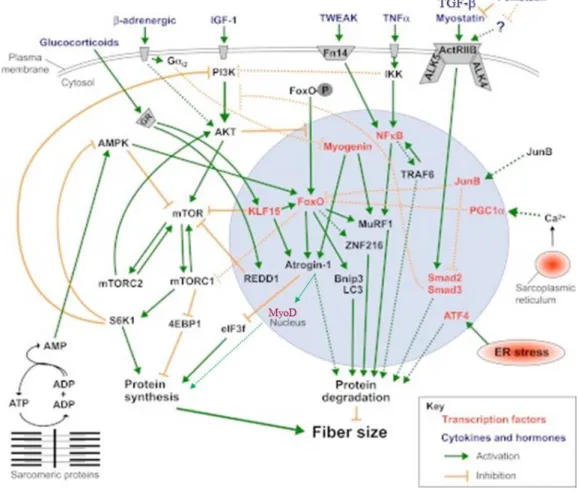
Figura 1. Principais vias sinalização de síntese e degradação de proteínas musculares.
Síntese e degradação de proteína são reguladas por diferentes estímulos, na qual ativa múltiplas vias de sinalização, na qual convergem para um intermediário comum e/ou através de um outro mediador. Fonte: adaptado de Bonaldo and Sandri (2013).
inibir a proliferação de mioblastos e células satélites, sem causar aparente diferenciação e aumento de apoptose (Thomas et al., 2000). Vários mecanismos já foram identificados, na qual a miostatina é capaz de regular o crescimento do músculo esquelético. Ela induz a perda de massa muscular, agindo sobre o sistema proteolítico ubiquitina-proteassomo, evidenciando a regulação positiva de genes como atrogina-1, FOXO1, e MuRF-1; e afeta a massa muscular com a regulação da miogênese, através da diminuição da MyoD (para revisão ver Snijders et al., 2015). Sua avaliação pode ser considerada como um bom indicador da atrofia muscular (Zhang et al., 2006).
Além da atrofia muscular, outra alteração importante é a fibrose que se instala em músculos desnervados. É caracterizada pelo acúmulo anormal de matriz extracelular (MEC), de modo a interferir com a função muscular (Lieber e Ward, 2013). O TGF- é considerado o principal indutor de processos fibróticos (Verrecchia, Mauviel, 2007), pois o aumento da
expressão gênica de TGF-1 é seguido pelo acúmulo de colágeno em músculos desnervados
(Zhang et al., 2008). Além de atuar na atrofia muscular, a miostatina atua na fibrose (Zhu et al., 2007), provavelmente, através da estimulação e da proliferação de proteínas da MEC e dos fibroblastos (Li et al., 2008), como também aumento da resistência dos fibroblastos à apoptose (Bo et al., 2012).
Os processos de remodelamento e reorganização da MEC ocorrem ao redor das fibras musculares atrofiadas (Peviani et al, 2010; Silva-Couto et al, 2012) e, requerem a ação de enzimas proteolíticas, as metaloproteinases (MMPs), que regulam a taxa de produção e degradação de conteúdo de colágeno (Ozawa et al., 2013). As MMPs são uma família de enzimas proteolíticas dependentes de zinco, sintetizadas e secretadas no músculo esquelético pelas células de Schwann, células satélites, e fibroblastos nas junções neuromusculares e ao redor das fibras musculares (Kherif et al., 1998; Carmeli et al., 2004). As MMP-2 e 9 (gelatinases A e B, respectivamente) são as principais metaloproteinases envolvidas na remodelação do músculo esquelético, sendo que mudanças na demanda muscular (hipertrofia/atrofia) provocam alterações em sua atividade proteolítica (Carmeli et al., 2004). Há pouca informação sobre o papel das MMPs durante reinervação muscular, especialmente nas fases iniciais (Ozawa et al., 2013).
As MMPs têm uma interação complexa com citocinas pró-inflamatórias, como fator
de necrose tumoral-alfa (TNF-), que regula a expressão de MMP-2 e MMP-9 (Shubayev et
muscular, como por exemplo nas doenças crônicas (Costamagna et al., 2015). Um potente indutor de perda de massa muscular esquelética é o sistema TWEAK / Fn14. TWEAK (fator
de necrose tumoral indutor de apoptose) é um membro da superfamília TNF- e parece ser a primeira citocina envolvida na perda de massa muscular em condições de desuso. Ele ativa várias vias proteolíticas e estimula a degradação da proteína miofibrilar in vitro e in vivo (Bhatnagar e Kumar, 2012). Esta citocina atua preferencialmente no seu receptor, o receptor do fator de crescimento de fibroblastos 14 (Fn14).
O papel do sistema TWEAK-Fn14 na atrofia por desuso foi confirmada pelas observações de que a perda da massa muscular esquelética, induzida por desnervação, foi significativamente inibida em ratos knockout para TWEAK (Mittal et al., 2010). Músculos desnervados de ratos que hiperexpressam TWEAK apresentam uma profunda perda de massa (Hindi et al., 2014). Recentemente, Tajrishi e colaboradores (2014) mostraram que o aumento da expressão do gene Fn14 após a desnervação, induziu a atrofia muscular, confirmando o envolvimento deste sistema, em resposta a desnervação. Chen e colaboradores (2012) sugeriram que o sistema de TWEAK / Fn14 aumenta a proliferação celular e a síntese de
colágeno através da ativação da via NFB e aumento da atividade de MMP-9 na fibrose do
miocárdio.
Apesar de marcantes, a atrofia muscular e a fibrose não são os únicos processos ativados em músculos desnervados. Há uma tentativa de sinalização das fibras musculares para que novos axônios as reinervem. Este processo dependerá de fatores produzidos tanto pelo nervo como pelo músculo, procurando se reestabelecer a interação neuromuscular. Uma importante mudança que ocorre no músculo após um LNP, visando o reestabelecimento da inervação, é o aumento dos receptores de acetilcolina (RACh) no sarcolema. Os receptores de acetilcolina são normalmente expressos na junção neuromuscular (JNM), em músculos inervados. Contudo, quando a fibra muscular se torna desnervada, estes receptores passam a ser expressos ao longo de toda a fibra. Esta estratégia permite que o axônio possa estabelecer uma nova placa motora em qualquer ponto da fibra muscular e não apenas na placa motora degenerada (Lieber, 2002).
Esse gene é expresso nas células musculares e, uma vez ativada, MuSK estimula vias para o agrupamento e ancoramento dos receptores de acetilcolina e proteínas musculares essenciais para a contração muscular (DeChiara et al., 1996). A diminuição da expressão desse gene leva a desestabilização da JNM e da contração muscular (Kong et al., 2004; Hesser et al., 2006).
Figura 2. Axônio motor terminal liberando agrina e acetilcolina. Fonte: Burden et al., 2013.
Para que MuSK seja ativada, é necessário que ocorra uma fosforilação induzida por agrina, uma proteoglicana produzida pelos motoneurônios. Parece que é justamente esta ligação entre agrina e MuSK que estabiliza a JNM e aciona a agregação de receptores de acetilcolina (Burden et al., 2011; 2014). Contudo, para que esta agregação ocorra de forma adequada é necessária a atuação de uma proteína de membrana chamada de Dok-7 (Okada et al., 2006). Animais knockout para Dok-7 apresentaram problemas na JNM e declínio na expressão de MuSK. Desse modo, MuSK, Dok-7 e RACh estão envolvidos em uma cascata de regulação e, são indispensáveis para a transmissão do impulso nervoso e o trofismo muscular.
profissionais da área de saúde para atuar na recuperação destes músculos e facilitar os processos de reinervação. Grande parte das pesquisas desenvolvidas sobre esta temática estão relacionadas a técnicas cirúrgicas de reparo nervoso, contudo, nas últimas décadas, um interesse constante sob o efeito de recursos físicos usados na reabilitação de músculos desnervados pode ser observado.
A reabilitação do indivíduo com músculos desnervados tem como objetivos terapêuticos centrais amenizar a atrofia muscular e a fibrose, bem como estimular o crescimento axonal e reinervação das fibras musculares, receptores e glândulas. Contudo, a literatura científica evidencia uma falta de consenso sobre a eficácia e a segurança da recomendação de recursos físicos para a recuperação de músculos desnervados. Por exemplo, trabalhos experimentais mostraram que o alongamento intermitente de músculos desnervados pode induzir o aumento da fibrose e acentuar a atrofia muscular (Faturi et al., 2015), mas também pode amenizar as alterações musculares deletérias decorrentes da desnervação (Agata et al., 2009) de acordo com os parâmetros de tratamento escolhidos, como número de alongamentos, tempo de descanso entre cada alongamento, etc.
Outro recurso amplamente utilizado na prática clínica para a reabilitação de músculos desnervados é a estimulação elétrica (EE). Geralmente aplicada com eletrodos de superfície, é proposto que a EE seria capaz de induzir contrações musculares, na ausência do estímulo nervoso, e assim deter ou amenizar a atrofia. Também se espera que os estímulos elétricos pudessem guiar o crescimento axonal. Contudo, alguns trabalhos do nosso grupo mostraram que a EE, apesar de modular uma série de genes musculares, não é capaz de deter a atrofia ou a proliferação de tecido conjuntivo de músculos desnervados (Russo et al., 2007; 2008 e 2010), ao contrário, ela pode induzir alterações deletérias, como atrapalhar a recuperação da função e acentuar a atrofia (Gigo-Benato et al., 2010). Estudos de outros grupos mostraram que 200 contrações diárias seriam suficientes para deter a atrofia muscular e a diminuição da força (Dow et al., 2008). Contudo, não há informação sobre a segurança da EE sobre o processo de reinervação muscular. Desta forma, avaliar protocolos clínicos de EE em modelo animal são relevantes para a prática baseada em evidências, garantindo sua eficiência e segurança.
moleculares integradas evidenciaram que a EE, de acordo com os parâmetros utilizados, pode atrapalhar o processo de reinervação, acentuar a atrofia muscular, induzir fibrose e atrapalhar a recuperação funcional em ratos.
REFERÊNCIAS
Agata N; Sasai N; Inoue-Miyazu M; Kawakami K; Hayakawa K; Kobayashi K; Sokabe M (2009). Repetitive stretch suppresses denervation-induced atrophy of soleus in rats. Muscle Nerve, 39:456-462.
Bonaldo P and Sandri M. Cellular and molecular mechanisms of muscle atrophy (2013). Disease Models & Mecahnisms 6, 25-39.
Burden S.J. SnapShot: neuromuscular junction (2011). Cell. 144: 826-826.
Burden S.J.; yumoto N.; Zhang W. The role of MuSK in synapse formation and neuromuscular disease (2013).Cold Spring harbor Perspectives in biology;5:a00 9167. Bhatnagar S and Kumar A. The TWEAK-Fn14 system: breaking the silence of cytokine-induced skeletal muscle wasting (2012). Current Molecular Medicine, 12, 3-13.
Carmeli, E., M. Moas, A. Z. Reznick and R. Coleman (2004). Matrix metalloproteinases and skeletal muscle: a brief review. Muscle Nerve 29(2): 191-197.
Chen Z.L.; Yu W.M.; Strickland S. Peripheral regeneration (2007). Annu Rev Neurosci; 30: 209-233.
Chen HN; Wang DJ; Ren MY; Wang QL; Sui SJ. TWEAK/Fn14 promotes the proliferation and collagen synthesis of rat cardiac fibroblasts via the NF-small-ka Cyrillic B pathway (2012). Mol Biol Rep 39 (8): 8231-41.
Costamagna D; Costelli P; Sampaolesi M; Pennsa F. Role of inflammation in muscle homeostasis and myogenesis.(2015). Mediators of inflammation 2015 1-14.
DeChiara T. M.; Bowen D.C.; Valenzuela D.M.; Simmons M. V.; poueymirou W.T.; Thomas, S. et al. The receptor tyrosine kinase MuSK is required for neuromuscular junction formation in vivo (1996). Cell. 85:501-512.
Dow D.E.; Cederna P.S.; Hassett C.A,; Kostrominova T.Y.; Faulkner J.A.; Dennis R.G. Number of contractions to maintain mass and for of a denervated rat muscle (2004). Muscle Nerve. 30:77-86,
Dow D.E.; Cederna P.S.; Hasset C.A.; Dennis R.G. and Faulkner J.A. Electrical stimulation prior to delayed reinnervation does not enhance recovery in muscles of rats (2007). Restor Neurol Neurosci; 25: 601-610.
Gigo-Benato, D.; Russo T.L.; Geuna S.; Domingues N.R. S.R.; Salvini, TF.; Parizotto, N.A. Electrical stimulation impairs early functional recovery and accentuates skeletal muscle atrophy after sciatic nerve crusch injury in rats (2010). Muscle & Nerve. 41:685-693.
Hesser B.A.; Henschel O.; Witzemann V. Synapse disassembly and formation of new synapses in postnatal muscle upon conditional of MuSK. (2006). Mol. Cell Neurosci. 31:470-480.
Hyatt JPK; Roy RR; Baldwin, KM; Wernig, A, Edgerton, R. Activity-unrelated neural control of myogenic factors in a slow muscle (2005). Muscle Nerve, v. 33, n. 1, pp. 49-60.
Hindi SM; Mishra V; Bratnagar S; Tajrishi MM; Ogura Y; Yan Z; Burkly LC; Zheng TS;
Kumar A. Regulatory circuitry of TWEAK-Fn14 system and PGC-1 in skeletal muscle
atrophy program (2014). The FASEB Journal 2: 1398-1411.
Ishido M.; Kami K.; Mitsuhiko M. In vivo expression of myoD, P21 and RB proteins in myonuclei and satellite cells of denervated rat skeletal muscle (2004) .Am J Physiol Cell Physiol. 287:C484-C493.
Kherif S, Dehaupas M, Lafuma C, Fardeau M, Alameddine HS. Matrix metalloproteinases MMP-2 and MMP-9 in denervated muscle and injured nerve (1998). Neuropathology and Applied Neurobiology. 24:309 –319.
Kong X.C.; Barzaghi P.; Ruegg M.A. inhibition of synapse assembly in mammalian muscle in vivo by RNA interference (2004). EMBO Rep. 5: 183-188,
Lieber R. Skeletal Muscle Structural, Function, &Plasticity. Lippincott Williams & Wilkins. 2nd edition, Philadelphia, 2002.
Lieber RL and Ward SR. Cellular mechanisms of tissue fibrosis. 4. Structural and functional consequences of skeletal muscle fibrosis (2013). Am J Physiol 305:C241-C252.
Mittal A; Bhatnagar S; Kumar A; Lach-Trifilieff; Wauters S; Li H; Makonchuk DY; Glass DJ; Kumar A. The TWEAK-Fn14 system is a critical regulator of denervation-induced skeletal muscle atrophy in mice (2010) JBC.188 (6).
McPherron A, Lawer A, Lee S. Regulation of skeletal muscle mass in mice by a new TGF- superfamily member (1997). Nature.;386:83-90.
Madduri S.; Gander B.; Schuwann cell delivery of neruotrophic factors for peripheral nerve regeneration (2010). J Peripher Nerv Sust 15:93-103.
Misgeld T. Death of an axon loss in development and disease (2005). Histochem Cell Biol; 124: 189-196.
Okada, K.; Inoue, A.; Okada M.; Murata Y.; Kakuta S. et al. The muscle protein DoK-7 is essential for neuromuscular synaptogenesis (2006). Science. 312.
Ozawa J; Kurose T; Kawamata S; Kaneguchi A; Moriyama H. Regulation of connective tissue remodeling in the early phase of denervation in a rat skeletal muscle. 2013.
Raducan A.; Mirica S.; Duici, O.; Raducan S.; Muntean D.; Fira-Miladinescu, O. and Lighezan R. Morphological and functional aspects of sciatic nerve regeneration after crush injury (2013). Rom J Morphol Embryol, 54 (3Suppl): 735-739.
Russo T.L.; Peviani S.M.; Freria C.M.; Gigo-Benato D.; Geuna S.; Salvini T.F. Electrical stimulation based on chronaxie reduces atrogin-1 and myoD gene expressions in denervated rat muscle (2007). Muscle Nerve. 35:87-97,
Russo T,L.; Peviani S.H.; Durigan, J.L.Q.; Salvini, T.F. Electrical stimulation increases matrix metalloproteinase -2 gene expression but does not change its activity in denervated rat muscle (2008). Muscle & Nerve; 37(5): 45-57.
Russo T,L.; Peviani S.H.; Durigan, J.L.Q.; Gigo-Benato D.; Delfino G.B.; Salvini, T.F. Stretching and electrical stimulation reduce the accumulation of MyoD, myostatin and atrogin-1 in denervated rat skeletal muscle (2010). J Muscle Res Cell Motil. 31:45-57.
Scaakxs, D.; Kalbermatten D.F.; Raffoul W.; Wiberg M. and Kingham P.J. Regenerative cell injection in denervated muscle reduces atrophy and enahces recovery following nerve repair (2013). Muscle & Nerve; 47:691-701.
Silva-Couto MA; Gigo-Benato D; Tim CR; Parizotto NA; Salvini TF; Russo TL. Effects of low-level laser theraphy after nerve reconstruction in rat denervated soleus muscle adaptation (2012). Rev Bras Fisioter; 16(4):320-7.
Shubayev VI and Myers RR. Anterograde TNF transport from rat dorsal root ganglion
to spinal cord and injured sciatic nerve. Neuroscience Letters (2002), 320: 99-101.
Snijders T; Nederveen JP; Mckay BR; Joanisse S; Verdijk LB; van Loon LJC; Parise G. Satellite cells in human skeletal muscle plasticity (2015). Front. Physiol. 6:283.
Tajrishi MM; Shin J; Hetman M; Kumar A. DNA methyltransferase 3a and mitogen-activated protein kinase signaling regulate the expression of fibroblast growth factor-inducible 14 (Fn14) during denervation-induced skeletal muscle atrophy. (2014). The Journal of biological chemistry, 389: (29): 19985-19999.
Thomas M, Langley B, Berry C, Sharma M, et al. Myostatin, a negative regulator of muscle growth, functions by inhibiting myosblast proliferation (2000). J. Biol. Chem., 275: 40235-40243.
Wood M.D.; Kemp S.W.P.; Weber C.; Borschel G.H. and Gordon T. Outcome of
peripheral nerve regenation (2011). Annals of Anatomy (193): 321-333.
Zhang D.; Liu M.; Ding F.; Gu X. Expression of myostatin RNA transcript and protein in grastrocnemius muscle of rats after sciatic nerve resection (2006). J Muscle Res Cell Motil. 27: 37-44,.
MANUSCRITO 1
____________________________________________________________
Title:
Electrical stimulation delays reinnervation in denervated rat muscle
Authors:
Clara M Pinheiro1 PhD, Bruna T Erbereli1, Davilene Gigo-Benato1 PhD, Paula A T S Castro2
PhD, Thiago L Russo1 PhD.
1 Laboratory of Neurological Physiotherapy Research (LaFiN), Department of Physiotherapy
(DFisio), Federal University of São Carlos (UFSCar), São Carlos, São Paulo, Brazil;
2 Laboratory of Muscle Plasticity, DFisio, UFSCar, São Carlos, São Paulo, Brazil.
Running title: Electrical stimulation and muscle reinnervation
Corresponding author: Prof. Dr. Thiago Luiz Russo, Laboratório de Pesquisa em Fisioterapia Neurológica (LaFiN), Departamento de Fisioterapia, Universidade Federal de
São Carlos (UFSCar), Rodovia Washington Luis, Km 235, C.P. 676 – CEP: 13565-905. São
Abstract
Peripheral nerve injury disrupts the normal functions of neurons and leads to rapid and progressive alterations in structural skeletal muscle causing functional deficits and electrical stimulation (ES) has been recommended to treat denervated muscles. It is not clear if ES can in fact affect reinnervation of denervated muscles. Thus, this study aimed to verify if ES, applied to denervated muscles by surface electrodes, can affect neuromuscular recovery after nerve crush injury in rats. Functional recovery at day 14 post-denervation was significantly lower in DES15d (15-day denervation and ES) compared to D15d (15-day denervation. The DES15d had chronaxie values significantly higher on days 6 and 14 compared to D15d, which indicates a decreased in muscle excitability in the DES15d. ES reduce the accumulation of mRNA of atrogenes, but further increased muscle fiber atrophy in DES15d than other denervated groups. N-CAM content decreased in DES7d compared to other denervated groups. ES altered gene expression of important markers of NMJ organization and maintenance as MuSK, Dok-7, nAChR. All together, these results provide evidence that ES can delay the reinnervation process by modulating factors related to NMJ stability and organization, as well as induced disability and muscle atrophy, and decreased muscle excitability.
Introduction
Peripheral nerve injuries (PNI) are usually related to disability and reduced quality of life (Schaakxs, et al., 2013). Muscle denervation post-PNI is a devastating condition, which produces atrophy (Russo, et al., 2010) and paralysis (Wood, et al., 2011). According to severity, PNI can be classified into three main types: neuropraxis (momentary loss of nerve impulse, nerve compression slight), axonotmesis (axon transection with both perineurium and epineurium remaining intact with spontaneous recovery, moderate injury), and neurotmesis (nerve transection where the continuity of the epineurium is disrupted requiring surgical intervention, severe injury) (Wood, et al., 2011).
Regenerative process after moderate and severe PNI includes Wallerian degeneration with distal fragmentation of axon and myelin sheaths made through macrophages and Schwann cells which phagocyte degraded materials. Fine sprouts the proximal axonal end occurs through restructuring the extracellular matrix, allowing axonal reconnection with target cells, maturation of the nerve fiber, reinnervation of muscle fibers, glandules, and sensorial receptors (Navarro, et al., 2007; Misgeld, 2005; Madduri, et al., 2010).
The innervation and reinnervation of skeletal muscle leads to the formation/reorganization of neuromuscular junctions (NMJ). Morphologically, three compartments form the NMJ. The presynaptic compartment where the vesicles containing acetylcholine (ACh) and Schwann cell are found in the nerve endings; the extracellular compartment filled with basal lamina; and the postsynaptic compartment composed of
muscle fibers’ sarcolemma and junctional folds, as well as the sarcoplasm that provides
structural and metabolic support to the postsynaptic region (Engel, 2003; Malomouzh, 2012). The postsynaptic surface sarcolemma contains a high density of nicotinic ACh receptors (nAChRs). When presynaptically released, the ACh activates those receptors, cations are permitted to flow through the nAChR pore, leading to depolarization of the muscle membrane and creating an end-plate potential. If the end-plate potential is large enough, an action potential is reached and ultimately causes muscle contraction (Wang, et al., 2010).
The presence of nAChR in NMJ is critical for muscle function as it determines robustness and reliability of NMJ (Strack, et al., 2015). The nAChR are integral membrane
et al., 2007). Those subunits are presented in the NMJs postsynaptic membrane of the innervated muscle, which are up regulated in response to peripheral nerve lesions.
In addition, there are NMJs proteins to stimulate the pathway of the nAChR clustering and anchoring in the postsynaptic membrane. That is accomplished through coordinated activity, such as muscle-specific receptor tyrosine kinase (MuSK) and cytoplasmic protein Downstream of kinase-7 (Dok-7) (DeChiara, et al., 1996; Okada, et al., 2006). In order to promote synapse stability, the neural cell adhesion molecule (N-CAM) is up-expressed (Polo-Parada, et al., 2001; Vitureir,a et al, 2012 in Chipman, et al, 2014).
For promoting muscle fiber reinnervation, N-CAM expression and protein content around and inside the muscle fiber is increased as well as there is an increase of the nAChR along all the muscle fiber (Wang, et al., 2010; Burden et al., 2011, 2013). N-CAM is a potent regulator of synaptic stability and strongly influences neurotransmission (Polo-Parada et al., 2001; Vitureira, et al, 2012 in Chipman, et al, 2014) (Chipman, et al., 2010; Enriquez-Barreto, et al, 2012; Chattopadhyay,a et al., 2013). Therefore, N-CAM is an important molecule to study the interaction between muscle and nerve.
Furthermore, the post-PNI causes muscle fibers denervation experiencing a complex process of unbalance between anabolic and catabolic processes (Bodine, et al., 2014). That process reflects in both the reduction of the synthesis and the increase of the protein degradation, leading to a muscle mass loss (Jackman and Kandarian, 2004). The expression of muscle-specific genes, involved the ubiquitin-proteasome pathway, such MuRF1 and MAFBx (atrogin-1) is increased in denervated muscles (Bodine, et al., 2001, 2013, 2014).
MuRF1 and MAFBx are muscle-specific E3-ubiquitin ligases that play an important role in the ubiquitin-proteasome system, since those enzymes mark selective substrates to ubiquitination and subsequent degradation by the 26S proteasome. Those two genes are actually the best markers of muscle atrophy and could be considered the master genes to muscle wasting, associated the muscle mass loss (Bodine, et al., 2014).
Due to many neuromuscular disorders that occur in PNI, the research on therapeutic resources that can favor the process of muscle reinnervation and atrophy attenuation are
important to clinical practice. Among the physical resources used on those injuries’
However, some studies from our group showed that the ES, although modulating a variety of muscle genes, is not able to hold atrophy or connective tissue proliferation of denervated muscles (Russo, et al, 2007; 2008 and 2010). On the other hand, it can induce deleterious changes, as impairment of the function recovery; and accentuated muscle atrophy (Gigo-Benato, et al., 2010).
There is controversy about the use of the ES in denervated muscle treatment, some investigators showed 200 daily contractions are effective to prevent mass and strength loss in denervated muscles (Dow, et al., 2007). Others, showed 600 contractions per day, 5 days a week, resulting a higher number of functional motor units and enhanced functional recovery; however, muscle mass and force remained unchanged (Willand, et al., 2015). These studies used implanted electrodes, differing from the clinical practice, and contradicting studies that have shown the muscle electrical stimulation provides no functional benefit with stimulation being applied infrequently or with subthreshold amplitudes being used that could not elicit strong contractions (Sinis, et al., 2009; Gigo-Benato, et al., 2010). On the other hand, Tam and Gordon (2003) and Salmons and cols (2004) suggested that perhaps a more moderate activity protocol is needed to obtain optimal results and concluded that denervated muscles, due to their little excitability, require larger amplitude and pulse duration to obtain contraction.
Because of that, more studies are needed to characterize the effects, to identify more efficient and safe parameters, and to show an effective protocol to prevent muscular atrophy after denervation. Based on previous results (Gigo-Benato, et al, 2010), the hypothesis of this study was that ES sessions may interfere negatively in the muscle reinnervation process and thus impair muscle recovery and functionality, through modulating muscular atrophy pathways (atrogin-1 and MuRF1) as well as maintaining the NMJ (MuSK, Dok-7, N-CAM and nAChRs) in denervated muscles after axonotmesis.
Material and Methods
Animal care and experimental groups
Thirty-five male 3-month-old Wistar rats weigthing 230 ± 3g were housed in plastic cages in a room with controlled environmental conditions and had free access to water and standard food. The experimental procedures were approved by the Ethics Committee of the Sao Carlos Federal University (033/2014) and conducted in accordance with the Guide for
Care and Use of Laboratory Animals.
Figure 1. Diagram of the experimental groups. In the upper part of the figure, a time line is show; in the middle and lower portions of the figure, the experimental groups and the procedures are show. At the right, the subtitle. At the end the ES protocol: electrical stimulation schematic representation of the TA muscle including electrical evaluation (EE). EE was performed at the initial moment followed by 4-sets of ES lasting 7.5 min each with a 10 min rest interval between sets. Legend: Ϯ: euthanized; §: electrical excitability evaluations; *: electrical stimulation.
Intraperitoneal injections of xylazine (12 mg/kg) and ketamine (95 mg/kg) anaesthetized the rats to the surgical and denervation processes, electrical excitability evaluations, ES treatment, and muscle removal. Two animals of D15d and DES15d groups died during the trial period, probably due to daily exposure to anesthesia. Then, an overdose of the anaesthetic on the 7th or 15th day of the experimental procedures euthanized the rats.
Denervation process
The denervation process consisted of a sciatic nerve axonotmesis conducted as previously done by Russo and cols (2010) and Gigo-Benato and cols (2010). Shortly after the rat was anaesthetized, a small incision was made through the skin and fascia (cleaned with 10% povidine iodine) near the right trochanter between the Gluteus maximus and the Femoris biceps muscles to expose the sciatic nerve. Next, a flat-tipped watchmaker´s tweezer number five (D.L. Micof) exerting a 54 N-force (Beer et al, 2011) was used to crush the sciatic nerve for a 30-second period. The nerves were kept moist at 37C in sterile saline solution throughout the surgical intervention. After that, the fascia and skin were sutured from distal to proximal, using silk thread. That procedure promoted distal hind-limb muscle denervation, including the TA muscle. The same researcher performed the crush to ensure that the damage was inflicted on the same area and with comparable duration and pressure in all denervated animal groups. After surgery, animals were housed in single cages and fed with rat chow and water ad libitum. For the first 3 days, paracetamol was added to the animals’ drinking water
for pain reduction. That model of nerve injury is characterized by axonal degeneration and no sign of muscle reinnervation during the first 10 days after the nerve injury were seen (Carmignoto et al., 1983; Carter et al., 1998).
Motor Function Evaluation
The assessment of nerve function recovery was carried out by calculating the sciatic functional index (SFI), as described by Brain et al. Animals were tested in a confined walkway that was 42 cm long and 8.2 cm wide, with a dark shelter at the end. A white paper was placed on the floor of the walkway. The hind paws of the rats were pressed down onto an ink-soaked sponge, and the animals were then allowed to walk down the walkway leaving hind-paw prints on the paper. Three measurements were taken from the hind-paw prints: (1) the print length (PL), which is the distance from the heel to the third toe; (2) the toe spread (TS), which is the distance from the first to the fifth toe; (3) the intermediate toe spread (ITS), which is the distance from the second to the fourth toe. The experimental (E) and normal (N) sides got those three measurements. The SFI was calculated according to the following equation:
Two weeks after surgery, all animals were euthanized with anesthesia overdose; the sciatic nerves and the TA muscles were carefully dissected and immediately weighed at a precision balance (Model 100a; Denver Instruments, Denver, Colorado).
Electrical Excitability Evaluation and Electrical Stimulation Procedures
Rats from the DES groups received ES treatment as previously described by Russo et al. (2004, 2007, 2008 e 2010; Gigo-Benato et al., 2010). ES equipment which allows changes in the electrical parameters was used to assess muscle excitability and for the ES treatment (NeMESys 941, Quark, Brazil). Briefly, before fixing the electrodes, the skin was shaved, cleaned, and covered with a layer of conductive gel, where two electrodes were positioned: an indifferent electrode (5 cm-round self-adhesive electrode) was positioned on the animal´s back, and an active electrode (3 mm-round metallic electrode) small enough to stimulate only the TA muscle. There was no hyperemia observed after the electrical procedures. During the ES procedure, the metallic electrode was in contact with the skin overlying the TA muscle and perpendicular to the muscle fibers. Furthermore, surface electrodes are more commonly used for therapeutic applications, and their position does not require procedures that are invasive to ES (Sheffler, 2007).
Before each evaluation of electrical parameters, we identified the site over the TA where the lowest stimulus amplitude fully activated the muscle. The electrical parameters were evaluated before each ES treatment to provide rheobase, chronaxie, and muscle accommodation values. Afterwards, the chronaxie values were used to determine the ES parameters applied to the TA muscle. These electrical indexes were previously reported (Russo et al., 2007 and 2010; Gigo-Benato et al., 2010).
contraction was selected. The selected stimulation frequency granted strong muscle contraction using low current amplitudes, as previously described (Cummings, 1990). Briefly, the D7d and DES7d groups were submitted to muscular excitability evaluations, one pre-denervation, and the other one on the day before euthanasia (sixth day); the same evaluations were conducted to D15d and DES 15d, and another one on the day before euthanasia (fourteen day).
After the muscle excitability evaluation, the amplitude necessary to induce a maximal contraction of the TA muscle (right ankle maximal full flexion) was identified. The ES sessions were applied daily for 6 (DES 7d) or 14 (DES 15d) days, beginning at 24h after denervation, and producing 200 TA muscle maximal contractions. Those maximal contractions were divided into four sets of 50 contractions, with a 7,5-min per set duration and a 10-min rest (without ES) between sets to minimize muscle fatigue (Cummings, 1990). Dow et al., 2004 has previously demonstrated that 200 muscle contractions are effective to maintain muscle mass and strength, and Kostrominova et al., 2005 has demonstrated that, to reduce the gene expression generally increased during denervation, 200 muscle contractions are also effective.
In our study, a small number of muscle contractions was used to reproduce what is usually applied during a single rehabilitation session for the recovery of human denervated muscles. Normally, in a single treatment session, the denervated muscle electrical treatment is associated with other interventions, such as physical exercise, muscle stretching, and passive movements. Therefore, we decided on 200 contractions using on-time 3s and off-time 6s, because more than that could provoke muscle fatigue, as all contractions were applied in a short time period during a treatment single session.
Sciatic nerve histology
Muscle evaluation
The muscles were split in half in the middle of the TA muscle belly. The distal fragment was used for the histological and morphometric measurements. The proximal fragment was divided in two, immediately frozen in liquid nitrogen, and stored at -80C (Forma Scientific, Marietta, OH) for the mRNA analysis as well as the western blotting analysis.
Morphometric analysis
Histological cross-sections (10 μm) were obtained in a cryostat microtome (Microm HE 505, Jena, Germany), along the TA muscles middle belly. For morphological evaluation by a light microscope (Axiolab, Carl Zeiss, Germany), tissue sections were stained using Toluidine Blue/1% Borax (TB). One histological cross-section of each TA muscle located in the central region of muscle injury was chosen to measure the cross-sectional area, using a light microscope and a software to evaluate general muscle morphology and morphometry (Axiovision 3.0.6 SP4, Carl Zeiss, Germany). One image central region of all muscle cross-sections was done at 20x low magnification; the image was obtained using a light microscope equipped with a digital camera (Carl Zeiss AxioCam HRc). From each picture, the CSA of 100 fibers was measured using the Axiovision 4.7.1.0 software (Carl Zeiss, Jena, Germany), with 100-muscle fibers per animal.
Immunofluorescence Analysis
sections were not incubated at the primary antibody and experimental results were considered only if those controls did not show immunoreactivity.
For qualitative measurements of immunoreactivity, images of five different regions
from the mid-belly of the TA muscles were captured using a fluorescence microscope
(Axiocam, Carl Zeiss, Jena, Germany) equipped with a rhodamine filter at a final magnification of 40x, with the microscopic setting kept the same for all slides.
RNA Isolation and Analysis
RNA was isolated from one frozen fragment of each TA muscle using 1 ml of Trizol reagent (Invitrogen, Carlsbad, CA), according to the manufacturer instructions. The extracted RNA was dissolved in tris-HCl and ethylenediaminetetracetic acid (TE) pH 7.6 as well as quantified by spectrophotometry. The purity was assessed by determining the absorbance ratio at 260 nm and 280 nm. All samples had 260/280 nm ratios above 2.0. The RNA integrity was confirmed by the ethidium bromide inspection (Invitrogen, Carlsbad, CA) which stained the 18S and 28S ribosomal RNA under ultra-violet light.
Total RNA (1µg) from each sample was treated with DNase I (Sigma, AMP-D1) to remove contaminating genomic DNA; then, the total RNA was reverse transcribed to synthesize cDNA using synthesis Kit (iScriptTM cDNA Synthesis Kit, Bio-Rad, CA). The
reaction contained 4µl of 5x iScript reaction mix, 1 µl iScript reverse transcriptase, and 1µg RNA. The final volume was adjusted to 20 μl with nuclease-free water. The reaction was
incubated for 5 minutes at 25C, 30 minutes at 42C, 5 minutes at 85C, and hold at 4C. The resulting cDNA samples were aliquoted and stored at -20C. The RNA transcript levels for the different experimental and control muscles were analyzed simultaneously, and the reactions were carried out in duplicate in the real time PCR (CFX 96 TouchTM Real Time
PCR Detection System, Version 3.0, Bio-Rad, CA), using fluorescent dye SYBR green detection (Thermo Scientific, US) and 180 nM of each primer in a final volume of 40µl. The thermal cycling conditions were 95C for 10 min and 40 94C-cycles for 15 s, 60C for 1 min, and 72C for 1 min. For each gene, all samples were amplified simultaneously in
duplicate using a final volume of 20 μL. Melting dissociation curves were plotted to confirm
that only a single product was amplified. Control reactions omitting cDNA template were run to check for reagent contamination. Gene expression was compared among individual
samples using the ΔΔCq method. The reference genes for the normalization was chosen using
phosphoribosyltransferase (HPRT), and peptidylprolyl isomerase A (PpIa). The sequences used were in table 1.
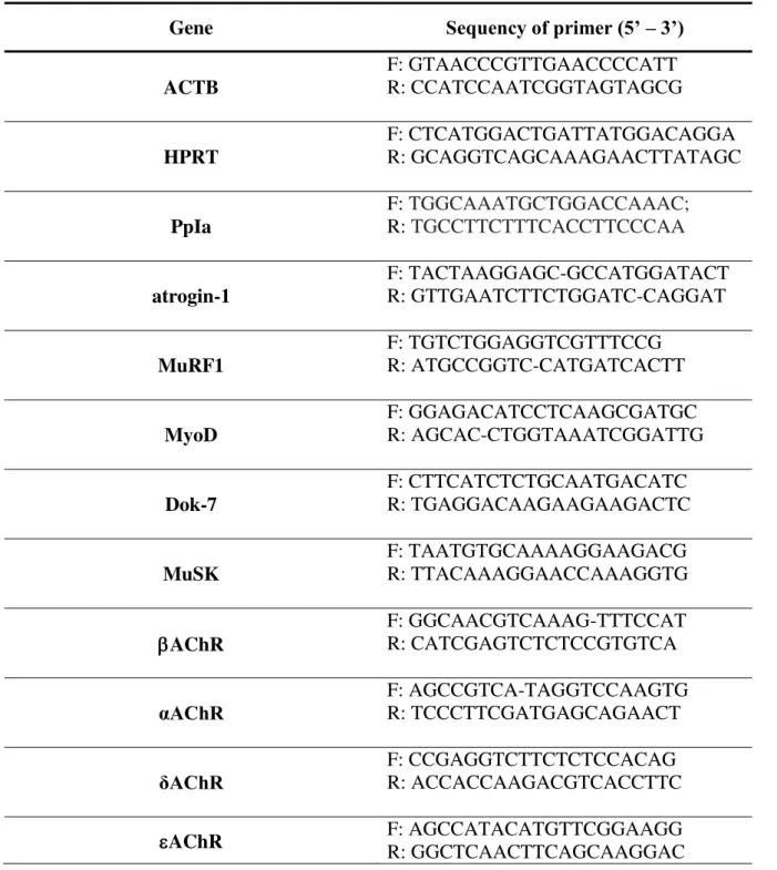
Table 1: Oligonucleotide primers used for real-time PCR amplification of reverse transcribed RNA
Gene Sequency of primer (5’ –3’)
ACTB F: GTAACCCGTTGAACCCCATT R: CCATCCAATCGGTAGTAGCG
HPRT F: CTCATGGACTGATTATGGACAGGA R: GCAGGTCAGCAAAGAACTTATAGC
PpIa F: R: TGGCAAATGCTGGACCAAAC;TGCCTTCTTTCACCTTCCCAA
atrogin-1
F: TACTAAGGAGC-GCCATGGATACT R: GTTGAATCTTCTGGATC-CAGGAT
MuRF1
F: TGTCTGGAGGTCGTTTCCG R: ATGCCGGTC-CATGATCACTT
MyoD
F: GGAGACATCCTCAAGCGATGC R: AGCAC-CTGGTAAATCGGATTG
Dok-7 F: CTTCATCTCTGCAATGACATC R: TGAGGACAAGAAGAAGACTC
MuSK F: TAATGTGCAAAAGGAAGACG R: TTACAAAGGAACCAAAGGTG
AChR
F: GGCAACGTCAAAG-TTTCCAT R: CATCGAGTCTCTCCGTGTCA
αAChR F: AGCCGTCA-TAGGTCCAAGTG R: TCCCTTCGATGAGCAGAACT
δAChR F: CCGAGGTCTTCTCTCCACAG R: ACCACCAAGACGTCACCTTC
N-CAM content assessed by Western blot
Protein content of N-CAM was quantified in TA muscle extracts through Western blot assays. Muscle samples were homogenized in lysis buffer (clapboard buffer: 10 mM Tris- HCl, pH 7.4; 150 mM NaCl; 1% Nonideto P-40, 1 % sodium deoxycholate , 0.1 % SDS , 10 % glycerol) with protease inhibitors (10 mM sodiopirofosfato; 10 mM NaF; 1 mM SoV4, 2 mM PMSF, 10 ml leupeptin inhibitor, trypsin, aprotinin and antipain). The samples were shaken and centrifuged for 30 min each at 4C. The total proteins were quantified using the Bradford method in the spectrophotometer at 550 nm and compared to a BCA concentration curve.Eighty micrograms of total protein were separated through one-dimensional SDS-PAGE and they were stained with Ponceau S red (Sigma Chemical) to confirm the equal loading of each sample. As a second approach to verify similar binding sites loads between the lines lanes, gels were loaded done in duplicate, and one of the gels was stained with Coomassie blue. Proteins were transferred from gel to a nitrocellulose membrane (Bio-Rad Laboratories, Hercules, CA, USA). Nonspecific binding sites were blocked with a 3% bovine serum albumin (BSA) solution in phosphate-saline buffer (PBS-T: 0.1 M NaH2PO4·H2O, 0.1 M Na2HPO4·7H2O, 0.15 M NaCl, 0.1% Tween-20, pH 7.4) for 10 min. Following blocking, the membranes were incubated overnight with specific primary antibodies against N-CAM (1:250, Millipore, Temecula, USA; #AB5032), in a 1% BSA solution. After four washing steps with PBS-T, membranes were incubated in a 1:5000 dilution of specific secondary antibodies (Santa Cruz Biotechnology, Santa Cruz, CA, USA), which were conjugated with horseradish peroxidase for 10 min. Finally, immunoreactivity protein signals were detected using Chemic DocTM MP Imaging System (Bio-Rad, CA) according to the
manufacturer’s instructions. The chemiluminescent signal was visualized and quantified by densitometry using the Gene Tools software, version 3.06 (Syngene, Cambridge, UK). The values were normalized by the values obtained through quantifying the alpha-tubulin protein.
Statistical Analysis
hoc test. Differences were considered significant when p<0.05. Statistical analysis was performed using the IBM SPSS Statistics 20.
Results
Sciatic Functional Index (SFI)
Table 2 shows the functional assessment results of the posttraumatic sciatic nerve recovery. Interaction (time and group) was observed in SFI (F (1,8) = 21.792, p = 0.001). The
pre-denervation (PD) moment showed no difference among the experimental groups (p>0.05). On the 6th day post-PNI, both D15d and DES15d reduced SFI compared to PD
values (p<0.05), indicating complete absence of sciatic nerve function, with no difference between them (D15d vs DES15d: p = 0.473). On the other hand, on the 14th day, D15 started
to recover functionality, increasing SFI, while DES15d remained similar to its values observed on the 6th day (Table 2). Furthermore, on the 14th day post-PNI, there was a
significant (p = 0.0001) difference between DES15d and D15d animals.
Table 2. Functional deficit in the D15d and DES15d groups assessed by sciatic functional index (SFI) calculation.
Group Median Minimum Maximum
PD D15d -14.88 -19.05 -8.80
DES15d -13.43 -16.39 -8.80
day-6 D15d -60,15 -78.00 -33.46
DES15d -57,16 -60.95 -21.31
day-14 D15d -30.66 -40.61 -16.28
Neuromuscular electrical excitability evaluation
Pre-denervation (PD) measurements of the D and DES groups obtained from the right TA muscles immediately before denervation were considered as normal muscle excitability values for rheobase, chronaxie and accommodation (Figure 2A-C). Interaction between time and group, was observed in chronaxie (F (1,8) = 42.904, p = 0.0001); but not in rheobase (F
(1,8) = 1.732, p = 0,218) and accommodation (F (1,8) = 0.072, p = 0.796).
The rheobase values declined at day-6 and -14 in both groups (D15d and DES15d), compared to PD values (p = 0.0001). Reduction was more pronounced 14 days after denervation, compared to 6 days (p = 0.0001; Fig. 2A). Both groups showed statistical difference (p=0.0001) in day 6 compared to day 14.
The chronaxie presented an important increase in both D and DES groups throughout 14 days (p<0.05) compared to PD values. Denervated electrical stimulated group presented higher chronaxie values on 6th and 14th days compared to denervated not stimulated one (Fig.
2B).
Figure 2. Electrical variables of the tibialis anterior (TA) muscles obtained from
electrical evaluations (EE) throughout 15 days of denervation. Pre-denervation data obtained from all denervated groups were considered as normal values of rheobase, chronaxie and accommodation. Data are the mean ± mean standard error (A) Rheobase, (B) Chronaxie, (C) Accommodation: a:represents statistical differences (p < 0.05) when day-6 and day-14 was compared to PD state b: represents statistical differences (p<0.001) when D15d was compared to DES15d at 6 and 14 days;c: represents statistical differences (p<0.05) between group D15d at 6 and 14 days; and d: represents statistical difference (p<0.01) between groups DES15d at 6 and 14 days. Chronaxie increased significantly in denervated muscles submitted or not to ES, and DES15d presented higher chronaxie values compared with D15d at days 6 and 14 days. Note that both rheobase and accommodation decreased after denervation.
Body and TA Muscle Weights
All groups increased body weight during the experimental period, nevertheless no difference was noted in the final body weight between D and DES groups (D7d: 258±12.1g; DES7d: 260±16.6g; D15d: 252.9±16.8g; DES15d: 254±13.6g; p>0.05); however they were different from the N group (N: 321±32.2g; p<0.05). Both D and DES groups decreased their muscle weights compared to the N group (p<0.05). The muscle weights presented differences when comparing the DES15d to the other groups (p<0.05).
Muscle Morphology and Muscle Fiber CSA
Figure 3 shows intense atrophy in the denervated TA muscle fibers in both the D (7 and 15 days) and DES (7 and 15 days) groups when compared to normal groups. The DES groups also showed central nuclei, and degenerated/regenerated muscle fibers. The CSA of the muscle fiber in DES15d animals was significantly smaller than the one in of denervated animals (p<0.05; Fig. 3). Due to e differences in body weight among groups, CSA data was normalized to the body weight of each animal in all groups and expressed as µm2 per gram of