Increased CSF levels of total Tau in
patients with subcortical cerebrovascular
pathology and cognitive impairment
Márcia Radanovic1, Florindo Stella1,2, Lis Gomes Silva1, Leda L. Talib1, Orestes V. Forlenza1
ABSTRACT. Cognitive impairment includes mild cognitive decline and dementia, such as Alzheimer’s disease (AD) and cerebrovascular-related pathologies. Objective: To investigate the profile of AD-related CSF biomarkers in a sample of cognitively impaired and unimpaired older adults with concomitant subcortical cerebrovascular burden. Methods:
Seventy-eight older adults attending an outpatient psychogeriatric clinic were enrolled. Diagnoses were based on clinical, neuropsychological, laboratory, and neuroimaging data. Participants were classified into: cognitively normal (controls, n = 30), mild cognitive impairment (MCI, n = 34), and dementia (AD, n = 14). All subjects were submitted to CSF analyses for determination of amyloid-beta (Aβ1-42), total tau (t-tau), phosphorylated tau (p-tau) and Aβ1-42/p-tau ratio according to the Luminex method. MRI was performed in all individuals, and was scored independently by two experts according to Fazekas scale. Statistical analyses were conducted with the aid of general linear model procedures, and the Chi-squared test. Results: T-tau levels were significantly associated with subcortical lesion pattern when Fazekas was considered as a group factor. CSF biomarkers were not associated with MCI, AD, or controls when considered separately. There was a tendency for reduction in CSF Aβ1-42 together with increasing Fazekas scores, but without statistical significance. Comparisons of Aβ1-42 and t-tau with each clinical group or with each neuroimaging pattern did not reach statistical differences. Likewise, Fazekas scores had no impact on CAMCOG scores. Conclusion: We found a significant association between t-tau levels and subcortical lesions when all Fazekas classifications were considered as a single group; comparisons of Fazekas subgroups and CSF biomarkers did not reach significance.
Key words: vascular cognitive impairment, subcortical vascular lesions, cerebrospinal fluid biomarkers, amyloid-β, tau protein
AUMENTO DOS NÍVEIS DE CSF DE TAU TOTAL EM PACIENTES COM PATOLOGIA SUBCUTÂNEA CEREBROVASCULAR E COMPROMETIMENTO COGNITIVO
RESUMO. O comprometimento cognitivo inclui alterações leves da cognição e demência, como doença de Alzheimer (DA) e patologias vasculares associadas. Objetivo: Investigar o perfil de biomarcadores da DA no líquor e doença cerebrovascular concomitante em idosos com e sem alterações cognitivas. Métodos: Foram incluídos 78 sujeitos de um ambulatório de psicogeriatria. Efetuaram-se os diagnósticos com base em dados clínicos, neuropsicológicos, laboratoriais e neuroimagem. Os participantes foram classificados em: cognitivamente normais (controles, n = 30), comprometimento cognitivo leve (CCL, n = 34) e demência (DA, n = 14). Todos foram submetidos ao exame liquórico para determinação de
β-amiloide (Aβ1-42), tau total (t-tau), tau fosforilada (p-tau) e razão Aβ1-42/p-tau, segundo o método de Luminex. RM foi efetuada em todos os indivíduos. Dois especialistas independentes avaliaram as imagens segundo a escala de Fazekas. As análises estatísticas basearam-se em modelo linear geral e teste qui-quadrado. Resultados: T-tau foi significantemente associada ao padrão de lesão subcortical quando o grau de Fazekas foi considerado como fator grupal. Não houve associação entre biomarcadores e diagnóstico clínico de CCL, DA e grupo controle, considerados individualmente. Observou-se uma tendência de redução de Aβ1-42 concomitante com elevação dos escores de Fazekas, sem correlação significante. Comparações entre Aβ1-42 e tau e diagnóstico clínico ou neuroimagem não foram significantes. Os resultados de Fazekas não influenciaram os escores do CAMCOG. Conclusão: Como principal resultado, observou-se associação significante entre os níveis de t-tau e lesões subcorticais quando as classificações de Fazekas foram incluídas em um único grupo. As comparações dos subgrupos de Fazekas e biomarcadores liquóricos não foram significantes.
Palavras-chave: comprometimento cognitivo vascular, lesões vasculares subcorticais, biomarcadores liquóricos,
β-amiloide, proteína tau.
This study was conducted at theLaboratório de Neurociencias LIM27, Departamento e Instituto de Psiquiatria, Hospital das Clinicas HCFMUSP, Faculdade de Medicina, Universidade de São Paulo, São Paulo, SP, Brazil.
1Laboratório de Neurociencias LIM27, Departamento e Instituto de Psiquiatria, Hospital das Clinicas HCFMUSP, Faculdade de Medicina, Universidade de São Paulo,
São Paulo, SP, Brazil. 2Biosciences Institute, Universidade Estadual Paulista (UNESP), Campus of Rio Claro, SP, Brazil.
Márcia Radanovic. Laboratório de Neurociências (LIM-27), Departamento e Instituto de Psiquiatria, Hospital das Clínicas da Faculdade de Medicina da Universi-dade de São Paulo. Rua Dr. Ovídio Pires de Campos, 785 / 1º andar / sala 7 – 05403-010 São Paulo SP – Brazil. E-mail: [email protected]
Disclosure: The authors report no conflicts of interest.
Received September 15, 2017. Accepted in final form November 07, 2017.
INTRODUCTION
M
ild cognitive impairment (MCI) is a clinically and pathologically heterogeneous entity with distinct characteristics related to particular cognitive changes, velocity and magnitude of clinical deterioration, time period of stability, or time to conversion to a speciic dementia, or recovery of normal cognition. Vascular cognitive impairment is a broad-spectrum concept that comprises distinct levels of clinical changes from mild manifestations of cognitive decline to established dementia; it also encompasses mixed entities such as vascular Alzheimer’s disease (AD)-related pathologies.1Rates per year of conversion from MCI to Alzheim-er’s disease (AD) vary widely. A Brazilian investigation identiied a 6% annual conversion,2 difering from rates
found in the original Petersen’s study (10 to 15%),3 and
from those described by other groups.4,5 Furthermore,
individuals with MCI who control cerebrovascular risk factors through a healthy life-style, cognitive resilience and appropriate mental health care, tend to remain cog-nitively stable, or attain reversion to normal cognition.6,7
Overall prevalence of cerebrovascular lesions tends to be higher with aging. hus, individuals aged 60-69 years had a frequency of 17.8% of these cerebrovascular events, whereas the proportion reached 38.3% in indi-viduals over 80 years; furthermore, strictly lobar micro-bleeds were signiicantly more frequent among APOE ε4 carriers than noncarriers.8 In addition to
underly-ing neurodegenerative mechanisms, cerebrovascular disease has been considered an important etiological constituent, associated with a more aggressive clini-cal course of MCI.9,10 Microbleeds can relect damaged
microvasculature, leading to decrease in blood low and subsequent ischemia with probable tau pathology and neuronal degeneration.11 Ischemic lesions are detectable
as subcortical hyperintensities using luid-attenuated inversion recovery (FLAIR) of the magnetic resonance (MRI) technique. Subcortical hyperintensities are very common in vascular dementia and aging, but also in AD, and have been related to impaired cognition, particularly executive functions.12,13
Regarding the cerebral distribution of cerebrovascu-lar events, a study supports the hypothesis that strictly lobar microbleeds are associated with amyloid angi-opathy,8,12 while these vascular events located in deep
gray matter or in infratentorial structures emerge from hypertension, diabetes, hypercholesterolemia, and may lead to atherosclerotic microangiopathy.8,11
A comprehensive study from the Alzheimer’s Disease Neuroimaging Initiative (ADNI) investigated the occur-rence of cerebrovascular lesions in patients with MCI,
AD, and in cognitively preserved individuals.11 Neither
diagnostic group was statistically diferent concern-ing these vascular occurrences. he authors identiied that patients having at least three microbleeds in brain lobar tissue had lower levels of CSF Aβ1-42 according to
statistical regression model analyses.11 hese indings
may relect a cerebral amyloidosis dose-response asso-ciation. On the other hand, the investigation failed to ind similar statistical signiicance in patients present-ing cerebrovascular lesions in deep gray matter or in infratentorial structures. Another interesting result of the study was the association between cerebrovascular events and phosphorylated tau (p-tau). Using logistic regression, the authors detected that patients having at least one lobar vascular lesion had more than double the odds of presenting abnormal levels of CSF p-tau. However, unlike amyloid-β, the authors did not observe a dose-response association between CSF p-tau levels and lobar vascular lesions. Moreover, gray or infratento-rial vascular lesions were not statistically correlated with CSF p-tau levels.11
A study investigating the risk of progression from MCI to AD used a methodological strategy combining regional cerebral blood low with cerebrospinal luid bio-markers.14 Patients were followed at least until they
con-verted to dementia or until they remained cognitively stable for more than 4 years, during a period of 5.2 years on average. MCI patients with both decreased parietal blood low and pathological levels of cerebrospinal tau and CSF Aβ1-42 were at very high risk of subsequent
progression to AD, as well as a further more accelerated neurodegenerative process.14
Determining a biomarker proile able to discriminate those individuals at risk of progression from MCI to AD is crucial for improving accuracy of diagnostic strate-gies.10 In this scenario, patients with MCI and
simulta-neous cerebrovascular disease are at increased risk of developing AD. 4
he main purpose of the present study was to com-pare CSF Aβ1-42 levels and t-tau and p-tau concentrations
with the degree of cerebrovascular lesions according to the Fazekas scale, in patients with MCI and AD, and in cognitively preserved subjects.
METHODS
the Elderly (CAMDEX) interview15,16 and full
neuro-psychological examination including the Rivermead Behavioural Memory Test,17,18 the Fuld Object-Memory
Evaluation,19,20 the Trail Making Test A and B,21,22 the
Short Cognitive Test,23,24 and the Wechsler Adult
Intel-ligence Scale-Revised (WAIS-R) Vocabulary and Block Design subtests.25,26 hese tests were applied for
cogni-tive assessment and diagnosis as a gold-standard proce-dure. he CAMCOG was considered in this study as a reference for the cognitive status of each subgroup, as its psychometric properties meet the requirements for such discrimination.2 he 21-item Hamilton
Depres-sion Scale (HAM-D)27,28 was administered to rule out
depressive comorbidity with euthymia deined as a score ≤ 7. Functional status was assessed using the Informant Questionnaire on Cognitive Decline in the Elderly (IQCODE),29,30 with a cut-of score of 3.41 being
used to discriminate dementia from MCI and normal function. Laboratory examinations included complete blood count and chemistry serum levels, syphilis test, thyroid function, folic acid and vitamin B12, and blood lipid proile. A multidisciplinary team established all diagnoses based on clinical, neuropsychological, labo-ratory and neuroimaging data. Subjects were grouped into controls (30 subjects), MCI (according to Petersen’s criteria – 34 subjects),31 and AD (according to the NIA-AA
criteria – 14 subjects).32 All subjects were submitted to
lumbar puncture and had their cerebrospinal luid levels of Aβ1-42 peptide, total tau (t-tau), phosphorylated tau
(p-tau), and Aβ1-42/p-tau ratio determined using the
INNo-Bia AlzBio3 assay (Innogenetics, Ghent, Belgium), a multiplex microsphere-based LuminexxMAP platform that allows simultaneous analysis of these biomarkers. MRI brain scans were performed in all participants
and were scored by two neurologists according to the Fazekas scale33 based on axial luid-attenuated inversion
recovery (FLAIR) acquisitions. CSF biomarker levels and Fazekas classiication were not taken into account for subjects’ diagnoses.
Statistical analysis. Intergroup comparisons of age,
education, CAMCOG scores, and CSF biomarkers levels were analyzed using a series of one-way analyses of variance (ANOVAs) followed by the Schefé post-hoc procedure to test for diference between means. he Chi-squared test was used to compare intergroup distribution for sex and Fazekas classiication. General linear model (GLM)-based analyses were used to verify the inluence of diagnostic status, biomarker levels, Fazekas classiication, and age on CAMCOG scores.
RESULTS
Demographic and clinical data for the sample are displayed in Table 1.
he distribution of biological data such as CSF bio-markers and deep white matter (DWM) hyperintensi-ties, as measured by Fazekas scale, among diagnostic groups is given in Table 2.
ANCOVA and multiple regression procedures showed that “diagnosis” (F = 4.471, p = 0.015) and “tau levels” (F = 5.791, p = 0.019) had an inluence on CAMCOG scores. Fazekas grade did not impact CAMCOG scores in any diagnostic group (F = 0.403, p = 0.752 for controls; F = 0.141, p = 0.935 for MCI; F = 3.984, p = 0.053 for AD). Age and CSF levels of Aβ1-42, p-tau or Aβ1-42/p-tau
levels also had no association with CAMCOG scores (all p > 0.05). An ANOVA procedure using CSF biomarker levels as the dependent variable and Fazekas grade as
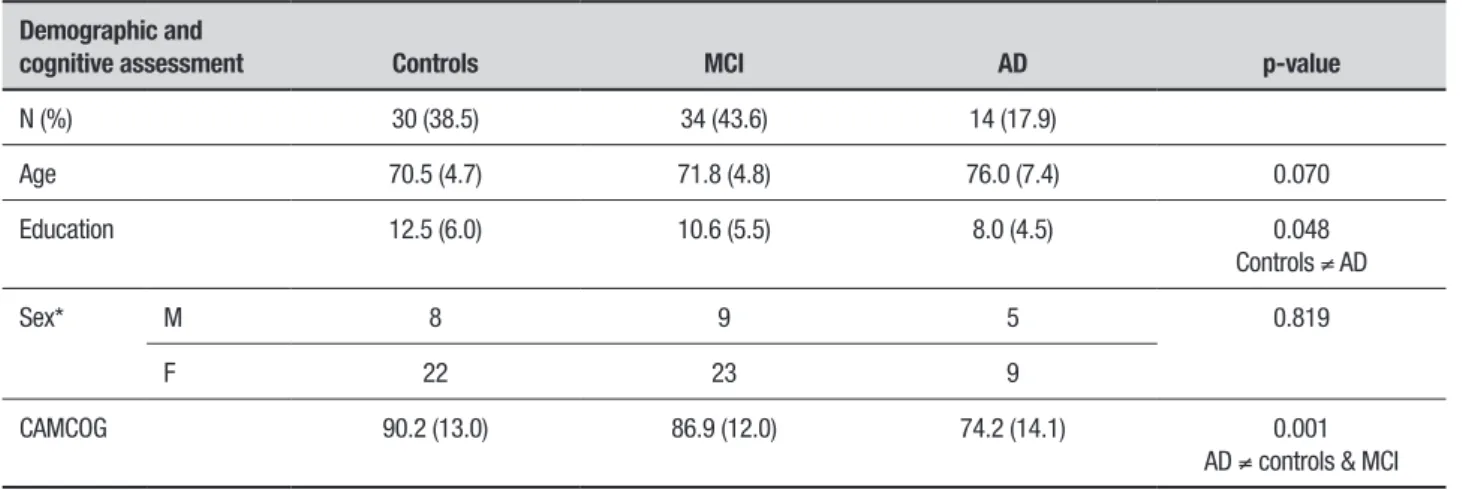
Table 1. Demographic and cognitive data by diagnostic subgroup.
Demographic and
cognitive assessment Controls MCI AD p-value
N (%) 30 (38.5) 34 (43.6) 14 (17.9)
Age 70.5 (4.7) 71.8 (4.8) 76.0 (7.4) 0.070
Education 12.5 (6.0) 10.6 (5.5) 8.0 (4.5) 0.048
Controls ≠ AD
Sex* M 8 9 5 0.819
F 22 23 9
CAMCOG 90.2 (13.0) 86.9 (12.0) 74.2 (14.1) 0.001
AD ≠ controls & MCI
the group factor showed that Tau-levels were associated with degree of DWM (Table 3).
AD patients were less educated than both controls and MCI patients and performed worse on the CAM-COG, as expected. Although not reaching statistical sig-niicance, there was a trend towards AD patients having lower CSF levels of Aβ1-42 and higher levels of t-tau, along
with lower Aβ1-42/p-tau ratios.
he distribution of deep white matter (DWM) hyperintensities, as measured by the Fazekas scale, among diagnostic groups is displayed in Table 3. Most subjects (56.4%) were classiied as Fazekas 1 (punctate foci) regardless of diagnosis, but there was no relation-ship between diagnosis and Fazekas classiication (Chi-square = 8.229, p = 0.221).
DISCUSSION
In the present study we addressed the proile of AD-related CSF biomarkers in the presence of concomi-tant subcortical vascular abnormalities in a
cross-section of older adults with varying degrees of cogni-tive impairment recruited from a specialized memory clinic. Regarding tau levels, our data suggest an asso-ciation between subcortical lesions and tau levels. Increased total tau levels were signiicantly associated with the degree of DWM changes when Fazekas grade was considered as a group factor, i.e., when all indi-viduals were aggregated into a single group, indepen-dently of subcortical lesion pattern. We also detected a progressive reduction in levels of Aβ1-42 as the degree of
DWM increased, i.e., from Fazekas = 0 (444.4 pg/mL) to Fazekas = 3 (367.5 pg/mL). However, diferences between biomarker groups and Fazekas scores were not statistically signiicant. As expected, our study showed a trend towards AD having lower CSF concentrations of Aβ1-42 and higher levels of t-tau, together with lower
Aβ1-42/p-tau ratio. Regarding MRI brain scans, most
subjects were classiied as Fazekas 1. Once more, we found no statistically signiicant diferences between levels of tau or Aβ1-42 and isolated neuroimaging group
Table 2. Biomarkers data by diagnostic subgroup.
CSF biomarkers and Fazekas Controls MCI AD p-value
Aβ1-42 pg/mL 426.2 (165.2) 423.2 (145.7) 374.9 (123.6) 0.535
P-tau pg/mL 46.8 (26.5) 50.1 (32.4) 48.3 (20.3) 0.896
T-tau pg/mL 85.6 (50.3) 107.1 (78.9) 127.2 (79.3) 0.179
Aβ1-42/p-tau 11.2 (6.3) 11.7 (7.9) 9.0 (5.8) 0.462
Fazekas (N) Frequency (%)
0 1 5 0 6 (7.7)
1 17 20 7 44 (56.4)
2 9 8 4 21 (26.9)
3 3 1 3 7 (9.0)
One-way ANOVA with Scheffé post-test; *Chi-squared test. AD: Alzheimer’s disease; MCI: mild cognitive impairment; CSF: cerebrospinal fluid; Aβ: amyloid-β; P-tau: phosphorylated tau; T-tau: total tau.
Table 3. CSF biomarker levels according to Fazekas grade.
Variable Fazekas = 0 (6) Fazekas = 1 (44) Fazekas = 2 (21) Fazekas = 3 (7) p-value
Aβ1-42 pg/mL 444.4 (130.7) 428.9 (157.1) 395.9 (141.6) 367.5 (149.3) 0.653
P-tau pg/mL 41.6 (18.6) 49.5 (28.5) 42.5 (19.6) 66.3 (46.2) 0.246
T-tau pg/mL 82.8 (30.8) 105.3 (75.9) 80.0 (30.9) 165.2 (102.5 0.038
3 ≠ 1 & 2 (p<0.05)
Aβ1-42/p-tau 13.0 (8.1) 11.0 (6.4) 11.5 (7.9) 7.6 (6.5) 0.515
of Fazekas, or between biomarkers and individual clin-ical diagnosis. In addition, Fazekas grade did not exert an impact on CAMCOG scores in any diagnostic group.
In contrast with our data, some cross-sectional stud-ies have demonstrated an increased deterioration of global cognition or executive dysfunctions in patients with cerebrovascular disease,12 as well as in patients with
lesions located strictly in lobar brain regions.34 Even
community-dwelling older adults showed subnormal cognitive performance associated with cerebrovascular disease.35
Neuropsychological assessments in patients with AD sharing cerebrovascular events tend to show worse cog-nitive impairment than evaluations among those with “pure” AD.11,36 hus, patients with multiple microbleeds
having lower CSF amyloid-β and higher t-tau and p-tau exhibited worse performance on cognitive assessments for global functions (Mini-Mental State Examination), object naming (Visual Association Test), language (Ver-bal Fluency Test), and digit span (forward and backward) than those without microbleeds.37
An intriguing issue is the impact of cerebrovascular disease on cognitive performance over time in patients with CSF abnormalities. he above-mentioned study11
used a linear mixed-efects model to analyze the asso-ciation between vascular events and cognition, adjust-ing for some covariates includadjust-ing CSF Aβ1-42 levels and
diagnostic group. Patients having at least one lobar vascular event had a 1.4 point decline per year on the Alzheimer’s Disease Assessment Scale – cognitive sub-scale (ADAS-Cog) when compared with those free from lobar vascular lesions, who showed a 0.8 point decline during the same period. In addition, cognitive decline reached 2.3 points per year on the scale among patients who had more than three lobar vascular lesions. Cogni-tive impairment persisted even after adjusting for CSF Aβ1-42 levels. he authors suggested that these
cerebro-vascular events per se might relect underlying amyloid angiopathy and contribute to cognitive deterioration.
Furthermore, longitudinal investigations involving individuals with cerebrovascular disease found worse cognitive global deterioration,38 a progressive decline in
memory impairment,39 an increased rate of conversion
from MCI to AD,40 as well as an elevated risk of
develop-ing incident dementia.41
Acute stroke also seems to alter levels of CSF Aβ
1-42 and tau. An analysis of these biomarkers found
increased levels of CSF tau among patients sufer-ing from acute ischemic events detected by combined clinical and MRI assessments.9 CSF p-tau levels were
similar between individuals with MCI and those with
stroke, suggesting some neurodegeneration within both groups, although this biomarker was more altered in AD. Furthermore, CSF Aβ1-42 levels were low among stroke
patients, as extensively described in AD.9 hese indings
suggest that changes in the CSF biomarker pattern can be caused by ischemic events themselves, independently of the neurodegenerative process alone.
Regarding molecular neuroimaging and brain per-fusion imaging, Bangen et al.42 found that older adults
with positive Aβ deposition, as measured by retention of lorbetapir in cortical regions, had higher cerebral blood low. Increased cerebral perfusion in positive Aβ deposition involved cortical regions mainly afected in AD, such as the hippocampus, posterior cingulate, and precuneus. Cognitively, these patients had poorer verbal memory. his hyperperfusion has been interpreted as indicating cellular and vascular compensatory activity in response to pathologic damage related to early stages of AD, notably in APOE ε4 carriers,43-45 and relects a
biological strategy to counteract memory challenges.44
Conversely, among negative Aβ load patients there was no signiicant association between resting cerebral blood low and global cognition, notwithstanding a trend toward higher perfusion in the posterior cingulate and better performance on verbal memory tasks.42
Interest-ingly, hyperperfusion in early MCI tends toward hypo-perfusion later in MCI, and exacerbates when approach-ing clinical diagnosis of dementia.42
A complex interaction of multiple mechanisms underlying the neurodegenerative process in AD has been recognized, even at early stages of the disease, and includes cerebrovascular events, molecular changes, and microstructural disorder; this phenomenon predicts brain atrophy and has an unfavourable impact on cog-nition and behaviour.12,13,17 herefore, damaged
micro-vasculature may lead to reduced blood low into brain regions, subsequently causing ischemia, tau pathology, and neuronal degeneration.11 Disruption of cerebral
blood vessels and disorder of the hemodynamic sys-tem induce a higher risk of developing AD in patients with MCI who present pathological levels of cerebro-spinal luid biomarkers.14 Decreased cerebral blood low
can elicit synaptic loss and neuronal death in cortical structures.46,47 Likewise, a post-mortem investigation
demonstrated that p-tau labelling was more intense around arteries and arteriole vessels with Aβ deposi-tion than around non-Aβ-laden vessels.48
Although subcortical hyperintensities have been associated with more pronounced pathological CSF biomarkers, cognitive decline could not be attributed to cerebrovascular disease per se.37 Level of education,
longer disease duration, and more severe cerebral atro-phy may contribute to the clinical course, although each isolated variable can only partially explain the cognitive worsening. Moreover, the reciprocal interference of cerebrovascular disease and AD downstream evolution warrants a more in-depth approach that highlights the magnitude of each biological mechanism and its impact on cognition.
In conclusion, in our study tau levels were associ-ated with degree of DWM when all individuals were aggregated into a single group, independently of clini-cal diagnosis. here was a progressive reduction in levels of Aβ1-42 as degree of DWM increased, although without
statistical signiicance. Again, there were no diferences between levels of tau or Aβ and isolated neuroimaging group of Fazekas, or between biomarkers and individual clinical diagnosis. he small number of subjects, espe-cially in the AD group, at least in part, could explain the absence of statistical diferences between diag-nosis groups regarding Fazekas scores and biomarker concentrations. Despite the limitation concerning the
small sample, the fact that all participants, including the control group, underwent MRI and especially CSF exams, represents a strength of this study. Future stud-ies should explore the current methodological strategy in a large sample, where this may provide more consis-tent data.
Author contribution. Marcia Radanovic: conception and
design, acquisition and interpretation of data, statis-tical analyses, intellectual contribution to the writing of the manuscript, discussion of data, and inal review. Florindo Stella: conception and design, intellectual contribution to the writing of the manuscript, discus-sion of data, and inal review. Lis Gomes: acquisition and analyses of data, and inal review. Leda L. Talib: analyses of data, and inal review. Orestes V. Forlenza: conception and design, organization of general execu-tion, intellectual contribution to the writing of the manuscript, discussion of data, and inal review. All authors approved the inal version of the manuscript.
Financial support and sponsorship. Fundação de Amparo
à Pesquisa de São Paulo (FAPESP Grant n8 09/52825-8, Brazil), Associação Beneicente Alzira Denise Hertzog da Silva (ABADHS).
REFERENCES
1. Dichgans M, Leys D. Vascular Cognitive Impairment. Circ Res. 2017;120:573-91.
2. Forlenza OV, Diniz BS, Talib LL, Radanovic M, Yassuda MS, Ojopi EB, et al. Clinical and biological predictors of Alzheimer’s disease in patients with amnestic mild cognitive impairment. Rev Bras Psiquiatr. 2010;32:216-22.
3. Petersen RC, Smith GE, Waring SC, Ivnik RJ, Tangalos EG, Kokmen E. Mild cognitive impairment: clinical characterization and outcome. Arch Neurol. 1999; 56:303-8.
4. Roberts R, Knopman DS. Classification and Epidemiology of MCI. Clin Geriatr Med. 2013;29:753-72.
5. Limongi F, Siviero P, Noale M, Gesmundo A, Crepaldi G, Maggi S. Dementia Registry Group. Prevalence and conversion to dementia of mild cognitive impairment in an elderly Italian population. Aging Clin Exp Res. 2017;29:361-70.
6. Etorre E, Cerra E, Marigliano B, Vigliotta M, Vulcano A, Fossati C, et al. Role of cardiovascular risk factors (CRF) in the patients with mild cogni-tive impairment (MCI). Arch Gerontol Geriatr. 2012;54:330-2. 7. Roh HW, Hong CH, Lee Y, Lee KS, Chang KJ, Kang DR, et al. Clinical
Conversion or Reversion of Mild Cognitive Impairment in Community versus Hospital Based Studies: GDEMCIS (Gwangju Dementia and Mild Cognitive Impairment Study) and CREDOS (Clinical Research Center for Dementia of South Korea). J Alzheimers Dis. 2016;53:463-73. 8. Vernooij MW, van der Lugt A, Ikram MA, Wielopolski PA, Niessen WJ,
Hofman A, et al. Prevalence and risk factors of cerebral microbleeds: the Rotterdam Scan Study. Neurology 2008;70:1208-14.
9. Kaerst L, Kuhlmann A, Wedekind D, Stoeck K, Lange P, Zerr I. Cerebro-spinal fluid biomarkers in Alzheimer’s disease, vascular dementia and ischemic stroke patients: a critical analysis. J Neurol. 2013; 260:2722-7. 10. Forlenza OV, Radanovic M, Talib LL, Aprahamian I, Diniz BS, Zetter-berg H, et al. Cerebrospinal fluid biomarkers in Alzheimer’s disease: Diagnostic accuracy and prediction of dementia. Alzheimers Dement. 2015;1:455-63.
11. Chiang GC, Cruz Hernandez JC, Kantarci K, Jack CR Jr, Weiner MW. A Alzheimer’s Disease Neuroimaging Initiative. Cerebral microbleeds, CSF p-tau, and cognitive decline: significance of anatomic distribution. Am J Neuroradiol. 2015;36:1635-41.
12. Gregoire SM, Scheffler G, Jäger HR, Yousry TA, Brown MM, Kallis C, et al. Strictly lobar microbleeds are associated with executive impairment in patients with ischemic stroke or transient ischemic attack. Stroke 2013;44:1267-72.
13. Radanovic M, Pereira FR, Stella F, Aprahamian I, Ferreira LK, Forlenza OV, et al. White matter abnormalities associated with Alzheimer’s disease and mild cognitive impairment: a critical review of MRI studies. Expert Rev Neurother. 2013;13:483-93.
14. O, Buchhave P, Zetterberg H, Blennow K, Minthon L, Warkentin S. Combined rCBF and CSF biomarkers predict progression from mild cogni-tive impairment to Alzheimer’s disease. Neurobiol Aging 2009;30:165-73. 15. Roth M, Tym E, Mountjoy CO, Huppert FA, Hendrie H, Verma S, et al.
CAMDEX: A Standardized Instrument for the diagnosis of mental disor-ders in the elderly with special reference to early detection of dementia. Br J Psychiatry 1986;149:698-709.
16. Bottino CM, Almeida OP, Tamai S, Forlezna OV, Scalco MS, Carvalho IAM. CAMDEX: The Cambridge Examination for Mental Disorders of the Elderly, Brazilian Edition. Translation and adaptation for Portu-guese language with authorization: of the Cambridge University Press. Cambridge University Press: UK, 1999.
17. Wilson B, Cockburn J, Baddeley AD. Rivermead Behavioural Memory Test. Suffolk: Thames Valley;1985.
18. Oliveira R, Schmidt SL. Teste Comportamental de Memória de River-mead. Cognição: Rio de Janeiro;1999.
19. Fuld P. Guaranteed stimulus processing in the evaluation of memory and learning. Cortex 1980;16:255-71.
21. Army Individual Test Battery. Manual of Directions and Scoring. War Department, Adjunt General’s Office Trail: Washington, DC 1944. 22. Montiel JM, Capovilla, AGS. Teste de Trilhas - Partes A e B. In: Capovilla
AGS, Capovilla FC. (eds.). Teoria e pesquisa em avaliação neuropsico-lógica. 1a. ed. São Paulo: Memnon, Capes, Inep, Fapesp, CNPq. 2007;
v. 1, p. 54-60.
23. Erzigkeit H. The Development of the SKT Project. In Dementia: Mole-cules, Methods and Measures, Hindmarch I, Hippius H, Wilcock G (eds). Wiley: England; 1991:101-8.
24. Flaks MK, Yassuda MS, Regina ACB, Cid CG, Camargo CH, Gattaz WF et al. The Short Test (SKT) - A Transcultural Test for Early Detection and Discrimination of Dementia: A Preliminary Study in Brazil. Int Psychoge-riatr. 2006;18:121-33.
25. Wechsler DI. Examiner’s Manual: Wechsler Adult Intelligence Scale -Revised. Psychological Corporation: New York;1981.
26. Nascimento E. WAIS-III: Escala de Inteligência Wechsler para Adultos - manual técnico. São Paulo: Casa do Psicólogo; 2005.
27. Hamilton M. Rating scale for depression. J Neurol Neurosurg Psychiatry 1960; 23:56-62.
28. Araújo RHS. Adaptação transcultural da GRID Hamilton Rating Scale for Depression (GRID-HAMD) para o português brasileiro e avaliação do impacto de um treinamento sobre a confiabilidade interavaliadores. Dissertação de Mestrado, Instituto de Ciências da Saúde, UFBA. Salvador, BA. Orientador: Oliveira IR; 2011, 140p.
29. Jorm AF, Jacomb PA. The Informant Questionnaire on Cognitive Decline in the Elderly (IQCODE): socio-demographic correlates, reliability, validity and some norms. Psychol Med. 1989; 19:1015-22.
30. Sanchez MA, Lourenço RA. Informant Questionnaire on Cognitive Decline in the Elderly (IQCODE): cross-cultural adaptation for use in Brazil. Cad Saúde Pública 2009; 25:1455-65.
31. Petersen RC. Mild cognitive impairment. J Int Med. 2004;256:183-94. 32. McKhann GM, Knopman DV, Chertkow H, Hyman BT, Jack Jr, CR,
Kawas CH, et al. The diagnosis of dementia due to Alzheimer’s disease: Recommendations from the National Institute on Aging-Alzheimer’s Association workgroups on diagnostic guidelines for Alzheimer’s disease. Alzheimers Dement. 2011;7:263-9.
33. Fazekas F, Chawluk JB, Alavi A et al. MR signal abnormalities at 1.5 T in Alzheimer’s dementia and normal aging. AJR Am J Roentgenol. 1987;149:351-6.
34. Poels MM, Ikram MA, van der Lugt A, Hofman A, Niessen WJ, Krestin GP, et al. Cerebral microbleeds are associated with worse cognitive func-tion: the Rotterdam Scan Study. Neurology 2012;78:326-33. 35. Takashima Y, Mori T, Hashimoto M, Kinukawa N, Uchino A, Yuzuriha T,
et al. Clinical correlating factors and cognitive function in
community-dwelling healthy subjects with cerebral microbleeds. J Stroke Cerebro-vasc Dis. 2011;20:105-10.
36. Pettersen JA, Sathiyamoorthy G, Gao FQ, Szilagyi G, Nadkarni NK, Geroge-Hyslop P, et al. Microbleed topography, leukoaraiosis, and cogni-tion in probable Alzheimer disease from the Sunnybrook dementia study. Arch Neurol. 2008;65:790-5.
37. Goos JD, Kester MI, Barkhof F, Klein M, Blankenstein MA, Scheltens P, et al. Patients with Alzheimer disease with multiple microbleeds: relation with cerebrospinal fluid biomarkers and cognition. Stroke 2009; 40:3455-60. 38. van der Vlies AE, Goos JD, Barkhof F, Scheltens P, van der Flier WM.
Microbleeds do not affect rate of cognitive decline in Alzheimer disease. Neurology 2012;79:763-9.
39. Ayaz M, Boikov AS, Haacke EM, Kido DK, Kirsch WM. Imaging cerebral microbleeds using susceptibility weighted imaging: one step toward detecting vascular dementia. J Magn Reson Imaging 2010;31:142-8. 40. Kirsch W, McAuley G, Holshouser B, Petersen F, Ayaz M, Vinters HV,
et al. Serial susceptibility weighted MRI measures brain iron and micro-bleeds in dementia. J Alzheimers Dis. 2009; 17:599-609.
41. Miwa K, Tanaka M, Okazaki S, Yagita Y, Sakaguchi M, Mochizuki H, et al. Multiple or mixed cerebral microbleeds and dementia in patients with vascular risk factors. Neurology 2014;83:646-53.
42. Bangen KJ, Clark AL, Edmonds EC, Evangelista ND, Werhane ML, Thomas KR, et al. Cerebral Blood Flow and Amyloid-β Interact to Affect Memory Performance in Cognitively Normal Older Adults. Front Aging Neurosci. 2017;9:181.
43. Dai W, Lopez OL, Carmichael OT, Becker JT, Kuller LH, Gach HM. Mild cognitive impairment and Alzheimer’s disease: patterns of altered cere-bral blood flow at MR imaging. Radiology 2009;250:856-66.
44. Wierenga CE, Dev SI, Shin DD, Clark LR, Bangen KJ, Jak AJ, et al. Effect of mild cognitive impairment and APOE genotype on resting cerebral blood flow and its association with cognition. J Cereb Blood Flow Metab. 2012;32:1589-99.
45. Zlatar ZZ, Wierenga CE, Bangen KJ, Lu TT, Jak AJ. Increased hippo-campal blood flow in in sedentary older adults at genetic risk for Alzheim-er’s disease. J Alzheimers Dis. 2014;41:809-17.
46. Maruyama M, Matsui T, Tanji H, Nemoto M, Tomita N, Ootsuki M, et al. Cerebrospinal fluid tau protein and periventricular white matter lesions in patients with mild cognitive impairment: implications for 2 major path-ways. Arch Neurol. 2004;61:716-20.
47. Lo RY, Jagust WJ. Vascular burden and Alzheimer disease pathologic progression. Neurology 2012; 79:1349-55.