Impaired abstract thinking may
discriminate between normal aging
and vascular mild cognitive impairment
Felipe Kenji Sudo1, Gilberto Sousa Alves1,Carlos Eduardo de Oliveira Alves1, Maria Elisa Lanna2, Letice Ericeira-Valente1, Denise Madeira Moreira3, Eliasz Engelhardt2, Jerson Laks1,4
ABSTRACT
Objective: Cerebrovascular disease (CVD) is associated with cognitive deficits. This cross-sectional study examines differences among healthy elderly controls and patients with vascular mild cognitive impairment (VaMCI) and vascular dementia (VaD) in performances on CAMCOG subscales. Method: Elderly individuals (n=61) were divided into 3 groups, according to cognitive and neuroimaging status: 16 controls, 20 VaMCI and 25 VaD. VaMCI and VaD individuals scored over 4 points on the Hachinski Ischemic Scale. Results:
Significant differences in total CAMCOG scores were observed across the three groups (p<0.001). VaD subjects performed worse than those with VaMCI in most CAMCOG subscales (p<0.001). All subscales showed differences between controls and VaD (p<0.001). Performance on abstract thinking showed difference between VaMCI and controls (p<0.001).
Conclusion: CAMCOG discriminated controls from VaMCI and VaD. Assessment of abstract thinking may be useful as a screening item for diagnosis of VaMCI.
Key words: CAMCOG, elderly, mild cognitive impairment, vascular dementia, cerebrovascular disease, abstract thinking.
O pensamento abstrato comprometido pode diferenciar o envelhecimento normal do comprometimento cognitivo leve vascular
RESUMO
Objetivo: A doença cerebrovascular (DCV) associa-se a déficits cognitivos. Este estudo transversal objetiva examinar diferenças entre controles saudáveis idosos e pacientes com comprometimento cognitivo leve vascular (CCLV) e demência vascular (DV) nas subescalas do CAMCOG. Método: Indivíduos idosos (n=61) foram divididos em 3 grupos, de acordo com o perfil cognitivo e com a neuroimagem: 16 controles, 20 CCLV e 25 DV. Pacientes com CCLV e DV pontuaram acima de 4 pontos no Escore Isquêmico de Hachinski. Resultados:
Diferenças significativas foram observadas entre os três grupos no resultado final do CAMCOG. Pacientes com DV obtiveram escores inferiores àqueles dos indivíduos com CCLV em quase todas as subescalas. Todas as subescalas mostraram diferenças entre DV e controles. O desempenho no item pensamento abstrato mostrou diferenças entre CCLV e controles. Conclusão. O CAMCOG diferenciou controles de pacientes com CCLV e DV. A avaliação do pensamento abstrato pode ser útil para discriminar CCLV de controles. Palavras-chave: CAMCOG, idosos, comprometimento cognitivo leve, demência vascular, doença cerebrovascular, pensamento abstrato.
Correspondence Felipe Kenji Sudo
Rua Ministro Alfredo Valadão 35/206 22031-050 Rio de Janeiro RJ - Brasil E-mail: [email protected]
Received 14 September 2009 Received in final form 12 November 2009 Accepted 22 November 2009
1Psychiatry Institute,Federal University Rio de Janeiro, Rio de Janeiro RJ, Brazil; 2Neurology Institute Deolindo Couto, Federal
Vascular mild cognitive impairment (VaMCI) can be deined as a cognitive impairment of vascular etiolo-gy that does not fulill criteria for dementia1. It has been
proposed that vascular-related cognitive impairment ex-ists throughout a continuum comprising VaMCI, vascu-lar cognitive impairment no-dementia (Va-CIND) and vascular dementia (VaD)2. In a sample of cerebrovascular
disease (CVD) patients with cognitive diiculties, Went-zel et al.3 reported a 50% rate of conversion to
demen-tia over a ive-year period. he early detection of VaM-CI may allow therapeutic intervention designed to halt or delay the progression of vascular lesions so as to pre-vent the conversion to dementia4,5. Presently, the
diag-nosis of mild cognitive impairment (MCI) requires im-provement in the sensitivity of conventional screening tests for dementia, since the rate of false-negative results is usually high for those individuals6,7. Studies attempting
to increase such sensitivity have shown that a combina-tion of diferent screening tests provides higher diagnos-tic accuracy compared to each test individually8,9. Diniz
et al.7 analyzed Mini-Mental State Examination (MMSE)
subtests in a sample of MCI subjects and managed to identify distinct proiles of cognitive deicits among the MCI subtypes. A higher rate of patients with MCI could be identiied when the item scores were analyzed, which was not possible when only MMSE inal scores were con-sidered. ROC curve analyses were performed to deter-minate cutof scores in the Cambridge Cognitive Exam-ination (CAMCOG) for MCI patients, but discrimina-tion between MCI subjects and controls with this meth-od showed low accuracy10.
It was suggested that some cognitive domains might be speciically impaired in MCI subjects and these as-pects could serve as diferential markers to distinguish this condition from normal aging. Rodríguez et al. found that individuals diagnosed as MCI performed signiicant-ly worse than controls in CAMCOG subtests assessing various areas, with higher signiicance levels correspond-ing to the variables memory, abstract thinkcorrespond-ing and ex-ecutive function11. In a previous study, Erkinjuntii et al.
observed impairment in memory, conceptual functions and arithmetical skills in a sample of individuals present-ing age-related cognitive changes12. Recently, one other
study showed that memory, constructive ability and ab-stract thinking were particularly impaired in MCI indi-viduals compared to controls13.
he purpose of this study is to evaluate the perfor-mance of VaMCI patients in comparison to cognitively unimpaired controls and VaD individuals on the CAM-COG tasks. We hypothesized that characterization of cognitive deicits using CAMCOG subtests may provide an increase in sensitivity in the screening for VaMCI, in addition to total test score. Furthermore, we intended to
verify if cognitive domains specially impaired in MCI in-dividuals, such as memory, abstract thinking and execu-tive function, could also serve as cogniexecu-tive markers to
di-agnosis ofVaMCI.
METHOD
Participants
Sixty-one elderly outpatients (mean age: 73.49±7.24 years; 68.85% female; mean education level: 6.86±4.57 years) were consecutively assessed at the Centre for Al-zheimer Disease and Related Disorders (CDA), Federal University of Rio de Janeiro (UFRJ), Brazil, between July 2006 and October 2008. he sample comprised both sub-jects who spontaneously demanded medical assistance due to cognitive complaints and those referred from oth-er clinics. Cognitive unimpaired controls woth-ere volunteoth-ers recruited from several sources who accepted invitation to participate in the study. Informed consent was obtained from participants or from a family member responsible prior to enrolment. his study is a branch of a larger proj-ect on vascular cognitive disorder, approved by the Eth-ics Committee of IPUB-UFRJ.
Clinical and neuropsychological assessment Patients and controls were examined by a multidis-ciplinary team, comprising psychiatrists, neurologists, one radiologist and one neuropsychologist. An interview with patient and caregiver was performed and those who had history of alcohol or drug abuse, psychiatric disor-ders (e.g. schizophrenia, bipolar mood disorder or life-time depressive disorder), non-corrected visual or audito-ry disorders, exposure to neurotoxic substances and cran-ioencephalic traumatism were excluded from the study. he cognitive assessment included the Cambridge
Cog-nitive Examination (CAMCOG)14, Mini-Mental State
Examination (MMSE)15, semantic verbal luency
(cate-gory animals)16,17, Trail Making Tests (TMT) A and B18
and the 12-item-Boston Naming Test19. Behavioral
eval-uation was assessed by the Neuropsychiatric Inventory (NPI)20,21. Depressive symptoms were measured with
Cor-nell Depression Scale22,23. Pfefer’s Functional Activities
Questionnaire (FAQ)24 was administered to informants
(a close relative or a caregiver) in order to identify evi-dence of functional decline. Risk factors for CVD were scored using Hachinski Ischemic Score (HIS)25.
Individ-uals were rated according to severity of cognitive deicits on the Clinical Dementia Rating scale (CDR)26,27.
Labora-tory tests were carried out to rule out reversible causes of cognitive decline, such as nutritional deiciencies, syphi-lis or diseases of the thyroid. All subjects underwent MRI scan of the brain.
Fourth Edition (DSM-IV)28 and the National Institute of
Neurological Disorders and Stroke and Association Inter-nationale pour la Recherché et l’Enseignement en Neuro-sciences (NINDS-AIREN)29 criteria for probable VaD.
he diagnosis of MCI was made according to Peters-en’s criteria30. MCI individuals scored below 4 points on
Pfefer’s FAQ and this cutof value was adopted to show functional activities largely preserved. Furthermore, VaMCI patients were identiied as those who obtained above 4 points on the HIS, showing high risk-factors for CVD, which were correlated with vascular subcortical le-sions and absence of signiicant cortical atrophy in MRI images.
he control group was submitted to the same pro-cedures described above. Subjects in this group did not present evidence of cognitive and functional impairment and had no or not signiicant neuroimaging abnormali-ties, such as vascular subcortical lesions or cortical atro-phy not correspondent to the expected for normal aging. Also, they had no history of major psychiatric disorders or substance-abuse.
Statistical analyses
Statistical analyses were made using SPSS for Win-dows version 11.5. Univariate Analysis of Variance (ANO-VA) was carried out to assess signiicant mean diferences for “Age” and “Educational Level”, followed by post-hoc Bonferroni analysis. Pearson’s Chi-square analysis was performed to evaluate diferences in the distribution of gender among the three groups. Analysis of covariance (ANCOVA) was performed to control for the potential confounding efects of schooling on cognitive variables.
RESULTS
Table 1 illustrates the sociodemographic data of indi-viduals among the three groups. Proportion of male and female subjects did not differ significantly among the groups. he control group showed a higher
education-al level when compared to VaD patients. his is depict-ed in Figure.
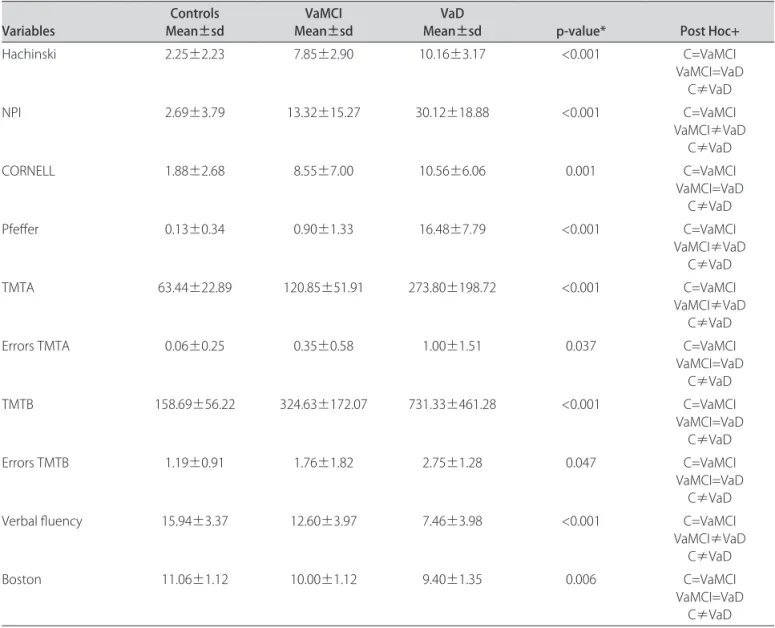
Results of the cognitive and behavioral evaluations are displayed in Table 2. Multiple comparison tests showed that VaD patients had worse performances in HIS, Cor-nell Depression Scale, changes in TMT A and TMT B (both time and errors) and Boston Naming Test as com-pared to controls. In comparison to controls and VaM-CI, VaD individuals had worse scores in NPI, FAQ, TMT A and Verbal Fluency.
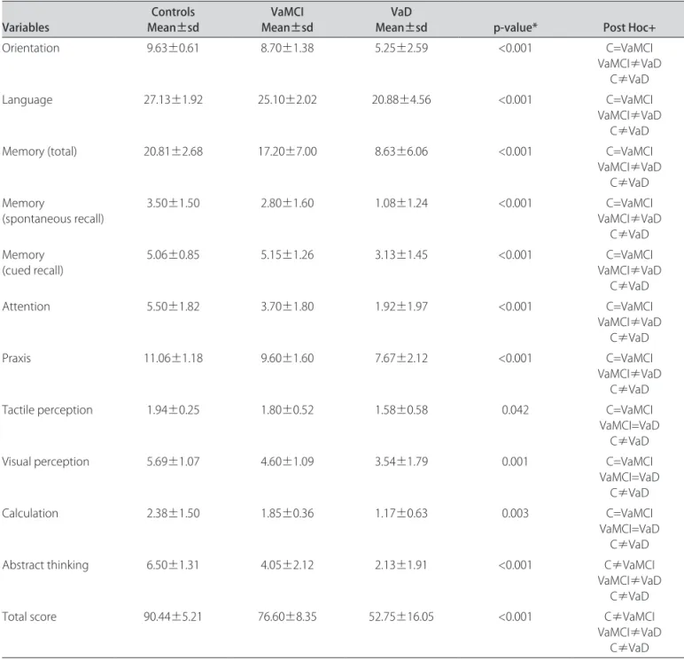
Multivariate analyses indicated that the three groups difered signiicantly in CAMCOG inal scores. Analyses of scores in the CAMCOG subtests demonstrated that abstract thinking was the only item that showed difer-ences among the three groups. Mean scores in orienta-tion, language, memory, attention and praxis indicated signiicant diferences between VaMCI persons and those with VaD, and also between controls and VaD individu-als. Visual and spatial perception, as well as calculation showed signiicant diferences only between controls and VaD subjects. hese data are shown in Table 3.
VaD VaMCI
Controls
Groups 20
15
10
5
0
S
c
h
o
o
li
n
g
Figure. Schooling (years) according to diagnostic groups.
Table 1. Demographic variables and MMSE according to diagnostic groups.
Controls VaMCI VaD p-value Post Hoc
Gender Male Female
N=4 (75%) N=12 (25%)
N=4 (80%) N=16 (20%)
N=11 (56%)
N=14 (44%) 0.186* –
Schooling (years) 9.44±3.88 6.55±4.60 5.48±4.43 0.022**
C=VaMCI VaMCI=VaDV
C≠VaD
Age (years) 73.00±7.83 72.95±6.78 74.24±7.44 0.803** C=VaMCI=VaD
MMSE (m±sd) 27.94±1.43 25.90±2.59 17.50±6.00 <0.001** MCI≠DVC=MCI
Table 2. Cognitive tests and behavioral evaluation according to diagnostic groups.
Variables Mean±sdControls Mean±sdVaMCI Mean±sdVaD p-value* Post Hoc+
Hachinski 2.25±2.23 7.85±2.90 10.16±3.17 <0.001 C=VaMCI
VaMCI=VaD C≠VaD
NPI 2.69±3.79 13.32±15.27 30.12±18.88 <0.001 C=VaMCI
VaMCI≠VaD C≠VaD
CORNELL 1.88±2.68 8.55±7.00 10.56±6.06 0.001 C=VaMCI
VaMCI=VaD C≠VaD
Pfefer 0.13±0.34 0.90±1.33 16.48±7.79 <0.001 C=VaMCI
VaMCI≠VaD C≠VaD
TMTA 63.44±22.89 120.85±51.91 273.80±198.72 <0.001 C=VaMCI
VaMCI≠VaD C≠VaD
Errors TMTA 0.06±0.25 0.35±0.58 1.00±1.51 0.037 C=VaMCI
VaMCI=VaD C≠VaD
TMTB 158.69±56.22 324.63±172.07 731.33±461.28 <0.001 C=VaMCI
VaMCI=VaD C≠VaD
Errors TMTB 1.19±0.91 1.76±1.82 2.75±1.28 0.047 C=VaMCI
VaMCI=VaD C≠VaD
Verbal luency 15.94±3.37 12.60±3.97 7.46±3.98 <0.001 C=VaMCI
VaMCI≠VaD C≠VaD
Boston 11.06±1.12 10.00±1.12 9.40±1.35 0.006 C=VaMCI
VaMCI=VaD C≠VaD *ANCOVA; +C: controls
DISCUSSION
he present study showed that VaMCI and controls had significantly different performances in regards to the CAMCOG total scores (p<0.001). Another interest-ing indinterest-ing was that impairment in abstract thinkinterest-ing was present in VaMCI but not in controls. Other subtests, such as orientation, language, memory, attention and praxis showed accuracy in separating VaMCI subjects from VaD individuals (p<0.001), whereas tactile and visu-al perception and cvisu-alculation could only identify VaD pa-tients from controls (p<0.001). MMSE failed in discrim-inating controls from VaMCI. VaMCI subjects and con-trols had similar overall performances on cognitive and behavioral evaluation, as depicted in Table 2.
Nunes et al.10 showed that CAMCOG presented a
sensitivity of 64% and a speciicity of 88%. One previous study, however, which explored psychometric properties (reliability, discriminative capacity and factorial structure) of the CAMCOG, found an excellent reliability of the
in-dividual subscales and high levels of sensitivity and spec-iicity of the instrument in diferentiating between de-mented and non-dede-mented individuals31.
his study shows some limitations that should be
out-lined and commented. Significant differences were
found in scores on Cornell Depression Scale between VaD individuals and controls. Depression is a common condi-tion observed in patients with CVD and it can be relat-ed to poorer performances in cognitive assessment, act-ing as a confoundact-ing factor to cognitive deicits primarily associated to dementia32,33. herefore, our results in
low scores in cognitive assessment associated to educa-tional level.
Moreover, our results do not agree with some stud-ies wherein the cognitive tests of MCI patients showed a ceiling efect, thus not being able to accurately recog-nize patients from controls. A possible explanation for the absence of a ceiling-efect could be the small sample, and similar studies with a larger sample should be per-formed. A great heterogeneity of individuals classiied as MCI following Petersen’s criteria has been described in
many studies34, and our indings may have been
inlu-enced by this limitation. Characterization of MCI may vary according to the diferent neuropsychological
instru-ments employed35, which may also explain the variation
of cognitive proiles of MCI among the studies. Difer-ently from the studies mentioned previously, our sample is constituted by individuals with vascular-related cogni-tive deicits, instead of those associated to neurodegener-ation, and this aspect may be related to the particularities of the results of this study. Furthermore, our sample was recruited at the Centre for Alzheimer Disease and Relat-ed Disorders (CDA-UFRJ), and population-basRelat-ed stud-ies, as well as studies with a larger sample are needed in order to replicate the results.
he assessment of abstract thinking requires the abili-ty of establishing similarities between objects. his capac-Table 3. CAMCOG subtests according to diagnostic groups.
Variables Mean±sdControls Mean±sdVaMCI Mean±sdVaD p-value* Post Hoc+
Orientation 9.63±0.61 8.70±1.38 5.25±2.59 <0.001 C=VaMCI
VaMCI≠VaD C≠VaD
Language 27.13±1.92 25.10±2.02 20.88±4.56 <0.001 C=VaMCI
VaMCI≠VaD C≠VaD
Memory (total) 20.81±2.68 17.20±7.00 8.63±6.06 <0.001 C=VaMCI
VaMCI≠VaD C≠VaD Memory
(spontaneous recall) 3.50±1.50 2.80±1.60 1.08±1.24 <0.001 VaMCI≠VaDC=VaMCI
C≠VaD Memory
(cued recall) 5.06±0.85 5.15±1.26 3.13±1.45 <0.001 VaMCI≠VaDC=VaMCI
C≠VaD
Attention 5.50±1.82 3.70±1.80 1.92±1.97 <0.001 C=VaMCI
VaMCI≠VaD C≠VaD
Praxis 11.06±1.18 9.60±1.60 7.67±2.12 <0.001 C=VaMCI
VaMCI≠VaD C≠VaD
Tactile perception 1.94±0.25 1.80±0.52 1.58±0.58 0.042 C=VaMCI
VaMCI=VaD C≠VaD
Visual perception 5.69±1.07 4.60±1.09 3.54±1.79 0.001 C=VaMCI
VaMCI=VaD C≠VaD
Calculation 2.38±1.50 1.85±0.36 1.17±0.63 0.003 C=VaMCI
VaMCI=VaD C≠VaD
Abstract thinking 6.50±1.31 4.05±2.12 2.13±1.91 <0.001 C≠VaMCI
VaMCI≠VaD C≠VaD
Total score 90.44±5.21 76.60±8.35 52.75±16.05 <0.001 C≠VaMCI
ity has been related to frontal lobe functioning35.
Impair-ment of abstract thinking has been considered one im-portant cognitive marker in the discrimination between MCI and normal aging11,13 and, since evaluation can be
considered simple, it may be a helpful item for clinical as-sessment of aged individuals with suspect of VaMCI. In conclusion, we propose that the analyses of each CAM-COG subtest, with emphasis on abstract thinking task, could be important as a screening item for early diagnosis of cognitive deicits in patients with high risk for CVD.
REFERENCES
1. Román G, Sachdev P, Royall D, et al. Vascular cognitive disorder: a new di-agnostic category updating vascular cognitive impairment and vascular de-mentia. J Neurol Sci 2004;226:81-87.
2. Garrett KD, Browndyke JN, Whelihan W, et al. The neuropsychological pro-ile of vascular cognitive impairment-no dementia: comparison to patients at risk for cerebrovascular disease and vascular dementia. Arch Clin Neuropsych 2004;19:745-757.
3. Wentzel C, Rockwood K, MacKnight C, et al. Progression of impairment in patients with vascular cognitive impairment without dementia. Neurology 2001;57:714-716.
4. Bowler JV, Hachinski V. Criteria for vascular dementia: replacing dogma with data. Arch Neurol 2000;57:170-171.
5. Engelhardt E, Laks J, Cavalcanti JL, Moreira DM, Madalen C. Demência vascu-lar. Rev Bras Neurol 2004;40:5-25.
6. De Jager CA, Milwain E, Budge M. Early detection of isolated memory deicits in the elderly: the need for more sensitive tests. Psychol Med 2002;32:483-491. 7. Diniz BS, Yassuda MS, Nunes PV, Radanovic M, Forlenza OV. Mini-Mental State Examination performance in mild cognitive impairment subtypes. Int Psy-choger 2007;19:647-656.
8. Lonie JA, Herrmann LL, Donaghey CL, Ebmeier KP. Clinical referral patterns and cognitive proile in mild cognitive impairment. Br J Psychiatry 2008;192: 59-64. 9. Heinik J, Shaikewitz D. The Clock Drawing Test-Modiied and Integrated Ap-proach (CDT-MIA) as an instrument for detecting mild cognitive impairment in a specialized outpatient setting. J Geriatr Psych Neurol 2009; [Epub ahead of print].
10. Nunes PV, Diniz BS, Radanovic M, et al. CAMCOG as a screening tool for di-agnosis of mild cognitive impairment in a Brazilian clinical centre of moder-ate to high education. Int J Geriatr Psychiatry 2008;23:1127-1133. 11. Rodríguez NR, Juncos-Rabádan O, Mayo FD. Cognitive markers to
discrimi-nate between mild cognitive impairment and normal ageing. Rev Esp Geriatr Gerontol 2008;43:291-298.
12. Erkinjuntti T, Laaksonen R, Sulkava R, Syrjäläinen R, Palo J. Neuropsycholog-ical diferentiation between normal aging, Alzheimer’s disease and vascular dementia. Acta Neurol Scand 1986;74;393-403.
13. Matsuda O, Saito M. Multiple cognitive déicits in patients during the mild cog-nitive impairment stage of Alzheimer’s disease: how are cogcog-nitive domains other than episodic memory impaired? Int Psychogeriatr 2009;21:970-976. 14. Roth M, Tym E, Mountjoy CO, et al. CAMDEX: a standardized instrument for
the diagnosis of mental disorder in the elderly with special reference to the early detection of dementia. Br J Psychiatry 1986;149: 698-709.
15. Folstein MF, Folstein SE, McHugh PR. Mini-Mental State: a practical method for grading the cognitive state of patients for the clinician. J Psychiatr Res 1975;12:189-198.
16. Rosen WG. Verbal luency in aging and dementia. J Clin Exp Neuropsychol 1980;2:135-146.
17. Brucki SMD, Malheiros SMF, Okamoto IH, Bertolucci PHF. Dados normativos para o teste de luência verbal categoria animais em nosso meio. Arq Neu-ropsiquiatr 1997;55:56-61.
18. Reitan RM. Validity of the trail making test as an indicator of organic brain damage. Percept Mot Skills 1958; 8:271-276.
19. Kaplan, E, Goodglass H, Weintraub S. The Boston Naming Test. Philadelphia: Lea & Febiger 1983.
20. Cummings JL, Mega M, Gray K, Rosenberg-Thompson S, Carusi DA, Gornbein J. The Neuropsychiatric Inventory: comprehensive assessment of psychopa-thology in dementia. Neurology 1994;44:2308-2314.
21. Camozzato Al, Kochhann R, Simeoni C, et al. Reliability of the Brazilian Portu-guese version of the Neuropsychiatric Inventory (NPI) for patients with Alzheim-er’s disease and their caregivers. Internat Psychogeriatrics 2008;20:383-393. 22. Alexopoulos GS, Abrams RC, Young RC, Shamoiam CA. Cornell scale for
de-pression in dementia. Biol Psychiatry 1988;23:271-284.
23. Carthery-Goulart MT, Areza-Fegyveres R, Schultz R, et al. Brazilian version of the Cornell depression scale in dementia. Arq Neuropsiquiatr 2007;65:912-915. 24. Pfefer RI, Kurosaki TT, Harrah CH Jr, Chance JM, Filos S. Measurement of func-tional activities in older adults in the community. J Gerontol 1982;37:323-329. 25. Hachinski VC, Ilif LD, Zilhka E, et al. Cerebral blood low in dementia. Arch
Neurol 1975;32:632-637.
26. Chaves Ml, Camozzato Al, Godinho C, et al. Validity of the Clinical Dementia Rating scale for the detection and staging of dementia in Brazilian patients. Alzheimer Dis Assoc Disord 2007; 21:210-217.
27. Hughes CP, Berg L, Danziger WL, et al. A new clinical scale for the staging of dementia. Br J Psychiatry 1982; 140: 566-572.
28. American Psychiatric Association. Diagnostic and statistical manual of men-tal disorders, 4th Ed. Washington, DC, 1994.
29. Román GC, Tatemichi TK, Erkinjuntti T, at al. Vascular dementia: diagnostic criteria for research studies: report of the NINDS-AIREN international work-shop. Neurology 1993;4:250-260.
30. Petersen RC, Doody R, Kurz A et al. Current concepts in mild cognitive im-pairment. Arch Neurol 2001;58:1985-1992.
31. Huppert FA, Jorm AF, Brayne C, et al. Psychometric properties of the CAM-COG and its efficacy in the diagnosis of dementia. Aging Neuropsychol 1996;3:201-214.
32. Wright SL, Persad C. Distinguishing between depression and dementia in older persons: neuropsychological and neuropathological correlates. J Geri-atr PsychiGeri-atry Neurol 2007;20:189-198.
33. Desmond DW. Vascular dementia: a construct in evolution. Cerebrovasc Brain Metab Rev 1996;8:296-325.
34. Norlund A, Rolstad S, Hellstrom P, Sjogren M, Hansen S, Wallin A. The Göte-borg MCI study: mild cognitive impairment is a heterogeneous condition. J Neurol Neurosurg Psychiatry 2005;76:1485-1490.