Angle’s Class II division 1 associated to mandibular retrusion and skeletal
open bite: a 5-year post-orthodontic/orthopedic treatment follow-up
Gustavo Tirado Rodrigues1
Obtaining long term stability allied to functional and aesthetic balance is the main goal of any orthodontic-orthopedic therapy. This case report describes the orthodontic therapy applied to a 7-year-9-month old child, who presented a Class II, division 1 malocclusion associated to skeletal open bite. Functional and skeletal corrections (sagittally and vertically) were obtained by means of mandible advancement achieved with a closed Balter’s bionator appliance followed by a fixed appliance. This approach showed to be efficient in accomplishing both functional and aesthetic goals, that were kept stable five years after the treatment was finished. This case report was presented to the Board of Directors of the Brazilian Board of Orthodontics and Facial Orthopedics (BBO), as partial requirement to becoming a Diplomate of the BBO.
Keywords: Angle’s Class II Malocclusion. Mandible Retrusion. Open bite. Corrective Orthodontics. Stability.
How to cite: Rodrigues GT. Angle’s Class II division 1 associated to mandibular retrusion and skeletal open bite: a 5-year post-orthodontic/orthopedic treatment follow-up. Dental Press J Orthod. 2017 Sept-Oct;22(5):98-112.
DOI: https://doi.org/10.1590/2177-6709.22.5.098-112.bbo
Contact address: Gustavo Tirado Rodrigues
Rua Terêncio Sampaio 429, Bairro Grageru – CEP: 49.025-700 – Aracaju/SE E-mail: [email protected]
» The author reports no commercial, proprietary or financial interest in the products or companies described in this article.
» Patients displayed in this article previously approved the use of their facial and intraoral photographs.
Submitted: July 03, 2017 - Revised and accepted: July 26, 2017
1 Universidade Tiradentes, Curso de Especialização em Ortodontia (Aracaju/SE,
Brazil).
DOI: https://doi.org/10.1590/2177-6709.22.5.098-112.bbo
INTRODUCTION
This case report describes the orthodontic treatment of a 7-year-9-month old male patient, during the second transitional period of mixed dentition, who presented at the clinic for treatment with the chief complaint of having excessively protruded teeth (“too lared”). Ac-cording to the mother, a slight advancement had been accomplished by the previous orthodontic interven-tion, during which a ixed palatal bar was used. Ater an otolanryngologic assessment, the boy was diagnosed as a partial mouth breather and presented a difuse nasal edema, with hypertrophic turbinates and adenoids. De-spite those indings, the case was treated non-surgically.
The clinical examination revealed satisfactory hygiene and a low cavity rate. Primary canines (53, 63 and 83) were prematurely lost.
DIAGNOSIS
Patient’s face presented marked features of chron-ic mouth-breathing, associated to a severe muscle hypotonia and an everted lower lip. A light protru-sive functional deviation was observed during mdible closure, together with speech impairment, an-teriorly positioned tongue and thumb sucking habit. Patient also lacked passive lip closure (5.0 mm), with a rather hypotonic upper lip. His lower lip was both A obtenção do equilíbrio funcional e estético em um contexto de estabilidade em longo prazo é o objetivo de qualquer abordagem ortodôntico--ortopédica. O presente relato aborda o tratamento ortodôntico realizado em uma criança com 7 anos e 9 meses de idade, portadora de Classe II, divisão 1, associada a mordida aberta esquelética. A correção funcional e esquelética (sagital e vertical) foi obtida por meio de avanço mandibular, com aparelho Bionator de Balters do tipo fechado, seguido de aparelho ortodôntico fixo. Essa abordagem foi eficiente para atingir as metas fun-cionais e estéticas, que continuaram estáveis cinco anos após a conclusão do tratamento. Esse caso clínico foi apresentado à Diretoria do Board Brasileiro de Ortodontia e Ortopedia Facial (BBO), como parte dos requisitos para a obtenção do título de Diplomado pelo BBO.
Figure 1 - Intraoral and facial initial pictures. hypotonic and everted. Although the profile was quite convex, given the severe mandible deficiency, nasolabial angle was normal and smile line was tend-ing from normal to low (Fig 1).
Intraoral assessment (Fig 1, 2) revealed a Class II division 1 malocclusion, with 9.7 mm overjet, anteri-or open bite and a 6.0 mm negative overbite between upper and lower central incisors. Lower midline pre-sented a 2.5 mm shift to the right due to the prema-ture loss of element #83, jeopardizing the space for the permanent successor (#43). During Class I model manipulation, a 5.5 mm cross-sectional maxillary de-ficiency was observed between first primary molars and the opposing teeth.
Initial panoramic radiograph (Fig 3) revealed addi-tional eruption diiculties related to permanent upper canines, imposed by the severe anterior diastemas, that by far exceeded the typical “ugly duckling” phase1. It was also veriied the presence of all permanent teeth, except for the upper third molars, still under development, as expected for that age group. Root contour, periodon-tal ligament space and bony crests showed no particular indings and presented quite normal.
ob-Figure 3 - Initial panoramic radiograph.
Figure 2 - Initial models.
served in patient’s proile through both SNGoGn (35o) and Axis Y (61o) would limit skeletal Class II and open bite corrections. There was a strong genetic factor associ-ated to the Class II, as his father’s proile was also seen to be severely convex from the skeletal
perspective (Con-vexity angle = 22o). Upper and lower incisors were pro-clined (Interincisal angle = 116o) and with increased axial inclination (1.NA = 26o; 1. NB = 30o, IMPA=99o).
Figure 4 - Initial profile cephalometric radiograph (A) and cephalometric tracing (B).
TREATMENT PLANNING
Due to the signiicant facial, skeletal and functional involvements, the planning was traced as follows: 1) max-illary cross-sectional approach, with modiied Haas ap-pliance, aiming at allowing for future mandible advance-ment without inducing posterior crossbite; 2) ixed ap-pliance for upper incisors (#12 and #22 in counter-angle position), as to manage the inter-incisors diastemas, fa-cilitating upper canines eruption; and 3) ater expander removal, closed Balter’s bionator appliance for mandible advancement, with constructive bite, reducing the over-jet by half, followed by a second appliance, posteriorly placed, in order to complete the correction.
The corrective orthodontic phase was planned with full fixed appliances in both arches (MBT, 0.022 x 0.028-in) and in order to solve the space dis-crepancy on the lower arch, pre-molars and canines interproximal stripping was planned, under lingual arch anchorage, welded to first molar bands (#36 and #46)2. Aligning and levelling were planned with 0.014-in and 0.016-in NiTi archwires, followed by 0.016-in, 0.018-in and 0.020-in stainless steel (SS) archwires, and 0.019 x 0.025-in SS finishing arches, with ideal shape, torque and coordination. During finishing, anterior vertical elastics could be indicated, if necessary, in order to overcorrect the overbite.
For the retention phase, an upper wraparound-like removable appliance, with palatal grid, for full time use,
except during meals, was planned for the irst 12 months, coupled with the lower ixed retainers (0.036-in stain-less steel) bonded to canines.
Lingual function and posture assessments were re-quested from a speech therapist, as well as diagnosis and treatment of the mouth-breathing condition.
The success of this planning would rely, besides to patient’s cooperation, on the cessation of the negative mouth habit (thumb sucking) as much as on his growth response. Nasal obstruction clinical therapy success and eiciency were key to both the growth response and to the compliance towards the closed Bionator therapy. The vertical growth tendency and the family compo-nent to the mandible retrusion would render the ortho-pedic response slightly less predictable.
TREATMENT PROGRESS
As estimated, the maxillary expansion promoted a transient increase of the open bite. On the other hand, it not only created space to accommodate both upper canines into the arch line but also prepared the maxilla for the incoming mandible advancement. The expander appliance itself was kept as a retainer for a period of four months, and removal was only promoted once the pala-tal suture was proven to be fully ossiied.
Upper anterior diastemas were managed by means of reciprocal forces, with the caveat to maintain the lateral incisors with the roots slightly inclined to the
mesial, what allowed for the canines to freely erupt over the alveolar ridge crest, without menacing the roots of the lateral incisors.
The mandible advancement with Balters bionator was necessary in two circumstances in order to make the process more gradual, optimizing adaptive condi-tions and treatment response.3,4 The wraparound acrylic splint was key towards correcting the tongue posture and the open bite.5 Patient cooperation was rather favorable. Ater wearing the irst appliance for 6 months, without any grinding of the acrylic body at the molar region, a second appliance was cast for the inal advancement and
it in an edge-to-edge incisors position (Fig 5). Vertical response required the patient to cease the thumb suck-ing habit, that was also incompatible with wearsuck-ing the appliance. At the end of 18 months with the Balters bi-onator6, facial improvements started to be observed, to-gether with changes in both sagittal and vertical aspects of the occlusion, leaving only the interdental stripping space adjustment to the corrective phase. When the intercep-tive phase was inished, patient was reassessed and a new set of orthodontic records was requested (Figs 6 to 9 and Tab 1). The inishing of the corrective phase required customized wire bending as to achieve the best root
allelism, aesthetic and functional adjustments. The cor-rective phase lasted one year and ive months, and elapsed without any intercurrences.
Retainers were used according to prescription and the speech therapist reassessment did not reveal the need for treatment, since tongue and perioral muscles had recov-ered normal status. Ater new radiographs and impres-sions had been taken, patient was requested to extract lower third molars. Once the upper retainer started to be used only overnight, the opening of a slight interincisal diastema demanded the bonding of a ixed 0.016-in SS retention wire to the palatal aspect of anterior teeth.
RESULTS
The orthodontic-orthopedic approach, allied to patient’s cooperation and good response to treat-ment, has allowed for a better and less concave facial profile. Passive lip closure was re-established, with a marked improvement of the lower lip position. Profile, however, was still kept somewhat concave, consistent with patient’s ethnical heritage. Smile line improved and allowed for a better exposure of upper teeth. Canine and molar excursion guides as well as improved overbite and overjet were visibly achieved (Figs 10 and 11).
Figure 8 - Intermediate panoramic radiograph.
Figure 9 - Intermediate profile cephalometric radiograph (A) and cephalometric tracing (B).
Figure 10 - Final intraoral and facial photographs.
The assessment of the skeletal condition can be seen in Figure 13 and Table 1. There has been a good sagittal response (ANB from 9.5o to 6.5o, and Wits from 4.5 mm to 3.5 mm) and improvements on the convexity of the skeletal profile (convexity angle from 22o to 17o). However, the skeletal changes pri-marily impacted the skeletal control of the maxilla (SNA from 83o to 77.5o). Mandible also exhibited changes, but only noticeable when assessing the fa-cial angle (from 82o to 84o). The discrete changes ob-served at the mandible were probably caused by the unfavorable clockwise rotation tendency (FMA from 26o to 27o and SNGoGn from 35o to 40o).
Upper incisors retraction resulting from the use of the orthopedic appliance was kept during the correc-tive phase (1.NA = 26o / 13o / 15o, 1-NA = 3.5/ 2/ 4 mm).
Lower incisors were also retracted during the ortho-pedic phase, despite having sufered a slight protrusion during the orthodontic phase (IMPA = 99o / 95o / 96o, and 1.NB = 30o / 27o / 29o). As a consequence, in-terincisal angle was tending towards normality (116o / 132o / 129o) (Tab. 1).
Mandible excursion guides were obtained for ante-rior movements and both let and right lateral move-ments. Neuromuscular balance was achieved including lip and tongue positions, as well as during swallowing and speaking. Besides that, periodontal and TMJ health were preserved.
The inal panoramic radiograph revealed nice con-touring of the roots and good parallelism, together with the alveolar bone crest heights, that were equally pre-served (Fig 13).
Figure 12 - Final profile cephalometric radiograph (A) and cephalometric tracing (B).
Figure 13 - Final panoramic radiograph.
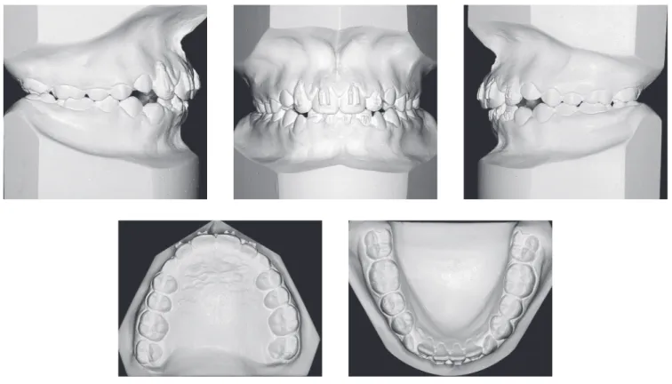
FIVE YEAR FOLLOW-UP AFTER TREATMENT The ive year follow-up ater the end of the active treatment (Figs 14 to 17) showed stability of the obtained results from both occlusal and skeletal/facial perspectives. The functional aspect is still very well balanced.
Figure 18 illustrates the skeletal behavior throughout the treatment, by means of the cephalometric tracings superimposition. Vertical growth was predominant in the face, following the intrinsic facial growth pattern. Maxillary anterior displacement control was efective. Mandible achieved a good sagittal gain, specially ater the use of the orthopedic appliance. During subsequent phases, a major inferior displacement was observed if compared to the anterior movement. Overbite and
overjet were efectively corrected, besides the relevant improvement of the facial proile.
Partial superimpositions revealed stable upper molars ater the orthopedic phase, followed by extrusion and some mesial displacement along the subsequent phases. Upper incisors presented a marked uprighting immedi-ately ater orthopedics and extrusion in all phases.
In the mandible, some extrusion was observed and slight mesial movement of the molars, besides extrusion and uprighting of the lower incisors, except during the retention phase, when a buccal movement was observed in these teeth. Mandible body and ramus observation re-vealed that an efective sagittal growth took place, with a good height gain along the ramus.
Figure 15 - Follow-up models, 5 years after orthodontic treatment.
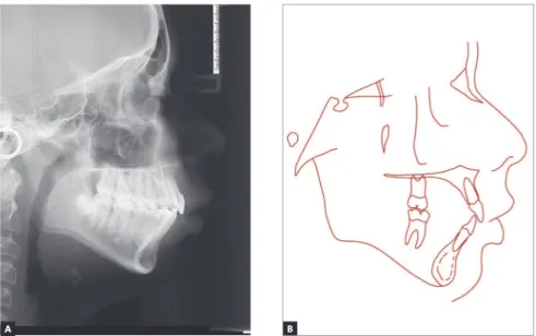
Figure 17 - Follow-up profile cephalometric radiograph (A) and cephalometric tracing (B), 5 years after orthodontic treatment.
Figure 18 - Total (A) and partial superimposition (B) of initial (black), intermediate (blue), final (red) and 5 years follow-up post-retention (green) cephalometric tracings.
B A
Table 1 - Cephalometric values: A) Initial, A1) intermediate, B) final and C) 5 years post containment.
Measurements Normal A A
1 B C Dif. A/B
Skeletal pattern
SNA (Steiner) 82o 83° 82° 77.5° 77.5° 5.5°
SNB (Steiner) 80o 73.5° 74.5° 71° 73° 1.5°
ANB (Steiner) 2o 9.5° 7.5° 6.5° 4.5° 3°
Wits (Jacobson) ♀ 0 ±2 mm
♂ 1 ±2 mm 4.5 mm 2 mm 3.5 mm 2.5 mm 1 mm
Angle of convexity (Downs) 0o 22° 19° 17° 12° 5°
Y-axis (Downs) 59o 61° 61° 60° 61° 1°
Facial angle (Downs) 87o 82° 83° 84° 84° -2º
SN-GoGn (Steiner) 32o 35° 36° 40° 37° -5°
FMA (Tweed) 25o 26° 27.5° 27° 25° -1°
Dental pattern
IMPA (Tweed) 90o 99° 95° 96° 99° 3°
1.NA (degrees) (Steiner) 22o 26° 13° 15° 20° -11°
1-NA (mm) (Steiner) 4 mm 3.5 mm 2 mm 4 mm 7 mm -0.5 mm
1.NB (degrees) (Steiner) 25o 30° 27° 29° 31° -1°
1-NB (mm) (Steiner) 4 mm 4.5 mm 7 mm 9.5 mm 10 mm -5 mm
1
1- Interincisal angle (Downs) 130o 116° 132° 129° 125° -13°
1-APo (Ricketts) 1 mm 0.5 3 5 7 -4.5
Proile
Upper lip — S-line (Steiner) 0 mm 6 mm 5 mm 6 mm 5 mm
-Lower lip — S-line (Steiner) 0 mm 5.5 mm 6 mm 6 mm 5 mm -0.5 mm
FINAL REMARKS
Treatment was based on the possibility of coupling skeletal correction to the orthopedic mandible ad-vancement.7-11 Anterior open bite was related both to functional changes (thumb sucking, mouth breathing, atypical swallowing and altered speech) as to the clock-wise rotation tendency presented by the mandible due to the strong vertical growth component — which by itself, was quite a limiting factor against more expres-sive sagittal gains.12 The age for intervention, despite a bit early from the pubertal growth spurt13-14 perspective, was adequate given the need for immediate action as to avoid the risk of trauma to anterior teeth and to improve patient’s self esteem, which had been deeply afected by the antisocial consequences of the malocclusion.
Thus, with patient’s cooperation, the orthopedic ad-vancement approach was shown to be a very eicient treatment strategy, providing the patient with a new
functional-skeletal condition, achieved by an unevent-ful corrective phase. Positive aspects were also seen on perioral muscle tonus and function. Skeletal goals were met despite the limitations imposed by the facial pat-tern, presenting vertical growth and mandibular retru-sion hereditary tendencies as additional hurdles.
REFERENCES
1. Broadbent BH. The face of the normal child. Angle Orthod. 1937;7(4): 183-208.
2. Zachrisson BU, Nyøygaard L, Mobarak K. Dental health assessed more than
10 years after interproximal enamel reduction of mandibular anterior teeth. Am J Orthod Dentofacial Orthop. 2007 Feb;131(2):162-9.
3. Falck F, Fränkel R. Clinical relevance of step-by-step mandibular advancement in the treatment of mandibular retrusion using the Fränkel appliance. Am J Orthod Dentofacial Orthop. 1989 Oct;96(4):333-41.
4. Du X, Hägg U, Rabie AB. Efects of headgear Herbst and mandibular
step-by-step advancement versus conventional Herbst appliance and maximal jumping of the mandible. Eur J Orthod. 2002 Apr;24(2):167-74.
5. Ortolani-Faltin C, Faltin Júnior K. Bionator de Balters. Rev Dental Press Ortod Ortop Facial. 1998 Nov-Dez;3(6):70-95.
6. Siqueira DF, Mondelli AL. Bionator de Balters: técnica de desgaste. Rev Clin Ortod Dental Press. 2002 Abr-Maio;1(2):9-16.
7. Bigliazzi R, Franchi L, Bertoz AP, McNamara JA Jr, Faltin K Jr, Bertoz FA. Morphometric analysis of long-term dentoskeletal efects induced by treatment with Balters bionator. Angle Orthod. 2015 Sept;85(5):790-8. 8. Bishara SE, Ziaja RR. Functional appliance: a review. Am J Orthod
Dentofacial Ortop. 1989 Mar;95(3):250-8.
9. Feitosa HO, Rodrigues GT. Tratamento da Classe II subdivisão em paciente com crescimento: relato de caso-parte I. Rev Clín Ortod Dental Press. 2012;11(2):118-27.
10. Franchi L, Pavoni C, Faltin K Jr, McNamara JA Jr, Cozza P. Long-term skeletal and dental efects and treatment timing for functional appliances in Class II malocclusion. Angle Orthod. 2013 Mar;83(2):334-40.
11. Malta LA, Baccetti T, Franchi L, Faltin K Jr, McNamara JA Jr. Long-term dentoskeletal efects and facial proile changes induced by bionator therapy. Angle Orthod. 2010 Jan;80(1):10-7.
12. Artese A, Drummond S, Nascimento JM, Artese F. Critérios para o diagnóstico e tratamento estável da mordida aberta anterior. Rev Dental Press Ortod Ortop Facial. 2011 Maio-Jun;16(3):136-61.
13. Baccetti T, Franchi L, McNamara JA Jr. The cervical vertebral
maturation (CVM) method for the assessment of optimal treatment timing in dentofacial orthopedics. Semin Orthod. 2005;11(3):119-29.