Alberto Consolaro*, Laurindo Furquim**
Intrusive mechanics generates inclination forces
and orthopedic stimulus followed by simultaneous
dental repositioning and bone remodelling
or
Intrusion forces are not applied in intrusive
mechanics, but intrusive effects are still obtained
* Full Professor, Bauru Dental School and Graduate Program of the Ribeirão Preto School of Dentistry, University of São Paulo, Brazil. ** Professor, State University of Maringá (UEM).
The low intensity and long duration aggressions to the periosteum induce the formation of new layers and can increase the volume of bone and change its shape. In intrusive mechanics, the natural inclination of the roots provides the tooth inclination movement. At the same time that it promotes compression forces on the periodontal ligament of teeth subjected to this kind of mechanics, in other areas, tension forces with deflection occur. These effects also involve the outer surfaces, since the thickness of the bone in the alveolar process is thin and can lead to the formation of new layers, including the cervical part of the alveolar bone crest. In intrusive me-chanics, there is an alveolar remodeling with orthodontic nature associated to a modification of bone internal and external structure, satisfying the demand for forces with orthopedic features. The intrusive effect on the so called intrusive mechanics may be the result of alveolar remodel-ing induced by the inclination forces, and of the modification of bone volume due to subperios-teal bone formation on the outer part of the alveolar process. Probably accurate imaging studies, with high precision CT, will be able to detect these subperiosteal phenomena in future studies involving patients before and after application of intrusive mechanics.
Abstract
Keywords: Intrusion. Orthodontics. Orthopedics. Periosteum.
How to cite this article: Consolaro A, Furquim L. Intrusive mechanics generates
inclination forces and orthopedic stimulus followed by simultaneous dental reposi-tioning and bone remodelling. Dental Press J Orthod. 2011 Sept-Oct;16(5):20-9.
root apex and at interradicular furcal regions.2
This risk would be greater if compared to other tooth movements. Despite the suggestion that apical root resorption depends on the inten-sity of orthodontic movements,10 many works
reveal that there is no relation between dental resorption and intrusion.3,5,6,7,11,12 Carrillo et al,2
in 2007, used specific appliances with full an-chorage bu means of plates and osseointegrated implants in dogs teeth and were not able to ver-ify root resorption through imaging. Although some intrusion effects did take place, resorp-tions observed were considered insignificant.
Experimental results obtained by Carrillo et al2 were similar to those obtained by other
au-thors,4,9,5 even when teeth were microscopically
observed after 4 to 7 month of intrusion. Mi-croscopically, root resorptions were small and in the apical and furcal areas of molar teeth of dogs and did not offer images reliable enough for a precise diagnosis. Clinical surveys with im-portant intrusive effects also revealed that the tooth resorption index was either very low or inexistent1,8.
Pure intrusive orthodontic forces are solely applied for experimental purposes and in these papers evidences reveal that whenever apical or furcal resorption take place on these teeth, they can only be observed microscopically, going un-diagnosed after imaging exams.2,4,5,9 Results of
these works also suggest that when intrusive forces are eliminated, surrounding tissues would promptly promote healing to the resorbed areas. In the present work, we have attempted to give the biological basis as to why purely intrusive forces are not generated in orthodontic therapies, even under the so known intrusive mechanics, through which clinically relevant intrusive effects are obtained. In order to understand the concep-tual fundamentals of intrusive mechanics it is para-mount to review the concepts of osseous biology,
option for orthodontic treatment planning.
bone And bone tissue: they Are not the sAme thing!
Bones represent anatomical structures com-prised of different tissues that interact to play specific roles, namely:
a) Bone tissue, a specialized connective tissue, in which an organic matrix is deposited and mineralized, forming two typical struc-tures: Cortical bone and trabecular bone. In the mineralized bone matrix there are numerous cells, osteocites (Figs 1, 2 and 3) embedded in gaps called osteoplasts. b) Bone marrow, a hematopoietic tissue that
produces blood cells and platelets. It may last until the end of one’s life or cease to function on an earlier stage. Adult’s jaws only persist as hematopoietically active in the maxillary tuber, mandibular angle and condyle; in macroscopic sections it pres-ents a red colour. As it becomes atrophic, the red bone marrow is gradually replaced by adipose tissue and/or fibrous connec-tive tissue and presents a yellowish colour. Bone marrow, especially the hematopoi-etically active, is very rich in undifferenti-ated cells or stem cells, containing also the primitive cells that generate many leuco-cytes, red blood cells and platelets.
FIGURE 1 - On the bone cortical surface (CS) periosteum reveals numer-ous bone cells in nature, particularly osteoblast and reserve cells with great osteogenic potential. Periosteum (P) still distributes blood vessels (V) and presents extra-cellular matrix externally, including collagen fi-bers that interact structural and functionally with skeletal muscles (SM) and tendons, besides other soft tissues. (H.E.; 20X).
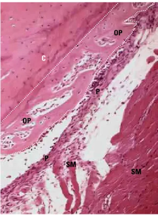
FIGURE 2 - When stimulated by long duration low intensity stimulus such as long duration light forces, associated or not to bone deformation, perios-teum (P) reacts apposing bone over the interface in a lamellar pattern and parallel to the cortical (C), a condition known as ossifying periostitis (OP, between dashed lines). More towards the periphery, skeletal muscle fibers are associated to the outermost portion of the periosteum (SM). (H.E.; 20X).
externally as hematopoietic medullary tis-sue, adipose or fibrous tissue.
periosteum concept And functions Bone surfaces are lined, covered and protect-ed by the periosteum (Figs 1 and 3). Externally, this connective tissue membrane is very fibrous, capable of offering considerable resistance to debridement during surgical procedures, which requires special tools. Periosteum is firmly at-tached to the bone through attachment collag-enous fibers that cross over its richly vascular-ized inner interface also rich in young and ma-trix producing cells. Blood supply mandatorily comes through the periosteum in order to get
to other bone structures (Fig 1).
The periosteum has many functions of which the soft tissue attachment stands out, particu-larly muscles and tendons. It is through contrac-tions and force transmission to bones that the body movements. In muscle attachment areas, bone structures are thicker and present denser trabeculae in order to meet the greater func-tional mechanical demand.
Collagenous fibers of the periosteal con-nective tissue unite to muscles in continuity to the epimisium (Figs 1 and 3), a connective membrane that lines or embraces muscle skel-etal fibers. Epimisium, on its turn, is attached to the sarcolema or muscle fibers citoplasmatic
P P
P
P
V
SM CS
C
OP
OP
P
P
SM
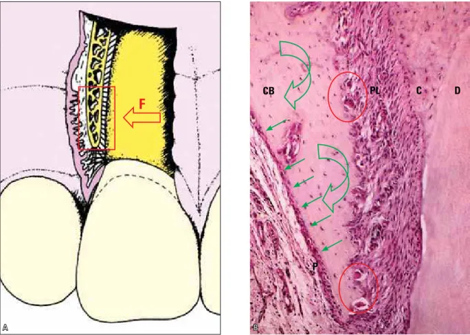
FIGURE 3 - During orthodontic movements, forces (F) may promote deflection (curved arrows) of the alveolar process and stimulate sub-periosteal new bone formation (arrows) along the interface between periosteum (P) and cortical bone (CB), while along the periodontal surface there is a frontal bone resorption characterized by bone remodeling (BMUs) units and its clasts (circles). C=cementum; D=dentin PL= periodontal ligament. (H.E.; 20X).
membrane through an extracellular matrix or membrane proteins.
When muscle contractions take place, mus-cle forces are transmitted to bones by the con-nective tissue junction between periosteum and epimisium. In some cases, forces are so intense and concentrated that they demand a stronger connective tissue, especifically organized for this purpose in the form of tendons. Tendons represent fiber stripes or cords through which muscles attach to bone or other organs. They are paramount to the maintenance of static and dynamic balance of the body through the trans-mission of muscle forces to bones and joints. The whole set of concerted forces from the
dif-ferent muscle groups produces flexion, exten-sion, rotation, abduction, adduction and transla-tion movements.
The periosteum’s reactional capacity in face of eventual injuries or long lasting low intensity stimulus promotes the apposition of new bone layers over the demanded area, arranged as an onion peel over the cortical interface (Figs 2 to 5). This may occur in deflections and deforma-tions thanks to the limited bone elasticity in the presence of stress, pressure, discrete or pro-longed inflammation processes, subperiosteal contusions, as well as surgical procedures or any other type of long lasting low intensity stimu-lus. During the dental movements induced on
CB PL
P
C D
F
FIGURE 4 - Intrusive mechanics promotes inclination forces due to the natural inclination of the roots of single-rooted anterior teeth. Bone resorption on the periodontal surface takes place along periodontal ligament compression areas (straight arrows) and bone apposition takes place along the area where fibers are being stretched (curved arrows), including the buccal periosteal interface. Bone deflection and deformation also induce or accelerate alveolar bone remodeling promoting a new position of the tooth inside the alveolus in relation to the other elements, creating an intrusive effect.
FIGURE 5 - Intrusive mechanics induces inclination forces due to the natural inclination from the cervical emergence of multi-rooted teeth. Bone resorption on the periodontal surface occurs in areas of peri-odontal ligament compression (straight arrows), while bone apposition takes place in the area where fibers are being stretched (curved ar-rows), including along the buccal periosteal interface. Bone deflection and deformation also stimulate or accelerate alveolar bone remodeling, promoting a new position of the tooth inside the alveolus in relation to adjacent elements, creating an intrusive effect.
buccal and lingual cortical ridges bone deflec-tions induce the formation of new bone layers underlying to the periosteum in these areas.
Human pathology, when analysing the for-mation of new cortical or subperiosteal layers as a reactional phenomenon, commonly refers to it as Garrè’s Osteomielitis, a terminology that should be exchanged by Ossifying Periostitis (Figs 2, 4 and 5). This picture neither represents an osteomielitis nor was described in the litera-ture by Garrè, as it is thought sometimes.
primAry And secondAry bone tissues: distinct functions And feAtures
Bone tissue may be classified according to its formation as primary or secondary. Primary
bone tissue is also known as embryonic or even immature bone. Its synthesis is performed by young and recently differentiated osteoblastic cells, which rapidly deposit a randomly dis-tributed matrix to fill up and occupy spaces with bone tissue.
Trabeculae formed by primary bone are short, rhomboid and rich in osteocytic cells, with numerous osteoblasts superficially ar-ranged in palisade. Trabeculae distribution in the primary bone tissue tends to be irregular or random, with the major function of filling up spaces originally occupied by blood clot, granulation tissue and young connective tissue. Meeting mechanical functional demands of a given area is not within the main attributions of primary bone tissue.
Bone resorption Alveolar boneremodeling
Bone apposition
Bone resorption
Bone apposition
degree and great regeneration and remodelling capacities. Primary bone represents a very im-portant tool for the organism to rapidly refill spaces resulting from fractures, bone surgeries and teeth alveoli after extractions. Once this recovery role has been accomplished by the primary bone, it is gradually replaced by sec-ondary bone.
Secondary bone is also known as adult or mature bone. Its major function is to supply me-chanical functional demands. In order to make it happen, it uses its constant remodelling ca-pacity to adapt the structures to forces and oth-er stimuli. Structures will be thinnoth-er, delicate or thick and robust according to the frequency and intensity of stimulus. This input may increase or decrease trabeculae density, forming bone struc-tures that can be more or less sclerotic. Cortical plates may vary in thickness in order to adapt to local functional demands.
Bone remodels itself regardless of mechani-cal input, although forces may accelerate this turnover. Constant bone remodelling is related to its mineral apposition function, maintaining calcium blood levels within the normal range to the overall tissue cell metabolism. Calcium represents the most important ion in our cell metabolism; since it is vital to the organism, its availability levels in the blood should be kept constant at all cost.
periodontAl ligAment: internAl AlveolAr periosteum
On the alveolar ridge bone surface peri-osteum is not as organized as in other bones. The periosteal role is played by the periodon-tal ligament, a highly organized connective tis-sue membrane containing a delicate, detailed and intricate collagen fiber arrangement (Fig 3). Periodontal ligament functionally connects root surface cementum to the bundle bone that
Out of the overall periodontal volume, 50% are blood vessels, most of which are very per-meable and thin walled venules. Periodontal ligament thickness ranges from 0.2 to 0.4 mm, with an average thickness of 0.25 mm.
Periodontal collagen fibers attach to the ce-mentum by merging or in continuity to this tis-sue, that is collagen-based and stratified in lamel-las parallely displayed along the root’s long axis. In the same way, periodontal collagen fibers fuse and merge to the bundle bone organic compo-nent that lines the internal alveolar surface.
Just like the collagen fibers and the peri-odontal vascular network, the ligament pres-ents a basket-like structure comprised of epi-thelial rests of Malassez, with threads 4 to 8 epithelial cells thick and 20 cells in length. The role of these rests lies in maintaining a minimum concentration of epithelial growth factor (EGF) in the periodontal ligament so that the alveolar surface is constantly stimu-lated to resorb itself and to maintain the aver-age periodontal ligament space. EGF is one of the local factors that stimulate bone resorp-tion under physiologic condiresorp-tions.
Whenever periodontal fibers are stretched, this is reflected over the external part of the alveolar bone, promoting the formation of new layers of subperiosteal bone (Figs 4 and 5). As soon as it happens, alveolar bone strain, de-flection, or deformation take place where the tooth is being inclined or pulled. This phenom-enon has therefore an orthopedic nature in the context of concepts that distinguish it from orthodontic phenomena.
bundle bone: the most externAl root component
many cementoblasts that end up embedded in the matrix and start to be called cementocytes. In the inner part of the cementum, gaps where cementocytes or cementoplasts lie are spider shaped and have numerous small channels filled by cytoplasmatic prolongations. Cementocytes look for a network intercommunication system through their extensions, particularly along the apical half of dental roots.
Cement, periodontal ligament and bundle bone have the same embryonic origin: Dental follicle or sac. Dental follicle presents itself as a tissue package of the tooth bud, being a part of it. In the centre of the tooth bud, lay the enamel organ and the dental papilla; the dental follicle establishes an interface with the surrounding bone structures.
The innermost part of the dental follicle originates the cementum and the most ex-ternal part originates the bundle or alveolar bone, which covers and lines the dental alveo-lus internal surface, merging with the alveolar ridge’s mature bone in a fully erupted tooth. The intermediate part of the dental follicle forms the periodontal ligament.
Microscopically, the structure of the bundle bone is similar to the cementum, although it is roughly disorganized. It presents with lamellar shaped apposition lines with periodontal col-lagen fiber bundles, that welds onto the inner surface. Apposition lines and periodontal fiber attachments create a bundle-like structure, with volumes or bands when this tissue is as-sessed under light microscopy, which justifies the name bundle bone.
Bundle bone can be considered as the plaster or liner of the dental alveolus originated from the tooth bud ectomesenchyma. In other words, the external part of a tooth from the morpho-functional point of view is made of bundle bone that stretches itself without any interface with the mature bone of the alveolar ridge.
By and large, almost all bone formed in
em-bryos and fetuses has the typical structure of primary bone, and therefore, its name: Embry-onic or immature bone. As movements and in-teraction with the external media gradually take place, remodelling also happens and it assumes the arrangement of mature, adult or secondary bone. Primary bone tissue only remains along tendon attachments and dental alveolus in the human skeleton. Not by chance, those are areas that require constant attachment and reattach-ment of collagen fiber bands in order to meet continuous functional demands. Along tendons and periodontal ligament, frequent and intense forces require a faster turnover of connective tissue attachment.
This faster turnover of periodontal tissue in relation to the other connective tissues in the body, together with bundle bone faster remod-elling process, help to cope with the local func-tional demand. It allows periodontal collagen fi-bers to be renewed and constantly attached and reattached, mitigating forces that hit the peri-odontal ligament. But even so many forces are able do deform, deflect or distort bone shape to the extent where subperiosteal tissues are stimulated to appose newly formed bone layers on top of the underlying cortical.
inclinAtion movement in intrusive mechAnics
necrosis — to neighboring areas, leaving the ex-tracellular matrix isolated and without renewal, this gives it a hyalinized aspect. If cementoblasts are destroyed, in the reconstruction of the area, root resorption may be observed before local full repair. Periodontal ligament compression — instead of focally disorganizing the periodontal ligament and promoting frontal bone resorp-tion — promotes great hyaline areas that, cell free, will suffer a peripheral reconstruction of the compressed area from outside inwards, far from where the phenomena should take place since the very beginning. Remodeling due to distance or peripheral bone resorption may gen-erate an area with undesirable contours, shape and height from an anatomical standpoint.
Periodontal ligament has a very organized distribution of its fiber bundles in order to soft-en ordinary forces of intrusive nature. The mor-phologic pattern of the periodontal ligament is not prepared to efficiently neutralize lateral forces and/or inclination of the tooth inside the alveolus. Proof of the difficulty to naturally dis-tribute lateral or inclination forces is in the very possibility of moving teeth orthodontically: Instead of dissipating and absorbing orthodon-tically applied forces, periodontal tissues re-organize themselves in order to adapt to the stimulus. The application of forces for lateral movements requires moderation and control since excessive forces may induce root and bone lesions because of excessive or morphologically uncontrolled resorption of mineralized tissues.
intrusion movements versus intrusive mechAnics
Periodontal ligament and its collagen fiber bands are finely prepared to receive and dissi-pate forces applied parallel to the root long axis. From the physiologic point of view, it absorbs intrusive forces and in orthodontic therapy,
sum up of masticatory forces. Despite its aver-age thickness is around 0.25 mm, not even the strongest of masticatory loads can compress the periodontal ligament in the apical third, and harm the neurovascular bundle that penetrates the pulp or promote tooth necrosis.
In orthodontic clinical practice, no mechanics are found to use solely intrusive forces, specific and parallel to the root long axis or even per-pendicular to the alveolus bottom. The so called intrusive mechanics applied might lead to this effect, although the forces that promote those effects are not intrusion but rather inclination forces (Figs 4 and 5). For a better understanding of this reasoning one should recall the absence of orthodontic devices that apply forces on the long axis of roots, especially if tooth positioning inside the dental alveolus and root inclinations in relation to the crown (Figs 4 and 5) are kept in mind, especially in multi-rooted teeth.
Whenever intrusive forces are applied, even if only in animals and on an experimental basis, those forces do not include inclination forces, as depicted by the Figures 4 and 5. Root in-clinations from its cervical emergence, lead experimental pure intrusion forces, in angles perpendicular to the occlusal surface of mo-lar teeth, to cause inclination forces over the roots. In these experimental works,2,4,5,9
when-ever resorptions are microscopically noticed, they are located on apical regions and buccal surfaces, facing the bifurcations.
formation of new bone layers over the cortical surfaces, which means in the cortical perios-teum interface (Figs 2 to 5). This reaction ca-pacity can modify the bone shape, increasing its volume and thickness.
Low intensity input promotes the apposition of new bone layers as periodontal ligament stress areas suffer straining of the fibers attached to the bundle bone during orthodontic treatment. Periodontal ligament primarily represents the alveolar surface periosteum and is very similar to the latter in the reaction capacity.
Alveolar process bone deflection induced by forces is capable of changing the final shape of jaws, since remodeling causes tissues to change in order to meet the new functional demands. Subperiosteal cortical resorption and apposition may take place on the external part of the alveolar processes where teeth are being intruded (Figs 4 and 5), what can also happen along the internal surfaces of the sinus walls and nasal cavity. This same phenomenon on endosteal surfaces changes and rearranges bone trabeculae, its spatial distribution, as much as thickness and length.
Intruded teeth in intrusive mechanics are repositioned in relation to the bone as a role and other teeth, due to orthodontic and ortho-pedic nature stimuli applied. Tooth structure may well suffer root resorption, as periodon-tal tissues may be altered in shape and height, without changing biologic widths of periodon-tal tissues and without affecting the biological feasibility of pulp tissues.
Intrusive mechanics promotes a special rear-rangement of the bone in relation to the tooth by means of so called orthopedic phenomena and teeth are concomitantly repositioned by means of inclination forces. As a result of this
synergy, teeth assume a new bone position and a new position in relation to its neighboring ele-ments in the dental arch.
conclusions
Bone has great reaction and adaptive capac-ity towards functional demands. Endosteal and periosteal surfaces stimuli may lead to the re-sorption and formation of new layers. The long lasting low intensity injuries to the periosteum induce the formation of new layers and may in-crease the overall bone volume and change its outer contours.
In intrusive mechanics, the natural inclina-tion of roots causes dental movements to be of inclination. Concomitantly to promoting com-pressive forces in some areas of periodontal liga-ment of teeth undergoing this type of mechan-ics, it induces stress and bone deflection forces in other areas and, extensively, in the surround-ing bone. These effects also involve external sur-faces, since bone thickness on the alveolar pro-cess is reduced and may lead to the formation of new layers, including the most cervical portion of the alveolar crest.
1. Andreoli FAM. Retração e intrusão anterior utilizando a técnica do arco segmentado [monograia]. Piracicaba (SP): Associação Paulista de Cirurgiões Dentistas; 2006. 2. Carrillo R, Rossouw PE, Franco PF, Opperman LA,
Buschange PH. Intrusion of multiradicular teeth and related root resorption with mini-screw implant anchorage: a radiographic evaluation. Am J Orthod Dentofacial Orthop. 2007;132(5):647-55.
3. Costopoulos G, Nanda R. An evaluation of root resorption incident to orthodontic intrusion. Am J Orthod Dentofacial Orthop. 1996;109(5):543-8.
4. Daimaruya T, Nagasaka H, Umemori M, Sugawara J, Mitani H. The inluences of molar intrusion on the inferior alveolar neurovascular bundle and root using the skeletal anchorage system in dogs. Angle Orthod. 2001;71(1):60-70.
5. Dellinger EL. A histologic and cephalometric investigation of premolar intrusion in the Macaca speciosa monkey. Am J Orthod. 1967;53(5):325-55.
6. Dermaut LR, Munk A. Apical root resorption of upper incisors caused by intrusive tooth movement: a radiographic study. Am J Orthod Dentofacial Orthop. 1986;90:321-6.
Contact address
Alberto Consolaro
E-mail: [email protected]
7. DeShields RW. A study of root resorption in treated Class II, division 1 malocclusions. Am J Orthod. 1969;39(4):231-44. 8. Moon CH, Wee JU, Lee HS. Intrusion of overerupted molars
by corticotomy and orthodontic skeletal anchorage. Angle Orthod. 2007;77(6):119-25.
9. Ohmae M, Saito S, Morohashi T, Seki K, Qu H, Kanomi R, et al. A clinical and histological evaluation of titanium mini-implants as anchors for orthodontic intrusion in the beagle dog. Am J Orthod Dentofacial Orthop. 2001;119(5):489-97. 10. Parker RJ, Harris EF. Directions of orthodontic tooth
movements associated with external apical root resorption of maxillary central incisor. Am J Orthod Dentofacial Orthop. 1998;114(6):677-83.
11. Phillips JR. Apical root resorption under orthodontic therapy. Angle Orthod. 1955;25:1-22.
12. Sameshima GT, Sinclair PM. Predicting and preventing root resorption: part II. Treatment factors. Am J Orthod Dentofacial Orthop. 2001;119(5):511-5.
Submitted: July 26,2011